Response to Vesicular Stomatitis Virus Vectors That Express Flagellin
Jason R. Smedberg,aMarlena M. Westcott,bMaryam Ahmed,cDouglas S. Lylesa
‹Departments of Biochemistryaand Microbiology and Immunology,bWake Forest School of Medicine, Winston-Salem, North Carolina, USA; Department of Biology,
Appalachian State University, Boone, North Carolina, USAc
Vesicular stomatitis virus (VSV) vectors that express heterologous antigens have shown promise as vaccines in preclinical stud-ies. The efficacy of VSV-based vaccines can be improved by engineering vectors that enhance innate immune responses. We pre-viously generated a VSV vaccine vector that incorporates two enhancing strategies: an M protein mutation (M51R) that prevents the virus from suppressing host antiviral responses and a gene encoding bacterial flagellin (M51R-F vector). The rationale was that intracellular expression of flagellin would activate innate immune pathways in addition to those activated by virus alone. This was tested with dendritic cells (DCs) from mice containing deletions in key signaling molecules. Infection of DC with either M51R or M51R-F vector induced the production of interleukin-12 (IL-12) and IL-6 and increased surface expression of T cell costimulatory molecules. These responses were dramatically reduced in DCs from IPS-1ⴚ/ⴚmice. Infection with M51R-F vector also induced the production of IL-1. In addition, in approximately half of the DCs, M51R-F vector induced pyroptosis, a proin-flammatory-type of cell death. These responses to flagellin were ablated in DCs from NLRC4ⴚ/ⴚmice but not Toll-like receptor
5-deficient (TLR5ⴚ/ⴚ) mice, indicating that they resulted from inflammasome activation. These results demonstrate that
flagel-lin induces additional innate immune mechanisms over those induced by VSV alone.
A
ctivation of innate immune cells is key to the activity ofvac-cine adjuvants as well as the success of live-virus vacvac-cines. Thus, a strategy for developing improved vaccines is to enhance their ability to activate the innate immune system. There are cur-rently multiple viruses being developed as live-virus vaccine vec-tors for the delivery of heterologous antigens that are effective activators of the innate immune system. Examples include the DNA viruses adenoviruses and poxviruses and the RNA viruses Newcastle disease virus, Sendai virus, and vesicular stomatitis
vi-rus (VSV) (1–4). In an effort to further stimulate the innate
re-sponse and trigger a greater adaptive rere-sponse, many viral vectors have also incorporated vaccine adjuvants. For example, influenza
virus, simian virus 5 (SV5), poxvirus, rabies virus (5), and as we
have reported, VSV, have been engineered to express bacterial
flagellin (6–10). The purpose of the current study was to test the
hypothesis that the expression of flagellin by VSV will lead to the activation of additional pathways not triggered by the virus alone. VSV is currently being developed as a vaccine vector for the
delivery of a wide array of heterologous antigens (1, 2, 11–15).
Although laboratory strains of recombinant VSV are not usually pathogenic in humans, the potential for VSV to cause disease in humans has been addressed by attenuation of VSV vectors. Mul-tiple methods of attenuation have been developed, including
in-troducing mutations into the viral M protein (5,16–18).
Substi-tution of arginine for methionine at position 51 of the M protein prevents VSV from inhibiting host gene expression, thus allowing cells to mount an antiviral response. This leads to an increased
immune response to VSV in the absence of neurovirulence (19).
VSV’s ability to elicit an immune response was further enhanced
by insertion of the gene for flagellin fromSalmonella entericaas a
separate gene transcript. This allows the expression of full-length
flagellin from the viral genome (6). We have previously reported
that VSV encoding flagellin induces higher antibody titers in mice
than does VSV vector without flagellin (6).
The hypothesis that flagellin activates additional pathways in
innate immune cells compared to those triggered by the virus alone was tested by analyzing the ability of VSV encoding flagellin to activate dendritic cells (DCs) from mice with deletions in key signaling molecules. DCs provide the critical link between the in-nate and adaptive immune systems. Previous studies have shown the ability of VSV that expresses flagellin to elicit a higher level of
cytokine production in human monocyte-derived DCs (6). VSV
has been shown to signal in DCs primarily through the pattern recognition receptors (PRR) Toll-like receptor 7 (TLR7) and
RIG-I (20,21). Not all subsets of DCs express TLR7, and in these
cells, the RIG-I pathway is likely the major inducer of antiviral
responses (21). The RIG-I response to VSV is mediated through
the mitochondrial adapter protein IPS-1 (22). In the case of
flagel-lin, two different pathways have been shown to mediate the
re-sponse in DCs (23–25). If the flagellin is extracellular, the response
is mediated by the Toll-like receptor TLR5. If the flagellin is intra-cellular, the NOD-like receptor NLRC4 mediates the response. Depending on which molecule detects flagellin, different out-comes for the cell can result. TLR5 activation leads primarily to
NF-B activation and cytokine production (26). NLRC4
activa-tion leads to formaactiva-tion of an inflammasome. The inflammasome is a multiprotein complex that activates innate immune response pathways. The composition of an inflammasome and the biolog-ical outcome is dependent on the sensor that is activated. In the case of NLRC4 activated by flagellin, the inflammasome is formed with pro-caspase-1 and adapter proteins, such as ASC. Formation of the NLRC4 inflammasome leads to processing and secretion of
Received2 October 2013Accepted31 October 2013
Published ahead of print6 November 2013
Address correspondence to Douglas S. Lyles, dlyles@wakehealth.edu.
Copyright © 2014, American Society for Microbiology. All Rights Reserved. doi:10.1128/JVI.02898-13
on November 7, 2019 by guest
http://jvi.asm.org/
tory molecules CD86, CD80, and CD40. Production of IL-1and
induction of pyroptosis were ablated in DCs from NLRC4⫺/⫺
mice but not TLR5⫺/⫺mice, indicating that they resulted from
inflammasome activation. In contrast, the production of IL-12 and IL-6 and expression of costimulatory molecules were dependent on IPS-1 and, thus, resulted primarily from antiviral signaling.
MATERIALS AND METHODS
Reagents.Recombinant mouse granulocyte-macrophage
colony-stimu-lating factor (GM-CSF) generated from a baculovirus vector was provided by S. Mizel, Wake Forest School of Medicine. Fluorescently labeled anti-bodies against mouse cell surface antigens (CD11c, clone N418; CD86, clone GL-1; I-Ab, clone AF6-120.1) were purchased from BioLegend. Fluorescently labeled antibodies against CD40 (clone 3/23) and CD80 (clone 16-1OA1) were purchased from BD Pharmingen. Caspase-1 p10 (M-20) antibody was purchased from Santa Cruz. IL-1antibody was purchased from R&D Systems. Flagellin antibody and purified flagellin were generously provided by S. Mizel. Lipopolysaccharide (LPS) from Salmonella entericaserovar Minnesota was purchased from Sigma (St. Louis, MO). Monoclonal anti-VSV N protein antibody 10G4 was pro-duced as described previously (31,32).
Mice.C57BL/6 mice were purchased from Charles River
(Wilming-ton, MA). With the permission of S. Akira, TLR5⫺/⫺mice were acquired from S. Mizel (Wake Forest School of Medicine), and IPS-1⫺/⫺mice were acquired from M. Schnell (Thomas Jefferson University). NLRC4⫺/⫺ mice were obtained from V. Dixit (Genentech). All mice were maintained and bred in the animal facility at Wake Forest School of Medicine, and experiments were conducted under protocols approved by the Institu-tional Animal Care and Use Committee and were in compliance with all relevant federal guidelines related to animal care and use.
Dendritic cells.GM-CSF-derived DCs were cultured as previously
described (33). Briefly, bone marrow was obtained from the femurs and tibias of 8- to 24-week-old mice. Red blood cells were lysed and washed, and progenitor cells were plated at a density of 5⫻105/ml in RPMI medium containing 10% fetal calf serum supplemented with 10 ng/ml of GM-CSF and 10g/ml of gentamicin. Cells were cultured at 37°C for 6 days and fed with fresh medium and GM-CSF on days 2, 4, and 5. On day 6, the nonadherent and loosely adherent cells were harvested. These cells were 90% (⫾5%) CD11c positive and showed low levels of CD40, CD80, CD86, and major histocompatibility complex class II (MHC-II) antigens, typical of immature DCs (34).
Virus growth and preparation.Recombinant M51R and M51R-F
vi-ruses were isolated from infectious VSV cDNA clones, and virus stocks were prepared and maintained on BHK cell monolayers as previously described (35). The M51R-F virus was purified by sucrose gradient cen-trifugation to remove extracellular flagellin.
Measurement of costimulatory molecule and cytokine expression
following viral infection.DCs were plated in a 48-well plate at a density of
5⫻105cells/ml in antibiotic-free medium and were infected with virus at the multiplicity of infection (MOI) indicated in the figures or legends for 6, 12, or 24 h. In a separate set of experiments, cells were treated with
membranes. Membranes were blocked and probed with antibodies against flagellin, caspase-1, IL-1, or actin followed by the appropriate secondary antibody conjugated to horseradish peroxidase. Chemiluminescent substrate was used for detection (Thermo Scientific, SuperSignal West Dura). Mem-branes were stripped and reprobed the following day until each protein had been analyzed. Pixel intensities were analyzed utilizing ImageQuant software.
Cell viability and death.DCs were infected at an MOI of 10 and at
the time points indicated in the figures or legends were analyzed for cell via-bility utilizing a 3-(4,5-dimethyl-2-thiazolyl)-2,5-diphenyl-2H-tetrazolium bromide (MTT) assay (Promega) or were stained with 7-aminoactinomycin D (7AAD) according to the manufacturer’s instructions (BD Pharmingen). Data were acquired using a BD FACSCalibur flow cytometer and analyzed using FlowJo software.
VSV N-protein detection.DCs were infected at an MOI of 10. At 24 h
postinfection, cells were harvested, fixed, permeabilized, and stained with VSV-N antibody followed by a fluorescently labeled secondary anti-body as described previously (36). Data were then acquired by flow cy-tometry as described above.
Statistical analysis.Comparisons were tested for statistical
signifi-cance by calculating thetstatistic and the probability (P) of the null hy-pothesis using Microsoft Excel software.Pvalues of⬍0.05 were consid-ered statistically significant.
RESULTS
Incorporation of a gene encoding bacterial flagellin has been shown to enhance the immune response and DC function for a
variety of live-virus vectors, including VSV (6–10). The
mecha-nism of enhancement is thought to be due to activation of innate immune pathways in antigen-presenting cells in addition to those activated by virus vectors that lack flagellin. This hypothesis was tested by analyzing the response to VSV vectors in murine myeloid DCs that lack key elements of the signaling pathways that respond to virus and flagellin. Construction of the M protein mutant VSV
vector and the vector encodingSalmonella entericaflagellin,
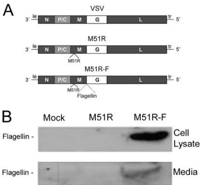
de-picted inFig. 1A, have been described previously (6). The M51R M
protein mutant vector has a single amino acid change, methionine to arginine, at position 51 of the M protein. This mutation pre-vents the virus from inhibiting host gene expression and allows infected cells to mount an antiviral response. The gene for flagellin
(thefliCgene) was inserted into the M51R backbone as a separate
transcriptional unit between the M and G genes. These vectors induce potent innate and adaptive immune responses in the ab-sence of viral pathogenicity in mice, whereas vectors with
wild-type (WT) M protein retain pathogenicity in mouse models (6,17,
19). The flagellin gene does not encode a eukaryotic signal
se-quence, so the flagellin would be expected to be primarily intra-cellular. The expression of flagellin was tested in myeloid DCs cultured from bone marrow precursors in the presence of GM-CSF. Cells were infected with the flagellin-expressing vector
on November 7, 2019 by guest
http://jvi.asm.org/
(M51R-F) or control vector (M51R), and flagellin expression in cell lysates or culture supernatants was determined by
immuno-blots (Fig. 1B). As expected, flagellin produced by cells infected
with M51R-F vector was present in the cell lysate and also the culture supernatant. When the relative amounts of flagellin in the supernatant and lysate were measured in the immunoblots and corrected for the relative volumes of the two samples, it was esti-mated that approximately 85% of the flagellin was in the superna-tant. This was likely released upon cell lysis.
The expression of flagellin by the M51R-F vector enhanced cytokine production by murine DCs over that induced by M51R
vector, similar to published results with human DCs (Fig. 2) (6).
DCs were infected with M51R and M51R-F at various MOIs for 24
h. Supernatants were analyzed for the cytokines IL-1, IL-6, and
IL-12 by ELISA. Infection of DCs with M51R vector did not result
in detectable levels of IL-1above the background of the ELISA.
In contrast, the M51R-F vector elicited robust production of
IL-1. DCs infected with M51R vector showed a strong IL-6 and
IL-12 response, which was increased in cells infected with M51R-F
vector at MOIs of 0.1 and 1.0 (Pvalues of 0.005 for IL-6 and 0.01
for IL-12, respectively). This shows that the presence of flagellin can enhance IL-6 and IL-12 production and is required to elicit
production of IL-1. Treatment of murine DCs with purified
flagellin has been reported to elicit low levels of cytokine production
compared to those seen with other TLR agonists (23,26). This was
confirmed in our experiments by comparing IL-6 and IL-12
produc-tion in response to flagellin with that in response to LPS (Fig. 2).
To determine the signaling pathways that contribute to cyto-kine production, DCs were cultured from bone marrow of WT,
TLR5⫺/⫺, NLRC4⫺/⫺, or IPS-1⫺/⫺mice and were treated with
M51R or M51R-F vector. Supernatants were analyzed for the
pro-duction of IL-1, IL-6, and IL-12 at 6 or 24 h postinfection. The
data obtained at 24 h are shown inFig. 3. As inFig. 2, only the
FIG 1VSV vector that expresses flagellin. (A) Diagram of genomes of WT and M51R VSV and M51R-F VSV. Shown are the genes encoding the viral N, P, C, M, G, and L proteins and the leader (le) and trailer (tr) RNAs. The gene for flagellin (thefliCgene) was inserted into the M51R backbone as a separate transcriptional unit between the M and G genes. (B) Murine DCs were infected with M51R or M51R-F vector (MOI⫽10) or mock infected. At 8 h postinfection, cell lysates (20 g of protein) and media (70g of protein) were analyzed for flagellin expression by immunoblotting. The volumes analyzed in the figure shown are as follows: cell lysates, 15.7l from a total 120l; media, 4.3l from a total 2 ml.
FIG 2Cytokine production by DCs following treatment with VSV vectors. DCs were treated at various MOIs for 24 h. Supernatants were collected, and cytokines were measured by ELISA. (A) IL-1production, (B) IL-6 produc-tion, (C) IL-12 production. Three independent determinations are shown for each treatment. The line indicates the mean; asterisks indicateP⬍0.05. Like symbols represent independent determinations for each treatment. In a sepa-rate set of experiments, DCs were treated with purified flagellin or lipopoly-saccharide (LPS) as described previously (6) and analyzed for production of IL-6 and IL-12.
on November 7, 2019 by guest
http://jvi.asm.org/
[image:3.585.300.539.63.627.2] [image:3.585.59.271.64.255.2]duced no detectable IL-6 or IL-12 above the levels seen for
mock-infected controls. IPS-1⫺/⫺cells treated with M51R-F virus did
pro-duce detectable amounts of IL-6 and IL-12, but they were well below the levels produced by cells of the other genotypes. Collectively, the
data in Fig. 3 indicate that production of IL-1 in response to
M51R-F vector primarily involves signaling through NLRC4, whereas production of IL-6 and IL-12 primarily involves signaling through IPS-1.
Production of IL-1requires stimulation of prointerleukin-1
(proIL-1) production, followed by caspase-1-mediated cleavage
to generate the mature form of the cytokine. The latter process results from inflammasome activation. To determine the
signal-ing pathways involved in expression of proIL-1, DCs from WT,
NLRC4⫺/⫺, or IPS-1⫺/⫺mice were treated with M51R or M51R-F
vector for 8 h, and cell lysates and supernatants were analyzed by
immunoblotting (Fig. 4). Only uncleaved proIL-1was detected
in cell lysates, and only the cleaved IL-1was detected in the
supernatants. There was a significant increase (P⫽0.003
com-pared to results of mock treatment) in proIL-1in the WT cells
treated with M51R-F vector, whereas cells treated with M51R vec-tor were similar to mock-treated cells, indicating that virus infec-tion alone in the absence of flagellin does not trigger producinfec-tion of
proIL-1. Cells deficient in NLRC4 and treated with M51R-F had
a significant reduction in proIL-1expression (P⫽0.007
com-pared to results for the WT), indicating that flagellin utilizes
NLRC4 to trigger proIL-1production. DCs deficient in IPS-1
also had a significant reduction in proIL-1expression (P⫽0.04),
although secreted levels of IL-1were similar between WT and
IPS-1⫺/⫺DCs treated with M51R-F vector, indicating that
expres-sion of total IL-1(proIL-1 ⫹cleaved IL-1) was higher than
that for mock- or M51R vector-treated cells.
An important indicator of DC maturation is the upregulation of costimulatory molecules and MHC-II molecules on the cell surface. To determine the effect of flagellin on expression of these surface markers, DCs were treated with M51R or M51R-F vector for 24 h and then harvested and stained for CD11c, CD80, CD86,
and MHC-II molecules (Fig. 5). The cells were analyzed by flow
cytometry with gating on CD11c-positive cells. Data are shown as a fold increase in mean fluorescence intensity (MFI) over that of mock-infected cells. WT cells treated with M51R or M51R-F vec-tor upregulated CD86, CD80, and MHC-II molecules. There was no significant enhancement due to the presence of flagellin. DCs from TLR5- and NLRC4-deficient mice showed no difference in comparison to WT cells. However, DCs deficient in IPS-1 failed to respond to either M51R or M51R-F vector, with a statistically
significant difference in the CD86 responses (P⬍0.04 for WT,
NLRC4⫺/⫺, and TLR5⫺/⫺mice versus IPS-1⫺/⫺, M51R-treated,
FIG 3Cytokine production by DCs with deletions of key signaling molecules. Dendritic cells were obtained from bone marrow of WT, TLR5⫺/⫺,
NLRC4⫺/⫺, or IPS-1⫺/⫺mice. DCs were infected for 24 h (MOI⫽10), and
cytokines were measured in supernatants by ELISA. (A) IL-1, (B) IL-6, (C) IL-12. Three independent determinations are shown for each treatment. The line indicates the mean. Like symbols represent independent determinations for each treatment.
on November 7, 2019 by guest
http://jvi.asm.org/
[image:4.585.47.279.63.651.2]and M51R-F-treated mice). Expression of CD80 and MHC-II
molecules by IPS-1⫺/⫺DCs also trended toward lower values but
did not reach statistical significance in these experiments. These results indicate that the upregulation of costimulatory molecules, particularly CD86, was dependent primarily on antiviral signaling through IPS-1.
Activation of the NLRC4 inflammasome can lead to cell death by pyroptosis in some cell types. The observation that DCs treated
with M51R-F vector elicit an IL-1response is an indicator of
activation of caspase-1, which is a requirement for pyroptosis. The major characteristics of pyroptosis that distinguish it from other forms of cell death are rapid permeabilization of the plasma
mem-brane, inflammasome activation, and IL-1release (29). The
re-lease of IL-1is shown inFig. 4. Evidence for rapid membrane
permeabilization and inflammasome activation is shown inFig. 6.
WT and mutant DCs were treated for 6 h with M51R or M51R-F vector and then stained with 7AAD to measure membrane permea-bilization. Cells were analyzed by flow cytometry for 7AAD fluores-cence and forward light scattering (FSC-H). In the WT cells, approx-imately 20% of both mock-treated and M51R vector-treated DCs were 7AAD positive. In contrast, approximately 50% of DCs treated with M51R-F vector were 7AAD positive. This indicates that DCs treated with flagellin-expressing virus elicited a rapid permeabiliza-tion of the plasma membrane that suggests the inducpermeabiliza-tion of
pyrop-tosis. When NLRC4⫺/⫺cells were treated with either vector, this early
membrane permeabilization did not occur, and all treatments resem-bled levels seen with mock-infected cells. Consistent with these
re-sults, flagellin was not detected in the supernatant of NLRC4⫺/⫺DCs
(data not shown). Therefore, the early cell death triggered by flagellin
is dependent on NLRC4. In both TLR5⫺/⫺and IPS-1⫺/⫺cells, the
effect of flagellin expression was similar to that in WT cells. This shows that signaling through NLRC4, but not IPS-1 or TLR5, plays a role in triggering cell death that is consistent with the induction of pyroptosis in response to M51R-F virus infection.
The observation that the M51R-F vector induces rapid mem-brane permeabilization raises the question of how many cells re-main viable after treatment with these viruses. DCs were treated with M51R or M51R-F vector, and cell viability was determined at
6, 12, and 24 h after infection using an MTT assay (Fig. 7). There
was lower cell viability after treatment with M51R-F than with M51R vector at each time point. However, there was no further
decrease in cell viability between 12 and 24 h, with M51R vector-treated cells stabilized at approximately 65% viability and M51R-F vector-treated cells at approximately 48% viability.
The cell viability data raise the question of whether the surviv-ing cells express viral antigens. Alternatively, they may be cells that are resistant to infection. To address these questions, DCs infected for 24 h were stained for surface CD11c and CD86 and intracellu-lar VSV N protein. Cells were analyzed by flow cytometry with
gating on live, CD11c-positive cells.Figure 8shows representative
histograms and data from multiple experiments expressed as the mean percentage of N protein-positive cells. At 24 h, roughly 70% of M51R vector-treated DCs and 55% of M51R-F vector-treated DCs were positive for viral N protein. This result was largely in-dependent of DC genotype. In addition, of those N protein-posi-tive cells, approximately 90% were also posiprotein-posi-tive for CD86 (data not shown). This demonstrates that M51R-flagellin-infected DCs not only were activated to mature but also expressed viral antigen.
DISCUSSION
The activity of bacterial flagellin as a vaccine adjuvant is well es-tablished both as a purified protein and when expressed in viral
vaccine vectors (reviewed in references 25and 26). There are
many cell types in the immune system whose activity is enhanced by flagellin. The experiments presented here address the mecha-nisms involved in the activity of DCs infected with VSV vectors that express flagellin. Previous experiments demonstrated the en-hanced activation of human DCs compared to VSV vectors that
do not express flagellin (6). The results presented here extend
these results to murine DCs, with which we demonstrated that the expression of flagellin in the intracellular compartment leads to the activation of pathways distinct from those activated by virus alone and leads to an increased production of mediators of in-flammatory responses. The enhanced release of inin-flammatory
me-diators included the production of IL-1(Fig. 2to4) and the
triggering of pyroptosis (Fig. 6and7). Additionally, it was shown
that the main pathway that contributes to IL-1production and
pyroptosis is the NLRC4 inflammasome (Fig. 3and7).
The pattern of expression of cytokines and costimulatory mol-ecules provided a clear distinction between pathways induced by virus and those induced by flagellin. The production of 6, IL-12, and costimulatory molecules was primarily through antiviral
FIG 4Production of proIL-1and mature IL-1following VSV treatment. DCs were cultured from bone marrow of WT, NLRC4⫺/⫺, or IPS-1⫺/⫺mice and
treated for 8 h with M51R or M51R-F vector (MOI⫽10). (A) Immunoblot of cells and media from infected cells, probed with antibodies to detect pro-IL-1, mature IL-1, and actin as an internal reference. (B) Quantitation of pro-IL-1in immunoblots. Integrated pixel densities were measured using ImageQuant software, and results are expressed as the maximal response (WT DCs treated with M51R-F vector). Data shown are means⫾SD from 3 experiments.
on November 7, 2019 by guest
http://jvi.asm.org/
[image:5.585.113.480.66.213.2]signaling, as shown by the effects of deletion of IPS-1. In contrast,
production of IL-1and induction of pyroptosis were primarily
through inflammasome activation, as shown by deletion of
NLRC4, which ablated both responses (Fig. 3and7). Deletion of
TLR5, the other major PRR for flagellin, had only minor effects in this study. There was certainly the potential for activation of TLR5, since a substantial proportion of the flagellin produced by infected DCs was released into the extracellular environment. In contrast to human DCs, which respond robustly to extracellular flagellin, murine DCs generated in culture respond less well to extracellular flagellin than other TLR agonists, presumably
be-cause of their low levels of TLR5 expression (6,23,25,26).
None-theless, it was important for us to test the role of TLR5, because of the potential for virus infection to stimulate its expression. TLR5
may play a larger rolein vivodue to the presence of subsets of DCs
that do express TLR5 as well as other cell types that respond
effec-tively to flagellin (23,25,26).
Previously published experiments have demonstrated VSV
triggering the production of IL-1 through activation of the
NLRP3 inflammasome in DCs similar to those studied here (37).
In contrast, we detected little if any IL-1production induced by
VSV vectors that do not express flagellin. The discrepancy in our results could be due to minor differences in the culture technique for these DCs. This might lead to a difference in differentiation of
FIG 5M51R and M51R-F vectors increase costimulatory molecule expression. WT, NLRC4⫺/⫺, TLR5⫺/⫺, and IPS-1⫺/⫺DCs were treated with M51R and
M51R-F vectors (MOI⫽10). At 24 h, the cells were harvested, stained for cell surface markers, and analyzed by flow cytometry. (A) Representative histograms depicting CD86 expression levels in arbitrary fluorescence units. Solid line, M51R treated; dotted line, M51R-F treated; shaded histogram, mock treated. (B) Mean fluorescence intensities (MFIs) from multiple experiments indicated as fold induction in MFI over that of mock-infected controls. Like symbols represent independent determinations for each treatment. The line represents the mean.
on November 7, 2019 by guest
http://jvi.asm.org/
[image:6.585.67.515.69.518.2]DCs prior to infection, which could lead to a different result postinfection. Despite these differences in results, it appears safe to conclude that activation of the NLRC4 inflammasome by
flagellin is a much more potent inducer of IL-1production than
activation of the NLRP3 inflammasome by VSV.
In addition to production of IL-1, the other important effect
of activation of the NLRC4 inflammasome in DCs by flagellin was the induction of cell death consistent with pyroptosis. This form
of cell death depends on activation of caspase-1 (which also cleaves
pro-IL-1to generate mature IL-1) or caspase-11 by NLRs
contain-ing Pyrin domains or caspase activation and recruitment domains
(CARDs), such as NLRC4 (29,30). Evidence for the induction of
pyroptosis was the NLRC4-dependent permeabilization of the plasma membrane by 6 h postinfection and by the rapid induction of
cell death as determined by MTT assay (Fig. 6and7). The principal
mechanism of cell death induced by VSV in the absence of flagellin expression is apoptosis, which is effectively induced by M51R viruses through activation of both the death receptor and mitochondrial
pathways (38–40). Induction of apoptosis in cell culture typically
leads to membrane permeabilization and cell lysis in the late stages
(38). However, apoptotic cells are usually taken up by phagocytic cells
in vivobefore their intracellular contents are released. In contrast to induction of cell death by apoptosis, induction of pyroptosis leads to
rapid release of intracellular contents bothin vivoand in cell culture
(29,30). Many of these intracellular contents represent “danger”
sig-nals that are potent activators of innate immune cells. These include metabolites such as ATP and uric acid, and proteins, such as heat shock proteins Hsp70 and Hsp90 and high-mobility group box 1
(HMGB1) protein (29,30). It is also possible that factors released
from infected cells induce cell death in adjacent cells. In addition to release of these activators of inflammatory responses, lysis of cells by pyroptosis leads to release of intracellular viral antigens, which can be
cross-presented by other classes of DCs, such as CD8⫹DCs (41,42).
For example, in the experiments inFig. 8, the nonviable cells were
positive for viral N protein. Cell death by pyroptosis has been de-scribed most often for myeloid cells infected with a variety of patho-gens, but it is likely that other nonmyeloid cells are also capable of undergoing pyroptosis in response to vectors that express flagellin
(29,30).
The results inFig. 7show that approximately 50% of DCs infected
with M51R-F virus remain viable at 24 h postinfection. This raises the question of why some cells survive the infection while others undergo cell death by pyroptosis. We ruled out the trivial explanation that the surviving cells were resistant to infection by demonstrating the
ex-pression of viral antigen in the surviving cells (Fig. 8). These were also
the cells that express costimulatory molecules such as CD86, which
were assayed at 24 h postinfection (Fig. 5). There is a similar
dichot-omy between cells that die as a result of apoptosis and those that
survive the induction of apoptosis. This is also apparent inFig. 7as
partial loss of cell viability following infection with M51R vector that does not express flagellin. In the case of the induction of apoptosis,
FIG 6Membrane permeabilization in DCs following viral vector treatment. DCs from WT, NLRC4⫺/⫺, IPS-1⫺/⫺, and TLR5⫺/⫺mice were treated with either
M51R or M51R-F vector (MOI⫽10) for 6 h. Cells were stained with 7AAD to measure membrane permeability and analyzed by flow cytometry. (A) Represen-tative dot plots depicting forward scatter (FSC-H) versus 7AAD in arbitrary fluo-rescence units. Numbers indicate the percentage of positive cells in the gate. (B) Results of multiple experiments expressed as percentage of CD11c positive that were also 7AAD positive (means⫾SD;nⱖ3 experiments per group).
FIG 7Viability of DCs following infection with M51R or M51R-F vector. WT DCs were infected with M51R or M51R-F vector for 6, 12, and 24 h. Cell viability was measured by MTT assay. Each point indicates the mean⫾SD from 3 experiments.
on November 7, 2019 by guest
http://jvi.asm.org/
[image:7.585.45.283.64.536.2] [image:7.585.336.505.65.186.2]as vaccine vectors and adjuvants for heterologous antigens (43) (http://clinicaltrials.gov/ct2/show/NCT01859325?spons
⫽profectus&rank⫽1, http://clinicaltrials.gov/ct2/show/NCT013 81744, http://clinicaltrials.gov/show/NCT01628640; sites ac-cessed 18 September 2013). VSV has several qualities that make it an attractive candidate vaccine vector, such as a low seroprevalence in humans and the ability to replicate in the cytoplasm without genetic shift or recombination, and thus far the safety profile has been favor-able. Flagellin also has many desirable qualities, such as retention of activity in the presence of preexisting antibody, when used as an ad-juvant in the form of intact flagellin or as fusion proteins with
heter-ologous antigens (25). Thus far, the safety profile for flagellin has also
been favorable, without excessive side effects related to the induction of inflammatory responses. However, there is no vaccine vector that incorporates both VSV and flagellin in current clinical trials, although this is an attractive approach for the next generation of VSV vectors. There are other viruses that are also being developed as vectors ex-pressing flagellin (SV5, influenza virus, rabies virus) that may also be subject to future clinical trials.
ACKNOWLEDGMENTS
We thank Matthias Schnell, Thomas Jefferson University School of Med-icine, and Vishva Dixit, Genentech Corporation, for providing IPS-1⫺/⫺ and NLRC4⫺/⫺mice, respectively. We thank Steven Mizel, Wake Forest School of Medicine, for providing TLR5⫺/⫺mice, purified flagellin, anti-body against flagellin, and GM-CSF.
This study was supported by NIH grants R01 AI032983 and P01 AI082325.
REFERENCES
1.Geisbert TW, Feldmann H.2011. Recombinant vesicular stomatitis vi-rus-based vaccines against Ebola and Marburg virus infections. J. Infect. Dis.204(Suppl 3):S1075–S1081.http://dx.doi.org/10.1093/infdis/jir349. 2.Schwartz JA, Buonocore L, Suguitan AL, Jr, Silaghi A, Kobasa D,
Kobinger G, Feldmann H, Subbarao K, Rose JK.2010. Potent vesicular stomatitis virus-based avian influenza vaccines provide long-term steril-izing immunity against heterologous challenge. J. Virol.84:4611– 4618. http://dx.doi.org/10.1128/JVI.02637-09.
3.Rollier CS, Reyes-Sandoval A, Cottingham MG, Ewer K, Hill AV.2011. Viral vectors as vaccine platforms: deployment in sight. Curr. Opin. Im-munol.23:377–382.http://dx.doi.org/10.1016/j.coi.2011.03.006. 4.Kurihara K, Takahara Y, Nomura T, Ishii H, Iwamoto N, Takahashi N,
Inoue M, Iida A, Hara H, Shu T, Hasegawa M, Moriya C, Matano T.
2012. Immunogenicity of repeated Sendai viral vector vaccination in ma-caques. Microbes Infect. 14:1169 –1176. http://dx.doi.org/10.1016/j .micinf.2012.07.016.
5.Clarke DK, Nasar F, Lee M, Johnson JE, Wright K, Calderon P, Guo M, Natuk R, Cooper D, Hendry RM, Udem SA.2007. Synergistic attenua-tion of vesicular stomatitis virus by combinaattenua-tion of specific G gene trun-cations and N gene translotrun-cations. J. Virol.81:2056 –2064.http://dx.doi .org/10.1128/JVI.01911-06.
FIG 8Viral N protein expression in DCs following treatment with VSV. WT, NLRC4⫺/⫺, TLR5⫺/⫺, and IPS-1⫺/⫺DCs were treated with M51R and M51R-F
vec-tors (MOI⫽10) for 24 h. Cells were labeled for CD11c and were then fixed, perme-abilized, and stained for VSV N protein. (A) Representative histograms depicting N protein expression in infected cells in arbitrary fluorescence units. (B) Results of mul-tiple experiments expressed as means⫾SD of the percentage of CD11c-positive cells that were also N protein positive (n⫽3 experiments per group).
on November 7, 2019 by guest
http://jvi.asm.org/
[image:8.585.64.259.60.648.2]6.Ahmed M, Puckett S, Arimilli S, Braxton CL, Mizel SB, Lyles DS.2010. Stimulation of human dendritic cells by wild-type and M protein mutant vesicular stomatitis viruses engineered to express bacterial flagellin. J. Virol.84:12093–12098.http://dx.doi.org/10.1128/JVI.00406-10. 7.Arimilli S, Johnson JB, Clark KM, Graff AH, Alexander-Miller MA,
Mizel SB, Parks GD.2008. Engineered expression of the TLR5 ligand flagellin enhances paramyxovirus activation of human dendritic cell func-tion. J. Virol.82:10975–10985.http://dx.doi.org/10.1128/JVI.01288-08. 8.Delaney KN, Phipps JP, Johnson JB, Mizel SB.2010. A recombinant
flagellin-poxvirus fusion protein vaccine elicits complement-dependent protection against respiratory challenge with vaccinia virus in mice. Viral Immunol.23:201–210.http://dx.doi.org/10.1089/vim.2009.0107. 9.Leng J, Stout-Delgado HW, Kavita U, Jacobs A, Tang J, Du W, Tussey
L, Goldstein DR.2011. Efficacy of a vaccine that links viral epitopes to flagellin in protecting aged mice from influenza viral infection. Vaccine
29:8147– 8155.http://dx.doi.org/10.1016/j.vaccine.2011.08.027. 10. Zhou M, Zhang G, Ren G, Gnanadurai CW, Li Z, Chai Q, Yang Y,
Leyson CM, Wu W, Cui M, Fu ZF.2013. Recombinant rabies viruses expressing GM-CSF or flagellin are effective vaccines for both intramus-cular and oral immunizations. PLoS One8:e63384.http://dx.doi.org/10 .1371/journal.pone.0063384.
11. Tani H, Komoda Y, Matsuo E, Suzuki K, Hamamoto I, Yamashita T, Moriishi K, Fujiyama K, Kanto T, Hayashi N, Owsianka A, Patel AH, Whitt MA, Matsuura Y. 2007. Replication-competent recombinant vesicular stomatitis virus encoding hepatitis C virus envelope proteins. J. Virol.81:8601– 8612.http://dx.doi.org/10.1128/JVI.00608-07.
12. Cobleigh MA, Buonocore L, Uprichard SL, Rose JK, Robek MD.2010. A vesicular stomatitis virus-based hepatitis B virus vaccine vector provides protection against challenge in a single dose. J. Virol.84:7513–7522.http: //dx.doi.org/10.1128/JVI.00200-10.
13. Schell JB, Rose NF, Bahl K, Diller K, Buonocore L, Hunter M, Marx PA, Gambhira R, Tang H, Montefiori DC, Johnson WE, Rose JK.2011. Significant protection against high-dose simian immunodeficiency virus challenge conferred by a new prime-boost vaccine regimen. J. Virol.85:
5764 –5772.http://dx.doi.org/10.1128/JVI.00342-11.
14. Kapadia SU, Rose JK, Lamirande E, Vogel L, Subbarao K, Roberts A.
2005. Long-term protection from SARS coronavirus infection conferred by a single immunization with an attenuated VSV-based vaccine. Virology
340:174 –182.http://dx.doi.org/10.1016/j.virol.2005.06.016.
15. Roberts A, Reuter JD, Wilson JH, Baldwin S, Rose JK.2004. Complete protection from papillomavirus challenge after a single vaccination with a vesicular stomatitis virus vector expressing high levels of L1 protein. J. Virol.
78:3196 –3199.http://dx.doi.org/10.1128/JVI.78.6.3196-3199.2004. 16. Flanagan EB, Zamparo JM, Ball LA, Rodriguez LL, Wertz GW.2001.
Rearrangement of the genes of vesicular stomatitis virus eliminates clinical disease in the natural host: new strategy for vaccine development. J. Virol.
75:6107– 6114.http://dx.doi.org/10.1128/JVI.75.13.6107-6114.2001. 17. Ahmed M, McKenzie MO, Puckett S, Hojnacki M, Poliquin L, Lyles DS.
2003. Ability of the matrix protein of vesicular stomatitis virus to suppress beta interferon gene expression is genetically correlated with the inhibi-tion of host RNA and protein synthesis. J. Virol.77:4646 – 4657.http://dx .doi.org/10.1128/JVI.77.8.4646-4657.2003.
18. Braxton CL, Puckett SH, Mizel SB, Lyles DS.2010. Protection against lethal vaccinia virus challenge by using an attenuated matrix protein mu-tant vesicular stomatitis virus vaccine vector expressing poxvirus antigens. J. Virol.84:3552–3561.http://dx.doi.org/10.1128/JVI.01572-09. 19. Ahmed M, Marino TR, Puckett S, Kock ND, Lyles DS.2008. Immune
response in the absence of neurovirulence in mice infected with M protein mutant vesicular stomatitis virus. J. Virol.82:9273–9277.http://dx.doi .org/10.1128/JVI.00915-08.
20. Lund JM, Alexopoulou L, Sato A, Karow M, Adams NC, Gale NW, Iwasaki A, Flavell RA.2004. Recognition of single-stranded RNA viruses by Toll-like receptor 7. Proc. Natl. Acad. Sci. U. S. A.101:5598 –5603. http://dx.doi.org/10.1073/pnas.0400937101.
21. Kato H, Sato S, Yoneyama M, Yamamoto M, Uematsu S, Matsui K, Tsujimura T, Takeda K, Fujita T, Takeuchi O, Akira S. 2005. Cell type-specific involvement of RIG-I in antiviral response. Immunity23:
19 –28.http://dx.doi.org/10.1016/j.immuni.2005.04.010.
22. Kawai T, Takahashi K, Sato S, Coban C, Kumar H, Kato H, Ishii KJ, Takeuchi O, Akira S.2005. IPS-1, an adaptor triggering RIG-I- and Mda5-mediated type I interferon induction. Nat. Immunol.6:981–988. http://dx.doi.org/10.1038/ni1243.
23. Bates JT, Uematsu S, Akira S, Mizel SB.2009. Direct stimulation of tlr5⫹/⫹ CD11c⫹cells is necessary for the adjuvant activity of flagellin. J. Immunol.
182:7539 –7547.http://dx.doi.org/10.4049/jimmunol.0804225.
24. Zhao Y, Yang J, Shi J, Gong YN, Lu Q, Xu H, Liu L, Shao F.2011. The NLRC4 inflammasome receptors for bacterial flagellin and type III secretion apparatus. Nature477:596 – 600.http://dx.doi.org/10.1038/nature10510. 25. Mizel SB, Bates JT.2010. Flagellin as an adjuvant: cellular mechanisms
and potential. J. Immunol. 185:5677–5682. http://dx.doi.org/10.4049 /jimmunol.1002156.
26. Honko AN, Mizel SB.2005. Effects of flagellin on innate and adaptive immunity. Immunol. Res.33:83–101.http://dx.doi.org/10.1385/IR:33:1:083. 27. Koizumi Y, Toma C, Higa N, Nohara T, Nakasone N, Suzuki T.2012. Inflammasome activation via intracellular NLRs triggered by bacterial in-fection. Cell Microbiol. 14:149 –154. http://dx.doi.org/10.1111/j.1462 -5822.2011.01707.x.
28. Lamkanfi M, Dixit VM.2011. Modulation of inflammasome pathways by bacterial and viral pathogens. J. Immunol.187:597– 602.http://dx.doi.org /10.4049/jimmunol.1100229.
29. Aachoui Y, Sagulenko V, Miao EA, Stacey KJ.2013. Inflammasome-mediated pyroptotic and apoptotic cell death, and defense against infec-tion. Curr. Opin. Microbiol.16:319 –326.http://dx.doi.org/10.1016/j.mib .2013.04.004.
30. Lamkanfi M, Dixit VM.2012. Inflammasomes and their roles in health and disease. Annu. Rev. Cell Dev. Biol.28:137–161.http://dx.doi.org/10 .1146/annurev-cellbio-101011-155745.
31. Lefrancois L, Lyles DS.1982. The interaction of antibody with the major surface glycoprotein of vesicular stomatitis virus. I. Analysis of neutraliz-ing epitopes with monoclonal antibodies. Virology121:157–167. 32. McCreedy BJ, Jr, Lyles DS. 1989. Distribution of M protein and
nucleocapsid protein of vesicular stomatitis virus in infected cell plasma membranes. Virus Res.14:189 –205.http://dx.doi.org/10.1016 /0168-1702(89)90001-4.
33. Inaba K, Inaba M, Romani N, Aya H, Deguchi M, Ikehara S, Mura-matsu S, Steinman RM.1992. Generation of large numbers of dendritic cells from mouse bone marrow cultures supplemented with granulocyte/ macrophage colony-stimulating factor. J. Exp. Med.176:1693–1702.http: //dx.doi.org/10.1084/jem.176.6.1693.
34. Merad M, Sathe P, Helft J, Miller J, Mortha A.2013. The dendritic cell lineage: ontogeny and function of dendritic cells and their subsets in the steady state and the inflamed setting. Annu. Rev. Immunol.31:563– 604. http://dx.doi.org/10.1146/annurev-immunol-020711-074950.
35. Ahmed M, Brzoza KL, Hiltbold EM.2006. Matrix protein mutant of vesic-ular stomatitis virus stimulates maturation of myeloid dendritic cells. J. Virol.
80:2194 –2205.http://dx.doi.org/10.1128/JVI.80.5.2194-2205.2006. 36. Westcott MM, Ahmed M, Smedberg JR, Rajani KR, Hiltbold EM, Lyles
DS.2013. Preservation of dendritic cell function during vesicular stoma-titis virus infection reflects both intrinsic and acquired mechanisms of resistance to suppression of host gene expression by viral M protein. J. Virol.87:11730 –11740.http://dx.doi.org/10.1128/JVI.00680-13. 37. Rajan JV, Rodriguez D, Miao EA, Aderem A.2011. The NLRP3
inflam-masome detects encephalomyocarditis virus and vesicular stomatitis virus infection. J. Virol.85:4167– 4172.http://dx.doi.org/10.1128/JVI.01687-10. 38. Kopecky SA, Willingham MC, Lyles DS.2001. Matrix protein and
an-other viral component contribute to induction of apoptosis in cells in-fected with vesicular stomatitis virus. J. Virol.75:12169 –12181.http://dx .doi.org/10.1128/JVI.75.24.12169-12181.2001.
39. Gaddy DF, Lyles DS.2007. Oncolytic vesicular stomatitis virus induces apoptosis via signaling through PKR, Fas, and Daxx. J. Virol.81:2792– 2804.http://dx.doi.org/10.1128/JVI.01760-06.
40. Gaddy DF, Lyles DS.2005. Vesicular stomatitis viruses expressing wild-type or mutant M proteins activate apoptosis through distinct pathways. J. Virol.
79:4170 – 4179.http://dx.doi.org/10.1128/JVI.79.7.4170-4179.2005. 41. Shortman K, Heath WR.2010. The CD8⫹dendritic cell subset. Immunol.
Rev.234:18 –31.http://dx.doi.org/10.1111/j.0105-2896.2009.00870.x. 42. Joffre OP, Segura E, Savina A, Amigorena S.2012. Cross-presentation by
dendritic cells. Nat. Rev. Immunol.12:557–569.http://dx.doi.org/10.1038 /nri3254.
43. Turley CB, Rupp RE, Johnson C, Taylor DN, Wolfson J, Tussey L, Kavita U, Stanberry L, Shaw A.2011. Safety and immunogenicity of a recombinant M2e-flagellin influenza vaccine (STF2.4xM2e) in healthy adults. Vaccine 29:5145–5152.http://dx.doi.org/10.1016/j.vaccine.2011 .05.041.