Copyright 01974 AmericanSociety for Microbiology Printed in U.S.A.
Replication Process of the Parvovirus H-1
II. Isolation and Characterization
of
H-1
Replicative
Form
DNA
SOLON L. RHODE III
PutnamMemorial Hospital InstituteforMedicalResearch, Bennington, Vermont Received forpublication 31 August 1973
TheParvovirus H-1replicates autonomously in hamster embryo cells. A DNA
synthetic event, called HA-DNA synthesis, uponwhich subsequent viral RNA
andviralhemagglutinin synthesis is dependent, isinitiated in lateSphase of the
infected cell (18). It was postulated that HA-DNA represents parental viral
replicative form DNA (RF DNA). This study describes the isolation and
characterization of H-1 RF DNA as part of the continuing study of the
mechanisms and control of DNA replication intheeukaryotic cell. The H-1 RF
DNA isalinearduplexmolecule containing the viral strand and itscomplement.
Thecomplementarystrands of the RF DNA have beenseparatedby equilibrium
density gradient centrifugation. The RF DNA has abuoyant density of1.705 in
neutral CsCl and an estimated guanine plus cytosine (GC) content of 45.9%.
It has asedimentation coefficient of17S.Thecalculated molecularweight of3.7
x 106 is twicethat of the
single-stranded
virionDNA. H-1 virions contain DNAthat is homogeneousand free ofcomplementary strands.
H-1 virus is a member of the subgroup of
Parvoviruses including H-3, RV andMVM that
are able to replicate without helper virus in
some cells (33). This replication process has
been showntobeassociated with host cell DNA
replication (23, 27, 28, 31,32). In the firstpaper
ofthisseries aparasynchronous cell systemwas
used to show that synthesis of a particular
DNA, presumably viral in origin and termed
HA-DNA, was necessary for subsequent
pro-duction of H-1 viral antigen. Synthesis of
HA-DNA was initiated only inlate S phase of the
infected cell (18, 27, 28).
Since the Parvovirusesareknowntocontaina
single-strandedDNA(19, 20, 21, 35), this DNA
was postulated to be a viral replicative form
(RF) consisting ofadouble-strandedDNA
mol-ecule containing the parental viral strand and
the newly synthesized complementary strand.
Thismodelisinagreementwith thereplication
process of other viruses in which the virus
genome is asingle-strandedpolynucleotide. The
requirement of a late S phase function for the
initiation ofsynthesis oftheHA-DNAsuggests
thepossibilityof aspecificinitiationprocessfor
HA-DNA synthesis that is homologous to an
unidentifiedhostcell initiation event for alate
replicating replicon. This requirement would be
inadditiontoany requirement oftheviral DNA
forthehost cell apparatus for DNAreplication.
Thus theParvovirus maybe amolecular probe
for the initiation of a subgroup of host cell replicons.
MATERIALS AND METHODS
Virus and cell culture. Virus infection and cell culture methods were aspreviously described (18).
DNA labeling. Labeling of the DNA was per-formed by incubating cultures in complete medium (without tryptose phosphate broth) containing the indicated amounts of labeled precursor:
3H-thymi-dine (19Ci/mmol, Amersham/Searle); '4C-thymidine (62mCi/mmol, Amersham/Searle). At the end of the
labeling period the medium was withdrawn, the cell layerswere washed once with ice cold Tris-buffered saline, and theywereprocessed as described below.
Extraction of viral DNA. (i) CPG chromatography. Cell nuclei were isolated by wash-ing cultures containwash-ing 2 x 10⁷ cells with 5 ml of 50 mM Tris, pH 7.4, and 0.15 M NaCl, and then harvestingthecells with a rubber policeman into 10 mlof 0.32M sucrose,50mMTris,pH7.5, 1mM Mg Cl2,and25mM KCl. Thecellswerehomogenizedina
Dounce-type homogenizer, and the nuclei were
col-lectedby centrifugation for 10 minat 2,000 x g and
consecutivelyresuspendedandresedimentedin 10ml of1M sucrose(7,000 xg for10min) and10ml of0.32
Msucroseinthesamebuffer.Thenuclear pelletwas
suspended in4 ml of50 mM Tris, pH 6.7 (TB, 6.7), andlayeredover a0.5-cmhigh bedofCPG-10, 80 to 120 mesh, 17 nm pore (Controlled Pore Glass,
Corning Glass Works, Corning, N.Y.) in a plastic column (1-cm diameter). Additional controlled pore glass (CPG)wasdropped through the nuclear suspen-sion; nucleirapidlyadsorbtotheglassandacolumnof
400
on November 10, 2019 by guest
http://jvi.asm.org/
REPLICATION OF PARVOVIRUS H-1. II.
about 3 to 4 cm inheight was produced. The column was washed with 10 ml of TB 6.7 and the nuclei lysed
byelution with 25 ml of 2 M NaCl and 50 mM Na ace-tate, pH 6.0, run slowly for about 30min. All of the above operations were at 0 to 4 C. The column was then eluted with 25 ml of 1% SDS, 0.1 M NaCl, 50 mM Tris, pH 7.4, and10-I M EDTA at room temperature. Thiselution contained the bulk of the viral DNA, but the host cell DNA is largely retained by thecolumn. In some cases the 2 M NaCl elution was omitted. After the final elutions the column was dismantled andmixed with 5 ml of 0.1 N NaOH. Samples were taken fortrichloroacetic acid precipitation and mea-surement of theradioactivity retained on the column. Inthis way, final totals of radioactivity recovered were equal to the total activity applied, which was deter-mined on portions of the nuclear suspension. Pronase was added to the eluants containing viral DNA to a final concentration of 100 gg/ml and was incubated for 16 h at 37C. The sample was then extracted with phenol, and the DNA was precipitated with 2 volumes ofcold ethanol overnight at -20 C. (ii) In some cases, viral DNA wasextracted by the method of Hirt (11). Radioactivity was assayed by precipitation of samples with trichloroacetic acid, final concentration 5%, collection of the precipitate on membrane filters, and liquid scintillation spectrometry as previously de-scribed (7). A toluene-base scintillation fluid was used throughout these studies.
Sedimentationanalysis.DNAwasdissolved in50
mM Tris,pH 8.0, 0.5%Sarkosyl, 0.1 MNaCl, 10' M EDTA, andwaslayeredon 5to 20%sucrosegradients, generally using 50 mM Tris, pH 8.0, 1 M NaCl, 10-i
MEDTA, and 0.2% Sarkosyl as solvent. Variations are
ascited in Results. Centrifugation was carried out in
an SW25 rotorfor 16h at23,000 rpm at4C. Tubes
were fractionated by puncture through the bottom and radioactivity was determined by solubilizing a
smallportionwith NCS solubilizer(Amersham/Searle)
and by countingdirectly or, in thecase of Hirt
ex-tracts,by acidprecipitation of thesamples.
Equilibrium centrifugation. CsCl and CsCl-ethidiumbromidegradientswereperformedas
previ-ously described(17).
Cs2SO4 gradients were carried out by adding an
appropriate volume ofasaturatedCs,SO4solution in watertothesample containing DNA and centrifuging
toequilibrium asdescribed in thetext.All gradients weredone in polyallomer tubes and fractionated by puncture through the bottom. Densities were deter-minedbyrefractiveindex and corrected for
tempera-tureand solvent (4), and radioactivity was determined
by assyingsamples on3-MM filter paper. The filter paperswerewashed twice with cold 5% trichloroacetic acid(4C)and twice with cold acetone,dried,treated with0.2ml ofNCS, and countedby liquid
scintilla-tionspectrometry.
Hydroxylapatite chromatography. The batch methoddescribedbyFanshieretal.wasused(9).
DNA-DNA hybridization and nuclease
digestion. Single-stranded DNA wasselectively
di-gested with micrococcal nuclease (Worthington Bio-chemical Co., Freehold, N.J.). DNA samples were
dissolvedin 10mMTris, pH8.0,0.8MNaCl,andan
equal volume of enzyme, 200
,g/ml
in 10 mM Tris, pH 8.0, 0.02MMgCl2,and 0.3 mMCaCl2wasadded(D.Kacianand S. Spiegleman, manuscript in prepara-tion). All solutions and the incubation mixturewere
maintained at 0 C. At higher temperatures, this nucleasepreparationdigested double-stranded DNA under theseconditions,possibly dueto a contaminat-ing nuclease. However, at 0C no solubilization of native hamster embryo DNA to acid precipitation occurred after 26 h ofincubation with micrococcal nuclease.
In thecompetitive hybridization experiments, the digestion went on for 4 h at 0C and terminated by precipitation with trichloroacetic acid (final concen-tration 5%). Bovine serum albumin (200ag/sample)
was used as a carrier, and the precipitates were collected on membrane filters for assaying acid-insoluble radioactivity.
H-1 viral DNA was obtained by lysing purified virus with 1%SDS at100C for 90 s, extracting twice with freshly distilled phenol, extraction of phenol from the aqueous phase with ether, and collecting
theDNAbyethanolprecipitation.Theconcentration ofDNAwasestimatedby adsorptionat 260 nm(Eho mg/ml = 28). This is the extinction coefficient for
4,X174DNA, which is
single
stranded and similar insizeandcompositiontoH-1 (29).
This investigation describes a new method of
ex-tracting viral DNA from nuclei of infected cells, its
application to the isolation ofH-1 RFDNA, and the initial characterization of viral RF DNA.
RESULTS
Isolation of viral DNAfrom infected cells.
Toisolate viral RFDNA, anewmethod under
study in our laboratory for other purposeswas
adapted to this problem. This method, which
utilizes theadsorption ofnucleito porous glass
particles preparedascolumnsfor
chromatogra-,phy, allows elution of the small viral DNA with
SDSwhilethe bulk of thecellDNA remainson
the column.
To illustrate the application of this method,
cultures of hamster embryo cells were
prela-beled by incubation with "C-thymidine (0.002
MCi/ml) during growth toconfluence. The cells
werewashed with Hanks balanced salt solution
and stimulated and infected as described in
Materials and Methods.Theywerelabeled from
20 to 21.25 hpostinfection (PI)with
3H-thymi-dine (10
oCi/ml).
Nuclei were isolated andanalyzed by chromatography on CPG. The
elutions used and resultsobtainedaredescribed
in Table 1. The DNA eluted with sodium
dodecyl sulfate (SDS) hasa
3H/P4C
rationearly10-fold higher than the DNA retained on the
column. The DNA elutedin thepreceding2M
NaCl fraction has a 3H/14C ratio slightly less thanthat of thetotalDNArecovered,whichwas
4.7. The recoveries are nearly quantitative
(> 95%) bythis method.
401
VOL.13,1974
on November 10, 2019 by guest
http://jvi.asm.org/
TABLE 1. CPGchromatography of H-1 DNAa
Counts per min of Total (%) Eluantand volume
3H 14C 3H/14C 3H 14C
SampleinTB,10ml ... ... 656 955 0.69 0.1 0.6
2MNaCl, 0.05 M acetate,pH 6.0, 25 ml 500,475 19,900 25.1 7.6 12.3
1%SDS, 0.1 MNaCl, 50 mM Tris, pH 7.5,
10-3EDTA, 25 ml ... ... 1,468,225 4,800 305.8 22.2 3.0
TB, 10 ml ... 130,626 695 188 2.0 0.4
Columnresidual ... 4,475,880 135,510 33.1 67.8 83.6
aCPGchromatography ofH-1 infected cells prelabeledwith
"4C-thymidine
andlabeled with3H-thymidine20to21.25 h PI asdescribed inthetext. The values shown arethe totalcountsper minute recovered in each eluant and retained by the column as determined by trichloroacetic acid precipitation ofsamples of each fraction, the3H/14Cratio, and the percentage oftotal countsper minute recoveredforeachisotope.
A more detailed analysis ofthis method and the elution behavior of DNA from uninfected cells is in preparation. It should be mentioned
here that prelabeled DNA of uninfected cells
shows a similar elution pattern to that of
infected cells andimprovements ofthemethod
have decreased the elutionofcell DNAto
gener-ally less than2%intheNaClandSDS fractions.
Sufficient strand breakage ofcell DNA allows
its elution with NaCl from the column. After
producing some strand breakage by shear or
nuclease action, nascent DNAlabeled forbrief
intervals (1 to 10 min) shows a preferential
retention overprelabeled DNAduring the NaCl
elution, as was found here for the 17S DNA
labeled for 75 min inthe H-1 infected culture;
only 10% of the total eluted with NaCl (c.f.
Table 1 and Fig. 1A, B, notethe scale change:
and unpublishedresults).This isthoughttobe
the result ofdifferencesinthe interactionofthe
replication sitesin nascent DNA in comparison
to bulk DNA with the components of the
column. Boththe 2 M NaCl fraction and SDS
fractionswere deproteinized by Pronase-phenol
treatment (as described in Materials and
Methods) and analyzed by velocity
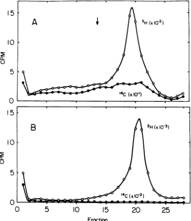
sedimenta-tion insucrosegradients (Fig. 1). The sedimen-tation of3H-thymidine-labeled DNA extracted
from purified virions ofH-1, which has an
s20,w
value of 27.8 (14), was analyzed in a parallel
gradient. Itsposition is indicatedbythe arrow.
A relatively homogeneous peak is present in
both eluants that is not associated with a
corresponding peak of prelabeled host cell
DNA. The'H/'4Cratiosreach amaximumof 52 for theNaCl elution and 620 for theSDS elution in the 17S region of the gradient. The
3H/'4C
ratios in other areas fall in the range 16 to 18. Thus the peak fractions have up to a 34-fold increase in the
3H/'4C
ratio. A sample of thepooled fractions 19 to 23 of the SDS elution
15
10
B
a_
0
B
a.
C-0 5 10 15 20 25
Fracton
FIG. 1. Sucrose gradient analysis of DNA ex-tracted by CPG chromatography. The 2 M NaCI eluant (A) and the 1%SDS eluant(B) weredigested withPronase,werephenolextracted, andwere precip-itated with ethanol. Theprecipitates weredissolved and sedimentedthrough5 to 20%sucrose in50mM Tris, pH 8.0, 10-3 MEDTA, 1 M NaCI, and 0.2%
Sarkosyl for16 h at23,000 rpminthe SW25.1rotor at 4 C.Fractionswerecollectedthroughthebottom and assayedforradioactivity byscintillation.Symbols:0, counts per minute of 3H-thymidine; 0, counts per minuteof14C-thymidine.
(Fig. 1B) was precipitated by trichloroacetic acid to obtaina more accurateestimate of the "4Ccontent, asthegradientsampleshadnearly
backgroundlevels of"C. Thissamplecontained
29,014 counts/min of 3H and 19counts/min of "4C with a 8H/14C ratio of 1,527. Because the 3H-labeled DNA is of a much higher specific
on November 10, 2019 by guest
http://jvi.asm.org/
[image:3.493.264.452.237.454.2]REPLICATION OF PARVOVIRUS H-1. II.
activity (at least 16- to 18-fold) the 17S peak
may be contaminated on aweight basiswith a
substantialamountof hostcell DNA. Only0.3%
ofthetotal'"C-prelabeledDNA remained in the
pooled 17S DNA fractions. Its estimated s20.
value is 16.5S, which indicates a molecular weight of 3.7 x 10⁶ if the DNA is double stranded (3, 30). Thus, this DNA, if it has a
duplex structure, has a molecular weight near
twice thatestimated forthe single-strandedH-1 DNA (1.7 x 10⁶). Extraction of H-1 infected
cellsby the methodofHirtyieldsasimilar 17S
DNAinsucrose gradient analysis. Thismethod
may be expected tofail to extract
membrane-bound viral DNA, as it is very similar in
principletothe detergentprecipitation method
ofTremblayetal.(34) and SDS modificationsof
itdescribed by Ormerod and Lehman(15). These
methods preferentially isolate
"membrane-bound DNA" and nascent DNA by virtue of
their association with precipitated detergent.
Forthese and other reasons, notdiscussed here,
theCPG methodwas notabandonedinfavorof
theHirt method, which istechnically simpler.
It should be noted that incorporation of
3H-thymidine into a DNA species sedimenting
as progeny DNA is inconspicuous orabsent in
these preparations and this will be examined
later.
The17S DNA extracted from infected cells is
identified below as a viral RF by DNA-DNA
hybridization andforsimplicity will be referred
to hereafter as RF DNA. The RF DNA was
analyzed by chromatography on
hydroxylapa-tite to determine its strandedness. A batch
method described by Fanshier et al. (9) was
used. 3H-thymidine-labeled hamster embryo
DNAs, both native and heat-denatured, were
used as controls. The results were: 98% ofthe
native hamster embryo double-stranded DNA
eluted inthe last fraction with0.4 MPO, and
afterheat denaturation, 86% oftheDNAeluted
in thepreceding eluantwith 0.2 MP04; the RF
DNA fraction eluted as double-stranded DNA
(89% at0.4 MP04).
Nature of 17S virion DNA. Since
hydrox-ylapatitechromatographywould beexpectedto
retain single-stranded molecules with portions
containing duplex structure during elution of
single-stranded DNA, competitive
hybridiza-tion studies with unlabeled H-1 DNA were
conductedby analyzing the product with a
nu-cleaseselective forsingle-strandedDNA.Before
conductingtheseexperimentsaprior
considera-tion wasthepossibilityof thepresence of virions
containingDNAstrands complementaryto the
majorviralstrand,whichwouldcomplicatethe
competitive hybridization. Adenoassociated virus consists of nearly equal portions of both strands (20).
The DNA of rat virus (RV) was reported to yield a minor 17.5S peak in velocity sedimenta-tion by May and May (13). To demonstrate the possible existence of virionscontaining comple-mentary strands, virion DNA labeled with 3H-thymidine was analyzed in high salt sucrose gradients after lysis of virions at 100 C for 90 s followed by quick chilling (Fig. 2A) or by an in-cubation periodconducive to DNA-DNA hybrid-ization, 65 C for 60min(Fig. 2B). Theviral DNA which was extracted under minimal renaturing conditionsshowedmainly27SDNA. The virion
DNAsubjectedtoannealing conditions shows a
significant increase in material sedimenting at 17S, from 27 to 57%. A smaller increase (28 to 37%) occurred after incubation in 50% forma-mide for 4 h at 40 C. To distinguish between duplexformation and strand breakage of single-stranded 27S DNA samples ofthe 27 and 17S virion, DNA peaks wereannealed for 60 min at 65 C in 0.8 M NaCl, and were analyzed for
8 27S 17S'
6 a
4
2
0 27S 17S
1: B
6 4
2 0
0 5 10 15 20 25
[image:4.493.250.440.333.582.2]Fraction
FIG. 2. Sucrose gradient analysis of H-1 virion
DNA. Purified H-I virus propagated on hamster
embryo cells and labeled with 3H-thymidine and
lysedbyheatingto100 Cfor1minin 50 mMTris, pH
8.0,0.5%Sarkosyl, 0.1 MNaCI,10- 3MEDTA.After
quenching in ice (A)orheatingto65Cfor60min(B)
theyweresedimentedinsucrose asinFig. 1.
VOL.13,1974 403
on November 10, 2019 by guest
http://jvi.asm.org/
single-stranded DNA by digestion with
mi-crococcal nuclease both with and without
re-heating at 100C for 90 sandby chillinginice. Both 17S virion DNA fractions showed similar rates ofdigestion by the nuclease and werethe same asthe rate for theannealed27S DNA. The
reheated 27S DNA was digested slightly more
rapidly. Both 27 and 17S DNA had a portion
equalto8% of the total thatwasnotsolubilized further after6 to26h ofincubation.Thus,these
data indicate that the 17S DNA fraction is
largely single stranded and doesnotresultfrom
virion DNA being annealed to its
complemen-tarystrand. It is mostlikely thatthe 17S DNA
generated from virion DNA is the result of
strand breakage.
CharacterizationofH-I RF DNA.(i)
Sepa-ration of complementary strands. If virion
populations do contain a minor portion with
DNA complementary to the major species, a
direct demonstration ofthis would be possible
by separation of the viral strand from its
com-plementary strand in equilibrium density
cen-trifugation. When BUdR issubstitutedfor
thy-midine by propagation ofvirus in the presence
of BUdR, as described by Berns and Rose for
adenoassociated virus (2), the viral strand
be-comes denser than its complement because of its higher thymidine content. H-1DNA was
reported by McGeoch et al. to contain 29.3%
thymidine and 25.5% adenine (14), andwehave
obtained an average value of 32% thymidine,
25%adenineforH-1DNAsedimentingat27S in
sucrose gradients (J.R. Kongsvik,J. F.Gierthy,
and S. L. Rhode, unpublished results).
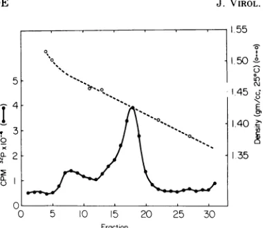
Virus labeledwith 32P and containing BUdR
was prepared as described by Berns and Rose
(2) and was purified as previously described
(18). The final step of the purification was
equilibirium density centrifugation afterwhich
theCsCl gradient wasfractionated and the 32P
activities were determined (Fig. 3). Two forms
ofH-1 werefound,one at adensityof 1.425and
one at 1.475. These two density forms of
com-plete H-1 virions are usually found for
thymi-dine-containing virus at 1.410 and 1.465. Two
similar forms of DNAcontainingRV and minute virus of mice (MVM) have been previously
re-ported (5, 13, 19). Virus containing 32P-BUdR
DNA from the fraction of 1.425 and 1.475 and
3H-BUdRRF DNA(seelegendFig. 4A, B) were
heatedat 100C for3minin 1mMTris, pH8.0, 0.005MNaCl, 1mMEDTA,and0.1%Sarkosyl,
andwerechilledinice.Thesampleswerediluted
with the samebufferat 0 Cto avolume of 1.29 mland mixed with1.89ml of saturatedCs2SO4,
also at 0 C. The samples were subjected to
equilibrium densitycentrifugation at 4C inthe
I
0~
a-0
1.55 150 °
U Z3
145 ;
E
1.40 l 35
0 5 10 15 20 25 30
[image:5.493.264.454.60.225.2]Fraction
FIG. 3. Equilibrium density gradient centrifuga-tion of H-1 virions containing 32P-BUdR-labeled DNA. Virus was purified as previously described (18). TheCsCl gradientwasfractionated into scintillation vials,radioactivitvwasassayed by measuring Ceren-kov activity, and densities were determined by refrac-tive index. The fractions of virus at 1.475 and 1.425
werepooled anddialyzed againstwaterand stored at
-20C.
SW50rotor,and fractionswerecollectedand
as-sayed for radioactivity. The results are
illus-trated in Fig. 4A and B. The 32P-BUdR viral DNA from virus of both densities (1.425 and 1.475; notshown) bandedat a densityof 1.530, 37% of the 3H-BUdR RF banded at the same
density as the virion DNA, and another two
fractions, 31% and 21% of thetotal, werefound
at adensityof 1.505and 1.474,respectively. No
evidence of this light DNA was found in the
32P-BUdRvirion DNA. To obtain greater
reso-lutionofthe fractionsobtained with theBUdR
RFDNA, it was subjectedto equilibrium
cen-trifugation in Cs2SO4 in the type40fixedangle
rotor. A portion was heat denatured, chilled,
and diluted with buffer and Cs2SO4 to a final
density of 1.50 and an unheated sample was
similarly prepared. The resultsof this analysis
are illustrated in Fig. 4C and D. The native
BUdR RF DNA described above (Fig. 4) yielded
two fractions at 1.472 and 1.444. Denaturation of RF DNAproducedtwoadditional peaksand
a reduced amount ofthe twooriginal moieties. Theheaviest bandedat adensity of1.530 asdid virion DNA, and the lightest banded at 1.505.
Competitive hybridization studies reported in
the next section confirm the presence of virion DNAinthe RFDNA, sothat the DNA of 1.530 is the viral strand.Ifthe material released from the RF DNA at 1.505 iscomplementary to the viral strand, then reannealing would result in
reformation ofduplexDNA with the density of the native BUdR RF DNA. Samples of the
on November 10, 2019 by guest
http://jvi.asm.org/
1.530 1.505 1.472 p(gm/cc,25°C)
A
B
15 20
Fraction
25 30
16
12 8
4
x
I
0L
U
1.530 1.505 1.472
0 5 10 15
Fraction
16
12
8
cm o
x
4
O
4
2
0
10
15
20
25
3035
Fraction
FIG. 4. Equilibriumdensity gradient analysis of 32P-BUdR-substitutedH-i virionDNA and RFDNA. Virus
was lysedand centrifuged to equilibrium in Cs2SO4 asdescribed in thetext (A). Sampleswereassayed for
radioactivity onfilterpapers and densities were determined. 3H-BUdR RF DNA was isolated by the Hirt
methodafterincubating infectedcells with mediumcontainingFUdR(0.5,g/ml)andBUdR(10gg/ml)14to16 hPI and then withmediumFUdR and3H-BUdR(10 MCi/ml)16to18 hPI. RFDNA labeledwith 3H-BUdRwas
similarly analyzed afterheat denaturationasdescribedin thetext(B). Centrifugationwas35,000rpmfor60 h in the SW50rotor at4C. 3H-BUdR RFDNAwasheatdenatured(D)asinBoruntreated(C)andcentrifugedto equilibirium in a type 40 fixed-angle rotor. The gradients were fractionated as previously described.
Centrifugationwas35,000rpmfor48 hat4C. Pooledfractions9 to 11(D)and thepeak tube, fraction13,were
usedfor further analysis.Asample of fractions9 to 11wasmixedwithasample of fraction13 ina5:4proportion (E)andfractions9to11alone(F)wereannealed 24 hat65 C in1MNaCIasinthetext.Thesampleswerethen diluted and rebanded inCs,SO4asabove.Centrifugationwas35,000rpmfor48 hat4Cin thetype40 rotor.
405
16 o 12
x
to 8
4
0 10
8
04
2 0
P(gm/cc,250C)
C
8
4
1.444
20 25
-1
on November 10, 2019 by guest
http://jvi.asm.org/
[image:6.493.53.440.45.561.2]fraction at 1.530 and 1.505 were mixed in the ratio 5:4 by 3H content to approximate equal amountsofeachstrand diluted1:5with10mM Tris, pH 8.0, 10-3 M EDTA, 1 M NaCl, and
0.2% Sarkosyl, to a final volumeof 0.5ml. The
samples were incubatedat 65 C for24h. They
were thenchilled, brought to afinal density of
1.50,and rebanded (Fig. 4E).TheDNAbanded at1.470, somewhat less than the density of the
heavy form of the native RF DNA (1.472). A
portion of the 1.530 DNA only was similarly
treated and it rebanded at 1.527, a little less
than its original density (Fig. 4F). Using this
DNA as a marker for the previous
centrifuga-tion conditions, the density of the annealed
mixture corrected (to 1.472) that of the heavy
RF DNA. These resultsareconsistent with the
1.530 and 1.505 strands of the RF DNA being
the viral and complementary strands,
respec-tively. RF DNA labeled with 3H-thymidinewas
subjected to equilibrium centrifugation in the
presence of a sample of3H-BUdR RF DNA of
density1.472 todetermine thedensity of
unsub-stituted RF DNAinCs2SO. Itbandedat1.423;
thus, it canbeestimated that the thymidineof
the DNA at 1.472 was substituted 80% with
BUdR and the DNA of intermediate orhybrid
densityat 1.444substituted 37.5% (1). Thefinal
yieldof isotope inthevirionstrand released by
heat denaturation ofRF DNA was 50% ofthe
total and 35% of the total rebanded at the
densities of native RF, probably due to
rean-nealing during the centrifugation (Fig. 4D).
Thus, both density forms of RF must have
contained the viral strand substituted with
3H-BUdR.
(ii) RF DNA contains viral DNA. To
estab-lish the presence ofviral DNA in the putative
RFDNA, competitivehybridizationinthe
pres-ence of varying amounts of unlabeled virion
DNA was carried out. When unlabeled viral
strands are in excess, 3H-labeled viral strands
present in the RF DNA prepared by CPG
extractionwill be prevented fromannealing to
their complementary strands. The results are
presentedinFig. 5. With increasing amounts of
unlabeledvirionDNA, anincreasingfraction of
3H-labeled RF DNA is made susceptible to nuclease digestion and is thus single-stranded DNA. The addition of virion DNA after the
periodofannealing didnotrender the annealed
RF DNAsusceptibletonucleasedigestion. It is thus confirmed that the 17S DNA extracted from infected cells is a double-stranded DNA
containing the viral strand. The 17S DNA,
extractedaccordingtothemethod of Hirt (11), was shown to contain viral DNA in the same manner. ThisDNAincorporateslabeled
precur-sors into both strands and is therefore an RF
DNA. The competition approaches saturation
atabout40%of theradioactivity. Theprevious
results with strand separation by equilibrium
density centrifugation would indicate that
greater than 50% of the radioactivity would be
intheviralstrand, but thelabeling period used
to prepare the RF DNA used therebegan 20h
PI, later than the introduction ofBUdRinthe
previously cited experiment, sothat this
differ-ence mightaccount forthediscrepancy.
Density and composition of H-1 RF DNA.
H-1 RF was obtained by preparative sucrose
gradient centrifugation and bandedto
equilib-rium inneutral CsCl and inethidium bromide
CsClgradients (17). The results areillustrated
in Fig. 6.
In neutral CsCl a buoyant density of 1.705
wasobtained. Using the standardequation, this
wouldindicatea GCcontentof45.9%(24).The
expected GC content of H-1 RF based on a
molar content of G and C of 22.6 and 22.6%,
respectively, as reported by McGeoch et al.
would be 45.2% (5). The buoyant density in
ethidium bromide CsCl was 1.545, and only a
small portion (fraction 13) might bein aclosed
circular configuration. The shoulder on the
denser scale of the peak in Fig. 6B was not
present on a similar ethidium bromide CsCl
gradient of this DNA in thetype 40fixedangle
rotor.
Rapidly labeled viral DNAspecies. Newly
synthesized viral DNA wasexamined by
label-50 40 30 20 10 0
0 0.0046 0.046
9g H-IDNA
0.460.92 FIG. 5. Competitive hybridization of 17S DNA (RF DNA) and H-1viral DNA. 17S DNAwaslabeledwith
3H-thymidineandprepared as in Fig. 2B. The labeled
DNA washeat denaturedat 100C for 90sin 0.01 M
Na2HPO4, pH 7.0, quenched in ice, and samples
weremixed withvaryingamountsofunlabeled H-1 vi-ral DNA. The samples were brought to 0.8 MNaCI
andincubatedfor16hat65C. Thesampleswerethen
digestedwith micrococcal nuclease.Acontrol sample had0.92 jg ofH-i DNA addedafter annealing and before digestion.Thepoints represent the percentage
oftotal counts perminute remaining acid-insoluble after4hofdigestion with nuclease.
75
z
on November 10, 2019 by guest
http://jvi.asm.org/
[image:7.493.263.452.419.533.2]REPLICATION OF PARVOVIRUS H-1. II.
'4
0
*0
8 6
4 2
10
O
-0o
5 10 15 20 25 30
Froction
1.76 1.74 1.72
-0
1. 70 _
u
1.68 \
18
E
_
1.80 c
0
0
1.70
[image:8.493.50.239.56.302.2]1.60 1.50 1.40
FIG. 6. Equilibrium density centrifugation ofH-i
RF.RF DNA purified by sucrose gradient and labeled with3H-thymidine was sedimented to equilibrium in neutral CsCI (A) and in an ethidium bromide CsCI gradient (B) as described (17). Centrifugations were (A) 60 h at 33,000 rpm in the S W50 rotor and (B) 24 h
at43,000 rpm in theSW50rotor.
ing infected cultures for short periods with
'H-thymidine and analyzing the DNA as
be-fore. The result of such an experiment is
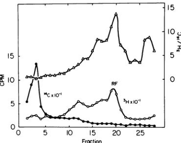
illus-trated in Fig. 7. Parasynchronous cultures of
HE cells prelabeled with "C-thymidine were
labeled for5 min at 25 C with 3H-thymidine(20
MuCi/ml). DNA was extracted by CPG
chroma-tographywithout a 2M NaClelution, Pronase
digested and collected by precipitation with
ethanol, and then analyzed on a high salt
sucrose gradient. Examination of the 'H/'4C
ratios and comparison with results obtained
with a labeling time of 75 min (c.f. Fig. 1)
re-vealsarelative increase in viral DNA
sediment-ing more rapidly than 17S RF DNA. The
'H/
'4C ratiofor the DNA retainedby thecolumn was 0.57, which was about that for the
fast-sedimenting DNA at the bottom of the
gradi-ent. The rapidly labeled replicative
intermedi-atesofviralDNAareunderstudy.
Does RFDNAexistinvivo. Thepossibility
must be entertained that the RF DNA
de-scribed here results from annealing of
comple-mentary strands during the extraction
proce-dure. If this occurs, the presence of exogenous
viral strandDNA added atthe time ofthe cell
lysis would resultintheincorporation ofa
por-tion ofthesesequences into RFDNA, andsome
of the endogenous viral strand DNA would
re-mainsingle stranded. This was tested by lysing
H-1 infected cells, labeled 14 to 16 h PI with
FUdR, 0.5
gg/ml,
and "4C-thymidine (1.5 x 10' M, specific activity 40 mCi/mmol) withthe SDS buffer containing 47,500 counts/min
oflysed 3H-thymidine-labeled H-1 virus (100C
for5min). The Hirt extractionwasthen carried
outandthe DNAwassedimentedin ahigh salt
sucrose gradient. Foracontrol,asecondculture
was similarly extracted exceptthat the 'H-H-1
DNA was added to the Hirt supernatant just
prior to centrifugation. Sixty percent of the
'H-H-1 DNA remained in the Hirt pellet, and
there was full recovery of the supernatant 'H
from the sucrose gradients. Thesedimentation pattern of the experimental and control were
similar with 72and76% ofthe'H sedimenting
at27S, and28and24%trailingthrough the 17S
region, respectively. Thespecific activity of the
'H-H-1 DNA was 25 times that of the 14C RF
DNA, and based on the counts recovered, the
"4C-RF DNA was present in12-foldexcess over
the 3H-H-1 DNA. Therefore, if the 17S DNA
was a result of annealing, the 'H-DNA would
have been quantitatively converted to 17S.
Only a 4% shift to 17S was found. The 14C
recovered as RF DNA represented 25% ofthe
total incorporation, so that figure represents a
minimum efficiency of the extraction. As
fur-ther confirmation that the 'H-DNA was not
incorporated into hybrids, the 27 and 17S
10 5
15 IOU
"I 1on 5 0
0 5 10 15 20 25
Froction
FIG. 7. Sucrose gradientanalysis ofrapidly labeled DNA.Parasynchronous culturesprelabeledwith
14C-thymidine were infected with H-i and pulsed with 3H-thymidinefor5minatroomtemperatureat 16h PI.A CPGextract wasanalyzedon a 5to20%/,ohigh
saltsucrosegradientasinFig.1.Symbols:0,counts
perminuteof3H-thymidine; *, countsper minuteof
14C-thymidine; A, 3H/14C ratio. Sedimentation was for16hat23,000 rpm in theSW25.1rotor.
VOL.13,1974 407
O L
on November 10, 2019 by guest
http://jvi.asm.org/
[image:8.493.254.439.422.568.2]pooledfractions ofboth gradientsweredigested
by single-strand-specific nuclease. After 4 h at
0C the 27and 17S 3H weresolubilized81 and
77%, respectively, in the experimental group
and 85 and 79%, respectively, in the control
gradient. Thus, the slight increase in resistance
tonucleasedigestioninthe17S 3H-DNAcannot
be attributed to annealing during the
extrac-tion. The "4C-RF DNA was resistant to
diges-tion.
DISCUSSION
Itisvirtuallyaxiomatic inmoleculargenetics
that genetic information is transferred and
replicated bytemplate-directed polynucleotide
synthesis. Therefore, the replication process of
theparvovirusH-1, whichcontains a genome of
single-stranded DNA, would be expected to
include astepinvolvingthesynthesisof a DNA
strand complementarytothe viral strand. The
resulting duplex DNA has been termed the
parental replicative form in the case of the
single-stranded DNA bacteriophages (16). In
this study I have described the synthesis of a
double-strandedDNA inH-1infectedcells with
a molecular weight ofabout 3.7 x 10⁶, about
twice that of the virion of a single-stranded
DNA. This DNA was shown to contain viral
DNA in onestrandasdemonstratedby
compet-itive hybridization. Thecompetitive
hybridiza-tionapproachedasaturationvalueof 40 to45%
of the3H activity renderedsusceptibleto
single-strand-specific digestion by micrococcal
nu-clease. Approximately 8 to 10% ofviral strand
DNA is resistant to digestion underthe
condi-tions used, so the saturation value can be
adjusted to 45 to 50%. The results with strand
separationdiscussed below indicateda
percent-ageof3H-BUdRin the viralstrand of 72% in a
different preparation of RF DNA, so that the
saturation value of 50% maybe a lowestimate
of the labeled viral strand content ofthe RF
DNA. The RFDNA was notfoundtobeaclosed
circular form by ethidium bromide density
centrifugation.Ithas adensityof1.705,
indicat-ing a GC content of45.9%aswould be expected for an RF DNA of H-1 (14). The incorporation
of 8H-BUdR into the viral complementary
strand in the RF DNAindicates that thisDNA
is undergoing replication at the time of the
pulse. Replication of RF DNA was occurring
at 16 to 18 h PI in parasynchronous infection, and RF DNA was labeled during intervals
rangingfrom 6 to 8 h PI to 22 to 24 h PI
(unpub-lished data). This is in agreement with the
previous suggestion that the DNA synthetic
eventupon whichsubsequentviral protein syn-thesis wasdependent (i.e., HA-DNAsynthesis)
and which initiatesonly in late S phase of the infected cell, about 8 h PI in this cell system,
maybe the synthesis ofparental RF. However,
the RF DNAexaminedinthisstudyislargelyor
entirely progeny RF. Analysis of the DNA in
equilibrium gradients showedtwodensity forms
containing viral DNA. Their differences in
den-sitycan besimply explained ifthe heavyform
represents aduplexofheavy-heavy (H-H) DNA and the intermediate form heavy-light (H-L)
DNA with the 3H-BUdR in the viral strand.
Reannealing of both heavy strands produced
the predicted duplex DNA with the density of
H-H (Fig. 4E). This interpretation has been
furtherconfirmedby density gradient
centrifu-gation ofdenatured H-H and H-L RF DNA to
be described in a subsequent report of this
series. This result would be theexpected conse-quence of the conservation ofthymidine con-tainingviralcomplementary strandDNA inthe
RFpool, which had beensynthesized before the
addition of BUdR at 14 h PI. The H-H DNA
would arise in the second generation of RF
undergoing semiconservative replication or
from progenystrandsynthesisonL-H RF DNA inthepresenceof BUdR and theH-L DNAfrom
either replication of the RF or synthesis of
progenyviralstrand DNA. Conservationofviral
strand DNA in the RF pool would result in
equal amounts of L-H and H-L in the hybrid
density RF DNA and thus equal amounts of
heavy 3H-BUdR-labeled viral andviral
comple-mentary strands in the RF pool. This was not the case, for less 3H was recovered in the
complementary strandthan in the viralstrand,
28% versus 73%. It was previously shown that
infectiousvirus production beganasearlyas 10
h PI, so that progeny strand synthesis can be
presumedto occur at thetime ofdensity
label-ing used here and result in preferential loss of
light viral-strandDNA from the RFpool.
Veloc-ity sedimentation analysis of viral DNA after
shortpulses of3H-thymidineyieldedan enrich-ment of forms sedimenting more rapidly than the 17SRF DNA.
The factors controlling the synthesis of the
parental RF and its possible requirement for a
late S phase function are unknown. It islikely
thatsynthesisofparentalRF isrequired before
transcription of viral DNA can occur and
syn-thesis of the mRNA for viral hemagglutin was previously shown to followclosely the synthesis of HA-DNA(18). Thespecific Sphase depend-ence of viral DNA synthesis suggests that a
homology between viral DNA and host cell
DNA may exist and results in a coordination of the initiation of viral DNA synthesiswith
cer-tain replicons of the host cell. During the
408
on November 10, 2019 by guest
http://jvi.asm.org/
REPLICATION OF PARVOVIRUS H-1. II.
preparation of this manuscript a report
ap-peared describing the isolation of RF DNA for
MVMvirusand evidence for homology between MVM and L-cell DNA (6). Experiments are in progress todetermine if H-1 DNA has homology
tolate replicating hamster embryoDNA.
A recent report by Salzman and White
de-scribed the formation of a duplex DNA of RV
within 60 min PI in asynchronous rat nephroma
cells (22). Inourhands, H-1 preparations have
particle/infectivityratios in excessof 101sothat
any chase experiment with prelabeled virus
would besubjecttohighbackgroundsof
nonin-fectious virions. I have reported the formation
at high ionic strength of a 17Sspecies ofviral
DNA. Further analysis indicated that H-1
vi-rionspropagatedinhamsterembryocells donot contain DNAcomplementarytothemajor viral DNA species as described for adenoassociated virus (2).IncubationofviralDNAunder condi-tions favorable for DNA-DNA hybridization
increased the yield of 17S DNA, but caused
littleincrease intheamountofdouble-stranded
DNA as assayed by nuclease digestion. Itthus appears that virion DNA undergoes strand
breakageonincubationat65 Cinhighsaltor at
40C in 50% formamide (unpublished data).
TheDNA ofH-1 may contain some self-comple-mentary sequencescontainingatleast8%ofthe
totalthymidine. Self-complementarysequences
have been reported for AAV (12), MVM (P.
Tattersal, Fed. Proc.31:913, 1972) and the
sin-gle-stranded DNA bacteriophage, fl (25, 26).
The possibilities of various configurational
forms of virion single-stranded DNAs and the presence of virions containing both strands of DNA must be considered in any analysis of
Parvovirus RF DNA. No evidence ofannealing
of complementary strands to form the 17S
RF DNA during the Hirt extraction was
ob-tainedinthe reconstruction experiment.
The two mostlikely hypotheses that account
forthecoordinationof viral DNAsynthesiswith
late S phase are: (i) parental RF synthesis
occursonlyinlateSphase;or(ii) parental RFis
synthesized earlier and subsequent
transcrip-tion requiresa late S phase function. In either
case, the replication of the Parvovirus H-1
providesaninterestingprobe ofhostcell
regula-tion of DNAreplication.Furtheranalysisofthe
synthesis ofviral HF DNA and especially the
synthesis ofviral complementary strands may
resolve between these alternatives.
ACKNOWLEDGMENTS
This research was supported by Public Health Service grants CA13414 and CA07826 from the National Cancer Institute, and by General Research Support Grant 1 SO1 RR05725fromthe National Institutes ofHealth.
I wish to thank Helene W. ToolanandK. A.0.Ellemfor
their helpful suggestions and for critically reviewing the manuscript. Ialso thankVeronica Lefevre and Jessica Brat-ton forexpert technicalassistance.
LITERATURE CITED
1. Baldwin, R. L., and E. M. Shooter. 1963. The alkaline transition ofBU-containing DNA and its bearing on thereplication of DNA. J. Mol. Biol. 7:511-526. 2. Berns, K. I., and J. A. Rose. 1970. Evidence for a
single-stranded adenovirus-associatedvirusgenome: isolation and separation of complementary single strands. J. Virol. 5:693-699.
3. Burgi, E., and A. D. Hershey. 1963. Sedimentation rate asa measure ofmolecular weight of DNA. Biophys. J. 3:309-321.
4. Chamberlin, M. J. 1966. Isolation, preparation and char-acterization ofnatural and synthetic nucleic acids, p. 513-519.In G. L. Canton and D. R. Davies (ed.), Pro-cedures in nucleic acid research, Harper & Row, N.Y. 5. Crawford, L. V. 1966. A minutevirus of mice. Virology
29:605-612.
6. Dobson, P. R., and C. W. Helleiner. 1973. A replicative formofthe DNA ofminute virus of mice. Can. J. Mi-crobiol. 19:35-41.
7. Ellem, K. A. 0. 1967. A dual-label technique for com-paringthe rates ofsynthesis of nucleic acid fractions separated by methylated albumin-Kieselguhr chro-matography from cells in different states of activity. Biochim.Biophys. Acta 149:74-87.
8. Ellem, K. A. O., and S. L. Rhode. 1969. The use of guani-dine thiocyanate to fractionate DNA-like RNA ad-sorbed onmethylatedbovine albumin-Kieselguhr col-umns.Biochim.Biophys.Acta174:117-123.
9. Fanshier, L., A. Garapon, J. McDonnell, A. Faras, W. Levinson, and J. M. Bishop. 1971. Deoxyribonucleic acid polymeraseassociated with avian tumorviruses secondary structure of the deoxyribonucleic acid product. J. Virol. 7:77-86.
10. Habener, J. F., B. S.Bynum,and J. Shack.1969.Unique secondary structure ofnewly replicated HeLa DNA. Biochim.Biophys. Acta186:412-414.
11. Hirt, B. 1967. Selective extraction ofpolyomaDNA from infectedmousecell cultures. J. Mol. Biol. 26:365-369. 12. Koczot, F. J., B. J. Carter, C. F. Garon, and J. Rose. 1973. Self-complementarity of terminal sequences withinplusor minusstrands ofadenovirus-associated virusDNA. Proc. Nat. Acad. Sci.U.S.A. 70:215-219. 13. May,P., and E.May.1970.The DNAofKilhamratvirus.
J.Gen. Virol.6:437-439.
14. McGeoch,D.J., L.V. Crawford, and E. A. Follett. 1970. The DNA's of three parvoviruses. J. Gen. Virol. 6: 33-40.
15. Ormerod, M.G., andA.R.Lehman. 1971. The release of high molecular weight DNA from a mammalian cell (L51784). Attachment of the DNA to the nuclear membrane. Biochim.Biophys. Acta 228:331-343. 16. Pratt, D. 1969. Genetics of single-stranded DNA
bac-teriophages. Ann. Rev.Genet. 3:343-362.
17. Radloff, R., W. Bauer, and J. Vinograd. 1967. A dye-buoyant-density method for the detection and isolation of closed circular duplex DNA: the closed circular DNA in HeLacells. Proc. Nat.Acad. Sci. U.S.A. 57: 1514-1521.
18. Rhode, S.L. 1973. Replication processoftheparvovirus H-1. I. Kinetics in a parasynchonous cell system. J. Virol.11:856-861.
19. Robinson, D. M., and F. M. Hetrick. 1969. Single-stranded DNA from the Kilham rat virus. J. Gen. Virol. 4:269-281.
20. Rose,J.A.,K. I.Berns,M. D.Hoggan,andF. J.Koczot. 1969.Evidencefor asingle-strandedadenovirus associ-atedvirusgenome:formation of aDNAdensity hybrid
VOL.13,1974 409
on November 10, 2019 by guest
http://jvi.asm.org/
410
onreleaseof viral DNA.Proc.Nat. Acad.Sci.U.S.A. 64:863-869.
21. Salzman, L. A., W. L. White, andT. Kakefuda. 1971. Linear, single-strandeddeoxyribonucleic acid isolated f'rom Kilhamrat virus.J. Virol. 7:830-835.
22. Salzman,L.A., andW.White.1973.In vivo conversion of' thesingle-strandedDNA of theKilhamratvirus to a double-strandedform.J.Virol. 11:299-305.
23. Salzman, L. A., W. L. White, and L. McKerlie. 1972. Growth characteristics of Kilham rat virus and its eff'ect oncellular macromolecularsynthesis. J. Virol. 10:573-577.
24. Schildkraut, C. L., J. Marmur, and P. Doty. 1962. Determinationofthebasecompositionof deoxyribonu-cleicacid from its buoyant density in CsCl. J. Mol. Biol.4:430.
25. Shishido, K., andY.Ikeda. 1971.Isolationof double-heli-cal regions rich inguanine-cytosine basepairingfrom bacteriophage fl DNA. Biochem.Biophys. Res. Com-mun.42:482-489.
26. Shishido, K., andY.Ikeda.1971.Isolationof double-heli-cal regionsrich inadenine-thyminebasepairingfrom bacteriophage flDNA. J. Mol. Biol. 55:287-291. 27. Siegl, G., and M. Gautschi. 1973. Themultiplicationof'
paravirus LUIII in a synchronized culture system. I. Optimum conditions for virus multiplication. Arch.
Gesamte Virusforsch. 40:105-118.
28. Sigel, G., and M. Gautschi. 1973.Themultiplication of parvovirus LUIII in a synchronized culture system. II. Biochemicalcharacteristics of virusreplication. Arch. Gesamte Virusforsch.40:119-127.
29. Sinsheimer, R. L. 1959. A single-stranded deoxyribonu-cleic acid from bacteriophage kX174. J. Mol. Biol. 1:43-53.
30. Studier, F. W. 1965. Sedimentation studies ofthe size andshapeofDNA.J. Mol.Biol. 11:373-390. 31. Tennant,R. W.,andR.E.Hand, Jr. 1970. Requirement
ofcellular synthesis forKilhamrat virus replication. Virology 42:1054-1063.
32. Tennant,R. W., R. E. Hand Jr., and K. B.Layman. 1969. Ef'fect of cell physiological state on infection by rat virus.J.Virol. 4:872-878.
33. Toolan,Helene W. 1968.The picodnaviruses: H, RV, and AAV,p. 135-180. InG.W.Ruhtez andM.A. Epstein (ed.), International reviewofexperimental pathology, vol.6. Academic PressInc., NewYork.
34. Tremblay, G. Y., M. J. Daniels, and M. Schaechter.1969. Isolation of acell membrane-DNA-nascent RNA com-plexfrombacteria.J. Mol.Biol. 40:65-76.
35. Usatequi-Gomez, M., F. Al-Lami, M. S. Hopkins, N. Ledinko, and H. W. Toolan. 1969. Single-stranded DNA fromtheparvovirusH-1.Virology 39:617-621.
on November 10, 2019 by guest
http://jvi.asm.org/