City, University of London Institutional Repository
Citation
:
Zuberbuhler, B (2010). Web-based system for outcome analysis and modification in laser vision correction. (Unpublished Doctoral thesis, City, University of London)This is the accepted version of the paper.
This version of the publication may differ from the final published
version.
Permanent repository link:
http://openaccess.city.ac.uk/17564/Link to published version
:
Copyright and reuse:
City Research Online aims to make research
outputs of City, University of London available to a wider audience.
Copyright and Moral Rights remain with the author(s) and/or copyright
holders. URLs from City Research Online may be freely distributed and
linked to.
City Research Online: http://openaccess.city.ac.uk/ [email protected]
Web-based system for outcome analysis and
modification in laser vision correction.
Bruno Zuberbuhler
A thesis submitted in fulfilment of the
requirements for a PhD in Health Informatics
Centre for Health Informatics
City University London
CONTENTS
Contents 2
List of tables 8
List of illustrations 9
Acknowledgement 11
Declaration 12
Abstract 13
Abbreviations 14
1. Introduction
1.1.Background 16
1.2.Aim of the research 17
1.3.Objectives of the research 17
1.4.Administrative organisation and settings 17
1.5.Opportunities 18
1.6.Organisation of the thesis 19
2. Domain of refractive laser eye surgery
2.1.Refractive Disorders 21
2.1.1.Myopia 21
2.1.2.Hyperopia 22
2.1.3.Astigmatism 23
2.2.Principles and techniques of laser vision correction 25
2.2.1.LASIK (Laser assisted in-situ keratomileusis) 26
2.2.2.LASEK (Laser Assisted Sub-Epithelium Keratomileusis) 31
2.2.3.Planning and programming laser treatments 32
2.3.Refractive outcome analysis 35
2.4.Summary 37
3. Review of literature, software and program languages
3.1.Literature review on refractive analysis 39
3.1.1.Waring Graphs (Reporting refractive outcomes) 41
3.1.2.Nomogram adjustments 43
3.1.3.Vector analysis with the Alpins method 43
3.2.Review of refractive analysis systems 44
3.2.1.Outcome Analysis 45
3.2.3.Refractive Surgery Consultant Elite 47
3.2.4.ASSORT 48
3.2.5.RSOIS 49
3.3.Review of program languages for system development 50
3.4.Summary 52
4. Prototype design and development
4.1.Needs assessment and task lists 55
4.2.User interface and system design 56
4.3.Implementation 57
4.4.Evaluation of the prototype and conclusions 59
4.5.Summary 61
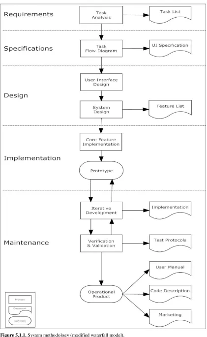
5. Design and development of the IBRA system
5.1.Needs assessment (task analysis) 66
5.1.1.Surgeon related needs 66
5.1.2.Educational needs 67
5.1.3.Promotional needs 68
5.1.4.Commercial and marketing needs 68
5.1.5.Maintenance and security needs 69
5.2.Specifications (task flow diagram) 70
5.3.User interface and system design 70
5.4.Overview of development 73
5.5.Modules of implementation in the operational product 75
5.5.1.Main menu 75
5.5.2.The „List of Cases‟ 76
5.5.3.Data recording 77
5.5.4.Single visual and refractive analysis 78
5.5.5.Vector analysis 79
5.5.6.Spherical equivalent outcome analysis 81
5.5.7.Defocus equivalent analysis 83
5.5.8.Visual acuity outcome analysis 83
5.5.9.Nomogram calculation 84
5.5.10. Satisfaction analysis 85
5.5.11. Multicentre functions 86
5.5.12. Download functions 86
6. System evaluation
6.1.Introduction 89
6.1.1.Clinical aims and evaluation methods 89
6.1.2.Non-clinical aims and evaluation methods 91
6.1.3.Organisation of the evaluation chapter 92
6.2.Refractive audits to evaluate general nomogram modifications 93
6.2.1.Introduction 93
6.2.2.Patients and methods 94
Methodology 94
Patient demographics 95
Laser treatment (LASIK) 97
Postoperative measurements 97
Data entry into IBRA 97
Determination of the treatment parameters 97
Optimising the treatment for the second Audit 98
Treatment parameters for the second Audit 99
Comparisons of the Audit outcomes 99
Main measure 99
Data analysis and results 100
Statistics 100
Presentations 100
6.2.3.Results 101
Refractive outcomes produced with IBRA 101
Refractive outcomes analysed with EXCEL 104
Statistical comparison between the outcomes 105
6.2.4.Discussion 107
Refractive groups 107
Outcome for patients with simple myopia 107
Outcome for patients with compound myopic astigmatism 109
Outcome for patients with simple hyperopia 109
Benefits and limitations of spherical nomogram adjustments 109
Benefits and limitations of IBRA 111
Future development of IBRA 111
6.2.5.Conclusions 112
6.3.Patient-individual modification of laser settings in the treatment 113
of astigmatic myopia with laser in-situ keratomileusis
Background and rational 113
Aims 114
Objectives 114
Study design 114
Research Funding 115
6.3.2.Methods 116
Summary of the original treatment protocol 116
Changes to the method and protocol 117
Settings and data collection 117
6.3.3.Recruitment 119
Recruitment of participants 119
Termination of recruitment and study 119
Follow-up assessments 119
Demographics 119
6.3.4.Results 121
Descriptive data 121
Primary outcome 121
Secondary outcomes 123
Complications 126
6.3.5.Discussion 126
Recruitment and selection 126
Clinical outcomes 127
Safety and enhancements 129
Strengths of the study 129
Weaknesses of the study 129
Limitations of the study and generalisability 129
6.3.6.Conclusions 130
Recommendations for health-care provision 130
Implications and recommendations for future research 131
Implications for IBRA and its development 131
6.3.7.Acknowledgements 131
6.3.8.Other information 131
6.4.Management of data entry processes 132
6.4.1.Introduction 132
6.4.2.Methods 134
Patient data / parameters 136
6.4.3.Results 138
Stepwise data collection 138
Learning effects 140
Data entry in one-go 141
Data entry intervals 142
6.4.4.Discussion 143
Primary outcomes 143
Learning curve effects 144
Limitations of the evaluation 144
Implications for future research and the IBRA development 145
Recommendations for new IBRA users 146
6.4.5.Conclusions 147
6.5.System functionality 148
6.5.1.Introduction 148
6.5.2.Methods 149
6.5.3.Results 151
6.5.4.Discussion 155
6.5.5.Conclusions 157
6.6.Survey on user satisfaction 158
6.6.1.Introduction 158
6.6.2.Methods 160
Determining the sample group 160
Writing the questionnaire 160
Administering the questionnaire 160
6.6.3.Results 161
Raw data 161
Participants reply 161
Outcomes 162
6.6.4.Discussion 164
General use of the system 164
Screen 164
Terminology 164
Learnability 165
Speed, reliability and security 165
Overall questions 165
6.6.5.Conclusion 166
Summary of strengths 166
Summary of weaknesses 166
Recommendations for future development 167
7. Discussion
7.1.Achievements related to the research objectives 169
Objective 1 - To develop a system ... 169
Objective 2 - To make the system ... 170
Objective 3 - To integrate means ... 171
Objective 4 - To test the system ... 172
Objective 5 - To analyse health changes ... 174
Objective 6 - To improve the understanding... 176
7.2.Publications and presentations 176
7.3.Limitations and recommendations 178
7.3.1.System related limitations and recommendations 178
7.3.2.User related limitations and recommendations 180
7.3.3.Patient related limitations and recommendations 182
8. Conclusions 186
References 188
Appendices
1. Clinical trial protocol 201
2. Participant information sheet 211
3. Participant consent form 214
4. Consent form Intra-LASIK 2008 216
5. Data collection sheet 220
6. Decision letter Ethics Committee 222
7. IBRA Treatment adjustment table 226
8. Declaration of the end of a study 228
9. Raw data from the clinical trial 232
10.User satisfaction questionnaire 235
11.Article on IBRA in EUROTIMES March 2009 240
City, University of London Northampton Square London EC1V 0HB United Kingdom
T +44 (0)20 7040 5060
THE FOLLOWING PARTS OF THIS THESIS HAVE BEEN REDACTED FOR
COPYRIGHT REASONS:
Figure 2.1.1. Anatomical image of an eye with myopia and with spectacle correction Pg. 22
Figure 2.1.2. Anatomical image of an eye with hyperopia Pg. 23
Figure 2.1.3. Anatomical image of an eye with corneal astigmatism ` Pg. 24
Figure 2.1.4. Topographic image of an eye with corneal astigmatism Pg. 24
Figure 2.2.1. Mechanical microkeratome (AMO) Pg. 27
Figure 2.2.2. Creating a corneal flap with a microkeratome Pg. 27
Figure 2.2.3. Creating a corneal flap with a Femto laser (Ziemer) Pg. 28
Figure 2.2.4. Removal of corneal tissue with the Excimer laser Pg. 29
Figure 2.2.5. Wavefront measurements of an eye Pg. 29
Figure 2.2.7. LASEK procedure Pg. 32
Figure 2.2.8. Planning stages of LASIK treatments Pg. 34
Figure 2.3.1. Stages of refractive outcome analysis Pg. 36
Figure 5.3.1. Structure of the hardware and software system Pg. 72
Figure 7.1.1. Ablation profiles for myopia and hyperopia Pg. 175
Appendix 11 Article on IBRA Iin Eurotimes March 2009 Pg. 240
LIST OF TABLES
Table 2.2.1. Indications for corneal procedures 25
Table 2.2.2. Complications, symptoms and treatments in LASIK 31
Table 3.1.1. Overview of selection process for the literature review 41 Table 3.2.1. Overview of commercially available refractive analysis software 45 Table 3.3.1. Overview of program languages reviewed for the system development 51
Table 5.1.1. Overview of IBRA versions 68
Table 5.4.1. IBRA development 74
Table 6.2.1. Exclusion criteria for the audits 96
Table 6.2.2. Patient demographics (Audit) 96
Table 6.2.3. Refractive disorder groups (Audit) 98
Table 6.2.4. Refractive results from Audit 2007 104
Table 6.2.5. Patient group adjustments 104
Table 6.2.6. Refractive results from Audit 2008 105
Table 6.3.1. Participants demographics 120
Table 6.3.2. Preoperative refractive and visual data 121
Table 6.3.3. Postoperative spherical equivalent and predictability 121
Table 6.3.4. Astigmatic changes 123
Table 6.3.5. Uncorrected and best-corrected visual acuity 124
Table 6.3.6. Enhancement rate 126
Table 6.4.1. MySQL database structure and data example (data entry management) 136
Table 6.4.2. Data collection sets (data entry management) 137
Table 6.4.3. Time requirements stepwise data entry management 138
Table 6.4.4. Time requirements „one-go‟ data entry management 142
Table 6.5.1. MySQL database structure (functionality) 150
Table 6.5.2. MySQL data export (functionality) 150
Table 6.5.3. Amount of performed analysis methods 152
Table 6.5.4. User individual use of analysis methods 154
Table 6.6.1. Overview of questionnaires for user satisfaction 159
Table 6.6.2. Raw data from questionnaire 161
Table 6.6.3. Questionnaire results for the different characteristics 162
Table 6.6.4. Overview of questionnaire results / scoring 163
Table 6.6.5. Summary of strengths and weaknesses 166
Table 7.2.1. Published articles on IBRA 176
Table 7.2.2. Articles submitted to Journals 176
Table 7.2.3. Articles in preparation 177
Table 7.2.4. Presentations of IBRA at international congresses 177
LIST OF ILLUSTRATIONS
Figure 2.1.1. Anatomical image of an eye with myopia and with spectacle correction 22
Figure 2.1.2. Anatomical image of an eye with hyperopia 23
Figure 2.1.3. Anatomical image of an eye with corneal astigmatism 24 Figure 2.1.4. Topographic image of an eye with corneal astigmatism 24
Figure 2.2.1. Mechanical microkeratome (AMO) 27
Figure 2.2.2. Creating a corneal flap with a microkeratome 27
Figure 2.2.3. Creating a corneal flap with a Femto laser (Ziemer) 28 Figure 2.2.4. Removal of corneal tissue with the Excimer laser 29
Figure 2.2.5. Wavefront measurements of an eye 29
Figure 2.2.6. Excimer laser (VISX) 30
Figure 2.2.7. LASEK procedure 32
Figure 2.2.8. Planning stages of LASIK treatments 34
Figure 2.3.1. Stages of refractive outcome analysis 36
Figure 3.1.1. Set of 6 standard Waring graphs 42
Figure 3.1.2. Double angle vector diagram 44
Figure 3.2.1. Screenshots „Outcome Analysis Software‟ for data collection 46 Figure 3.2.2. Screenshots „Outcome Analysis Software‟ for analysis 46
Figure 3.2.3. Screenshots „Datagraph-med‟ 47
Figure 3.2.4. Screenshots „Refractive Surgery Consultant Elite‟ 48
Figure 3.2.5. Screenshots „ASSORT‟ for data collection 49
Figure 3.2.6. Screenshot „ASSORT‟ with polar diagram 50
Figure 4.3.1. Screenshot „Proexcimer‟ patient list 57
Figure 4.3.2. Screenshot „Proexcimer‟ patient data collection 58 Figure 4.3.3. Screenshot „Proexcimer‟ integrated analysis methods 58 Figure 4.3.4. Uncorrected visual acuity chart produced with „Proexcimer‟ 59
Figure 5.1.1. System methodology of the IBRA development 64
Figure 5.2.1. Summary of functions of IBRA 70
Figure 5.3.1. Structure of the hardware and software system 72 Figure 5.4.1. Screenshot „Moorfields Intranet‟ with IBRA button 74
Figure 5.5.1. Screenshot IBRA Menu 75
Figure 5.5.2. Screenshot IBRA List of cases 76
Figure 5.5.3. Screenshot IBRA Excimer data 77
Figure 5.5.4. Screenshot IBRA Preoperative data 78
Figure 5.5.5. Screenshot IBRA Single analysis 79
Figure 5.5.6. Screenshot IBRA Astigmatism analysis 80
Figure 5.5.7. Scattergram and Centroid from vector analysis 81
Figure 5.5.8. Predictability chart and SE histogram chart 82
Figure 5.5.9. Screenshot IBRA Analysis module 82
Figure 5.5.10. Defocus equivalent chart 83
Figure 5.5.11. Uncorrected and best-corrected visual acuity chart (safety) 84
Figure 5.5.12. Screenshot IBRA Nomogram module 85
Figure 5.5.13. Patient satisfaction chart 85
Figure 5.5.14. Screenshot IBRA Download module 86
Figure 6.2.1. Methodology of refractive Audits 2007 and 2008 95
Figure 6.2.2. Determination of the standard treatment 98
Figure 6.2.3. General modifications (diagram) 99
Figure 6.2.4. Screenshot IBRA with selection criteria 101
Figure 6.2.5. Refractive results from Audit 2007 (IBRA) 102
Figure 6.2.6. Refractive results from Audit 2008 (IBRA) 103
Figure 6.2.7. Stability and visual results from refractive audit 2007 and 2008 (IBRA) 104 Figure 6.2.8. Boxplot with SE comparison 2007/2008 for myopic eyes 105 Figure 6.2.9. Boxplot with SE comparison 2007/2008 for myopic astigmatic eyes 106 Figure 6.2.10. Boxplot with SE comparison 2007/2008 for hyperopic eyes 106
Figure 6.2.11. Nomogram adjustment and Bansal-Kay nomogram 108
Figure 6.2.12. SE distribution for Audit 2007 and 2008 110
Figure 6.3.1. Research design RCT (Methodology) 117
Figure 6.3.2. Modification algorithm of standard treatment 118
Figure 6.3.3. Patient-individual modifications (diagram) 118
Figure 6.3.4. Modification formula 118
Figure 6.3.5. CONSORT diagram 120
Figure 6.3.6. Boxplot postoperative spherical equivalent 122
Figure 6.3.7. SE predictability, distribution and scattergram (IBRA) 123
Figure 6.3.8. Astigmatic predictability and changes (IBRA) 124
Figure 6.3.9. Boxplot postoperative uncorrected visual acuity 125
Figure 6.3.10. Visual acuity outcomes (IBRA) 125
Figure 6.4.1. Data logging process (data entry management) 135
Figure 6.4.2. Time requirements for data entry of demographic data (step-wise) 139 Figure 6.4.3. Time requirements for data entry of preoperative data (step-wise) 139 Figure 6.4.4. Time requirements for data entry of postoperative data (step-wise) 139
Figure 6.4.5. Learning curve in stepwise data entry 140
Figure 6.4.6. Boxplot learning curve effect 141
Figure 6.4.7. Time requirements for data entry of minimal data set (one-go) 142 Figure 6.4.8. Time requirements for data entry of extensive data set (one-go) 143
Figure 6.5.1. General usage of IBRA (analysis methods) 151
Figure 6.5.2. Usage of analysis methods 152
Figure 6.5.3. Follow-up periods (analysis methods) 153
Figure 6.5.4. International comparison of used analysis methods 154
Figure 6.6.1. Participants demographics (part 1) 161
Figure 6.6.2. Participants demographics (part 2) 162
Figure 6.6.3. Questionnaire scores for the different characteristics 162
Figure 7.1.1. Ablation profiles for myopia and hyperopia 175
Figure 7.3.1. General modifications (diagram) 182
Figure 7.3.2. Patient-individual modifications (diagram) 183
ACKNOWLEDGEMENT
I would like to thank my supervisor, Professor Abdul Roudsari, for his guidance over
the last years. He kept me on track, helped keep the research focused, and provided
advice along every step of the way.
I am very grateful to Professor Gartry for his support and his generosity to undertake a
randomised controlled trial in his private practice.
I would like to thank Suzanne Cabral, Wen Lee and Catey Bunce from the Research
Department at Moorfields Eye Hospital for their help with the clinical trial and
statistical advice.
I am thankful to Olivier Meylan for the introduction into PHP programming and for the
server support and maintenance.
My colleagues at City University are owed a great deal, especially Peter Weller, for
their friendship over the last years.
Finally, sincere thanks are due to my partner Kasia and my parents for those
DECLARATION
I grant powers of discretion to the University Librarian to allow this thesis to be copied
in whole or in part without further reference to me. This permission covers only single
ABSTRACT
Refractive laser eye surgery is a specialised field in ophthalmology which aims to correct the refractive disorder of an eye. The most established technique is LASIK, which has shown good results for the treatment of simple myopia. Complex refractive disorders, such as compound myopic astigmatism, have shown less predictable refractive outcomes, and in some cases the severe over- or under-correction can even worsen the preoperative situation and damage the eye.
In its first stage, this research aimed to develop a software system able to present and analyse refractive outcomes. Over 2 prototype stages, this research has led to an operational system named IBRA (Internet Based Refractive Analysis), offering web-based data collection and refractive and vector analysis.
In a second stage, Nomogram calculation formulas were developed and integrated into IBRA. These formulas were created from linear regression and best-fit analyses of spherical and cylindrical outcome data stored in IBRA. The purpose of the nomogram calculations was to provide surgeons with adjustment factors that could be used to improve the refractive outcome of patients with complex refractive disorders.
Two extensive clinical audits and a randomized controlled trial were performed at Moorfields Eye Hospital to evaluate the IBRA nomogram adjustments. This research showed that IBRA was able to achieve a positive health change. In addition, results from the audits and trial contributed to the knowledge of nomogram adjustments and provided a framework in which future investigations on nomogram and treatment modifications could be performed.
In addition to the above clinical studies, two evaluations were performed with the use of IBRA and data logging techniques to investigate users‟ behaviour relating to the management of data entry processes and the use of analysis functions. This research revealed the best method for entering refractive data, and was able to identify the most important analysis methods.
ABBREVIATIONS
AE Angle of Error
Ax Axis
BCVA Best-corrected Visual Acuity
BZ Bruno Zuberbuhler
CHI Centre for Health Informatics, CU
CI Correction Index
CU City University London
Cyl Cylinder (Astigmatism)
D Diopters
DBMS Database Management System
DE Defocus Equivalent
DSG David S. Gartry
DV Difference Vector
EHR Electronic Health Record
Femto Femtosecond (fs)
HID Hospital Identification Number
HTML Hypertext Markup Language
IBRA Internet Based Refractive Analysis
IOP Intraocular Pressure
IOS Index of Success
ISO International Organization for Standardization LASIK Laser-Assisted In-situ Keratomileusis
LASEK Laser-Assisted Sub-Epithelial Keratomileusis LogMAR Scale expressing angle of resolution
MEH Moorfields Eye Hospital
NID National Health Service Identification Number
OD Right eye (oculus dexter)
OS Left eye (oculus sinister)
PHP Hypertext Preprocessor
PID Patient Identification Number
QUIS Questionnaire for User Interface
RCO The Royal College of Ophthalmologist
RMS Root Mean Square
RSB Residual Stromal Bed
SD Standard Deviation
SE Spherical Equivalent
SIA Surgically Induced Astigmatism Vector
Snellen Eye chart (Dutch ophthalmologist H. Snellen)
Sph Sphere Value
SUMI Software Usability Measurement Inventory
TIA Target Induced Astigmatism Vector
Chapter 1
1. INTRODUCTION
1.1. Background
Refractive vision disorders such as myopia (short-sightedness) and astigmatism
(abnormality in the shape of the cornea) can be corrected with refractive laser eye
surgery.
A refractive laser unit consists of a laser source, a system of optical instruments and
controller software. The algorithm that describes the relation between the laser power
and the amount of surface ablation (removal of corneal tissue) necessary to achieve the
refractive change bases mainly on empirical data. Although mechanically perfected over
the last 10 years, the treatment algorithm of the controller software has not kept up and
suffers from optimisation. Imprecision in the treatment can result in an outcome that is
over- or undercorrected, and may even worsen the preoperative situation and damage
the patient‟s eye. As a consequence, intensive care and re-treatments are necessary.
The collection and analysis of refractive outcome data has become an increasingly
important requirement of refractive surgery practice. Not surprisingly, the forthcoming
Quality Standard and Revalidation initiatives of The Royal College of Ophthalmologists
underpin this importance in the provision of auditable outcomes of surgery.
One of the most critical factors that can affect the outcome after laser vision correction
is the nomogram that the surgeon uses. A nomogram is a unique group of detailed
treatment settings that is programmed into the laser. The best results are achieved when
the treatment is tailored for each person based on age, gender, prescription, and other
factors.
Although there are software systems on the market that can analyse refractive data and
perform nomogram optimisation, such systems use an unspecific general approach. A
system that performs both general and patient-individual nomogram modifications,
using 2-3 different calculation technologies, has not yet been developed. In addition, the
use of modified nomograms has never been evaluated scientifically with controlled
1.2. Aim of the research
The aim of the research is to record, analyse and improve the health outcome in patients
undergoing laser vision correction.
1.3. Objectives of the research Basic objectives:
To perform an extensive literature and software review.
To establish requirements in the area of refractive eye laser surgery. To analyse the management of data entry processes.
To evaluate user preferences and user satisfaction. To present and publish the results of this research.
Main clinical objectives of the research:
1. To develop a system that can combine data collection and analysis and offer
tools for standardised outcome presentation.
2. To make the system easily accessible, secure, safe, flexible and capable of future
developments.
3. To develop and integrate means (algorithms, formula) that can provide
information on how refractive laser treatments could be improved.
4. To test the system in a clinical (private practice) environment with real patient
treatments.
5. To evaluate the system impact on patient‟s health.
6. To improve the understanding of nomogram adjustments.
1.4. Administrative organisation and settings
A prototype of the system and a functional successor were developed and clinically
tested at the Eye Clinic, Cantonal Hospital, Lucerne, Switzerland, and further revised in
accordance to the needs.
Additional system development, methodological support and system evaluations were
performed at the Centre for Health Informatics, City University London, United
New treatment algorithms with formulas for individual nomogram calculations were
created and implemented in collaboration with the Refractive Laser Unit at Moorfields
Eye Hospital NHS Foundation Trust, London.
The clinical evaluation included a randomised controlled trial, which was performed in
accordance with National Ethical Commission guidelines and the Research &
Development Department at Moorfields Eye Hospital. A user-centred system evaluation
was performed at the Centre for Health Informatics, City University London.
The analysis of the results was performed at the Centre for Health Informatics, London.
Statistical support was provided by medical statisticians at Moorfields Eye Hospital.
Summary of participating organizations:
Centre for Health Informatics, School of Informatics, City University London. Refractive Surgery Unit, Moorfields Eye Hospital, London.
Department of Research and Development, Moorfields Eye Hospital Department of Corneal and External Eye Disease, Moorfields Eye Hospital Statistical Advice Unit, Moorfields Eye Hospital, London
Health and Safety Executive's Research Ethics Committee
1.5. Opportunities
This was the first research that had integrated different refractive outcome analysis
techniques in one system, and which undertook extensive user-centred and
patient-related system evaluations.
The main promises of this research were:
To create a calculation formula which can improve the patient‟s health outcome. To improve the understanding of laser treatments for refractive disorders. To have a major impact on the surgeon‟s management of eye laser surgery. To contribute to the knowledge of refractive data collection, data analysis and
nomogram adjustment.
To increase the cost-effectiveness of current treatments in reducing the
1.6. Organisation of the Thesis
This thesis is organised in 8 chapters. The first chapter states the background, aim and
objectives of the investigation.
Chapter 2 describes the domain of laser vision correction and provides important
information on refractive disorders, principles of laser treatments and refractive
outcome analyses.
Chapter 3 reports the results from the literature review on refractive analysis and
nomogram adjustments, presents the different refractive analysis software currently
available, and outlines the reviewed program languages evaluated for the system
development.
In Chapter 4 the thesis continues by discussing the initial needs for the analysis system
and presents the development of a prototype system called „Proexcimer‟.
Chapter 5 describes the range of new needs that led to the development of the research
system called „IBRA‟ (Internet Based Refractive Analysis). All parts of IBRA are
presented with screenshots and are discussed.
Chapter 6 reports on 5 evolutions performed to test the IBRA system from both a
clinical and a user point of view. The results of clinical audits, randomised clinical trial,
survey and data logging processes are presented in scientific format.
In Chapter 7 the results of system development and evaluation are discussed, and
compared with the aims and objectives of the research.
Chapter 8 provides a conclusion of the work carried out in this study and the results
Chapter 2
2. DOMAIN OF REFRACTIVE LASER EYE SURGERY
Refractive laser eye surgery is a specialised field of eye surgery which focuses on
improving the optical state of the eye using an excimer laser beam to reshape the
surface of the cornea. This change in the cornea compensates the ocular disease.
This chapter will provide basic information on topics from the field of refractive laser
eye surgery which were used in this research, including: Refractive disorders (Section 2.1.)
Principles and techniques of laser vision correction (Section 2.2.) Range of refractive data and refractive analysis (Section 2.3.)
2.1. Refractive disorders
Typical indications for laser treatment are refractive vision disorders such as myopia
(short-sightedness), hyperopia (long-sightedness) and astigmatism (an abnormality in
the shape of the surface of the cornea).
2.1.1. Myopia
If one thinks of the eye as camera, then the retina would be the film and the cornea the
lens (objective). The camera is able to produce a sharp image when the lens is able to
focus the light rays on the film plane. The eye works in a similar way. If the light rays
are focused on the retina the image is sharp.
Myopia is a vision disorder in which the light rays are focused on a single point that lies
in front of the retina (within the globe). The image is blurred. This can occur in 2
situations. Firstly, the axial length of the eye is too big; the eye “is too long”. This
situation is called axial myopia (Figure 2.1.1.). Secondly, the cornea is focusing the
Figure 2.1.1. Left image: In myopia, the light rays are focused on a single point that lies in front of the retina (within the globe). Images from distant objects are blurred. Right image: Correction of myopia with
a (biconcave) minus lens.
Mild to moderate myopia can be corrected with spectacles. The spectacles have to move
the point of focus further back so it can reach the retina. The types of lenses used for
this purpose have a biconcave curvature and are called minus (diverging) lenses.
The degree of myopia is measured in diopters [D] by the strength (or optical power) of a
corrective minus lens. Low myopia usually describes myopia of -3.00 D or less. Myopia
is common and is regarded as physiological if less than -6.00 D.
The incidence of myopia varies with age, country, sex, race, ethnicity, occupation,
environment, and other factors (Verma 2005 and Fredrick 2002). In Western Europe a
review found that 26.6% aged 40 or over have at least -1.00 D of myopia and 4.6% have
at least -5.00 D (Kempen 2004).
Of those with high myopia (-6.00 D or more) there is a subset who are at risk of
degenerative changes with increased prevalence of retinal detachment, choroidal
neovascularisation and open angle glaucoma (Oxford Handbook of Ophthalmology).
Myopia has also been found in association with genetic disorders like Down's
syndrome, Marfan‟s syndrome or albinism.
2.1.2. Hyperopia
The focal point of incoming light in the hyperopic eye, which is too “short”, lies behind
the retina (Figure 2.1.2.), which means that distant objects are seen fairly clearly, whilst
Figure 2.1.2. In hyperopia the light rays are focused behind the retina (outside the globe).
2.1.3. Astigmatism
Astigmatism is a refractive disorder that results from a common abnormality in the
shape of the cornea. The human cornea is usually dome-shaped, like part of a football,
but with astigmatism, the cornea has an ellipsoidal shape, more like part of a rugby ball.
This will cause blurred and distorted vision.
Similar to an ellipse being described by 2 axes (a major and minor axis) the ellipsoid
cornea is described by 2 radii. The meridian with the smaller radius is also called the
steeper axis, which lies perpendicular to the meridian with the bigger radius, which is
called the flatter axis. Each radius has a different refraction and all the light that passes
through an astigmatic cornea will therefore produce two focal planes, instead of one.
Usually, one of the focal planes is in front of the retina, with the other one behind
(Figure 2.1.3.). This situation is called mixed astigmatism. In simple astigmatism one of
the two focal points is focused on the retina.
Astigmatism can be combined with myopia. This situation is called myopic
astigmatism. Simple myopic astigmatism is a situation with one focal point in front of
the retina and one focal point on the retinal plane (Alio 1995).
Astigmatism is mainly hereditary and the prevalence increases with age (Robert 2003
and Asano 2005). Astigmatism is not a rare condition and remains lifelong.
astigmatism, but only 1% of eyes have more than 4 diopters. Higher amounts, especially
irregular forms of astigmatism, may cause blurred vision, squinting or headaches, and
occasionally can be very difficult to correct with spectacles or contact lenses.
The diagnosis of astigmatism is made by subjective refraction (the process to determine
the best corrective lenses) and corneal topography (a procedure that scans the shape of
the cornea, Figure 2.1.4.).
Figure 2.1.3. The “rugby ball” shaped cornea of an eye with astigmatism produces 2 focal planes. Left image: Both planes are in front of the retina in simple myopic astigmatism. Right image: The planes are
in front and behind the retina in mixed astigmatism (red and blue lines).
Figure 2.1.4. Topographic image of an eye with corneal astigmatism (bow tie figure). Left image: before laser surgery; Right image: same eye 3 months after LASIK showing flattening of the central cornea
Astigmatic corrections are more challenging than purely spherical corrections.
Astigmatism can be corrected by spectacles with a cylindrical lens, a lens that has
different radii of curvature. Such lenses are more complex to prescribe and more
expensive to produce. Patients with higher amounts of astigmatism may require contact
lenses to achieve good visual acuity.
2.2. Principles and techniques of laser vision correction
Glasses and contact lenses have drawbacks: they are a hindrance in certain professions
and activities (e.g. chef, actor, sports). Reducing the dependence on spectacles or
contacts promises an improvement in quality of life. For many people, the prospect of
going through life without glasses or contact lenses is reason enough to consider
intervention of this kind. Intolerance to contact lenses can be a further incentive for
wanting refractive surgery.
Over the past 20 years, refractive surgery has undergone a turbulent development.
Despite intensive scientific monitoring there is no long-term experience reaching back
more than 10-15 years of more recent procedures.
A wide range of corrective methods is available in refractive surgery. A basic
distinction is made between corneal procedures and lens procedures. In corneal
procedures (Table 2.2.1.) the refractive strength of the cornea can be modified using the
excimer laser. Most common procedures are Femto-LASIK (Laser Assisted In-Situ
Keratomileusis) and LASEK (Laser Assisted Sub-Epithelium Keratomileusis). Lens
procedures correct the vision disorder through an additional lens which is implanted
into the eye, or through a lens replacement.
Myopia Hyperopia Astigmatism
LASIK up to -8.0 D up to +3.0 D up to 4.0 D
LASEK up to -6.0 D up to +1.0 D up to 3.0 D
2.2.1. LASIK (Laser assisted in-situ keratomileusis)
Laser assisted in-situ keratomileusis (LASIK) is one of the most frequently performed
elective procedures to correct myopia. Around 100,000 LASIK procedures are
performed per year in the United Kingdom, and over 12 million procedures have been
performed worldwide since the introduction in 1993 (Maurino 2008). It is a highly
effective (private) outpatient procedure.
The majority of people with focusing errors of the eye are able to have LASIK.
However, some people are not able to have laser eye correction. Possible reasons for
this include ocular surface diseases, thin corneas, early cataract or focusing errors
outside the range that can be corrected by laser.
Generally, suitable patients for LASIK have:
An age of 21 or more (the eye is still growing until this age). Myopia up to -8 dioptres and hyperopia up to +3 dioptres. Regular astigmatism (up to 4 dioptres).
A stable spectacle or contact lens prescription for at least 12 months. Good vision in both eyes with glasses or contact lenses.
A typical LASIK procedure consists of multiple steps. The whole intervention takes
place using anaesthetic eye drops and lasts about 10-15 minutes per eye.
Flap creation
First of all, a thin flap of corneal tissue is prepared. For this procedure two different
techniques can be used: a mechanical microkeratome or a Femto laser. A
microkeratome is a precision surgical instrument with an oscillating blade designed for
creating a flap. The hand piece (Figure 2.2.1.) mainly consists of an engine connected to
a controller unit, which analyses the resistance of the oscillation of the blade with the
aim to prevent blockage. The head of the microkeratome contains the single use blade.
It can be mounted on a disposable holder unit for flap creation. During the cut, the eye
is temporarily fixed using a suction ring, which is felt as a slight pressure in the eye. A
negative pressure of up to 80 mmHg is used for this fixation, rarely leading to
conjunctival or choroidal haemorrhages. The blade of the microkeratome smoothly
[image:29.595.111.532.76.294.2]
Figure 2.2.1. Microkeratome hand piece (AMO) and head with holder and support unit, fixed to the cornea by a vacuum.
Figure 2.2.2. Creating a corneal flap with a microkeratome.
In recent years, the “bladeless” technique has gained popularity, mainly for hygienic
reasons. This method uses a Femto laser (Figure 2.2.3.) to create a corneal flap. A
Femto laser is an expensive piece of equipment (approximately £250,000 per unit) and
operates with a high energy laser with a wavelength of 1050nm.
The beam of the Femto laser is focused to create a micro air bubble (explosion) in the
corneal stroma. The bubble naturally expands and separates the corneal fibres and
layers. If multiple air bubbles are placed next to each other, two corneal planes can be
surgeon can customize the corneal flap for every individual patient. The term „Femto-LASIK‟ is used for LASIK treatments with Femto laser flap creation.
[image:30.595.271.533.135.433.2]
Figure 2.2.3. Creation of a corneal flap with a Femto laser (Ziemer).
Corneal ablation with the Excimer laser
To reshape the cornea an excimer laser is used. Computer-controlled pulses of excimer
laser light are applied to the inner layers of the cornea (also called corneal stroma). This
removal of corneal stromal tissue (also called ablation) reshapes the cornea and changes
the refractive power of the cornea (Figure 2.2.4.). This procedure takes between 30 and
90 seconds. In this process the smallest, unintentional eye movements are also
Figure 2.2.4. The laser beam removes corneal stromal tissue to change the refraction.
Once the ablation has been completed the corneal flap is replaced and positioned, and
antibiotic and anti-inflammatory eye drops are applied. Further fixation, e.g. with
sutures, is not necessary. Slightly blurred vision and slight watering of the eye are both
normal following the intervention. After just a few hours, sufficient visual acuity is
achieved so that glasses or contact lenses are no longer required. The vision stabilises
after 4-8 weeks.
Most new generation Excimer laser units offer wavefront (customised) treatments.
Using wavefront measurements (Figure 2.2.5.) the unique imperfections of each
individual eye, just like a fingerprint, can be determined. The wavefront data then is
used to calculate an individual treatment profile, allowing higher ablation precision.
From a technical and engineering point of view, the Excimer laser unit (Figure 2.2.6.) is
a highly expensive treatment unit (£350,000) and consists of hardware and software.
The hardware is the part that produces a laser beam with 192nm wavelength. This beam
is directed via a system of highly precise optical instruments (mirrors and lenses) to a
binocular microscope where it finally is focused on the patient's eye.
Figure 2.2.6. Excimer laser unit for laser vision correction used for this research (VISX S4).
The software part controls the mirrors, the lens positions and the laser power that is
needed for the refractive treatment. A calculation algorithm (nomogram) describes the
relation between the laser power and the surface ablation. The nomograms mainly base
on empirical data.
Risks and complications of LASIK
No surgical procedure is ever risk-free. Fortunately, sight-threatening complications
from laser vision correction are rare. Serious complications occur in less than 1% and
many LASIK complications can be resolved with additional surgery or medical
treatment. A list of possible complications is shown in Table 2.2.2. Visual aberrations
summarise symptoms such as glare, double vision, ghosting, halos, starbursts, loss of
Complications Symptoms Treatments Incomplete corrections Blurry, less-than-perfect
vision
Glasses or contact lenses; eye drops; re-treatment
Decentred ablations Visual aberrations Eye drops; re-treatment
Oversize pupils
(pupils > treatment zone)
Visual aberrations Eye drops; re-treatment
Haze Visual aberrations Eye drops; re-treatment
Irregular flap
(folds, wrinkles, striae)
Visual aberrations Surgical correction; second laser procedure
Dry eye Dry, itchy or scratchy eyes, often with redness and sense of foreign object in eye, and sometimes pain
Prescription dry eye medication; artificial tears; punctal occlusion (blockage of tear ducts in order to retain tear film on eye)
Diffuse lamellar keratitis (eye inflammation)
Visual aberrations Eye drops; surgical rinsing of cells
Epithelial ingrowth Visual aberrations Surgical removal of epithelium
Infection Redness, oozing of eyes, sometimes pain
[image:33.595.111.541.72.291.2]Eye drops; oral medications
Table 2.2.2. Complications, symptoms and treatments in LASIK (Keith Croes).
2.2.2. LASEK (Laser assisted in-situ keratomileusis)
LASEK is an alternative laser refractive procedure. LASEK is known as a „surface
procedure‟ and may be safer if the cornea is relatively thin, or if any other medical
conditions mean that LASIK is not the best option.
Instead of creating a corneal „flap‟ on the surface of the cornea, the very superficial
layer of corneal epithelial cells is treated with alcohol and moved to the side, allowing
the underlying cornea to be re-shaped by the refractive laser with wavefront technology
(Figure 2.2.7.). Afterwards, the epithelium can be smoothed over the lasered corneal
surface. The surface cells then grow back across the cornea within a few days. Finally, a
bandage contact lens protects the surface layer that has not yet grown together securely
until it has completely healed, and is then removed.
Generally, the recovery period is longer than for LASIK, and in the first days following
treatment, patients may experience a foreign body sensation and may suffer from eye
pain and photophobia. The improved vision is not appreciated until the epithelium has
fully healed, usually after about a week. The long-term results for low to moderate
Figure 2.2.7. LASEK procedure.
2.2.3. Planning and programming laser treatments
Refractive eye laser surgery requires careful preoperative assessment and an extensive
planning of the treatment (Figure 2.2.8.).
A typical procedure of a Femto-LASIK treatment consists of the following steps:
Pre-assessment and consenting
Deciding on a treatment plan, ablation pattern and target refraction. Programming the Femto laser for flap creation
Programming the Excimer laser for corneal ablation Follow-up visits
Pre-assessment and consenting
The pre-assessment for laser surgery comprises a comprehensive examination of the
eyes and a discussion of visual needs with a Consultant Ophthalmic Surgeon. The
examination includes an exact determination of best-corrected and uncorrected visual
acuity with refraction, measurement of intraocular pressure and nocturnal pupil
diameter, slit lamp examination of the eye lids, cornea, lens, optic nerve and retina, and
At the end of the assessment, the surgeon discusses the findings with the patient and
determines the suitability for laser vision correction. Sometimes the surgeon has to
advise against undergoing laser surgery. Additional information is provided on the
benefits, risks and possible complications (Table 2.2.2.) of laser vision correction, on
the predictability of visual and refractive outcomes, and on the cost of the treatment.
Finally, once the patient agrees to go ahead, a consent form is signed by the patient
acknowledging being informed completely and with understanding.
Deciding on a treatment plan, ablation pattern and target refraction
At this stage the surgeon can suggest a certain method of correction (LASIK or
LASEK) and has the chance to discuss treatment targets with the patient. In most cases,
patients request emmetropia, enabling them to see distant objects clearly without the
need for glasses. The spherical equivalent of emmetropia is zero.
Depending on the vision disorder, different patterns of ablation (the way the corneal
tissue is removed by the laser) are applied. For example, myopia is treated with a
spherical ablation. This is a straightforward treatment process with a uniform, disc-like
ablation of the corneal stroma.
The ablation pattern for the treatment of astigmatism is much more demanding.
Basically, the steeper axis of the cylinder is flattened by torical ablation. Flattening of
the steeper axis alone, without reshaping the flatter axis, results in hyperopic shift (the
eye gets less myopic). This can be a desired side effect in the treatment of myopia, but
is usually not desirable because of its unpredictability (McDonnell 1991).
Over the years newer ablation patterns have been developed, notably the bitoric ablation
pattern (Chayet 1998, DeOrtueta 2008) and the cross-cylinder ablation pattern
(Vinciguerra 2000 and 1999). In the cross-cylinder ablation technique the amount of
astigmatism is divided in two: half of the correction is treated on the flatter meridian
and half is treated on the steeper meridian. This method does not lead to unpredictable
hyperopic shifts. The treatment pattern for combined myopia and astigmatism is a
Figure 2.2.8. Planning stages of LASIK treatments.
Programming the Femto laser for flap creation
Modern Femto lasers allow programming and modifying of nearly every parameter
defining the corneal flap. The flap diameter and thickness are the most important
parameters determined for each patient individually. The flap diameter depends on the
ablation pattern and is slightly bigger for the treatment of hyperopic eyes. The thickness
depends on the surgeon‟s preference and experience. Thinner flaps allow more corneal
tissue to be treated but are more difficult to lift. Further parameters that can be
programmed include the shape of the flap (round or oval), the marginal profile (convex,
concave, perpendicular) and the position of the flap (centred, off-set). It requires years
of experience to find the ideal programming of the flap parameters for each patient
(Faktorovich 2008).
Programming the Excimer laser for corneal ablation
In a successful LASIK treatment, the induced refractive change equals the preoperative
refractive error. Although the excimer laser‟s nomogram provides good support in
ablation, also called the optical zone (OZ). The optical zone has to be increased in eyes
with bigger nocturnal pupil diameter, in hyperopic eyes and in eyes that receive a
peripheral blend zone. Finally, the amount of ablation can be boosted or decreased,
based on the surgeon‟s preference.
Follow-up visits
Following surgery, patients are seen the next day to check the position of the corneal
flap. Additional reviews can be arranged at 1 week and 1 month following surgery. The
refractive change of the eye usually stabilises after 4-6 weeks. The final review is
undertaken at 3 months, including comprehensive examination with refraction and
topography. Most patients are discharged at this time.
2.3. Refractive outcome analysis
Regular postoperative refractive analysis is good medical practice and can identify
factors which, together with individual treatment and wound healing factors, could
influence the refractive outcome.
The Royal College of Ophthalmologists (2007) and NICE (IPG164, 2006) have
published guidelines regarding good medical practice in refractive laser eye surgery.
They advise undertaking a careful audit of results following laser in-situ keratomileusis
(LASIK), on a regular basis.
Standards have been proposed regarding how refractive outcomes should be calculated
and presented (Waring 2000), and commercially available software facilitates this
outcome analysis and nomogram changes (see chapter 3).
The basic principle of outcome analysis and nomogram adjustment is a process
consisting of 3 stages (Figure 2.3.1.), normally facilitated by the refractive outcome
analysis software:
Refractive data collection: recording of treatment data, preoperative and
postoperative refractive and visual data, and data on complications and patient
Analysis of the data: results from demographics calculations, Waring graphs,
vector analysis, and others; more details on methods of analysis are given in
Chapter 3 and 5.
Transformation of the results into nomogram tables and adjustment factors: use
of linear and non-linear regression analysis, nomogram graphs and tables.
Other methods include back-calculation to model strategies for pre-treatment
adjustment of the ablation sphere to eliminate unpromising new approaches before
[image:38.595.113.545.325.641.2]clinical trials (Arnalich-Montiel 2009).
2.4. Summary
This chapter provides information on refractive vision disorders, types of laser
treatments, benefits and risks of laser vision correction, and the importance of
pre-assessment and postoperative outcome analysis.
Typical indications for a refractive eye laser treatment are vision disorders such as
myopia (short-sightedness) and astigmatism (an abnormality in the shape of the surface
of the cornea). Laser assisted in-situ keratomileusis with Femto laser flap creation
(Femto-LASIK) is the most frequently performed elective procedure and has become
the standard in laser vision correction.
Postoperative refractive outcome analysis is a complex procedure consisting of 3 main
stages: data collection, outcome analysis and nomogram calculation.
The current state of quality of refractive laser surgery has been presented in review
Chapter 3
3. REVIEW OF LITERATURE, SOFTWARE AND PROGRAM LANGUAGES
A review of literature and software was performed extensively at the beginning of the
prototype development in 2002, at the beginning of the evaluations in 2008 and
following completion of the evaluations in 2010.
3.1. Literature review on refractive analysis
The focus of the thematic literature review was on the handling and analysis of
refractive data, on the presentation of refractive outcomes, and on refractive calculation
models (nomograms) for treatment optimisation. We were selective in this review focus,
and only wanted to include articles that reported on refractive data in laser eye surgery,
such as LASIK or LASEK.
Between 2002 and 2008 many articles on laser eye treatments and techniques for their
improvement were published. Surprisingly, only a few new articles reporting on this
topic were found in a Medline search since 2008. This could be linked to the fact that
this field has not seen significant technical changes over the last 2 years, and that the
refractive outcomes from LASIK and LASEK have reached a high precision (Sutton
2010 and O'Keefe 2010).
For the search in Medline (PubMed) and Google we have used a range of different key
words, and their combination. These search criteria are listed in Table 3.1.1., which also
shows the results from the first selection round.
In total, 429 documents were found on refractive data handling, analysis and
optimisation. Although this seems to be a high number of publications, in fact, this
number is rather low when comparing to other fields of Ophthalmology dealing with
refractive outcomes, for example LASIK for myopia (2130 results) or standard cataract
surgery (11206 results). This relative shortage of evidence-based literature is mainly a
result of circumstances, given that laser vision correction is performed exclusively in
private practice. This business field has some distinctive characteristics, some of which
are:
Laser surgery is part of a highly competitive business with a strong view on
experimental work (research). This bears the risk of damaging reputations. The
general tenor is to remain out of studies.
If the outcome of a study is not good, it may not be published. This may help to
reduce the risk of negative news and damage to the prestige that could ultimately
bring about a significant cut to revenue.
If the research results are very good, the journal reviewers may not believe them.
Research results could be manipulated to promote private business (marketing
issues).
Research takes time, during which a surgeon could be consulting or operating on
patients, again resulting in extra income. Time used for research does not
provide earnings; on the contrary, it increases spending.
Dealing with private practice participants is more complicated than the
management of NHS participants in research projects.
We rejected 393 of 429 articles because the link to laser eye surgery was weak or
absent. The topics of the excluded articles showed a wide range, including refractive
analysis following cataract surgery, corneal grafting, corneal incisional surgery, testing
of refractive equipment, reporting on epidemiological findings, intraocular lens power
calculation, and refractive treatment of keratoconus.
The remaining 36 documents were marked for full inspection, and graded on their
relevance to this research (Table 3.1.1.). The detailed analysis of these articles showed
that some of the articles were presenting obsolete technology (e.g. older versions of
laser units and ablation profiles), or technology that has become standard in the
meantime (we described these articles with the term „historic‟). Many of the reviewed
articles presented results that are valid only for specific equipment, for example laser
units Schwind, Technolas, Nidek or Alcon; or the use of a specific (overnight) contact
lens called Paragon. Some of the articles presented results that can only be achieved in
combination with additional procedures, e.g. LASIK in combination with a cataract
operation, and some of the articles with important findings did not describe the
methodology well enough for us to repeat the technique or use the calculation algorithm
for our research. However, some of the articles graded as partially relevant (+) were
Search criteria / other key words
Res Exc Inc Full inspection Authors (Year) Relevance Refractive data / Calculations / Algorithm / Presentation Refractive outcome / Analysis / Reporting / Presentation
207 195 12 Kaye (2002)
Naeser (2001) Holladay (2001) Kaye (2001) Calossi (1993) Arbelaez (2009) Feltham (2008) Anderson (2003) Nakano (2003) Waring (2000) Huang (1999) Hefetz (1997) +
- (very complete) - (very complex) - (very complex) - (historic) - (only Schwind) - (mainly Technolas) - (only Technolas) - (only Nidek) +++
+
- (historic)
Vector analysis 125 115 10 Suominen (2003)
Thibos (2001) Alpins (2001) Huang (2000) Corones (1999) Alpins (1997) Shah (1997) Naeser (1997) Alpins (1997) Neumann (1989) +
- (very complex) +++
- (too general) - (historic)
- (integrated in 2001) - (historic)
- (too theoretical) - (integrated in 2001) - (for AK only) Nomogram
/ Outcome / Treatment / Adjustment / Improvement
97 83 14 Arnalich-Montiel (2009)
Lapid-Gortzak (2008) De Ortueta (2008) González-Méijome (2007) Mrochen (2006) Zaldivar (2005) Feiz (2005) Caster (2004) Anderson (2004) Ortiz (2003) Moniz (2002) Reviglio (2000) Ditzen (1999) Probst (1998) +
- (no details provided) - (only Schwind) - (only Paragon) +++
+
- (IOL related) - (only Alcon) - (only Technolas) + (but too general) - (too general) - (mainly results) - (historic) - (historic)
Total 429 393 36
Table 3.1.1. Overview of the selection process for the literature review, with the search criteria, the results (Res) from Medline search, the excluded documents (Exc), the remaining documents (Inc) that
were used for inspection, and the relevance.
Finally, only 3 articles matched the search terms and topic sufficiently, providing
specific, significant and generalisable information for this research. We believe that
these publications provide essential information on refractive analyses and were
therefore used as reference. For many refractive surgeons, these articles have become
part of their „key literature‟ in reporting refractive outcomes, calculating nomograms
and analysing refractive astigmatic data with the method of vector analysis.
3.1.1. Waring Graphs (Reporting refractive outcomes)
The main article linked to the presentation of refractive results is written by George
J Refract Surg, 2000;16:459-466. Mr Waring writes about his experience in refractive
analysis, and presents his ideas on how refractive data should be analysed and presented
in publications. He proposes a set of six standard graphs (Figure 3.1.1.) which should
be included in any paper reporting the results of a series of cases. Generally, the graphs
can easily be produced by anyone with widely available software (Microsoft EXCEL).
This standardized system of reporting outcomes allows comparison between the results
of different publications. The graphs of different articles can be arranged side by side,
allowing a direct visual evaluation of the outcomes of surgical procedures. Although
originally proposed by Prof Neuhann (Comment: Prof. Neuhann did not publish the
concept in a journal, but he spoke about the idea of graphs and refractive outcome
reporting at Congresses and in private personal communications), the concept of 6
standard graphs, as presented by Waring, has become a “gold standard” and the concept
has been taken on by many surgeons in their routine praxis. Therefore, any software that
analyses refractive data and produces outcome graphs has to implement at least some of
3.1.2. Nomograms adjustments
The key article for nomogram adjustment is authored by Michael Mrochen et al, and has
the title “Nomograms for the improvement of refractive outcomes”, published in
Ophthalmologe 2006;103:331-8. The authors of this study analysed the clinical
relevance and limitations of nomograms in case series on a theoretical basis. Their
results suggest that the use of individual nomograms can significantly improve the
predictability of refractive outcomes. However, the investigations demonstrate that a
homogeneous data distribution within cohorts was a key factor for predictable
nomogram calculations. The authors concluded that nomograms are helpful for
improving refractive outcomes, but are limited to approximately 90% of outcomes
within +/-0.5 D of the target.
3.1.3. Vector analysis with the Alpins method
The most important literature on vector analysis is authored by Noel Alpins with the
title “Astigmatism analysis by the Alpins method”, published in J Cataract Refract Surg,
2001;27(1):31-49. The aim of Mr Alpins‟s method is to determine the effectiveness of
correcting astigmatism by laser refractive surgery by a vectorial astigmatism outcome
analysis. For the calculations the method uses 3 fundamental vectors: the target induced
astigmatism vector (TIA), the surgically induced astigmatism vector (SIA) and the
difference vector (DV). TIA is the astigmatic change (by magnitude and axis) the
surgery was intended to induce. This can be seen as a golf scenario where the player
intends to hit a ball from a starting point into the hole. SIA represents the amount and
axis of astigmatic change the surgery actually induces. In “golf language” this would
mean where the ball effectively landed after the hit. Finally, DV is the astigmatic
change that would enable the initial surgery to achieve its intended target. This is the
required hit of the ball needed to bring it from the (unintended) landing point to the
target point (hole). The vectors can be drawn on a double angle vector diagram (Figure
3.1.2.). By examining individual vector relationships to the TIA (e.g. the correction
index, index of success, and flattening index), a comprehensive astigmatism analysis is
completed. Each index provides information necessary for understanding any astigmatic
Figure 3.1.2. Double angle vector diagram (produced with the IBRA system) showing TIA (green line), SIA (red line) and DV (black line).
3.2. Review of refractive analysis systems
A review of refractive analysis software over the years showed parallels to the literature
review, with minimal changes since 2008. Five different software systems were
available on the market in 2008. An April 2010 review showed that 4 of the 5 products
are still available, and 3 of 4 products are upgraded on a regular basis (Outcome
Analysis, Datagraph-med and ASSORT Software). The Refractive Surgery Outcomes
Information System was removed from the market in 2009 and its successor
(EUREQUO) has just been introduced, showing a delay of almost 1 year. The most
up-to-date review also showed that no other company has invested in the development of
outcome software in the field of refractive laser eye surgery.
In the following subsections we will summarise key figures of all 5 software systems
that had an influence on refractive surgeons over the last 10 years (Table 3.2.1.); and on
the IBRA system development generally. Each system is described with information
about the company, the surgeon involved in the development of the system, the features
and costs. Images from print screens (where available) show the user interface.