Copyright © 2003, American Society for Microbiology. All Rights Reserved.
T Cells Infiltrate the Brain in Murine and Human Transmissible
Spongiform Encephalopathies†
Hanna Lewicki,
1Antoinette Tishon,
1Dirk Homann,
1Honore´ Mazarguil,
2Franc¸oise Laval,
2Valerie C. Asensio,
3Iain L. Campbell,
3Stephen DeArmond,
4Bryan Coon,
1Chao Teng,
1Jean Edouard Gairin,
2and Michael B. A. Oldstone
1*
Department of Neuropharmacology (CVN-9)3and 1Division of Virology, Department of Neuropharmacology
(IMM-6),1The Scripps Research Institute, La Jolla, California 92037; 2Institut de Pharmacologie et de
Biologie Structurale-UMR 5089, CNRS, 31077 Toulouse, Cedex 4, France2; and Department
of Pathology, University of California—San Francisco, San Francisco, California 941434
Received 12 August 2002/Accepted 26 November 2002
CD4 and CD8 T lymphocytes infiltrate the parenchyma of mouse brains several weeks after intracerebral, intraperitoneal, or oral inoculation with the Chandler strain of mouse scrapie, a pattern not seen with
inoculation of prion protein knockout (PrPⴚ/ⴚ) mice. Associated with this cellular infiltration are expression
of MHC class I and II molecules and elevation in levels of the T-cell chemokines, especially macrophage
inflammatory protein 1, IFN-␥-inducible protein 10, and RANTES. T cells were also found in the central
nervous system (CNS) in five of six patients with Creutzfeldt-Jakob disease. T cells harvested from brains and spleens of scrapie-infected mice were analyzed using a newly identified mouse PrP (mPrP) peptide bearing the
canonical binding motifs to major histocompatibility complex (MHC) class IH-2borH-2dmolecules,
appro-priate MHC class I tetramers made to include these peptides, and CD4 and CD8 T cells stimulated with 15-mer
overlapping peptides covering the whole mPrP. Minimal to modest Kbtetramer binding of mPrP amino acids
(aa) 2 to 9, aa 152 to 160, and aa 232 to 241 was observed, but such tetramer-binding lymphocytes as well as CD4 and CD8 lymphocytes incubated with the full repertoire of mPrP peptides failed to synthesize intracellular
gamma interferon (IFN-␥) or tumor necrosis factor alpha (TNF-␣) cytokines and were unable to lyse PrPⴚ/ⴚ
embryo fibroblasts or macrophages coated with51Cr-labeled mPrP peptide. These results suggest that the
expression of PrPscin the CNS is associated with release of chemokines and, as shown previously, cytokines
that attract and retain PrP-activated T cells and, quite likely, bystander activated T cells that have migrated from the periphery into the CNS. However, these CD4 and CD8 T cells are defective in such an effector
function(s) as IFN-␥and TNF-␣expression or release or lytic activity.
Transmissible spongiform encephalopathies (TSE) (e.g., prion disease and scrapie) are progressive, fatal neurodegen-erative diseases of humans and other animals (10, 34). The hallmark of TSE diseases is the conversion of normal prion protein (PrPc) to an abnormal, protease-resistant form (PrPsc).
Characteristic features are spongiosis, astrocytosis, and neuro-nal loss in the central nervous system (CNS). Of the many enigmas concerning this process, three stand out. First, how is TSE inheritable information encoded and transmitted in PrP in the absence of discernible nucleic acids; second, what are the pathophysiologic events by which PrPsc causes CNS
dis-ease; and third, why is there no detectable immune response accompanying scrapie infection (4, 5)? In this report, we use a model of scrapie in mice to focus on the pathophysiologic response in the CNS and on the immune response.
Within the brain, TSE disease generates an accumulation of protease-resistant proteins, PrPscor PrPres, derived by a
post-translational event from a normal host-encoded protease-sen-sitive isoform, designated PrPcor PrPsen(10, 34). PrPcis
at-tached by a glycolipid anchor to the cell surface. In the CNS, PrPcconverts to PrPscin both neurons and astrocytes (10, 13).
In genetic experiments with PrP knockout (PrP⫺/⫺) mice,
hamster PrPc was expressed only in neurons after using a
neuron-specific enolase promoter (35) or in astrocytes upon using an astrocyte-specific glial fibrillary astrocyte protein (GFAP) promoter (37). In both instances, challenge with ham-ster scrapie resulted in TSE, thereby incriminating both neu-rons and astrocytes in the replication of PrPscand in the
dis-ease process. Still unclear are how PrPcconverts to PrPscin
these cells and how PrPscaccumulation gives rise to the
pro-found neurodegeneration characteristic of scrapie.
Associated with ongoing TSE disease is the expression of tumor necrosis factor-alpha (TNF-␣), interleukin-1 alpha (IL-1␣), IL-1, GFAP, and murine acute-phase response gene mRNA in the brain but not in peripheral tissues like spleen, kidneys, or liver (9). Absent or not altered in the TSE brains are IL-4, IL-5, gamma interferon (IFN-␥), IL-2, IL-6, and IL-3 mRNA (9). In addition to these cytokines, chemokines are present within and outside the CNS, where they function as soluble mediators possessing a spectrum of actions and che-motactic activities (3, 39, 46). Localized production of chemo-kines is possible from astrocytes and neurons, the two CNS cell types involved in conversion of PrPcto PrPsc. Yet expression of
chemokines in the CNS during scrapie infection is unknown. In this work, we evaluated the expression of chemokines as * Corresponding author. Mailing address: Division of Virology,
De-partment of Neuropharmacology, The Scripps Research Institute, 10550 N. Torrey Pines Rd., La Jolla, CA 92037. Phone: (858) 784-8054. Fax: (858) 784-9981. E-mail: mbaobo@scripps.edu.
† This is publication number 13546-NP from the Department of Neuropharmacology, The Scripps Research Institute, La Jolla, Calif.
3799
on November 8, 2019 by guest
http://jvi.asm.org/
five of six patients with clinical, neuropathologic, and biochem-ically defined Creutzfeldt-Jakob disease (CJD). Tetramer anal-ysis of T cells from scrapie-infected mice suggests that such T cells may be specific to MHC-restricted prion peptides but incapable of lytic responses or PrP peptide-stimulated IFN-␥
and TNF-␣production.
MATERIALS AND METHODS
Mouse strains, infectious agent, and infection protocol.C57BL/6 (H-2b),
BALB/WEHI or cdj (H-2d), FVB/N (H-2q), and SWR/J (H-2q) mice were bred
under specific-pathogen-free conditions and obtained from the Rodent Breeding Colony of The Scripps Research Institute. As described previously (36, 37), the
two sets of PrP⫺/⫺mice used included one group carrying a null mutation in PrP
that abolished PrP mRNA production (such mice were crossed toH-2bmice) and
another group with a truncation in PrP gene (crossed toH-2qmice). The former
group was obtained from Jean Manson, Institute for Animal Health, Edinburgh, United Kingdom, and the latter group was developed by Charles Weissmann, Institute for Molecular Biology, Zurich, Switzerland, and obtained from Stanley Prusiner, University of California, San Francisco. Genetically deficient CD4 or CD8 mice came from The Jackson Laboratories, Bar Harbor, Maine. Mice genetically deficient in MHC class I and class II originated from Michael Grusby at Harvard School of Public Health, Boston, Mass. These mice were bred in The Scripps Research Institute vivarium core and genotyped and phenotyped as reported previously (26, 35, 37, 43). Mice were inoculated orally, intraperitone-ally (i.p.), or intracerebrintraperitone-ally (i.c.) with the specified doses of Chandler RML strain of mouse scrapie made as a 10% solution of brain homogenate in PBS and cleared of debris by low-speed centrifugation. The i.p. inoculation volume was
100l, and the i.c. inoculation volume was 30l. Oral infection was in a volume
of 200 l administered via a small-diameter flexible polypropylene catheter
inserted over the base of the tongue about 1 to 2 cm into the esophagus as described previously (33). Infectious scrapie was quantitated after i.c. injection of serial 10-fold dilutions of a 10% brain homogenate into C57BL/6 mice (four mice/dilution).
The Armstrong (ARM) strain of lymphocytic choriomeningitis virus (LCMV), clone 53b, was also used (33, 40). LCMV was plaque purified three times on Vero cells, and stocks were prepared by a single passage on BHK-21 cells.
Eight-to twelve-week-old mice were infected with a single i.p. dose of 105PFU. For
secondary challenge, mice were inoculated with 106PFU of LCMV i.p.
Brain material and clinical and neuropathologic synopsis of CJD patients and immunochemical studies.The brains from six patients with sporadic CJD (sCJD) were examined and immunohistochemically stained for T- and B-cell markers (CD3 and CD20, respectively) in the Neuropathology Prion Disease Laboratory at the University of California in San Francisco. The patients’ ages ranged from 55 to 73 years. All had the characteristic neurohistopathological feature of CJD,
vacuolar (spongiform) degeneration of the gray matter. In addition, PrPscwas
identified in each of the cases by the hydrolytic autoclaving method applied to formalin-fixed, paraffin-embedded brain sections (30) and by the more sensitive and specific histoblot technique applied to unfixed, cryostat sections blotted to nitrocellulose paper (42).
Cell lines.Mouse embryo fibroblasts (MEF) were made from both PrP⫺/⫺
lines. MurineH-2bmutant RMA-S cells and human T2 cells transfected with
H-2Dbor transfected withH-2Ldmolecules were grown as described previously
(23). The murineH-2bcell line (MC57) andH-2dline (BALB Cl-7) were utilized
as reported previously (40, 45). Cells were grown in either RPMI 1640 (RMA-S,
MC-57, BALB Cl-7, and MEF) or Iscove’s modified Dulbecco’s medium (T2-Db
andT2-Ld) containing 8% bovine serum,L-glutamine (2 mM), and antibodies
(penicillin [10 U/ml] and streptomycin [10g/ml]). Geneticin (400g/ml) was
tion) induced by peptide added exogenously. The mutant cell line, in which transport of endogenous peptides to the endoplasmic reticulum is deficient,
RMAS (Db, Kb), transfected with Kdmolecules and the mastocytoma cell line
P815 (H-2d) were used to measure Db, Kb, Kd, or Ldstabilization, as described
previously (11, 23). Briefly, cells (5 ⫻105/well) were incubated at 37°C in
microtiter plates in the presence of increasing peptide concentrations (10⫺10to
10⫺5M). The expression of peptide-stabilized MHC molecules was analyzed
after a 4-h incubation period. Cells were incubated on ice for 1 h with 0.1 ml of hybridoma culture supernatant of mouse monoclonal antibody 28-14-8S
(anti-Db, anti-Ld), Y3 (anti-Kb), or SF1-1.1.1 (anti-Kd). As negative controls, the cells
were cultured in medium alone. After being washed once with ice-cold 1% bovine serum albumin–PBS and incubated for 1 h with the fluorescent secondary antibody (fluorescein isothiocyanate [FITC]-conjugated goat anti-mouse immu-noglobulin G [IgG]), the cells were washed twice and fixed in 1% paraformal-dehyde in 1% bovine serum albumin–PBS. Analysis followed in a fluorescence-activated cell sorter (FACScan; Becton Dickinson, San Jose, Calif.). The 50%
stabilizing concentration (SC50) was an amount of peptide that produced half the
maximal up-regulation. The peptides were considered to be MHC binders when
displaying affinity values of 50M or lower. Positive control peptides for Db, Kb,
Kd, and Ldbinding were LCMV ARM NP amino acids FQPQNGQFI (21),
Moloney murine leukemia virus peptide SSWDFITV (41), P198 tumor antigenic peptide KIQAVTTTL, and P29 peptide YPNVNIHNF (11), respectively.
Cytotoxic T-cell assay.mPrP peptides at a concentration of 20g/ml were
incubated with PrP⫺/⫺MEF labeled with51chromium (15, 40, 45). Lymphocytes
harvested from the brain (15) and spleen (40, 45) were added at effector-to-target cell ratios of 100:1, 50:1, and 25:1 for a 5-h assay as described elsewhere
(15, 40, 45). As a positive control, PrP⫺/⫺MEF were infected with LCMV
(multiplicity of infection, 1.0) for 48 h and added to lymphocytes harvested from scrapie-infected mice or from LCMV-infected mice as described elsewhere (15, 40, 45).
MHC class I tetramers.Kb-restricted mPrP amino acids (aa) 2 to 9, aa 152 to
160, and aa 232 to 241; Db-restricted LCMV GP aa 33 to 41; Db-restricted
LCMV NP aa 396 to 404; and Kb-restricted LCMV GP aa 34 to 43 were used as
allophycocyanin or phycoerythrin conjugates. Either these were obtained from the Tetramer Core Facility, Emory University, Atlanta, Ga. (1), or in some instances, biotinylated MHC-peptide monomers were made as tetramers in our laboratory (21) immediately before use. Staining with MHC class I tetramers was performed at a 1:50 to 1:100 dilution in the presence of various surface antibod-ies for 30 min at 4°C, and propidium iodide was added at a final concentration
of 5g/ml to allow analytical exclusion of dead cells (21).
Flow cytometry and cytokine ELISPOT.Single-cell suspensions of lympho-cytes were restimulated for 5 h with MHC class I- or class II-restricted peptides
(1 or 2g/ml, respectively) in the presence of recombinant human IL-2 (10 to 50
U/ml; PharMingen, La Jolla, Calif.) and brefeldin A (1g/ml; Sigma, St. Louis,
Mo.). Staining for cell surface antigen and intracellular antigens was performed as described previously (21). Negative controls were peptide-stimulated cells obtained from uninfected mice, cells restimulated for 5 h in the absence of viral peptides, and cells stained with conjugated cytokine-specific antibodies preincu-bated for 30 min at 4°C with an excess of recombinant cytokine. Cells were acquired with FACSort or FACSCalibur flow cytometers (Becton Dickinson) using Cell Quest software (Becton Dickinson). For five- and six-color analyses, a FACSVantage SE flow cytometer (Becton Dickinson) was used. FITC-, phyco-erythrin-, CyChrome-, peridinin chlorophyll-a protein-, or allophycocyanin-con-jugated, biotinylated, and/or purified antibodies (PharMingen) were used to evaluate CD4 (RM4-5) and CD8a (53-6.7) cells and evaluated as described elsewhere (21, 40, 45).
Immunohistochemical staining of CNS tissues.Brains removed from test mice were covered with Tissue-Tec OCT (Miles Diagnostics Division, Elkhart, Ind.),
snap-frozen at⫺80°C in isopentane, and then stored at⫺20°C.
Immunohisto-chemistry was performed on 6- to 10-m-thick cryostat sections that were fixed
in 100% ethanol for 15 min at 4°C and blocked with avidin and biotin (Vector
on November 8, 2019 by guest
http://jvi.asm.org/
Laboratories, Burlingame, Calif.). Staining was done with the following primary antibodies: anti-CD8 (anti-Ly-2 and Ly-3; PharMingen), anti-CD4 (anti-L3T4; PharMingen), anti-H-2 monotypic antigen (MHC class I), anti-Ia antigen (MHC class II), anti-B220 (Boehringer Mannheim, Indianapolis, Ind.), and anti-F4/80 (Serotec, Oxford, England). The second antibody was either a biotinylated anti-mouse IgG used in conjunction with the Vectastain Elite ABC (peroxidase) kit (Vector Laboratories) or anti-Ig-FITC. In the former case, staining was detected using diaminobenzidine as a chromogen. Sections were counterstained in May-er’s hematoxylin (Sigma) and mounted in Aqua-Mount (Lerner Laboratories, Pittsburgh, Pa.).
For light microscopy study, brain tissue was fixed in Bouin’s fixative or 10% formaldehyde, prepared in paraffin, sectioned, and stained with hematoxylin and eosin.
RESULTS
Presence of CD8 and CD4 T cells in brains of
scrapie-inoculated PrPⴙ/ⴙbut not PrPⴚ/ⴚmice. Twelve weeks after
i.c. injection of 100,000 infectious doses of Chandler strain mouse scrapie into 6- to 8-week-old C57BL/6 and BALB mice, both CD8 and CD4 T cells infiltrated the brain parenchyma, including regions of the hippocampus, cortex, and cerebellum (Fig. 1A and B). These cells were easily discernible by immu-nochemical staining of brain sections with monoclonal anti-bodies to CD4 and CD8 T cells (15) (four to six mice per group; experiment repeated twice). Such infiltration rarely was perivascular, and it was not appreciated by light microscopy. No infiltration was seen at 6 or 10 weeks after inoculation of PrP⫹/⫹ mice or at any time in mice injected with PrP⫺/⫺
(Table 1). Correspondingly, oral inoculation of 100,000 infec-tious doses of mouse scrapie into 8-week-old C57BL/6 mice led to classic clinical and histopathologic disease with conversion of PrPcto PrPscin the CNS within 282 to 300 days. All six of
these mice had CD8⫹and CD4⫹T-cell infiltrates as did all
four murine recipients of 100,000 infectious doses at 185 days after i.p. inoculation. To the contrary, age-matched control mice not injected with scrapie had no CD8 or CD4 T cells in their brains (group of four mice), nor did brains from mice given scrapie orally contain either PrPscor infiltrating CD8⫹or
CD4⫹T cells 100 days after administration.
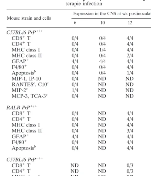
Other immunochemical analysis revealed a close temporal association of T-cell infiltration with the expression of MHC molecules, especially class I, and the presence of F4/80⫹
mi-croglia or macrophages. Enhanced expression of MHC tran-scripts after scrapie infection was noted previously by Duguid and Trzepacz (14). Apoptosis of neurons lagged behind the detection of T cells. All these studies included stringent
[image:3.603.133.451.70.182.2]con-trols for antibody specificity and infection of CNS tissues (15, 22, 32). RNase protection assay (RPA) analysis (2, 9) displayed in Fig. 2 shows elevations of the T-cell chemokines—most prominently MIP-1, IP-10, and RANTES—in C57BL/6 and BALB/c mice with the macrophage chemokine C10 upregu-lated in brains of C57BL/6 but only minimally so in brains of BALB/c mice. MIP-2 was also enhanced in most mice during scrapie infection in the brain at 18 weeks and in a few infected mice at 6 weeks post-scrapie inoculation (Fig. 2). Other che-mokines such as monocyte chemotactic protein 3 and TCA-3 were not elevated at any time point at either tissue site. FIG. 1. (A and B) CD8⫹T-cell infiltration in the parenchyma of a C57BL/6 (A) and BALB/WEHI (B) mouse 18 weeks after i.c. inoculation with scrapie. (C and D) Micrographs of sections of autopsied CNS from two distinct patients with CJD stained with antibody to human T cells. In these human tissues, T cells most often lay near blood vessels but occasionally appeared in the brain parenchyma.
TABLE 1. Immunohistologic study of the CNS during scrapie infection
Mouse strain and cells Expression in the CNS at wk postinoculation
a
6 10 12 18
C57BL/6 PrP⫹/⫹
CD8⫹T 0/4 0/4 4/4 4/4
CD4⫹T 0/4 0/4 4/4 4/4
MHC class I 0/4 1/4 4/4 4/4
MHC class II 0/4 0/4 2/4 3/4
GFAP⫹ 4/4 4/4 4/4 4/4
F4/80⫹ 0/4 0/4 4/4 4/4
Apoptosisb 0/4 0/4 1/4 4/4
MIP-1, IP-10 0/4 ND ND 4/4
RANTESc, C10c 0/4 ND ND 4/4
MIP-2c 1/4 ND ND 4/4
MCP-3, TCA-3c 0/4 ND ND 0/4
BALB PrP⫹/⫹
CD8⫹T 0/4 ND 4/4 4/4
CD4⫹T 0/4 ND 4/4 4/4
MHC class I 0/4 ND 4/4 4/4
MHC class II 0/4 ND 3/4 3/4
GFAP⫹ 4/4 ND 4/4 4/4
F4/80⫹ 0/4 ND 4/4 4/4
Apoptosisb 0/4 ND 4/4 4/4
C57BL/6 PrP⫺/⫺
CD8⫹T ND ND 0/3 0/3
CD4⫹T ND ND 0/3 0/3
MHC class I ND ND 0/3 0/3
Apoptosisb ND ND 0/3 0/3
aExpression of T cells in brains of mice and humans with TSE disease. Results
are given as number of positive mice/total number of mice. ND, not determined
bApoptosis of neurons.
cChemokines in brains. Spleens were negative for chemokines at 6 and 18
weeks. Analysis by RPA.
on November 8, 2019 by guest
http://jvi.asm.org/
[image:3.603.300.542.403.685.2]T-cell infiltration in humans with TSE disease. Six brains from well-studied CJD patients were reevaluated for T-cell infiltration. As before, no T-cell infiltration was apparent by light microscopy. However, the application of antibody to hu-man CD3 showed that T cells accumulated predominantly near or around blood vessels but also in the CNS parenchyma in five of the six brains studied. Shown in Fig. 1C and D are photomi-crographs of brain tissues from two of the five patients with sCJD showing CD3 immunopositive lymphocytes. Tissues are from a cerebello-thalamo-striatal variant of sCJD with PrP-immunopositive kuru-type plaques in the cerebellum and the thalamus, and from a cortico-striato-olivo-cerebellar variant of sCJD, respectively.
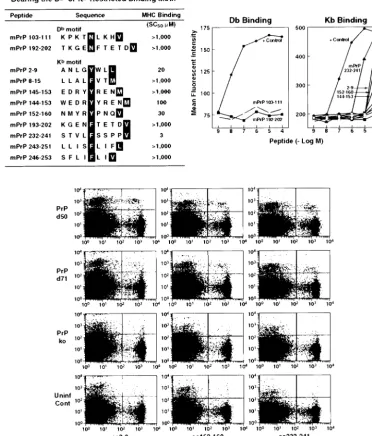
Mapping mPrP peptide sequences that bound to H-2b
(C57BL/6) or H-2d (BALB) MHC class I alleles. We then
analyzed mPrP for peptide sequence motifs that were candi-dates for binding to Kb, Db, Kd, or LdMHC class I alleles (23,
38, 41). Figures 3 and 4 display the prion sequences bearing murine MHC binding motifs: two for Db, nine for Kb, two for
Kd, and four for Ld. These 17 peptides were then synthesized
and tested for binding to their corresponding MHC class I alleles. For a MHC stabilization assay, RMAS cells (Db, Kb)
transfected with Kdmolecules and P815 (H-2d) were used to
measure Db, Kb, Kd, or Ld stabilization in the presence of
increasing peptide concentrations. Empty, unstable MHC mol-ecules reaching the cell surface can be stabilized by peptides added exogenously. The affinity of a peptide for an MHC allele is tightly correlated to its ability to stabilize it. On this basis, MHC stabilization assays have been developed and are now commonly used to estimate the MHC binding properties of a given peptide. In our study, the positive control peptides ex-hibited affinity values (SC50) of 0.01M (Dbbinding [Fig. 3]),
0.1M (Kbbinding [Fig. 3]), 1M (Ldbinding [Fig. 3]), and
0.3M (Kdbinding, not shown), respectively. The mPrP
pep-tides were considered to be potent MHC binders when dis-playing affinity values (SC50) of 50M or less. In summary, we
found that none of the peptides bearing the Db- or Kd-binding
motif bound to their respective MHC alleles (SC50⬎ 1,000
M), whereas one of the four Ldpeptides (mPrP aa 100 to 108:
KPSKPKTNL) bound to Ld(SC
50⫽20M) and three of the
nine Kbpeptides (mPrP aa 2 to 9: ANLGYWLL; aa 152 to 160:
NMYRYPNQV; and aa232 to 241: STVLFSSPPV) bound to Kbwith SC
50s of 20, 30, and 3M, respectively.
Binding of Kb tetramers to lymphocytes harvested from
scrapie-inoculated PrPⴙ/ⴙmice. Kb tetramers containing
ei-ther mPrP aa 2 to 9, aa 152 to 160, or aa 232 to 241 were made (1) and showed low to modest but consistent binding to splenic lymphocytes at day 7 (data not shown) and days 50 and 71 (Fig. 3) after scrapie inoculation. Similar inoculation into PrP⫺/⫺
mice gave lower levels (background) of lymphocyte binding to Kbtetramers at day 7 (data not shown) and days 50 and 71
post-scrapie infection (Fig. 3, data shown for three mice). On day 50, the mean lymphocyte-Kbtetramer binding values⫾1
standard error were these: for aa 2 to 9, that for PrP⫹/⫹mice
was 2.1⫾0.8, compared to 0.4⫾0.3 for PrP⫺/⫺mice; for aa
152 to 160, that for PrP⫹/⫹mice was 1.9⫾0.8 and that for
PrP⫺/⫺mice 0.2⫾0.1; and for aa 232 to 241, that for PrP⫹/⫹
mice was 1.6 ⫾0.8 and that for PrP⫺/⫺mice was 0.1 ⫾0.4
(average of three to four mice per group). The results at day 71 were as follows: for aa 2 to 9, that for PrP⫹/⫹mice was 1.2⫾
0.3 and that for PrP⫺/⫺mice was 0.5⫾0.1; for aa 152 to 160,
that for PrP⫹/⫹mice was 1.4⫾0.4 and that for PrP⫺/⫺mice
was 0.2⫾0.1; and for aa 232 to 241, that for PrP⫹/⫹mice was
0.9⫾0.5 and that for PrP⫺/⫺mice was 0.4⫾0.4. Values of Kb
FIG. 2. RPA analysis of brains harvested from scrapie-infected mice at 6 and 18 weeks post-i.c. inoculation. Individual mice were infected with scrapie (⫹) (pool of four).⫺, uninfected age- and sex-matched controls.
on November 8, 2019 by guest
http://jvi.asm.org/
tetramer binding to splenic lymphocytes harvested from PrP⫹/⫹mice 7 days after scrapie inoculation were as follows:
for aa 2 to 9, 2.6⫾0.3; for aa152 to 160, 2.1⫾0.3; and for aa 232 to 246, 1.2⫾0.08 (three mice per group). Binding by Kb
tetramers to lymphocytes from PrP⫺/⫺mice (three mice per
group) 7 days after scrapie infection showed background val-ues 30 to 50% lower than those to lymphocytes from PrP⫹/⫹
mice. Thus, binding values for each of these three Kbtetramers
to splenic lymphocytes from scrapie-free C57BL/6 mice were
0.6⫾0.8 or less at days 7, 50, or 71 of exposure. Because the Kb mPrP tetramers are relatively unstable, they were used
within 2 to 3 weeks of labeling or made fresh preceding each experiment. Lymphocytes obtained from brains of mice 12 to 18 weeks after scrapie infection failed to bind to Kbtetramers,
and other tetramers made to detect LCMV-specific CD8⫹T
cells (1, 21) failed to stain splenic lymphocytes from PrP⫹/⫹
mice (⬍0.4% of background values). Manufacture of Ld
[image:5.603.108.480.87.523.2]tet-ramers for mPrP was unsuccessful.
FIG. 3. Motif of mPrP Dband Kbpeptides that are appropriate for MHC class I binding (upper panels) and the data to show that only three
of these mPrP peptides bind at heightened affinity to the corresponding MHC molecules. The lower panel displays data from three independent
H-2b(C57BL/6) mice showing that their splenic lymphocytes bound to Kbtetramers containing mPrP aa 2 to 9, aa 152 to 160, and aa 232 to 241.
Background binding for C57BL/6 control mice and C57BL/6 PrP⫺/⫺mice is shown. The positive control for Kbbinding was Moloney murine
leukemia virus peptide SSWDFITV (41), and that for Dbbinding was LCMV ARM NP peptide FQPQNGQFI (21). For binding, the horizontal
axis of the graph shows the reciprocal log peptide dilution, while the vertical axis reflects the mean fluorescence intensity. Abbreviations: ko, knockout; Uninf Cont, uninfected control.
on November 8, 2019 by guest
http://jvi.asm.org/
Effector function(s) of lymphocytes obtained from
scrapie-inoculated PrPⴙ/ⴙmice. Next we examined whether CD4 or
CD8 T cells infiltrating the CNS and the enhanced MHC expression contributed to the pathogenesis of scrapie. For these studies we employed several groups of mice made genet-ically deficient to CD4, CD8, MHC class I, or MHC class II molecules. However, deletion of CD4 or CD8 T cells did not significantly decrease the kinetics or incidence of developing scrapie, as shown in Table 2 and in agreement with a report by Klein et al. (24). Mice null for CD4 and CD8 T cells died or showed severe morbidity at 156⫾8 or 154⫾8 days (mean⫾
1 standard error), respectively, compared to 148⫾8 days for CD4 and CD8 T-cell-competent mice. Similarly, MHC class I and class II knockout mice developed severe disease at days 165⫾5 or 158⫾7, respectively, incubation periods similar to those of genetically normal mice. However, mice lacking both CD4 and CD8 T cells took 193 ⫾ 5 days to show signs of clinical scrapie infection, a significantly longer period (P ⬍
0.01) than that observed for wild-type mice or those lacking only one component, i.e., CD4⫺/⫺, CD8⫺/⫺, MHC class I⫺/⫺,
or MHC class II⫺/⫺ alone. These data were confirmed in a
repeat experiment. In the limited number of MHC class I and MHC class II double-knockout mice available (total of four receiving i.c. and three receiving i.p. inoculations of scrapie) also showed prolonged incubation periods for developing clin-ical disease, taking longer than single-knockout MHC I or MHC II mice. All mice inoculated i.c. took at least 204 days to become moribund, and mice inoculated i.p. became clinically ill at or after 254 days. Within 8 days of becoming ill, all these MHC double-knockout mice died or were sacrificed because of severe morbidity, and neuropathologic and biochemical evi-dence of scrapie infection was found.
The final studies evaluated the effector functions of lympho-cytes from scrapie-infected mice. Initially, lympholympho-cytes were harvested from C57BL/6 or BALB/WEHI mice at 7, 14, 51, 70, or 150 days after primary inoculation of 100,000 PFU of scrapie or after two or three inoculations given i.p. 3 weeks apart. These splenic lymphocytes were then cultured with51
Cr-labeled PrP⫺/⫺ MEF or, from PrP⫺/⫺ mice, macrophages
coated with the relevant Kb-restricted aa 2 to 9, aa 152 to 160,
or aa 232 to 241 or Ld-restricted aa 100 to 108 prion
peptide-peptide PrP⫺/⫺ cells. No specific 51Cr release indicative of
CTL activity was seen, although when lymphocytes originating from syngeneic mice infected 7 days earlier with LCMV were added to PrP⫺/⫺targets either infected with LCMV or coated
with relevant LCMV peptide, significant lysis occurred (Fig. 5A). Similar negative results occurred with lymphocytes har-vested from brains of scrapie-infected mice, whereas lympho-cytes harvested from LCMV-infected mice (15) lysed synge-neic LCMV-infected targets. In subsequent experiments, lymphocytes were harvested on days 7, 50, or 71 after PrP⫹/⫹
mice were infected with scrapie, and mPrP aa 2 to 9, aa 152 to 160, and aa 232 to 241 failed to induce cytoplasmic expression of IFN-␥ or TNF-␣ (data not shown) (8, 21, 29), although again expression of those cytokines was easily induced in mice at days 7, 50, or 120 after LCMV infection when their splenic lymphocytes were similarly incubated with relevant LCMV Kb,
Db, or Ldpeptides (21, 40). In addition, neither IL-2, IL-6, nor
IL-10 expression was induced in lymphocytes harvested from PrP⫹/⫹mice at similar times after scrapie infection and
[image:6.603.46.280.68.416.2]incu-bation with mPrP aa 2 to 9, aa 152 to 160, or aa 232 to 241, FIG. 4. Motif of mPrP Kdand Ldpeptides that are appropriate for
MHC class I binding and data to show that only one of these peptides, mPrP 100 to 108, binds to LdMHC molecules. The positive control for
Ldbinding is the P29 peptide YPNVNIHNF (11), and the negative
control is the PB1 peptide VSDGGPNLY (12). For binding, the hor-izontal axis of the graph shows the reciprocal log peptide dilution, while the vertical axis reflects the mean fluorescence intensity.
⫾
MHC class II⫺/⫺ 6 158⫾7 ND
aMice were inoculated with 100,000 PFU of scrapie as described in Materials
and Methods and sacrificed when moribund. The presence of scrapie was con-firmed by pathogenomic findings on brain sections viewed by light microscopy (spondiosis, gliosis, and neuronal dropout) and by demonstration of conversion
from PrPcto PrPscby Western blotting (33, 35).
bND, not done.
cP⬍0.01.
on November 8, 2019 by guest
http://jvi.asm.org/
[image:6.603.301.542.89.194.2]although IL-4, IL-6, and IL-10 were expressed in lymphocytes after LCMV infection (21, 40, 44).
Next, as shown in Table 3, we obtained 15-mer overlapping peptides of mPrP and arranged them in 14 groups for testing against splenic lymphocytes from scrapie-inoculated PrP⫹/⫹
mice. After one or three i.p. inoculations of 100,000 infectious doses of Chandler scrapie, each spaced 3 weeks apart, spleens were harvested, and single-cell suspensions of lymphocytes made and incubated with the various mPrP cocktails displayed in Table 3 and then stained and fixed to allow detection of intracellular cytokines IFN-␥and TNF-␣as well as molecules bound to CD4 or CD8 T cells. Although the accompanying control splenocytes from LCMV-immunized mice concurrently tested but with appropriate LCMV peptides gave positive re-sults, we were unable to detect any convincing intracellular
expression of IFN-␥(Fig. 5B) or TNF-␥(data not shown) in either CD8 or CD4 T cells from scrapie-inoculated mice. No-tably, similar inoculation of murine scrapie into PrP⫺/⫺mice,
due to a deletion of mPrP, also failed to reveal IFN-␥ or TNF-␣cytoplasmic expression in CD8 or CD4 T lymphocytes. Further, such lymphocytes from scrapie-infected PrP⫹/⫹ or
PrP⫺/⫺mice failed to lyse syngeneic PrP⫺/⫺51Cr-labeled MEF
coated with the peptide cocktails (Fig. 5).
DISCUSSION
We found T cells in brains of mice and humans with TSE. Our kinetic studies, possible in the murine PrP⫹/⫹model using
the Chandler RML scrapie strain, located both CD4 and CD8 T cells within the brain by 12 weeks (84 days) and later after i.c. FIG. 5. (A) Lymphocytes obtained from the spleens of mice either primed once or three times with scrapie fail to lyse syngeneic PrP⫺/⫺target cells coated with 20g of those peptides which when separately placed in Kbtetramers bound such T cells (see Fig. 3). In contrast, T cells harvested
from littermates primed seven days (Po) earlier with LCMV ARM 105PFU i.p. or 60 days after initial priming receiving a second injection of
LCMV ARM (So) lyse these PrP⫺/⫺target cells when they were infected with LCMV ARM 2 days earlier or coated with 20g of the LCMV GP peptide aa 33 to 41. See Materials and Methods and reference 21 for details of51Cr-release assay and immunizations. Abbreviations: ko, knockout;
CTL, cytotoxic T lymphocyte; E:T, effector-to-target cell; INF, infected; ND, not determined; spl, spleen; 3⫻inoc, inoculated three times. (B) T cells obtained after multiple (three) inoculations with scrapie fail to generate intracytoplasmic IFN-␥when stimulated with the various pools of peptides that cover the mPrP sequence (see Table 3). Data are from a single mouse and representative of four additional mice. The positive control shows IFN-␥cytoplasmic staining for CD8 and CD4 T cells obtained 7 days after an inoculation with 105PFU of LCMV ARM. Spleen lymphocytes
were incubated with CD8 T-cell peptide LCMV GP aa 33 to 41 or CD4 T-cell peptide LCMV GP aa 61 to 80. See Materials and Methods and reference 21 for details.
on November 8, 2019 by guest
http://jvi.asm.org/
[image:7.603.76.508.78.429.2]T cells were not found in the brain 8 weeks after RML Chan-dler scrapie inoculation, these authors (6, 7) found T-cell in-filtrates at that time but used a different strain of mouse scrapie.
Since only activated T cells are believed to cross the blood-brain barrier to enter the CNS, presumably scrapie infection was responsible for the initial T-cell activation. The cause could be generation of antigen-specific scrapie T cells or, al-ternatively or concomitantly, chemokine and cytokine che-moattractants induced during scrapie infection in the brain. We (20) and others (reviewed in references 3, 39, and 46) have incited cytokine and chemokine expression focally in selected tissues using transgenic approaches with cell-specific promot-ers and reported the resulting infiltration of lymphocytes into the target area. Our studies here are unable to discriminate between these two possibilities at present. The detection here of suspected PrP⫹/⫹antigen-specific T cells by tetramers, but
at low levels, was not associated with intracellular cytokine expression when such lymphocytes were incubated with the appropriate prion peptide(s). This finding is in accord with those of Zajac et al. (47), who reported an example of disas-sociation between tetramer-positive staining and intracellular cytokine staining in a viral model, while Field and Shenton (16) previously reported peripheral lymphocyte sensitization with TSE infection.
After entering the brain, activated T cells remain for approx-imately 10 to 14 days and then circulate out unless the recog-nition T-cell antigen appears expressed in the CNS milieu or appropriate chemokines are continuously present (15, 18). Al-though the presence of CD8⫹and CD4⫹T cells and scrapie
infection in the CNS bear a direct association, the lack of marked inflammatory cell recruitment to that site, coupled with the failure of T cells obtained from the periphery and the CNS to display detectable effector functions in vitro as cyto-toxicity or expression or release of Th1 cytokines, suggests that these cells are minimally or not at all significant to the disease process. In agreement are earlier studies in which several im-munosuppressive strategies or separate deletion of either CD4 or CD8 T cells did not alter the kinetics or outcome of scrapie infection (reviewed in references 4, 5, and 24). Further, the Th1 cytokine IFN-␥was not found in brains of mice succumb-ing to scrapie infection (9). However, as shown here, deletion of both CD4 and CD8 T cells significantly delayed the onset of disease, although once disease occurred, it was uniformly fatal. Hence, the delayed progression of disease and expression of TNF-␣ in brains of scrapie-infected mice (9) raise the possi-bility that the T-cell response may modestly influence the in-fection.
Why is it difficult to detect prion-specific T-cell response with scrapie infection? Arguments (reviewed in references 4
9 QGSPGGNRYPPQGGT
10 GNRYPPQGGTWGQPH
11 PQGGTWGQPHGGGWG
12 WGQPHGGGWGQPHGG
13 GGGWGQPHGGSWGGP
14 QPHGGSWGGPHGGSW
15 SWGGPHGGSWGQPHG
16 HGGSWGQPHGGGWGQ
17 GQPHGGGWGQGGGTH
18 GGWGQGGGTHNQWNK
19 GGGTHNQWNKPSKPK
20 NQWNKPSKPKTNLKH
21 PSKPKTNLKHVAGAA
22 TNLKHVAGAAAAGAV
23 VAGAAAAGAVVGGLG
24 AAGAVVGGLGGYMLG
25 VGGLGGYMLGSAMSR
26 GYMLGSAMSRPMIHF
27 SAMSRPMIHFGNDWE
28 PMIHFGNDWEDRYYR
29 GNDWEDRYYRENMYR
30 DRYYRENMYRYPNQV
31 ENMYRYPNQVYYRPV
32 YPNQVYYRPVDQYSN
33 YYRPVDQYSNQNNFV
34 DQYSNQNNFVHDCVN
35 QNNFVHDCVNITIKQ
36 HDCVNITIKQHTVTT
37 ITIKQHTVTTTTKGE
38 HTVTTTTKGENFTET
39 TTKGENFTETDVKMM
40 NFTETDVKMMERVVE
41 DVKMMERVVEQMCVT
42 ERVVEQMCVTQYGKE
43 QMCVTQYGKESQAYY
44 QYGKESQAYYDGRRS
45 SQAYYDGRRSSSTVL
46 DGRRSSSTVLFSSPP
47 SSTVLFSSPPVILLI
48 FSSPPVILLISFLIF
49 VILLISFLIFLIVG
Pool no.
1 1,⫹2,⫹3,⫹4,⫹5,⫹6,⫹7
2 8,⫹9,⫹10,⫹11,⫹12,⫹13,⫹14
3 15,⫹16,⫹17,⫹18,⫹19,⫹20,⫹21
4 22,⫹23,⫹24,⫹25,⫹26,⫹27,⫹28
5 29,⫹30,⫹31,⫹32,⫹33,⫹34,⫹35
6 36,⫹37,⫹38,⫹39,⫹40,⫹41,⫹42
7 43,⫹44,⫹45,⫹46,⫹47,⫹48,⫹49
8 1,⫹8,⫹15,⫹22,⫹29,⫹36,⫹43
9 2,⫹9,⫹16,⫹23,⫹30,⫹37,⫹44
10 3,⫹10,⫹17,⫹24,⫹31,⫹38,⫹45
11 4,⫹11,⫹18,⫹25,⫹32,⫹39,⫹46
12 5,⫹12,⫹19,⫹26,⫹33,⫹40,⫹47
13 6,⫹13,⫹20,⫹27,⫹34,⫹41,⫹48
14 7,⫹14,⫹21,⫹28,⫹35,⫹42,⫹49
aPeptides comprising pools 1 through 14 were incubated at a concentration of 20
g/ml for 5 h with a single-cell suspension of lymphocytes obtained from spleens of
mice inoculated three times with murine scrapie (see Materials and Methods and reference 21) in the presence of recombinant IL-2 (10 to 50 U/ml) and brefeldin A
(1g/ml) and stained with antibodies to either CD4 or CD8 and with antibody to
IFN-␥conjugated to a difference fluorochrome. Cells were studied by
fluorescence-activated cell sorting using two-color analysis (see Fig. 5).
on November 8, 2019 by guest
http://jvi.asm.org/
[image:8.603.46.286.79.668.2]and 5) to explain such a phenomenon range from proposing a state of tolerance due to identical amino acid sequences be-tween PrPc and PrPsc that is not broken either by scrapie
infection or immunization with scrapie prions injected in ad-juvant and the unusual protease resistance of PrPsc, which
might prevent its degradation and processing in antigen-pre-senting cells. Yet, immune responses are reproducibly gener-ated against other “self antigens” when the self protein or one of its peptide fragments is inoculated with adjuvants or during infections. For example, immune responses to CNS proteins like myelin basic protein, proteolipid protein of myelin, and myelin-associated oligodendrocyte basic protein, etc., are eas-ily generated and often associated with corresponding automune diseases (17, 19, 28, 48). Further, specific antiviral im-mune responses can be detected against endogenous murine retroviruses (31) that have existed in their hosts over thousands of years, most often infecting thymi and peripheral lymph nodes. Heterologous PrP in adjuvant can elicit immune (anti-body) responses, suggesting that antigen processing of the PrP molecule can occur (25).
CD8 and CD4 T cells generated during scrapie infection, under the experimental conditions used here, were unable to mount effector functions. Of interest, this inability to act as lytic agents or cytokine producers also occurred when scrapie was inoculated into PrP⫺/⫺mice with a deletion of the PrP
gene as well as PrP⫺/⫺mice that were unable to transcribe PrP
because of an engineered mutation. However, it may be pos-sible using other immunizing strategies or vehicles to express prions (i.e., DNA vaccination) that at least scrapie-specific CD4 T-cell responses can be generated, since antibodies to prions can be made. As for scrapie-specific CD8⫹T cells, we
mapped potential peptide motifs for MHC class I H-2b and
H-2d molecules. Found were three CD8 T-cell motifs that
bound well to Kb(SC
50, 3, 20, and 30M) and one that bound
to Ld(SC
50, 20M) molecules. Kbtetramers made with these
three mPrP peptides—aa 2 to 9, aa 152 to 160, and aa 232 to 241–bound, albeit to a modest extent, to splenic lymphocytes at days 7, 50, and 71 after scrapie inoculation. However, these lymphocytes from PrP⫹/⫹mice were unable to synthesize the
intracellular cytokines IFN-␥or TNF-␣when stimulated with appropriate peptides and were unable to lyse 51
chromium-labeled PrP⫺/⫺MEF coated with mPrP peptides. These
find-ings are reminiscent of those from recent studies of CD4⫺/⫺
mice infected with LCMV (47) and from a report characteriz-ing circulatcharacteriz-ing T cells for tumor-specific antigens (27). Lastly, the accumulation of T cells in the brains of scrapie-infected mice likely mirrors other models in which T cells accumulated and resided in the CNS following antigen-specific stimulation, when the recognized antigen was continuously expressed in the brain (15, 18) and/or when attracted by chemokines expressed in the CNS (3, 39, 46). Further analysis of these T cells and their induced genetic profiles should be of interest.
ACKNOWLEDGMENTS
This work was supported by the National Institutes of Health grants AG04342 (M.B.A.O., J.E.G., and D.H.), AG02132, AG10770 (S.D.), and MH50426 (I.L.C.); training grant AG00080 (D.H.); and postdoc-toral fellowships from the National Multiple Sclerosis Society (V.C.A.) and the Juvenile Diabetes Foundation (D.H.).
We thank Denis Hudrisier, INSERM, Toulouse University, France, for kind assistance in peptide prediction and MHC binding studies;
John Altman, Vaccine Center Yerkes, Emory University Medical School, and the NIH Tetramer Core, for advice and assistance in making and providing prion tetramer reagents; Rick Race, NIH Rocky Mountain Laboratories, Laboratory of Persistent Viral Diseases, for assistance with the PrPscanalyses; and John Alcantara for technical
assistance.
REFERENCES
1. Altman, J. D., P. A. H. Moss, P. J. R. Goulder, D. H. Barouch, M. G. McHeyzer-Williams, J. I. Bell, A. J. McMichael, and M. M. Davis.1996.
Phenotypic analysis of antigen-specific T lymphocytes. Science274:94–96.
2. Asensio, V. C., and I. L. Campbell.1997. Chemokine gene expression in the
brains of mice with lymphocytic choriomeningitis. J. Virol.71:7832–7840.
3. Asensio, V. C., and I. L. Campbell.1999. Chemokines in the CNS:
pluri-functional mediators in diverse states. Trends Neurosci.22:504–512.
4. Aucouturier, P., R. I. Carp, C. Carnaud, and T. Wisniewski.2000. Prion
diseases and the immune system. Clin. Immunol.96:79–85.
5. Berg, L. J.1994. Insights into the role of the immune system in prion
diseases. Proc. Natl. Acad. Sci. USA91:429–432.
6. Betmouni, S., and V. H. Perry.1999. The acute inflammatory response in CNS following injection of prion brain homogenate or normal brain
homog-enate. Neuropathol. Appl. Neurobiol.25:20–28.
7. Betmouni, S., V. H. Perry, and J. L. Gordon.1996. Evidence for an early inflammatory response in the central nervous system of mice with scrapie.
Neuroscience74:1–5.
8. Butz, E. A., and M. J. Bevan.1998. Massive expansion of antigen-specific
CD8⫹T cells during an acute virus infection. Immunity8:167–175.
9. Campbell, I. L., M. Eddleston, P. Kemper, M. B. A. Oldstone, and M. V. Hobbs.1994. Activation of cerebral cytokine gene expression and its
corre-lation with onset of molecular pathology in scrapie. J. Virol.68:2383–2387.
10. Chesebro, B.1999. Prion protein and the transmissible spongiform
enceph-alopathy diseases. Neuron24:503–506.
11. Corr, M., L. F. Boyd, S. R. Frankel, S. Kozlowski, E. A. Padlan, and D. H. Margulies.1992. Endogenous peptides of a soluble major histocompatibility complex class I molecule, H-2Lds: sequence motif, quantitative binding, and
molecular modeling of the complex. J. Exp. Med.176:1681–1692.
12. DiBrino, M., T. Tsuchida, R. V. Turner, K. C. Parker, J. E. Coligan, and W. E. Biddison.1993. HLA-A1 and HLA-A3 T cell epitopes derived from influenza virus proteins predicted from peptide binding motifs. J. Immunol.
151:5930–5935.
13. Diedrich, J. F., P. E. Bendheim, Y. S. Kim, R. I. Carp, and A. T. Haase.1991. Scrapie-associated prion protein accumulates in astrocytes during scrapie
infection. Proc. Natl. Acad. Sci. USA88:375–379.
14. Duguid, J., and C. Trzepacz.1993. Major histocompatibility complex genes have an increased brain expression after scrapie infection. Proc. Natl. Acad.
Sci. USA90:114–117.
15. Evans, C. F., M. S. Horwitz, M. V. Hobbs, and M. B. A. Oldstone.1996. Viral infection of transgenic mice expressing a viral protein in oligodendrocytes leads to chronic central nervous system autoimmune disease. J. Exp. Med.
184:2371–2384.
16. Field, E. J., and B. K. Shenton.1975. Cellular sensitization in kuru, Jakob-Creutzfeldt disease and multiple sclerosis: with a note on the biohazards of
slow infection work. Acta Neurol. Scand.51:299–309.
17. Hammerling, G. J., G. Schonrich, F. Momburg, N. Auphan, M. Malissen, B. Malissen, A.-M. Schmitt-Verhulst, and B. Arnold.1991. Non-deletional mechanisms of peripheral and central tolerance: studies with transgenic mice with tissue-specific expression of a foreign MHC class I antigen. Immunol.
Rev.122:47–67.
18. Hawke, S., P. G. Stevenson, S. Freeman, and C. R. M. Bangham.1998. Long-term persistence of activated cytotoxic T lymphocytes after viral
infec-tion of the central nervous system. J. Exp. Med.187:1575–1582.
19. Holz, A., B. Bielekova, R. Martin, and M. B. A. Oldstone.2000. Myelin-associated oligodendrocytic basic protein: identification of an
encephalito-genic epitope and association with multiple sclerosis. J. Immunol.164:1103–
1109.
20. Holz, A., K. Brett, and M. B. A. Oldstone.2001. Constitutive beta cell expression of IL-12 does not perturb self-tolerance but intensifies established
autoimmune diabetes. J. Clin. Investig.108:1749–1758.
21. Homann, D., L. Teyton, and M. B. A. Oldstone.2001. Differential regulation
of antiviral T cell immunity results in stable CD8⫹but declining CD4⫹T
cell memory. Nat. Med.7:913–919.
22. Horwitz, M. S., C. F. Evans, F. G. Klier, and M. B. A. Oldstone.1999. Detailed in vivo analysis of interferon-gamma induced major histocompati-bility complex expression in the central nervous system: astrocytes fail to express major histocompatibility class I and II molecules. Lab. Investig.
79:235–242.
23. Hudrisier, D., H. Mazarguil, F. Laval, M. B. A. Oldstone, and J. E. Gairin.
1996. Binding of viral antigens to major histocompatibility complex class I H-2Db molecules is controlled by dominant negative elements at peptide non-anchor residues: implications for peptide selection and presentation.
J. Biol. Chem.271:17829–17836.
on November 8, 2019 by guest
http://jvi.asm.org/
mechanisms in experimental autoimmune encephalomyelitis: implications
for immunotherapy of human autoimmune diseases. FASEB J.5:2560–2566.
29. Murali-Krishna, K., J. D. Altman, M. Suresh, D. J. Sourdive, A. J. Zajac, J. D. Miller, J. Slansky, and R. Ahmed.1998. Counting antigen-specific CD8 T cells: a reevaluation of bystander activation during viral infection.
Immu-nity8:177–187.
30. Muramoto, T., T. Kitamoto, J. Tateishi, and I. Goto.1992. The sequential development of abnormal prion protein accumulation in mice with
Creutzfeldt-Jakob disease. Am. J. Pathol.140:1411–1420.
31. Oldstone, M. B. A., T. Aoki, and F. J. Dixon.1972. The antibody response of mice to murine leukemia virus in spontaneous infection. Absence of classical
immunologic tolerance. Proc. Natl. Acad. Sci. USA69:134–138.
32. Oldstone, M. B. A., H. Lewicki, D. Thomas, A. Tishon, S. Dales, J. Patterson, M. Manchester, D. Homann, and A. Holz.1999. Measles virus infection in a transgenic model: virus-induced central nervous system disease and
immu-nosuppression. Cell98:629–640.
33. Oldstone, M. B. A., R. Race, D. Thomas, H. Lewicki, D. Homann, S. C. Smelt, A. Holz, P. A. Koni, D. Lo, B. Chesebro, and R. A. Flavell.2002. Lymphotoxin-alpha- and lymphotoxin-beta-deficient mice differ in suscepti-bility to scrapie: evidence against dendritic cell involvement in
neuroinva-sion. J. Virol.76:4357–4363.
34. Prusiner, S. B.2001. Prions, p. 3063–3087.InD. M. Knipe and P. M. Howley (ed.), Fields virology, 4th ed. Lippincott Williams and Wilkins, Philadelphia, Pa.
35. Race, R. E., S. A. Priola, R. A. Bessen, D. Ernst, J. Dockter, G. F. Rall, L. Mucke, B. Chesebro, and M. B. A. Oldstone.1995. Neuron-specific expres-sion of a hamster prion protein minigene in transgenic mice induces
suscep-tibility to hamster scrapie agent. Neuron15:1183–1191.
36. Raeber, A. J., S. Brandner, M. A. Klein, Y. Benninger, C. Musahl, R. Frigg,
model: role of cross-reacting viruses and quantitation of effector T cells
needed to cause disease J. Virol.74:3284–3292.
41. Sijts, A. J. A. M., M. L. H. DeBruijn, M. E. Ressing, J. D. Nieland, E. A. M. Mengede, C. J. P. Boog, F. Ossendorp, W. M. Kast, and C. J. M. Melief.
1994. Identification of an H-2 Kb-presented Moloney murine leukemia virus
cytotoxic T-lymphocyte epitope that displays enhanced recognition in H-2
Dbmutant bm13 mice. J. Virol.68:6038–6046.
42. Taraboulos, A., K. Jendroska, D. Serban, S.-L. Yang, S. J. DeArmond, and S. B. Prusiner.1992. Regional mapping of prion proteins in brains. Proc.
Natl. Acad. Sci. USA89:7620–7624.
43. Tishon, A., H. Lewicki, G. Rall, M. G. von Herrath, and M. B. A. Oldstone.
1995. An essential role for type 1 interferon gamma in terminating persistent
viral infection. Virology212:244–250.
44. von Herrath, M., J. Dockter, and M. B. A. Oldstone.1994. How virus induces a rapid or slow onset insulin-dependent diabetes mellitus in a transgenic
model. Immunity1:231–242.
45. von Herrath, M. G., D. P. Berger, D. Homann, T. Tishon, A. Sette, and M. B. A. Oldstone.2000. Vaccination to treat persistent viral infection.
Virology268:411–419.
46. Wang, J., V. C. Asensio, and I. L. Campbell.2002. Cytokines and chemokines as mediators of protection and injury in the central nervous system assessed
in transgenic mice. Curr. Top. Microbiol. Immunol.265:23–48.
47. Zajac, A. J., J. N. Blattman, K. Murali-Krishna, D. J. Sourdive, M. Suresh, J. D. Altman, and R. Ahmed.1998. Viral immune evasion due to persistence
of activated T cells without effector function. J. Exp. Med.188:2205–2213.
48. Zaller, D. M., and V. S. Sloan.1996. Transgenic mouse models of
experi-mental autoimmune encephalomyelitis. Curr. Top. Microbiol. Immunol.206:
15–31.