A STUDY ON VENTILATOR ASSOCIATED PNEUMONIA WITH SPECIAL REFERENCE TO MULTIDRUG RESISTANT
PATHOGENS IN A TERTIARY CARE HOSPITAL.
Dissertation submitted to
THE TAMILNADU DR.M.G.R.MEDICAL UNIVERSITY In partial fulfillment of the regulations
for the award of the degree of M.D. (MICROBIOLOGY) BRANCH - IV
MADRAS MEDICAL COLLEGE
THE TAMILNADU DR. M.G.R. MEDICAL UNIVERSITY CHENNAI – TAMILNADU.
CERTIFICATE
This is to certify that this dissertation titled “
A STUDY ON
VENTILATOR ASSOCIATED PNEUMONIA WITH SPECIAL
REFERENCE TO MULTIDRUG RESISTANT PATHOGENS IN A
TERTIARY CARE HOSPITAL.”
is a bonafide record of work done by
DR. K.VASANTHI,
during the period of her Post Graduate study from
2013 to 2016 under guidance and supervision in the Institute of
Microbiology, Madras Medical College and Rajiv Gandhi Government
General Hospital, Chennai- 600003, in partial fulfillment of the
requirement of
M.DMICROBIOLOGY
degree Examination of The
Tamilnadu Dr. M.G.RMedical University to be held in April 2016.
Dr.R. VIMALA., M.D Dr.MANGALA ADISESH M.D.,
Dean Director,(I/C)
DECLARATION
I
declare that the dissertation entitled “
A STUDY ON
VENTILATOR ASSOCIATED PNEUMONIA WITH SPECIAL
REFERENCE TO MULTIDRUG RESISTANT PATHOGENS IN A
TERTIARY CARE HOSPITAL.
” submitted by me for the degree of
M.D. is the record work carried out by me during the period of
October
2014–August 2015
under the guidance of
Dr. R.Vanaja M.D.,
Professor,
Institute of Microbiology, Madras Medical College, Chennai. This
dissertation is submitted to The Tamilnadu Dr.M.G.R. Medical
University, Chennai, in partial fulfillment of the University regulations
for the award of degree of M.D., Branch IV (Microbiology) examination
to be held in April 2016.
Place : Chennai Signature of the candidate
Date: (Dr. K.VASANTHI)
Signature of the guide
Prof.Dr.R.VANAJA.,MD,
Professor
Institute of Microbiology
ACKNOWLEDGEMENT
I humbly submit this work to the Almighty who has given the health
and ability to pass through all the difficulties in the compilation and
proclamation of this blue print.
I wish to express my sincere thanks to our Dean,
Dr. R.VimalaM.D.,
for
permitting me to use the resources of this institution for my
study.
I owe special thanks to
Prof. Dr. Mangala Adisesh, M.D.,
Director (i/c) and Professor, Institute of Microbiology for her support,
valuable suggestions, erudite guidance in my study and for being a
source of inspiration in my endeavours.
I express my sincere thanks to our
professor Dr.S.Vasanthi M.D.,
for
her guidance and support.
My sincere thanks to
Dr.Ragunandanan M.D.,
Professor, Department of
Medicine for permitting me to carry out my study.
I express my gratitude to our former Director,
Prof. Dr. G. Jayalakshmi,
M.D.,DTCD,
for her guidance and support.
I would like to thank my former Professor,
Dr.T.Sheila Doris MD.,
for her support and guidance.
I
feel
fortunate
to
work
under
the
guidance
of
Prof.Dr.R.VanajaM.D.
for her valuable suggestions and great support
throughout my study.
I would like to thank my
Professors Dr.S.Thasneem Banu M.D.,
I extend my whole hearted gratitude and special thanks to my Assistant
Professor
Dr.R.Deepa M.D.,
for her most valuable guidance, constant
support and encouragement in my study.
I also express my thanks to our Assistant professors
Dr. T.Usha Krishnan M.D., Dr.N.Rathna Priya M.D., Dr. David Agatha
M.D., Dr. C. SriPriya M.D., Dr.N. Lakshmi Priya M.D.,
Dr.K.G.Venkatesh M.D,
and
Dr.B.Natesan M.D.,DLO.,
for their
immense support in my study.
I hereby express my gratitude to all the technical staff for their help
throughout my study.
I would like to thank my department colleagues and friends for their
constant support and co-operation.
I would like to thank the Institutional Ethics Committee for approving my
study.
TABLE OF CONTENTS
S.NO TITLE PAGE
NUMBER
1 INTRODUCTION 1
2 AIMS AND OBJECTIVES OF THE STUDY 3
3 REVIEW OF LITERATURE 4
4 MATERIALS AND METHODS 24
5 RESULTS 39
6 DISCUSSION 67
7 SUMMARY 76
8 CONCLUSION 79
9 COLOUR PLATES
10 APPENDIX-I ABBREVATIONS
11 APPENDIX-II STAINS, REAGENTS AND MEDIA
12 ANNEXURE-I CERTIFICATE OF APPROVAL
13 ANNEXURE-II PROFORMA
14 ANNEXURE-III PATIENTS CONSENT FORM
15 ANNEXURE-IV MASTER CHART
DISSERTATION TITLE: A study on ventilator associated pneumonia with
special reference to multidrug resistant pathogens in a tertiary care hospital.
ABSTRACT :
Background:
Ventilator Associated Pneumonia (VAP) is the most frequent intensive care unit (ICU) acquired infection. The aetiology of VAP varies with different patient populations and types of ICUs.
Methodology:
Endotracheal aspirates/bronchioalveolar lavage were collected from patients on mechanical ventilation for > 48hrs and processed quantitatively to determine the various aetiological agents causing VAP and the prevalence of multidrug resistant (MDR) pathogens.Combination disc method, Modified Hodge test, EDTA Combined disc test and AmpC disc test were performed for the detection of extended spectrum beta lactamases (ESBL), carbapenemases, metallo betalactamases (MBL)and AmpC β
lactamases respectively. Results:
The incidence of VAP was 16 per 1000 ventilator days.In this study,34.8% of the cases were early onset VAP,while 65.2% were late onset VAP.
Klebsiella pneumonia,Klebsiella oxytoca and Pseudomonas aeruginosa were more
common in early onset VAP, while non fermenters (Acinetobacter baumannii and
Pseudomonas aeruginosa)were predominantly associated with late onset VAP.70% of
respectively. MBL was produced by 33% of P. aeruginosa and 33% of Acinetobacter
baumannii.AmpC betalactamases were produced by 17% of Pseudomonas
aeruginosa,22% of Acinetobacter baumannii and 33% of Klebsiella pneumonia.Of the S. aureus isolates, 100% were methicillin resistant. Prior antibiotic therapy,reintubation,Emergency intubation and hospitalization of five days or more were common risk factors associated with VAP.
Conclusions:
VAP is increasingly associated with MDR pathogens. Production of ESBL, AmpC betalactamases and metallo betalactamases were responsible for the multidrug resistance of these pathogens. Increasing prevalence of MDR pathogens in patients with late onset VAP indicate that appropriate broad spectrum antibiotics should be used to treat them.It is useful in implementing simple and effective preventive measures including precaution during emergency intubation, minimizing the occurrence of reintubation, and judicious use of antibiotics.
Key words:Ventilator Associated pneumonia,Intensive care unit,Extended spectrum
1 INTRODUCTION
Ventilator-associated pneumonia (VAP) is defined as pneumonia that occurs more than 48 hrs following endotracheal intubation and initiation of mechanical ventilation.It is characterized by the presence of a new or progressive radiographic infiltrate, fever, altered white blood cell count,changes in sputum characteristics and detection of a causative pathogen.(1)
VAP is the frequent ICU acquired infection among patients on Mechanical Ventilation.(1)VAP is a subgroup of Hospital Acquired Pneumonia.It occurs in 9-27% of patients on ventilator.(2)India has an overall crude mortality of 67.4% in patients with pneumoniain ICU, with 40% of the mortality is attributable to infection alone.(6)
VAP is usually categorised as early onset and late onset VAP.Early onset VAP occurs within first 4 days of mechanical ventilation (MV),usually carries a good prognosis and are likely to be due to organisms sensitive to antibiotics. Late onsetVAPdevelops five (or more) days after initiation of Mechanical ventilation.It is caused by MDRpathogens and is associated with increase in patient mortality and morbidity (3).
2 The diagnosis of pneumonia in mechanically ventilated patients is based on the combination of clinical, radiological and microbiological criteria.(7)The lower
respiratorytract samples obtained either by
bronchoscopic(eg.BAL,PSB)ornonbronchoscopic methods(Endotracheal aspirates) are used in the diagnosis of VAP.The endotracheal aspirates are easy to collect and have a high sensitivity.(5)
The common pathogens causing VAP includePseudomonas aeruginosa, Acinetobacterspecies, Klebsiellapneumoniae, Enterobacterspecies, and MRSA (methicillin Resistant Staphylococcus aureus).Among them Pseudomonas and Acinetobacter species are often multidrug resistant which is attributed to the production of ESBL(Extended spectrum beta lactamases),Amp C beta lactamases and metallo beta lactamases.(2)
Thus ,VAP poses grave complications in endotracheally intubated patients in ICU’s worldwide.It leads to adverse clinical outcomes and increase in healthcare costs.(1) The causes of VAP are different among different patient populations and also in different type of Intensive care units.Hence the local microbial flora associated with VAP and their sensitivity pattern should be studied in all clinical setting which may guide in the effective and rational utilization of antimicrobial agents.Our Institution is a tertiary care hospital providing critical carefacilities,where many patients routinely undergo assisted mechanical ventilation.
3 study their antibiotic susceptibility patterns with specialemphasis on multidrug resistant pathogens.
AIMS AND OBJECTIVES Aims:
1. To identify the bacterial and fungal etiological agents associated with Ventilator Associated Pneumonia(VAP).
2. To evaluate the antimicrobial susceptibility pattern for the isolates.
3. To determine the frequency of Multidrug resistant (MDR) pathogens among the VAP patients.
4. Toanalyse the risk factors associated with VAP.
5. To calculate the Ventilator associated pneumonia rate per 1000 ventilator days.
6. Toasses the clinical outcome in VAP patients. Objectives:
1. To monitor adult patients on mechanical ventilator in Medical Intensive care unit for the development of VAP by clinical and radiological criteria
2. To Process endotracheal aspirates and BAL samples quantitativelyfor the identification of causative organism.
3. To detect the presence of ESBL, AmpC beta lactamase, and (MBL) Metallobetalactamase production among the MDR pathogens.
4 5. To monitor the total number of patientson mechanical ventilator and the
totalventilator days exposed by the patients inMICU. 6. To follow up the patients with VAP for prognosis. REVIEW OF LITERATURE.
History:
The Roman physician Galen may have been the first to describemechanical ventilation: "If you take a dead animal and blow air through its larynx [through a reed], you will fill its bronchi and watch its lungs attain the greatest distention.(11).Vesalius too describes ventilation by inserting a reed or cane into the trachea of animals(10).
The iron lung, also known as the Drinker and Shaw tank, was developedin 1929 and was one of the first negative-pressure machines used for long-term ventilation. It was refined and used in the 20th century largely as a result of the polio epidemic that struck the world in the 1940s. The machine is effectively a large elongated tank, which encases the patient up to the neck. The neck is sealed with a rubber gasket so that the patient's face (and airway) are exposed to the room air.(11)
5 Definition:
The exact definition of VAP is still a matter of debate ,because of the lack of criteria which is able to distinguish itfrom other pulmonary conditions in patients who are critically ill.
In 2005, the American Thoracic Society and InfectiousDiseases Society of America jointly published practicalguidelines on hospital-acquired infection which defines(HAP)Hospital acquired pneumonia as pneumonia that occurs 48 hours or more after admission, which was not incubating at the time of admission whereas VAP isdefined as pneumonia that arises more than 48-72 hrs after endotracheal intubation.(2)The 48-hours time frame was set to differentiate any newinfection from processes already ongoing at the moment ofintubation. VAP is categorised into an early and late onsetVAP,due to the difference in epidemiological features and treatment options available for the two forms (2).
6 ThoracicSociety/Infectious Diseases Society of America definition,leading to an increase in VAP incidence.
Pneumonia acquired within 48 hours after hospital admission as aconsequence of emergency intubation, aspiration due to decreased level ofconsciousness and coma, or cardiopulmonary resuscitation are excluded fromdefinition of VAP .(2)
Categorisation of VAP:(3) Early-onset VAP
Ventilator associated pneumonia occurring in the first four days ofendotracheal intubation and initiation of mechanical ventilation is called as early onset VAP anditaccounts for a better prognosis.
Late-onset VAP
Ventilator associated pneumonia developing after four days of mechanicalVentilation is defined as late onset VAP andis associated with higher mortality and is often caused by multidrug resistant bacteria.(2)
Incidence:
7 A study was conducted with Athenians ,which identified patients at a greater risk of developing VAPand the incidence of VAP among patients on mechanical ventilator was found to be around eight percent.
(12)Bowton DL et al(24) (2006) showed in their study that the nosocomial pneumonia rate in mechanically ventilated patients varies from 9% to 68% andmortality rates varies from 33% to 71%.
Wagh H andAcharya D stated the rates of VAP to range from 9 to 27% with the presence ofMDR pathogens and associated with high morbidity and mortality rates.VAP also increased length of ICU stay by 28%.(2)
A study from India evaluated 51 patients in the critical care unit and found a mortalityrate of 37% attributable to VAP, which also correlated very well with higher APACHEIII scores; 33% of the cases were early onset, and 67% were late onset.Themortalityrates of patients with or without VAP in different studies were ; 71% and 28% (20) 55% and 25%,(19)and 33% and 19% (21) respectively.
There is increased risk of acquiring pneumonia when there is increase in duration of mechanical ventilation.(23). The prognosis for VAP caused by Gram negative aerobic bacilli is considerablyworse than that for infection with Gram positive pathogens.According to Study by Fagon et al(20) mortality associated withPseudomonas or Acinetobacter pneumonia was 87% compared to 55% for pneumoniadue to other organisms.
8 higher mortality rate(65%) when compared to patients with VAP due to other organisms (31%).
Risk factors for VAP
During the first four days of artificialventilation , insertion of multiple central venous line,emergency intubation and intravenous sedatives were found to be independent risk factors of Ventilator Associated Pneumonia,whereas after 4 days of mechanical ventilation the risk factors of VAP are tracheostomy, reintubation and treatment with H2 receptor antagonists.(12,15)
Host Factors: Surgery:
Post surgical patients are at increased risk for the development of VAP(4).Risk for VAP differs among various types of surgicalICU’s.Cardiothoracicsurgery and trauma patients were more prone to develop VAP.(4)
Burns:
VAP is more common in serious thermal injury patients,especially if there is co-existent inhalation injury or if the patient is intoxicated at the time of admission.(30) Sinusitis:
9 Patients who areimmunosuppressedfrequently develop pulmonary infectious complications leading to respiratory failure,which necessitate mechanical ventilation.Immunocompromised patients are at risk fordeveloping VAP due to opportunistic microbes as well as the common pathogens.(30)
Intervention factors: Intubation:
Intubation is the most important risk factor associated with a 3 to 21 fold risk for developing VAP.It increases the risk by:
1.causing trauma to nasopharynx or oropharynx 2.impairing swallowing of secretions
3.Increasing bacteriological adherence and colonization 4.causing ischemia secondary to cuff pressure
5.impairingciliary clearance and cough
6.causing pooling of contaminated secretions and leakage of secretions around the cuff.
7.requiring frequent suctioning.(31) Tracheostomy and reintubation:
Aspiration during reintubation and the presence of tracheostomy may contribute to the development of VAP.(29,34)
Nasal intubation:
10 Duration of mechanical ventilation:
The incidence of VAP increases with duration of mechanical ventilation.The risk of VAP is highest early in the course of hospital stay and is estimated to be 3% per day in the first week of MV,2% per day in the second week and 1% per day later(32).As mechanical ventilation is most often short term,about half of all episodes of VAP are of early onset type(4).
Nasogastric tube:
Nasogastric tube may increase oropharyngeal colonization and cause stagnation of secretions.It also increases gastro-esophageal reflux and hence the risk of aspiration.(4,33)
Supine position:
Seriously ill patients who spend greater time at backrest elevations of less than 300 during the first day of intubation are more prone to develop VAP.(34)
Antacids:
Patients receiving H2receptor antagonists were at high risk for developing VAP(4). Prior antibiotic therapy:
Prior antibiotic therapy appears to have an interesting dual effect.Thoughthe use of antibiotics prophylactically reduces the risk of early onset VAP (due to antibiotic susceptible bacteria),it may predispose to late onset VAPdue to colonization and infection with multi drug resistant pathogens.(4,31,30)
Respiratory equipment:
11 compared and VAP rate was found to be lowerin patients treated with the closed system when compared with those patients with the open system .(35,36)
Mechanical ventilators with humidifying cascades have high levels of tubing colonization as well as condensate formation that may be a risk factors for pneumonia.(37)
As most of the tubingcolonization was derived fromsecretions of the patients, the highest bacterial counts wereseen near the endotracheal tube.
The use of heat-moisture exchangers (HMEs) has been studied by various studies in placeof conventional heated-water humidification systems.
HMEs are associated with lower incidence of VAP than heated humidifiers(38).(Lorente L et al )(39) suggests that using HMEs instead of heated humidifiers, may increase the VAP rate.
Kollef MH et al (40) suggested improper hand washing results in crosscontamination of patients which is the major personnel related risk factor for VAP. Patientswho are in mechanical ventilation often need interventions such assuctioning of secretions or manipulation of the ventilator circuit.
12 Etiological agents:
Microorganisms causing VAP may differ according to the specificdiagnostic methods used ,populationunder study, the durations of stay in hospital and ICU’s. The type of organism causing VAP usually depends on mechanical ventilationduration.Early onset VAP is caused by antibiotic sensitive pathogens, whereas late onset VAP is caused by multi drug resistant pathogens.
Bacteria causing early onset VAP areHemophilusinfluenzae, Streptococcus pneumoniae,methicillin-sensitiveStaphylococcusaureus(MSSA),Escherichia
coli,Proteusspecies,Klebsiellapneumoniae,Enterobacter species,and Serratiamarcescens.
MDR bacteriasuch asmethicillin-resistant S. aureus(MRSA), Acinetobacter sp., Pseudomonas aeruginosa, and extended-spectrum beta-lactamase producing bacteria (ESBL) are typically pathogens of late onset VAP (33)
Oropharyngeal commensals likeStreptococcus viridans, Corynebacterium, coagulase negative staphylococcus (CONS) and Neisseriaspecies can attain clinically significant numbers in the lower airways.
13 than pneumonia inimmunocompetentpatients.It rarely requires antifungal therapy.(2)
Multidrug resistant pathogens:
The pathogens causing ventilator associated pneumonia like Acenetobactersp,pseudomonassp,(nonfermentors) and other GNB‘s producing ESBL, AmpCbetalactamases, display high levels of resistance to antibiotics.These bacteria are called as multidrug resistant pathogens.(3)
Pathogens causing VAP, their frequency and their possible mode of multi drug resistance, if any, are listed below (2)–(4):
1. Pseudomonasspecies(24.4 %): (Upregulation of efflux pumps, decreased expression of outer membrane porinchannel, acquisition of plasmid mediated metallobetalactamases).
2.Staphylococcusaureus(20.4 %, of which > 50 % MRSA): Production of a penicillin-binding protein (PBP) with reduced affinity for beta-lactam antibiotics. Encoded by the mecA gene.
3. Enterobacteriaceae (14.1 % – includes Klebsiella spp., E. coli, Proteus spp.,Enterobacter spp., Serratia spp., Citrobacter spp.): Plasmid mediated production of ESBLs, plasmid mediated AmpC-type enzyme.
4. Streptococcusspecies (12.1 %). 5. Hemophilusspecies (9.8 %).
6.Acinetobacterspecies(7.9 %): Production of metalloenzymes or carbapenemases. 7. Neisseriaspecies (2.6 %).
14 9. Coagulase-negative staphylococcus (1.4 %).
10. Others (4.7 % – includes Corynebacterium, Moraxella, Enterococcus, fungi).
Pathophysiology:
VAP occurs by four main routes:
1.Aspiration of infectious secretions,either directly from the oropharynx or secondarily,by reflux from the stomach,
2.Inhalation of contaminated air or infectious aerosols
3.The development of biofilm acts as a bacterial reservoir for inoculum in to lung. 4.Hematogenous spread of microbes to the lung from a distant focus of infection. The inspired air is filtered and humidified in the upper airways.The presence of antimicrobial agents in saliva ,an intact mucociliary clearance and cough reflex acts as a normal defence mechanism in the host to prevent invasion of bacteria.In ICU patients who are critically ill,these defences are altered which favours the pathogens to reach the distal lung and multiply to cause an invasive disease.(4).
A well structured biofilm develops rapidly within hours of tracheal intubation.Bacteria easily attach to the polyvinychloride (PVC) surface of the ETT, where they multiplyand differentiate their phenotype within the extracellular self-produced matrix (8).
15 endotracheal tube atthe moment of intubation due to leakage of secretions outside the cuff, or following Endo tracheal suctioning.
There is increased bacterial resistance to antimicrobial agents due to biofilm formation, which is probably related todifferent cellular and extracellular mechanisms.
Biofilm can act as a reservoir for highly infectivemicroorganisms that can detach themselves and enter the lungs as aconsequence of endotracheal aspiration or inspiratory flow during mechanical ventilation.(8).
Diagnosis:
Clinical diagnosis:
The clinical diagnosis of VAP is made when a radiographic infiltratethat is progressive or new plus at least 2 of the following 3parameters-leukocytosis, orfever or purulent tracheal secretions.
An alternative approach to diagnose VAP clinically is suggested byPugin et al.,based on fever, leukocyte count, purulent tracheal secretions,difference in oxygenation, radiographic changes, Gram stain and culture results is calculation of Clinical Pulmonary Infection Score (CPIS).(37,38).
A CPIS of more than 6 was associated with a clinical definition of pneumonia which has 93% sensitivity and 100% specificity compared withquantitative BAL culture.
16 variables,whichhelps to stop antibiotics in those patients with a (CPIS<6)a low scorepersistently after three days of treatment.(39).
Fartoukh et al(2) suggested that Gram stain results should be incorporatedinto the score which increase CPIS sensitivity.
The Clinical Pulmonory Infection Score (CPIS) Ref(1)
Assessed Parameter Result Score
Temperature ≥38.5°C & ≤ 38.9°C Point 1
>39°C or <36°C Point 2
Blood leucocyte count (cells/mmᶾ)
<4000 or >11000. Point 1
+ >50% band forms Point 2
Oxygenation(mmHg) Pa02/Fio2 <240 and no ARDS Point 2
Chest X-ray No infiltrates Point 0
Patchy or diffuse infiltrates Point 1
Localised infiltrates Point 2
Tracheal secretions (subjective visual scale)
Mild/non purulent Point 1
Purulent Point 2
Culture & Gram stain of endotracheal aspirate.
Moderate or heavy growth Point 1 Same morphology on Gram
stain
Point 2
17 The findings in chest radiograph(eg.progressiveinfiltrate,rapidcavitation,single air bronchogram etc.) were associated with a specificity of 96% for diagnosis of VAP. Specific findings in radiograph are relatively uncommon, help in excluding the VAP diagnosis,when it is normal(7).
Other conditions like emphysema, chemical
pneumonitis,cardiopulmonaryedema,drug reaction, pulmonary contusion,atelectasis etc. may show similar radiographic abnormalities consistent with VAP.(7,4,33) Laboratory diagnosis:
The microbiological diagnosis is based on microscopy and culture of secretions obtained from the lower respiratory tract(41) as suggested below.
1)The samples should be collected preferably before starting antibiotics. 2) Adequate amount of sample is essential.
3) Specimenprocessing within 30 min is ideal,otherwiserefrigerated in case of delay of few hours.(4,41,42).
Microscopy:
The DirectGramstain is used to detect bacteria as well as yeast cells insamples from respiratory tract.The presence of greater than 10 squamous epithelial cells per low power field in gram stain is used to reject the endotracheal aspirate sample from processing.The number of pus cells is generally not indicative of a good specimen in patients with VAP(43,44).
18 Culture:
Qualitative culture:
There is high possibility of false positivity in Qualitative endotracheal aspirateculture .This is because of the growth of lower respiratory tract colonizers.(31)It is used to rule out the VAP diagnosis if negative culture is obtained.(15)
The treatment based only on qualitative culture report will result in unnecessary overuse of antibiotics.(46,47)
Semiquantitative culture:
19 Quantitative culture:
Quantitative culture is done by serially diluting the specimen.Culturereportsare given innumber of colony forming units per milliliter (CFU/ml).If it is more than the threshold value,it is diagnosed as pneumonia. The commonly used threshold values for diagnosis of VAP by quantitative culture are≥105for ETA,≥104for BAL,and≥103CFU/ml for PSB, respectively(4,48)Quantitativecultures are preferredfor making decisions regarding treatment of VAP.
Bronchoscopic specimens:
TheBAL and protected specimen brush (PSB) are the commonly used bronchoscopictechniques .(4) In critically ill patients ,there is mild risk for development of hypoxemia, cardiac arrhythmias, and bronchospasm(4).
Non bronchoscopic specimens- Endotracheal aspirates:
Endotracheal aspirates(ETA) cultured quantitatively is a good method to diagnose VAP asit is an non invasive approach which is inexpensive and can be used widely.(15).
Non-bronchoscopic vs. bronchoscopic specimens:
Quantitatively cultured Endotracheal aspirate and bronchoscopically collected specimens have a very good correlation.
Role of blood and pleural effusion cultures:
20 The blood culture has a sensitivity of only 26%fordiagnosing the VAPpathogens.(49).Hence it is recommend to take blood and pleural effusion culturesin suspected VAP patients,when unable to find the other source of infection(4).
Role of biomarkers as diagnostic and prognostic markers of VAP:
Biomarkers likeprocalcitonin (PCT),C-reactive protein(CRP),endotoxin, soluble triggering receptor expressed on myeloid cells-1 (sTREM-1) are used as diagnosticbiomarkers whereas proadrenomedullin, endothelin-1 precursor peptides andcortisol levels are used as prognostic markers.(50).
Treatment:
The guidelines suggested by American Thoracic Society forVAP treatment depends on the presence or absence of risk factors for MDR pathogens which is summarized below.(2).
Initial Empirical Treatment for VAP(2) VAP without any risk factors for
MDR pathogens
VAP associated with risk factors for MDR pathogens
Ceftriaxone
AntipseudomonalCephalosphorin(Cefipime ,Ceftazidime)
Or Or
Levofloxacin,Moxifloxacin or Ciprofloxacin
AntipseudomonalCarbapenem(Imipenem or Meropenem)
Or Or
Ampicillin/Sulbactum Or
Beta Lactum/Beta lactamase inhibitor(Piperacillin-Tazobactum) Plus
21 The durationof empiric antibiotic therapy is traditionally fourteen to twenty one days,maybe shortened in those patients with good clinical recovery to7 days, except in case of infection withnonfermenters especiallyPseudomonas aeruginosa and Acinetobacterspecies.(3)
Initial Intravenous adult dose of antibiotics for empiric therapy of VAP with late onset disease or risk factors for MDR pathogens(2)
Antibiotic Dosage *
AntipseudomonalCephalosphorin
Cefipime 1-2 g every 8-12 h
Ceftazidime 2g every 8h
AntipseudomonalCarbapenam
Imipenem 500mg every 6h or 1g every 8h
Meropenem 1g every 8h
Beta Lactum/Beta lactamase
inhibitor
Piperacillin-Tazobactum 4.5 g every 6h
Antipseudomonalfluoroquinolone
Ciprofloxacin 400mg every 8h
Levofloxacin 750mg every day
Aminoglycoside
Amikacin 20mg/kg per day
(Ciprofloxacin or Levofloxacin) Or
Aminoglycoside
(Amikacin,gentamicinorTobramicin) Plus
Linezolid or Vancomycin (In MRSA infection.)
22
Gentamicin 7mg/kg per day
Tobramicin 7mg/kg per day
Linezolid 600mg every 12h
Vancomycin 15mg/kg every 12h
MDR=Multidrug resistant.
*Dosages are based on normal renal and hepatic function.
A new approach based on‘de-escalation’strategyhasbeen suggested for effective treatment without the antibioticoveruse.(51)
The use of clinical and microbiological data to change from an initial broad spectrum treatment to therapy witha narrower spectrum agents and withfewer antibiotics is referred as (51).In a study evaluating the nebulized colistin treatment, it is found to be safe and effective for treating Multi drug resistant organisms like Acinetobacterbaumannii and pseudomonas aeruginosa.(52).
Prevention of VAP:
There are multiple recommended measures for prevention of VAP.These measures are summarized below(1). Institutions or ICUs may observe a reduction in VAP rates by utilizing a ‘VAP-bundle’ approach. (54,55,57,58).
Suggested measures for prevention of ventilator-associated pneumonia- (1,56)
S.no. ICU focused measures Institution focused measures
1 Alcohol-based hand washing policy .
Surveillance program for pathogen profiling and creation of “antibiogram”
2 Early discontinuation of invasive devices
Frequent educational programs to
Reduce unnecessary antibiotic prescription. 3 Reduce reintubation rates Propagate use of non-invasive positive
pressure ventilation(NIPPV) 4 Use of oropharyngeal vs.
nasopharyngeal feeding tubes
Endotracheal tubes (ETTs) with potential benefit
23 Silver/antibiotic coated ETT
Aspiration of subglottic secretions. 5 Semi-recumbent patient
positioning (30–45°)
Maintain policy for oral decontamination, Selective digestive decontamination (SDD) 6 Endotracheal tube cuff
pressure ~ 20 cm H2O
Early weaning and extubation 7 Small bowel feeding instead
of gastric feeding
Daily sedation holds
8 Prophylactic probiotics Preference on using heat-moisture exchangers over heater humidifiers
9 Early tracheostomy Mechanical removal of the biofilm (e.g., the mucus shaver)
The 5-element (Institute of Healthcare Improvement)IHI VAP bundle (57).includes: 1.oral care with chlorhexidine
2.Head of bed elevation, 3.stress ulcer prophylaxis,
4.daily sedation assessment and spontaneous breathing trials. 5.deep venous thrombosis prophylaxis.
Implementation of VAP prevention bundle significantly reduce VAP rates, antibiotic use and MRSA acquisition (53).
The IHI emphasizes the need for high (95 %) overall compliance rates with VAP bundles although this particular study reported overall bundle compliance rates of 70%.
24 MATERIALS AND METHODS:
Ethical consideration:
This study was approvedbyinstitututionalethicscommittee and informed consent was obtained from the study population.
Study design: Cross sectional study.
Study period: The study period is from October2014 to August 2015
Study setting:
The study was conducted at the Institute of Microbiology, Madras Medical College in association with other Departments (Intensive Medical Care Unit (IMCU)&Toxicology Unit) of Rajiv Gandhi Government General Hospital, Chennai.
Sample size: 100 patients
Study population:
The study was done in patients on ventilatory support for more than 48hrs in the IMCU & Toxicology Unit with the following inclusion criteria.
25 Patients older than 18 years.
Patients undergoing mechanical ventilation for more than 48hrs,with the radiological and clinical parameters indicative of Ventilator Associated pneumonia. (The parameters are presence of a new or progressive radiographic infiltrate plus atleast two of the following features which include fever greater than 38̊C,leucocytosis or leukopenia and purulentlower respiratory secretions)(2)
Exclusion criteria:
Patients who are severely immunocompromised such as Acquired immune
deficiency syndrome(AIDS), organ transplant patients, terminal stages of
malignancy are excluded.
Patients with pneumonia prior to mechanical ventilation or within 48 hours of
Mechanical ventilation.(2)
Data collection
The various patient data such as age, gender,address, date of admission ,level of consciousness,risk factors(presence of nasogastric tube,enteralnutrition,antacid or histamine type 2(H2) blocker therapy) involved, underlying diseases, date of intubation/ tracheostomy, duration of mechanical ventilation, prior antibiotic therapy etc.wererecorded.The clinical condition ofpatientswasfollowed up from the time of inclusion in the study to the date of discharge from MICU& Toxicology unit.
26 1.Endotracheal Aspirate
2.Bronchioalveolar lavage. 3.Blood.
Under strict aseptic precautions,samples were collected from the patients and transported immediately to the laboratory in appropriate settings and sample processing done.
Collection of Endotracheal aspirates(ETA):
Under aseptic precautions endotracheal aspirates were obtainedusing a 22-inch,No.12F suction catheter and collected in a mucus collector. The catheter was gently introduced through the endotracheal tube for at least 25-26cmlength.Gentle aspiration was then performed without instilling saline and the catheter was withdrawn from the ET tube, 2mL of normal saline was injected with a sterile syringe to flush the exudate into a sterile container for collection.(16)
Collection of Broncheoalveolarlavage(BAL):
During this procedure,a high volume of saline (100 to 300ml) was infused in to a lung segment through a bronchoscope by bronchoscopist,to obtain cells and proteins of pulmonary interstitium and alveolar spaces.It is estimated that more than one million alveoli are sampled during this process.The saline is then aspirated in a sterile containerand sent for microbiological processing.(60)
27 Respiratory(ETA&BAL) Samples were mechanically homogenised by vortexing for 1 min and then subjected to the following microscopic examination using standard laboratory techniques.(62)
Microscopy : Direct Gram stain:
Direct examination of Gram stained preparations were performed and studied for the presence of squamous cells, polymorphonuclearcells,bacteria(Gram positive and Gram negative) and their morphology.
For Gram stain results, the thresholds for the diagnosis of VAP with the ETA samples were as follows:(5)
>10 polymorphonuclear neutrophils (PMN) / high power field (HPF)
≥ 1 bacteria / oil immersion field .
presence of intracellular bacterial inclusions.
Criteria used to reject endotracheal aspirates from adult patients by Gram’s stain:(60)
1.Greater than 10 squamous epithelial cells per low power field. 2.No organism seen under oil immersion field.
KOH mount:
10% potassium hydroxide (KOH) mount is performed for the identification of fungal pathogens.(60)
Culture:
28 diluted with sterile normal saline as 1/10dilution, 1/100dilution, 1/1,000 dilutions and 0.01 ml of above dilutions were inoculated on to 5% sheep blood agar,Macconkey agar and Chocolate agar. After incubation at 37̊C for 24 to 48 hours,colony count was done and expressed as number of colony forming units per ml (CFU/ml).(18).
The number of bacteria in the original sample is expressed in colony forming unit per millilitre.(cfu/ml)=number of colonies ×dilution factor×Inoculation factor.
Bacterial growth with a colony count ≥105 CFU/ ml (for Endotracheal aspirate) and ≥10⁴cfu/ml(for BAL) were considered aspathogens.Growth of any organisms below the threshold were categorised as colonizers or contaminants.(4,16).
The plates whichshowed threshold growth were studied by colony morphology, Gram reaction and identified using standard biochemical reactions.Afterinitial characterisation of the isolates by colony morphology and Gram stain, species identification and susceptibility testing were done.
The sample was also inoculated on to two tubes of Sabouraud’s dextrose agar and incubated at 25°C and 37°C.The slants were inspected daily during the first week and twice weekly during the next three weeks for growth.
The macroscopic appearance of the colonies in SDA were studied and the yeast isolates wereidentified by Gram’s stain morphology andgerm tube test.
Blood culture:(62).
29 minute and then 10ml of blood sample was collected with a sterile syringe and added into a sterile screw capped blood culture bottle containing 50 ml of sterile Brain Heart Infusion broth(BHI broth) at the bed side and transported immediately to the laboratory.
Brain Heart Infusion (BHI) Broth was incubated at 37°C aerobically and examined for turbidity at 24 and 48 hours. If turbidity or haemolysis was observed in BHI, subcultures were done onto Blood Agar and MacConkey Agar.
These plates were incubated aerobically at 37°C for 24 hrs. Any growth observed was identified up to species level by colony morphology, Gram staining, catalase test, oxidase test, motility and biochemical reactions.Subcultures were done every third day for a period of 10 days and a negative report was given if no growth was observed.
Interpretation of clinical and microbiological Criteria:
The patients satisfying both the clinical and microbiological criteria were diagnosed with VAP.(18,22)Modified clinical Pulmonary infection Score >6
Positive Gram stain (more than 10 polymorphonuclear cells/high power field and ≥ 1 bacteria per oil immersion field) and quantitative endotracheal aspirate culture results showing ≥105CFU/ml.
VAP pathogenswere identified as follows:
Identification of the organisms were done by various biochemical tests like
30 Gramnegative bacilli producing bluegreenpigment,Nonfermenting, motile, oxidase positive, nitrate reducing, were identifiedasPseudomonasaeruginosa.
Gramnegativecoccobacilli ,Nonfermenting, non motile, oxidase negative, nitrate non-reducing, producing acid from OF glucose and 10% OF lactoseoxidatively, growth at 42̊ C, were identified as Acinetobacterbaumannii.
Gramnegative bacilli, fermenting glucose and othercarbohydrates,Oxidasenegative, catalase positive, nitrate reducing, nonspore forming, were identified as members of Enterobacteriaceae.
Gram-positive cocci in clusters, with characteristic golden yellow pigment,Catalasepositive, mannitol fermenting, coagulase producing were identified as Staphylococcus aureus.
Anti microbial susceptibility testing:
Anti microbial susceptibility testing is done by Kirby Bauer’s disc diffusion method 0n Mueller Hinton agar based on CLSI guidelines.(62,63)
Preparation of inoculum for sensitivity testing:
31 Escherichia coli(ATCC 25922),Pseudomonas aeruginosa(ATCC 27853)andStaphylococcusaureus (ATCC 25923)
Panel of antibiotics included for testing antimicrobial sensitivity of Gram negative bacilli.(63)
Antibiotic (Disc content )
Diameter of Zone of inhibition in mm.
Sensitive Intermediate Resistant
Amikacin (30 µg) ≥ 17 15-16 ≤ 14
Cefotaxime (30 µg)
Enterobacteriaceae
Acinetobacter sp.
≥26 23-25 ≤22 ≥23 15-22 ≤14 Ceftazidime (30 µg)
Enterobacteriaceae
P.aeruginosa&Acinetobacter sp.
≥21 18-20 ≤17 ≥18 15-17 ≤14 Cotrimoxazole (1.25 μg / 23.75 μg) ≥16 11-15 ≤10
Ciprofloxacin (5 μg) ≥21 18-20 ≤17
Gentamicin (10 μg) ≥15 13-14 ≤12
Imipenem (10 μg)
Enterobacteriaceae
P.aeruginosa
Acinetobacter sp.
32 The panel of antibiotics included in the antimicrobial sensitivity testing for Staphylococcus aureuswere .(63)
Antibiotics Disc content Zone of inhibition in mm
Sensitive Intermediate Resistance
Amikacin 30μg ≥17 15-16 ≤14
Gentamicin 10μg ≥15 13-14 ≤12
Ciprofloxacin 5μg ≥21 16-20 ≤15
Cotrimoxazole 1.25/23.75μg ≥16 11-15 ≤10
Chloramphenicol 30μg ≥18 13-17 ≤12
Penicillin 10units ≥29 - ≤28
Erythromycin 15μg ≥23 14-22 ≤13
Tetracyclin 30μg ≥19 15-18 ≤14
Cefoxitin 30μg ≥22 - ≤21
The VAP pathogens were screened for the production of (ESBL) Extended spectrum beta lactamases.
Extended spectrum ß- lactamase (ESBL) detection method:
Meropenem(10μg) ≥ 18 15-17 ≤14
33 Gram negative bacilli ( Enterobacteriaceae family) showing reduced zone of inhibition around Ceftazidime(30μg) &Cefotaxime(30μg) discs were further confirmed by combination disc method.
Antibiotic Zone diameter for ESBL producing strain
Ceftazidime(30μg) ≤22mm
Cefotaxime(30μg) ≤27mm
Phenotypic confirmation method: Combination disc test:
Procedure:
Using a sterile loop, four or five colonies of similar morphology were picked up, inoculated to peptone water & incubated at 37° C for 2-4 hours until turbidity matched that of McFarland 0.5 turbidity standard (1.5 x 10⁸cfu/ml) . Lawn culture was done on Mueller-Hinton agar plates and antibiotic disc ceftazidime(CAZ 30µg) and ceftazidime /clavulanic acid (CAZ/CA 30µg/10µg) discs were placed on to the plate.
Interpretation :
Zone of inhibition was measured around the disc.Anincrease of≥5mm in zone of inhibition in a disc containing clavulanic acid compared to the drug alone is considered as ESBL producer.(63)
AmpC beta lactamase enzyme detection:(64,65) Screening method:
34 distance of 20mm from each other.Afterincubation,isolatesshowing blunting of ceftazidime or cefotaxime zone of inhibition adjacent to cefoxitin disc or showing reduced susceptibility to either of the above drugs andcefoxitin(30µg) were considered as “screen positive” and selected for detection of AmpC β-lactamases by AmpCdisc test.
AmpC Disc Test:
A lawn culture of E. coli ATCC 25922 was prepared on MHA plate. Sterile discs (6mm) were moistened with sterile saline (20μl) and inoculated with several colonies of test organism. The inoculated disc was then placed beside a cefoxitin disc (almost touching) on the inoculated plate. The plates were incubated overnight at 37°C. A positive test appeared as a flattening or indentation of the cefoxitin inhibition zone in the vicinity of the test disc. A negative test had an undistorted zone.
Metalloß lactamase ( MBL) detection method: Screening for MBL:
Isolates resistant tocarbapenem(Imipenem or Meropenem)were further subjected to confirmatory tests for MBL detection.
Imipenem-EDTA combined disc test:(66)
35 This suspension of test organism was then inoculated on to Mueller-Hinton Agar (MHA) plates by performing lawn culture with a sterile cotton swab.Imipenem (10µg) disc and (10 µg)Imipenem disc containing 750 µg of EDTA were placed 20mm apart in the plate.
Afterovernight incubation at 37°C,enhancement of the zone of inhibition of Imp-EDTA combination disc of ≥7mm when compared to Imipenem disc alone was interpreted as a positive result(MBL production).
Modified Hodge Test:(63)
Two to three identical colonies of Escherichia coli (ATCC 25922) were inoculated into saline and incubated at 37° C for 4 to 6 hours to obtain optical density matching that of 0.5 McFarland turbidity standards.
A lawn culture of E.coli ATCC 25922 was done on to the Mueller-Hinton Agar (MHA) plates with a sterile cotton swab. A 10μg Meropenem disc was placed at the centre and the test organism was streaked in a straight line from the edge of the disc to the edge of the plate. The plate was incubated overnight at 37° C.
The presence of distorted zone of inhibition or clover leaf type of indentation at the intersection of the test organism and E.coli 25922, within the zone of inhibition of the Meropenem susceptibility disc was interpreted as positive result.
METHODS FOR DETECTION OF MRSA
:
Cefoxitin disc method:(63)
36 placed on the surface of lawn culture of both isolates and incubated at 33–35 °C in ambient air for 16–18 hours.
The Interpretation was done as follows: For Staphylococcus aureus:
Zone of inhibition :≥22mm-MSSA (mec A negative) Zone of inhibition :≤21mm-MRSA(mec A positive)
MINIMUM INHIBITORY CONCENTRATION BY EPSILOMETER TEST (E-TEST): (62)
All MRSA isolates were subjected to MIC estimation for Vancomycin, by using E-test(Epsilometer) method (HI-MEDIA).
The E-test strips contains antimicrobial agent with a continuous exponential gradient of antibiotics immobilized on paper material and MIC values printed on both sides identically.
Procedure:
The strains were inoculated into tubes containing 2ml of peptone water. The suspension was streaked onto the Mueller Hinton Agar with 2% Nacl to give a lawn culture. E-test strips were placed on the inoculated plates.The plates were incubated at 37°C for 24 hours and reading was taken the next day.
37
MINIMUM INHIBITORY CONCENTRATION BY MACROBROTH
DILUTION METHOD:(for vancomycin and meropenem)
Preparation of stock antibiotic solution: (62)
Antibiotic stock solution was prepared using the formula 1000 × V × C = W.
P
Where P= potency of the antibiotic in relation to the base. (For vancomycin, P= 950/1000 mg; Himedia)
(For Meropenem, P=750/1000mg)
V = volume of the stock solution to be prepared (10ml) C =final concentration of the antibiotic solution (1024μg/ml) W = weight of the antibiotic to be dissolved in the volume V.
In a sterile screw capped bottle,10ml of distilled water is taken and the appropriate weight of drug (eg.vancomycin,Meropenem) is added to prepare stock solution with a final concentration the of antibiotic solution as 1024 μg/ml.
Inoculum preparation for the test and ATCC control strain:
To 9.9 ml of Mueller Hinton broth in a sterile container , 0.1 ml of 0.5 Mcfarland turbidity matched test organism was added and mixed well.Similarly ATCC control strain inoculum was prepared.
Procedure:
38 each row,mixedwell and from this concentration(512µg/ml), 1ml was transferred to the second tube (256µg/ml) ,then it is serially diluted till the last row.The various concentration of antibiotics in the following tubes are 128,64,32,16, 8, 4, 2, 1, 0.5, 0.25, 0.125(µg/ml).Using sterile pipette, 1 ml of the above inoculum was transferred to each antibiotic containing tubes in the first row and also to the growth control tube.
The first row of tubes were inoculated with test organism.
The secondrow of tubes were inoculated with ATCC control strain.
ATCC Pseudomonas aeruginosa 27853 was used as the control strain for testing Meropenem.ATCCStaphylococcus aureus25923 was used as the control strain for testing Vancomycin.
1 ml of the antibiotic free broth was placed in the last tube in each row as growth control.
1 ml of antibiotic solution were kept as sterility control. These tubes were incubated at 37°C overnight.
Observation &Interpretation:
The MIC of ATCC control strain were observed, they were within sensitive range, hence the test was considered to be valid. The lowest concentration of the antibiotic in which there was no visible growth was taken as the MIC of the drug for the test organism.
Interpretation:Minimum Inhibitory Concentration(MIC).
Drug Susceptible Intermediate Resistant.
39
Meropenem ≤ 2μg/ml 4μg/ml ≥ 8 μg/ml
Statistical analysis:
SPSS for windows Version SPSS 20 is used for data entry analysis. All P values <0.05 were considered to be significant statistically.
RESULTS:
This study was conducted in the MICU setting of Government General Hospital. Chennai from October 2014 to August 2015.
A total of 100 patients who full filled the inclusion criteria were taken into the study.
Table1:Ageand Gender distribution of suspected VAP patients.(n=100).
Age Group Male Female Total Percentage
< 30 13 12 25 25%
31-40 7 5 12 12%
41 – 50 12 7 19 19%
40
61-70 9 4 13 13%
>70 8 1 9 9%
Total 67 33 100 100%
Among the suspected VAP patients,majority(25%) of patients belong to less than 30 years and 22% belongs to 51-60 years.
Gender Distribution of suspected VAP Patients.(N=100) 25%
12%
19%
22%
13%
9%
0% 5% 10% 15% 20% 25% 30%
< 30 31-40 41 - 50 51 -60 61-70 >70
41 Out of the 100 patients included in the study,67 (67%) were males &33(33%) were females.
Table-2 Clinical Spectrum of Patients included in the study(N=100)
Diagnosis Total N=100 Percentage
Poisoning 33 33%
Cardiovascular Diseases 17 17%
Intra-abdominal diseases 13 13%
Neurological Disorders 11 11%
Sepsis 10 10%
CNS Infections 9 9%
Head injury 5 5%
Neurotoxic Snakebite 2 2%
67% 33%
Gender Distribution -suspected VAP patients
Male
42 The clinical spectrum of patients included in the study was shown in Table-2.It indicates that the maximum number of cases enrolled in the study were of poisoning (33cases) followed by Cardiovascular diseases (17),Intra abdominal diseases(13),Neurological disorders(11),Sepsis (10),CNS infections (9),head injury(5),Neurotoxic snake bite(2)
Clinical Spectrum of Patients included in the study:(N=100)
Table 3 Distribution of samples among the patients(N=100)
S.no Samples Count
33 17
13 11 10 9 5
2
0 5 10 15 20 25 30 35
Poisoning Cardiovascular Disease Intra abdominal diseases Neurological Disorders Sepsis CNS Infections Head injury Neurotoxic Snakebite
43 1
Respiratory Sample
a. Endotracheal aspirates(ETA)
100
b. BAL(Broncheoalveolar lavage) 11
2 Blood 100
Both endotracheal aspirates and Bronchioalveolar lavage specimen were collected from 11 patients out of 100 patients.
Table 4 Correlation between pus cells in Gram stain and growth in quantitative culture:
Gram Stain
Quantitative Culture of ETA Total
≥ 105 Cfu/ml (Pathogens) N=23
<105Cfu/ml (colonizers) N=44
NG N=33 No. of pus
cells/HPF
>10 19 83% 6 14% 0 29
1-10 4 17% 31 70% 9 40
0 - - 7 16% 24 31
[image:52.595.101.539.82.187.2]44 Table 5 Results of Quantitative culture of respiratory samples.
Same pathogens were isolated from both endotracheal aspirates and bronchioalveolar lavage in 3 patients.
Correlation of Gram stain findings with quantitative culture.
Gram stain Findings Quantitative culture
Pathogens Colonizers NG
Pus cells +,organism+ 19(83%) 2 4.5% 0
Pus cells +,organism - 4(17%) 39 88.5% 5(15%)
Pus cells - ,organism + 0 3 7% 0
Pus cells -,organism - 0 0 0 28(85%)
The presence of >10 pus cells /HPF with ≥1 bacteria per oil immersion field is an useful method for presumptive diagnosis of VAP.
Table7 Clinical Pulmonary Infection Score (CPIS Score) Threshold of
Pathogens
Threshold of Colonizers ETA
≥ 105 Cfu/ml
BAL ≥ 104 Cfu/ml
ETA <105 Cfu/ml
BAL <104 Cfu/ml
Endotracheal aspirates (ETA).N=100
23 NA 37 NA
Broncheoalveolarlavage(BAL).N=11
NA 3 NA 7
[image:53.595.99.545.482.669.2]45
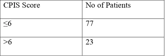
CPIS Score No of Patients
≤6 77
>6 23
The patients with CPIS score of >6 were diagnosed as VAP patients.
[image:54.595.99.373.85.177.2]The mean CPIS of confirmed VAP cases(8.48±1.238) were significantly higher than that of No VAP group (3.55±0.804)(The two tailed p value is <0.0001).
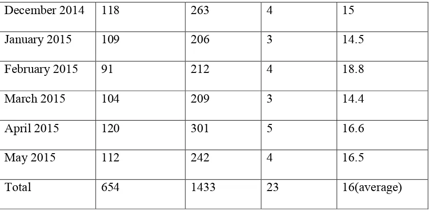
Table 8 Calculation of VAP rate per 1000 ventilator days:
Month No of Patients
on Mechanical Ventilation.
Duration of mechanical ventilation (In days)
No. of VAP cases
diagnosed.
VAP Rate per 1000 ventilator days.
77% 23%
CPIS score
46
December 2014 118 263 4 15
January 2015 109 206 3 14.5
February 2015 91 212 4 18.8
March 2015 104 209 3 14.4
April 2015 120 301 5 16.6
May 2015 112 242 4 16.5
Total 654 1433 23 16(average)
[image:55.595.100.541.83.300.2]In this study ,the VAP rate was 16 per 1000 ventilator days
Table9 Age and Gender distribution of confirmed VAP patients.(N=23)
Age Group
Male N=17
Female N=6
Total
N=23 Percentage
< 30 1 3 4 17%
31-40 2 0 2 9%
41 – 50 2 2 4 17%
51 -60 7 0 7 31%
61-70 3 1 4 17%
>70 2 0 2 9%
47 Table10 Clinical Spectrum of confirmed VAP patients.N=23
Diagnosis Total N=23 Percentage
OPC Poisoning 7 30%
Cardiovascular Diseases 3 13%
Intra-abdominal diseases 4 17%
Neurological Disorders 2 9%
Sepsis 3 13%
CNS Infections 1 4%
Head injury 3 13%
The highest percentage of VAP occurrence was seen among patients with Organo phosphorus poisoning (30%) followed by intra abdominal diseases (17%),Sepsis(13%),head injury(13%).
74% 26%
Gender distribution confirmed VAP Patients
Male
Table 11.VAP onset.
NS-Not Significant.
VAP onset.
Out of 23 confirmed VAP patients, 8(35%) were & 15(65%) were categorised under late onset VAP.
Table 12 Risk factors in patients included in the study: VAP Onset Number of Patients (N
Early 8
Late 15
Out of 23 confirmed VAP patients, 8(35%) were categorised under early onset VAP & 15(65%) were categorised under late onset VAP.
Risk factors in patients included in the study:((N=100)
Early Onset
34.8%
Late Onset
65.2%
Onset of VAP
Number of Patients (N-23) Percentage 34.8% 65.2%
48 categorised under early onset VAP
((N=100)
Early Onset
[image:57.595.99.543.248.640.2]49
Risk factors Total VAP Percentage Pvalue
Prior antibiotics 26 16 61.5% 0.023(S)
Tracheostomy 19 8 42% NS
Stress ulcer prophylaxis 37 7 19% NS
Impaired consciousness 17 6 35% NS
IV sedation 12 6 50% NS
Reintubation 7 5 71% 0.025(S)
Nasogastric tube 17 4 24% NS
Emergency intubation 5 3 60% 0.045(S)
S-Significant,NS-Not significant.
VAP Pathogens: 0
5 10 15 20 25 30 35 40
Risk factors
VAP
50 The majority of the bacterial isolates were found to be gram negative bacilli(91%),of which Non fermentors(65%) were the predominant pathogens isolated from confirmed VAP patients in our study.The gram positive organism accounts for 9% of the VAP isolates ,of which all were methicillin resistant staphylococcus aureus. Of the 23 patients diagnosed as VAP pathogens,21 (91%) patients had monomicrobial infection and 2 (9%) patients had polymicrobial infection.
Table 13 VAP Pathogens(N=23)
Sr.no VAP Pathogens(N=23) Count. (N=23) Percentage
1 Gram negative bacilli 21 91%
Table-14 - Etiological Agents of VAP(n=23) Organism
Acinetobacterbaumannii Pseudomonas aeruginosa
KlebsiellaPneumoniae
KlebsiellaOxytoca
Etiological Agents of VAP(n=23)
Total n=23 Percentage
Acinetobacterbaumannii 9 39%
aeruginosa 6 26%
KlebsiellaPneumoniae 3 13%
2 9%
GNB 91% GPC
9%
VAP Pathogens
51 Percentage
39% 26% 13% 9%
52
E.Coli 1 4%
Staphylococcus aureus 2 9%
[image:61.595.98.545.594.749.2]The most frequently isolated organisms in VAP patients were Acinetobacterbaumannii (39%) followed by Pseudomonas aeruginosa (26%),Klebsiellapneumoniae (13%),Klebsiellaoxytoca(9%),E.coli(4%), Staphylococcus aureus(9%).
Table 15 – Distribution of pathogens among early and late onset VAP
Organism Early (n=8) percentage
Late
(n=15) Percentage
Acinetobacterbaumannii 1 12.5% 8 53%
Pseudomonas aeruginosa 2 25% 4 27%
53
KlebsiellaOxytoca 2 25% 0 -
E.Coli 1 12.5% 0 -
Staphylococcus aureus 0 - 2 13%
The predominant organism in the late onset VAP was
Acinetobacterbaumannii(53%) followed by Pseudomonas aeruginosa(27%).The predominant organism in the early onset VAP group were Pseudomonas aeruginosa(25%),Klebsiella pneumonia(25%) and klebsiellaoxytoca(25%).
Table:16 Distribution of respiratory tract Colonizers in mechanically ventilated patients.(N=100)
1 2 2 2 1
8 4 1 2 0 1 2 3 4 5 6 7 8 9 10 Acinetobacter baumannii Pseudomonas aeruginosa Klebsiella Pneumoniae Klebsiella Oxytoca E.Coli Staphylococcus aureus Distribution of pathogens among early and late onset VAP
[image:62.595.99.546.82.177.2]54
Sno Organism(Colonizer) Count Percentage
1 Acinetobacterbaumannii 10 23%
2 Acinetobacterlwoffii 3 7%
3 KlebsiellaPneumoniae 10 23%
4 KlebsiellaOxytoca 2 4.5%
5 Pseudomonas aeruginosa 8 18%
6 Pseudomonas fluorescens 1 2%
7 Staphylococcus aureus 4 9%
8 Staphylococcus epidermidis 2 4.5%
9 Escherichia coli 2 4.5%
10 Candida albicans 2 4.5%
Total 44 23%
55 Table 17: Distribution of etiological agents causing bacteremiaamong confirmed VAP Patients (n=23)
S.no Blood Culture VAP % P value
1 Acinetobacterbaumannii 1 4.3
0.721 Not 2 KlebsiellaPneumoniae 1 4.3
3 KlebsiellaOxytoca 1 4.3 4 Pseudomonas aeruginosa 2 9
0 5 10 15
Acinetobacter baumannii Acinetobacter lwoffii Klebsiella Pneumoniae Klebsiella Oxytoca Pseudomonas aeruginosa Pseudomonas fluorescens Staphylococcus aureus(MSSA) Staphylococcus epidermidis Escherichia coli Candida albicans
[image:64.595.101.539.84.398.2]56 5 Staphylococcus aureus 3 13.1 Significant
Total 8 35
Blood culture:
Out of 23 VAP cases, blood culture was positive in 8 patients.The organisms isolated were Acinetobacterbaumannii, KlebsiellaPneumoniae,KlebsiellaOxytoca, Pseudomonas aeruginosa, Staphylococcus aureus.The sensitivity of blood cultures for the diagnosis of VAP is low and also if positive,the organisms may originate from an extrapulmonary site of infection.
[image:65.595.97.478.84.146.2]Antimicrobial susceptibility pattern of the Gram negative and Gram positive isolates causing VAP is shown below.Most of the Gram negative organisms and gram positive organisms isolated were multidrug resistant.
Table 18: Antimicrobial sensitivity pattern of Gram negative isolates
Organism A K C O T
CIP CT
X C A Z C A C C X G M IM P M E R P T Acinetobacterb aumannii(n=9)
44% (
4) 2 2 % (2 ) 33% ( 3)
- 11
%
(1
)
- 44
57 Pseudomonas aeruginosa (n=6) 67% ( 4)
- 17% (
1)
- 33
%
(
2
)
- 50
% (3 ) 5 0 % ( 3 ) 6 1 % ( 4 ) 6 1 % ( 4 ) 5 0 % ( 3 ) Klebsiellapneu
moniae (n=3)
67% ( 2) 0 % 33% ( 1) 0 % 0 % 6 7 % ( 2 ) 6 7 % ( 2 ) 3 3 % ( 1 ) 1 0 0 % ( 3 ) 1 0 0 % ( 3 ) 6 7 % ( 2 ) Klebsiellaoxyt oca (n=2) 50% ( 1) 0 % 50% ( 1) 0 % 0 % 1 0 0 % ( 2 ) 1 0 0 % ( 2 ) 5 0 % ( 1 ) 1 0 0 % ( 2 ) 1 0 0 % ( 2 ) 1 0 0 % ( 2 )
E.Coli (n=1)
100% (
1)
0
%
0% 0% 0% 10
0 % ( 1 ) 1 0 0 % ( 1 ) 1 0 0 % ( 1 ) 1 0 0 % ( 1 ) 1 0 0 % ( 1 ) 1 0 0 % ( 1 ) AK-Amikacin,COT-Cotrimoxazole,CIP-ciprofloxacin,CTX-cefotaxime,CAZ-ceftazidime,CAC-Ceftazidime&clavulanic acid,CX-cefoxitin, GM-Gentamicin,IMP-Imipenem,MER-Meropenem, PT-Piperacillintazobactum.
The nonfermentors showing resistance to carbapenemswere further subjected to Macrobroth dilution method for determining the MIC.
Table 19.Sensitivity to meropenem by Disc diffution method and Macrobroth dilution method
[image:66.595.100.539.82.368.2]58
Method Acinetobacterbaumannii
.
Pseudomonas aeruginosa
Count Percentage Count Percentag
e Kirby Bauer’s Disc Diffusion
method.
3 33%
(3/9)
2 33%
(2/6)
Macrobroth dilution method 3 33% 2 33%
The isolates of nonfermentors showing resistance to carbapenem (meropenem) by Disc Diffusion method also showed resistance by Macrobroth dilution method with a MIC value of >8µg/ml.
59
Antimicrobial sensitivitypattern of Pseudomonas aeruginosa
67% isolates of Pseudomonas aeruginosa were sensitive to amikacin,67% to carbapenems and 50% to Piperazilintazobactum.
Antimicrobial sensitivity pattern of Enterobacteriaceae 44% 22% 33% 11% 44% 22% 67% 67% 44% 0% 10% 20% 30% 40% 50% 60% 70% 80%
AK COTRI CIP CAZ CX GM IMP MER PT
Acinetobacter baumannii
Sensitive 67% 17% 33% 50% 50% 67% 67% 50% 0% 10% 20% 30% 40% 50% 60% 70% 80%AK CIP CAZ CX GM IMP MER PT
Pseudomonas aeruginosa
60 The isolates of Enterobacteriacea were 67% sensitive to Amikacin83% to Piperazilin-tazobactum.All were sensitive to carbapenems.