0022-538X/02/$04.00⫹0 DOI: 10.1128/JVI.76.14.7150–7162.2002
Copyright © 2002, American Society for Microbiology. All Rights Reserved.
Herpes Simplex Virus Type 1/Adeno-Associated Virus
rep
⫹
Hybrid
Amplicon Vector Improves the Stability of Transgene Expression in
Human Cells by Site-Specific Integration
Y. Wang,
1* S. M. Camp,
2M. Niwano,
1† X. Shen,
1J. C. Bakowska,
2X. O. Breakefield,
2and P. D. Allen
1Department of Anesthesia, Brigham & Women’s Hospital,1and Molecular Neurogenetics, Department of Neurology, Massachusetts
General Hospital,2Boston, Massachusetts
Received 2 January 2002/Accepted 16 April 2002
Herpes simplex virus type 1 (HSV-1) amplicon vectors are promising gene delivery tools, but their utility in gene therapy has been impeded to some extent by their inability to achieve stable transgene expression. In this study, we examined the possibility of improving transduction stability in cultured human cells via site-specific genomic integration mediated by adeno-associated virus (AAV) Rep and inverted terminal repeats (ITRs). A
repⴚHSV/AAV hybrid amplicon vector was made by inserting a transgene cassette flanked with AAV ITRs into
an HSV-1 amplicon backbone, and arepⴙHSV/AAV hybrid amplicon was made by insertingrep68/78outside
therepⴚvector 3ⴕAAV ITR sequence. Both vectors also had a pair ofloxPsites flanking the ITRs. The resulting
hybrid amplicon vectors were successfully packaged and compared to a standard amplicon vector for stable transduction frequency (STF) in human 293 and Gli36 cell lines and primary myoblasts. Therepⴙ, but not the
repⴚ, hybrid vector improved STF in all three types of cells; 84% of Gli36 and 40% of 293 stable clones
transduced by therepⴙhybrid vector integrated the transgene into the AAVS1 site. Due to the difficulty in
expanding primary myoblasts, we did not assess site-specific integration in these cells. A strategy to attempt further improvement of STF by “deconcatenating” the hybrid amplicon DNA via Cre-loxPrecombination was tested, but it did not increase STF. These data demonstrate that introducing the integrating elements of AAV into HSV-1 amplicon vectors can significantly improve their ability to achieve stable gene transduction by conferring the AAV-like capability of site-specific genomic integration in dividing cells.
The elucidation of the molecular bases of inherited or ac-quired diseases and the completion of the human genome map have challenged vector development in the field of gene ther-apy. In many gene therapy paradigms, successful long-term gene transduction has not been achieved due to the loss of transgene expression over time, particularly when nonintegra-tive vectors, such as those based on adenovirus or herpes simplex virus type 1 (HSV-1), were used. Although the mech-anisms involved are not well understood, some evidence sug-gests that down-regulation of the transgene promoter (46, 50, 54), degradation or extrusion of transgene DNA (50), and rejection of transgene-expressing cells by the immune system (20, 21) may all lead to the instability of transgene expression. The most common approaches to this problem have been to attempt to maintain the transgene vectors as stable episomal elements, e.g., with Epstein-Barr virus or mammalian artificial chromosome sequences, or to promote transgene integration into the cellular genome of the host (for a review, see reference 27). The present study was designed to address the latter ap-proach using elements of adeno-associated virus (AAV) and P1 bacteriophage loxP in the context of a plasmid-based HSV-1 amplicon vector.
HSV-1 amplicon vectors are highly versatile vectors with very attractive aspects in terms of gene therapy. They can very efficiently infect a broad range of dividing and nondividing cells with minimal multiplicities of infection (MOIs) (51, 53) and can deliver up to 153 kb of DNA into mammalian cells (51). A major drawback of all HSV-1 vectors is the transient nature of transgene expression that is most likely related to the nonepi-somal and nonintegrated state of amplicon DNA in the host cells. AAV is a human parvovirus with a 4.7-kb single-stranded linear DNA genome, which consists of two open reading frames, encoding four overlapping regulatory proteins (Rep78, -68, -52, and -40) and three capsid proteins, flanked by 145-base inverted terminal repeats (ITRs) (45). This virus has the unique ability during latent infection to undergo site-specific integration of its viral DNA into the AAVS1 region in chro-mosome 19 (19q13.3-qter) of the human genome (26, 39). The mechanism of the site-specific integration has not been fully elucidated. However, there is consistent evidence from several laboratories suggesting that the ITRs and the two large Rep proteins (Rep68 and Rep78) are responsible for this function. Recombinant AAV (rAAV) vectors which are devoid of all AAV sequences except the two ITRs lose their site-specific integration ability, although they are still capable of random integration (31, 35, 37, 60). However, when Rep68, Rep78, or Rep68 and Rep78 is expressed incisortrans relative to the ITRs, the AAV ITRs and ITR-flanked DNA can be “rescued” from the plasmid backbone of AAV-derived plasmids and in-tegrated into the AAVS1 site of transduced cells (1, 28, 48, 57). It has not been possible to includerepand transgene together * Corresponding author. Mailing address: Department of
Anesthe-sia, Brigham & Women’s Hospital, 75 Francis St., Boston, MA 02115. Phone: (617) 732-6879. Fax: (617) 732-6927. E-mail: Yaming@zeus .bwh.harvard.edu.
† Present address: First Department of Surgery, Facility of Medi-cine, Kyoto University, Kyoto, Japan.
7150
on November 8, 2019 by guest
http://jvi.asm.org/
in recombinant AAV vectors because of their relatively small packaging capacity (⬍5 kb), but in recent years, hybrid vectors have been created which incorporate the AAV ITRs andrep
genes in combination with larger transgene capacities. These include the hybrid baculovirus-AAV vector (BAC-AAV), the hybrid adenovirus/AAV ITR vector (Ad/AAV) in combination with an adenovirus/AAVrep68/78 helper vector, and the hy-brid HSV-1 amplicon vector (HSV/AAV) (23, 34, 36). With the BAC-AAV and Ad/AAV vectors, both enhanced stable transduction frequencies (STFs) over the nonhybrid parent vectors and site-specific integration were achieved. In the pre-vious versions of the HSV/AAV hybrid vectors, only partial ITR sequences were included inadvertently, and in the later version of the vectors, the green fluorescent protein (GFP)-expressing HSV/AAV hybrid vector, there were no Rep pro-teins being expressed due to a construction error. Therefore, although improved stability of transgene expression was ob-served both in cultured cells and in vivo, site-specific integra-tion was not evaluated (6, 10, 23).
The P1 bacteriophage Cre-loxPsystem has been widely used for in vitro and in vivo genomic manipulation. This system includes the 38-kDa recombinase (Cre) and the 34-bp loxP
target sequence (47). When two parallelloxPsites exist in the same DNA molecule, Cre will first bind these two sites to-gether to form a circular protein-DNA complex and then me-diate a synaptic union of these twoloxPsites. This results in two smaller circular molecules, each of them containing one
loxPsite (13).
HSV-1 amplicons are plasmid-based vectors containing only two cis-acting HSV-1 elements, a packaging signal and an origin of DNA replication (12, 43, 44). The packaged amplicon vectors have a⬃152-kb DNA genome consisting of multiple head-to-tail copies of the amplicon (amplicon concatemer). Hybrid HSV/AAV amplicons also include multiple copies of the AAV ITR-flanked and rep-containing units. Structurally, this is very different from wild-type (wt) and recombinant AAV in which the ITRs exist at the two ends of the linear genome. Theoretically, Rep should first rescue the ITR-flanked frag-ment from the vector genome and then target it to the AAVS1 site. Since rescue and integration are two contradictory func-tions of Rep proteins and each of them might require a dif-ferent level of Rep expression (2), we predict that HSV/AAV vectors might have a reduced efficiency of integration com-pared to that of wt AAV. Based on this analysis, we hypothe-size that if the ITR-flanked transgene were first excised from the vector concatemer by means of Cre-loxPrecombination, Rep expressed at low levels could facilitate site-specific inte-gration.
In this study, we reengineered the HSV/AAV hybrid ampli-con to ampli-contain the full-length ITRs flanked byloxPsites, with or withoutrep68/78. This was done to test the hypothesis that inclusion of this AAV “integration machinery” into an HSV-1 hybrid amplicon vector could promote the transgene integra-tion into the host genome and thereby stabilize the transgene expression. The HSV/AAV hybrid vectors were evaluated for STFs as well as site-specific integration events in three differ-ent types of human cells. In addition, we tested our hypothesis that deconcatenation of the amplicon DNA via Cre-loxP re-combination would further enhance site-specific integration. Our results show that a Rep-expressing HSV/AAV hybrid
vec-tor (rep⫹) enhanced the STF in all cell types tested. The site-specific integration rate was as high as 84% in the stably trans-duced Gli36 colonies examined. There was no additional improvement in STF after deconcatenation with Cre-loxP re-combination, but the vector DNA was appropriately targeted to AAVS1 in the clones transduced byrep⫹vector at a slightly higher frequency than without deconcatenation. The ability to achieve site-specific integration of transgene via HSV/AAV hybrid vectors should have a significant impact on stable and reproducible levels of transgene expression in transduced cells that contain the AAVS1 site.
MATERIALS AND METHODS
Cell culture. Human embryonic kidney 293 cells were obtained from the American Type Culture Collection (Manassas, Va.). 293T/17 cells express the simian virus 40 large T antigen and were selected specifically for high transfec-tion efficiency (provided by David Baltimore, Massachusetts Institute of Tech-nology, Cambridge). Human glioma Gli36 cells were provided by Anthony Cam-pagni (UCLA School of Medicine). African green monkey kidney VERO 2-2 cells were provided by Rozanne Sandri-Goldin (University of California, Irvine). Human primary myoblasts were provided by Emanuela Gussoni (Children’s Hospital, Boston, Mass.). The HEK293, 293T/17, Gli36, and VERO 2-2 cells were grown in Dulbecco’s modified Eagle medium with 10% fetal bovine serum (Sigma, St. Louis, Mo.) supplemented with 100 U of penicillin/ml and 0.1 mg of streptomycin (Sigma)/ml. VERO 2-2 cell growth medium was further supple-mented with 0.4 mg of G418 (Gibco/BRL, Rockville, Md.)/ml. Primary myoblasts were grown in Ham’s F10 media supplemented with 20% fetal bovine serum and 5 ng of basic fibroblast growth factor (Promega)/ml. All cells were incubated at
37°C and 5% CO2with a humidified atmosphere.
A Cre recombinase-expressing HEK293 cell line was generated by infecting wt HEK293 cells with a retroviral vector carrying Cre recombinase and puromycin expression cassettes (kindly provided by Philippe Leboulch, Massachusetts
In-stitute of Technology). Infected cells were selected with 2g of puromycin/ml for
1 week. Puromycin-resistant colonies were isolated and expanded. The expres-sion of Cre recombinase in stable cell lines was detected by immunocytochem-istry with anti-Cre antibody (Babco, Richmond, Calif.).
Vector constructs.AAV ITR sequences with unique restriction sites at each end were synthesized manually (Midland Certified Reagent Co.). Plasmid pHS-VloxP was created by replacing the polylinker of pHSVprPUC (gift of Howard Federoff, University of Rochester, Rochester, N.Y.) with a new oligonucleotide
polylinker. This contained a pair ofloxPsites near each end flanked byAscI and
PshaI (5⬘) andEcoRV (3⬘) and hadNotI,PacI,BglII,SalI, andSgfI restriction
sites in betweenloxPsites. Plasmid pHSVLGN was constructed by inserting the
BglII-flanked cytomegalovirus promoter-driven enhanced GFP (eGFP)
expres-sion cassette and simian virus 40 promoter-driven neomycin (neo)-resistance protein expression cassette derived from plasmid pHyRGN (10) into pHSVloxP
(Fig. 1A). Plasmid pHLIGN was constructed by sequentially inserting the 3⬘
AAV ITR into theSalI andSgfI sites, the eGFP and neo-resistance cassette
fragment into theBglII site, and the 5⬘AAV ITR into theNotI andPacI sites of
pHSVloxP (Fig. 1B). The AAVrep68/78expression cassette with its p5 promoter
(derived from pRep68/78 in which the expression of Rep52 and Rep40 had been aborted with a missense mutation of their starting codon; a gift of Cornel Fraefel, Institute of Virology, University of Zurich, Zurich, Switzerland) was inserted in
both orientations either in thePshAI site upstream of the 5⬘loxPsite or in the
EcoRV site downstream of the 3⬘loxPsite of pHLIGN. Of these four plasmids,
the only one used in further experiments was pHLIRGN, in whichrep was
inserted in the forward orientation after the 3⬘loxPsite (Fig. 1C) as the other
yielded low titers on packaging. The integrities of the ITRs in pHLIGN and pHLIRGN were determined by sequential digestion with either of the enzymes that digest the sites flanking ITRs, followed by digestion with enzymes that digest
the sites inside of ITR (MscI andSmalI sites exist inside of the ITRs as
palin-dromes). ITR fragments were resolved by electrophoresis on 5% Nusieve aga-rose (FMC, Rockland, Maine) gels.
Vector packaging. The helper virus-free packaging system developed by Fraefel et al. (11) was used to package all amplicons. In brief, VERO 2-2 cells were transfected with a mixture of amplicon plasmid, a set of five cosmids, and Lipofectamine (Gibco/BRL). The five cosmids spanned the entire HSV-1
ge-nome, with only the cleavage-packaging signals (“a” sequences containing the
pacsignals) deleted. Amplicon vectors were harvested 60 h later, freeze thawed
three times, sonicated, and centrifuged at 1,000⫻gfor 10 min. Amplicon vectors
on November 8, 2019 by guest
http://jvi.asm.org/
were then concentrated and purified by centrifugation at 55,000⫻gfor 2 h onto a 25% sucrose gradient. Amplicon vector stock titers were determined as
trans-duction units per milliliter (TU/ml) by infecting 3⫻105293T/17 cells per well in
24-well plates and then counting the GFP-positive cells 18 h postinfection at a
20⫻magnification.
Western blot analysis. The expression of Rep proteins was evaluated by Western blot analysis. 293T/17 cells transfected with amplicons pHLIRGN, pH-LIGN, pHSRepN1 (from David Jacoby, Massachusetts General Hospital, Bos-ton, Mass.), and pPSSV9 (from Richard J. Samulski, University of North Caro-lina at Chapel Hill, Chapel Hill, N.C.) were lysed in a buffer containing 1% NP-40, 50 mM Tris (pH 8.0), 150 mM NaCl, and Complete-Mini Cocktail of protease inhibitors (Boehringer Mannheim, Indianapolis, Ind.). Protein concen-trations were determined using the Coomassie Plus protein reagent assay (Pierce, Rockford, Ill.) and a bovine serum albumin standard (Bio-Rad,
Her-cules, Calif.). Equal amounts of cell lysates (300g) were denatured and
sepa-rated by electrophoresis on 10% polyacrylamide gels with sodium dodecyl sul-fate. Rainbow Markers (Amersham Life Sciences, Arlington Heights, Ill.) were used as molecular weight markers. Proteins were transferred to nitrocellulose
membrane (Bio-Rad Trans-blot Transfer Medium Pure; 0.45-m pore size) in
transfer buffer (25 mM Tris, 192 mM glycine, pH 8.3) by using a Bio-Rad Trans-blot Cell for 3 h at 0.5 mA at 4°C. Membranes were stained with 0.2% Ponceau S (Sigma) to ensure proper transfer and equal loading of samples. After staining, membranes were blocked overnight in 10% nonfat dry milk in TBS-T (150 mM NaCl, 50 mM Tris-HCl [pH 7.9], 0.01% Tween 20). The following day, membranes were washed twice for 15 min and twice for 5 min in TBS-T and then incubated for 1 h at room temperature with primary antibodies in 2% nonfat dry milk in TBS-T. Mouse anti-AAV Rep clone 303.9 (ARP Scientific, Belmont, Mass.) was developed against all four Rep proteins (with molecular masses of 40, 52, 68, and 78 kDa) and used at dilutions of 1:25. The membranes were then washed as before and incubated for 30 min with a 1:10,000 dilution of anti-mouse immunoglobulin G (IgG) horseradish peroxidase-conjugated secondary antibody (Amersham Life Sciences) in TBS-T with 5% nonfat dry milk. After washing, as described above, the blots were developed using enhanced chemiluminescence (ECL) reagents (Amersham Life Sciences). Membranes were then exposed to film for 3 min.
Cloning efficiency and STF.One day prior to infection, 293 cells, Gli36 cells,
or human primary myoblasts were plated at a density of 5⫻105cells per
60-mm-diameter dish. On the following day, cells were infected at an MOI of 0.1 or 1 TU/cell with either HLIRGN or HLIGN hybrid vectors. In parallel, all cell types were infected with the nonhybrid HSVLGN vector as a control for the presence of AAV ITRs, and 293 cells were also infected with HSVGN as a
control for the presence ofloxPsites. The day after infection, all
vector-contain-ing medium was replaced with fresh growth medium. Forty-eight hours postin-fection, cells were harvested and then subjected to fluorescence-activated cell sorter (FACS) analysis for GFP-positive cells, as appropriate. The unsorted cells
were replated at specific cloning densities and grown in media with or without 1 mg of G418 (Gibco/BRL)/ml for 293 and Gli36 cells or 0.4 mg/ml for human primary myoblasts for 10 to 20 days. All clones in the plates with or without G418 selection were counted, and the cloning efficiencies and stable transduction frequencies (STFs) were determined, respectively. Cloning efficiency was defined as the number of clones formed without drug selection divided by the number of cells plated. STF was defined as the number of neomycin-resistant clones after G418 selection divided by product of the number of cells plated times the
percentage of GFP⫹cells in the original sample times the cloning efficiency. In
addition, one group of 293 cells was also FACS sorted for GFP-positive cells and plated at cloning density in two groups without G418 selection. One group was removed from their plates at 1 and 2 weeks postsorting and reanalyzed each time
by FACS for the percentage of GFP⫹cells. The other group had the number of
GFP-positive and -negative clones counted at 2 weeks postsorting. The STF of this group of cells without the influence of G418 selection was then determined using the same formula used above.
FACS and analysis.All cell sorting and dynamic GFP expression analysis was done using a FacsCalibur analyzer (Becton-Dickinson, Franklin Lakes, N.J.).
Hirt DNA extraction and bacteria transformation.Five million Cre-expressing or wt 293 cells were infected by HLIGN at an MOI of 3. Forty-eight hours after infection, the cells were harvested and the episomal DNA was extracted accord-ing to a modified Hirt DNA extraction procedure (18). Five microliters of Hirt
DNA was used to transform DH5␣-competent bacteria (Invitrogen) according to
the manufacturer’s instructions. The resulting plasmid DNA was analyzed by digestion with restriction enzymes and followed by gel electrophoresis.
Southern blot analysis.Isolated GFP⫹colonies from Gli36 and 293 cells
infected by the each of the three vectors (HLIRGN, HLIGN, or HSVLGN) were expanded, and genomic DNA was extracted according to standard methods.
Twenty micrograms of genomic DNA was digested withEcoRI, which doesn’t cut
theloxP-flanked transgene cassette or the amplicon backbone but does cut the
repgene and also generates a native 8.2-kb AAVS1 fragment from human
genomic DNA. After electrophoresis in 0.7% agarose gels, the digested DNA was transferred onto a nylon membrane. The membrane was first hybridized
using a 0.7-kbPmeI fragment derived from the eGFP cDNA as probe, and then
the blot was stripped off. The blot was exposed to film for 24 h to ensure no
radioactive signal was left and then rehybridized using a 1.1-kbBamHI/PvuII
fragment derived from the AAVS1 locus (kindly donated by R. M. Kotin,
Na-tional Institutes of Health) as probe. All probes were labeled with [32P]dCTP
using a Radprime DNA labeling system (Invitrogen). The genomic DNA from
colonies infected with therep⫹amplicon was also cut withKpnI, which cuts the
vector twice, generating 11- and 0.5-kb bands. This blot was hybridized using a
1.1-kbBamHI/XhoI fragment from pRep68/78 as probe to examine the incidence
ofrepintegration. Some of the Hirt DNA from Cre⫹or wt 293 cells was digested
withNheI, a unique cutter in the transgene region, and hybridized with the eGFP
probe. Finally, two sets of genomic DNA from Gli36 and 293 colonies infected
withrep⫹amplicon were cut withStuI, the enzyme that cuts once inside of the
transgene, and were hybridized with the 1.8-kb amplicon backbone probe or 0.7-kb eGFP probe to check the integration of amplicon backbone and the existence of a concatemer of ITR-flanked transgene units.
RESULTS
Construction and packaging of HSV and HSV/AAV hybrid amplicon vectors. The ITRs of AAV and a transcriptional cassette for the two large AAV Rep proteins were inserted into HSV-1 amplicons. In addition, a pair ofloxP sequences was also added into the amplicons flanking the ITRs (in the case of hybrid vector) or into the transgene cassette (in the case of standard amplicon). The resulting HSV/AAV hybrid vectors and a control vector lacking AAV ITRs, all withloxPsites, are shown in Fig. 1.
Since the highly homologous sequences between the two AAV ITRs can cause recombination events leading to muta-tions of the ITR during plasmid replication and because vari-ous deletions were detected in the ITR fragments in plasmids that were made available to us, we synthesized the ITRs (Mid-land Certified Reagent Co.) flanked by unique restriction sites for insertion into our hybrid amplicons. After the final ampli-cons were completely ampli-constructed, the integrities of the ITRs FIG. 1. Vector constructs. (A) A standard amplicon with a pair of
loxPgenes flanking GFP and neomycin resistance transgene cassettes. (B) Therep⫺HSV/AAV hybrid amplicon was made by inserting a pair
of AAV ITRs into the standard amplicon flanking the transgene cas-settes. (C) Therep⫹HSV/AAV hybrid amplicon was made by adding
Rep68/78 into therep⫺hybrid amplicon downstream of the 3⬘loxPsite.
on November 8, 2019 by guest
http://jvi.asm.org/
were verified by digestion with restriction enzymes that flank an individual ITR and then byMscI andSmaI that cut inside the ITRs, and the restriction products were resolved using 5% Nusieve agrose gel electrophoresis. Analysis of the restriction products from all hybrid vectors, including pHLIGN and pH-LIRGN, demonstrated the presence of all expected restriction products (data not shown), indicating that the ITRs were in-tact.
All vectors were packaged using the five-cosmid helper vi-rus-free packaging system (11). Therep⫺HLIGN vector and both control vectors all had titers of 1⫻106to 6⫻106TU/ml
in crude stocks and 1⫻108to 3⫻108TU/ml after purification.
Of the vectors that contained therep68/78gene, only when the
repgene was placed downstream of the 3⬘ITR at the forward orientation was a comparable amplicon vector titer achieved. This amplicon, pHLIRGN orrep⫹hybrid vector (Fig. 1C), was the only one of these fourrep-containing vectors to be used for further studies, and its range of titers was 0.4⫻105to 2⫻105
TU/ml in crude stock and 0.5⫻ 107to 1⫻107TU/ml after
purification.
The expression of the Rep68 and Rep78 proteins in cells transduced with the HSV/AAV rep⫹ amplicon, pHLIRGN, was confirmed by Western blotting. 293T/17 cells were trans-fected with pHLIRGN, pHLIGN, pHSRepN1, or pSSV9. Plas-mids pHSRepN1 and pSSV9 express all four Rep proteins. Mouse anti-AAV Rep clone 303.9 primary antibodies were used to detect the expression of Rep proteins. Four Rep pro-teins were shown in the lysates of 293T/17 cells transfected by both HSRepN1 and pSSV9. Only Rep68 and -78 were detected in the lysates of pHLIRGN-transfected cells and, as expected, no Rep proteins were expressed in pHLIGN-transfected cells (data not shown).
Evaluation of AAV hybrid function. (i) Stability of transgene expresion. The aim of this study was to obtain more stable transgene expression through the function of ITRs and Rep proteins of AAV within the context of the HSV-1 amplicon vector. This function was assessed in three cultured human cell types, 293, Gli36 (glioma cells), and primary myoblasts.
First, we examined the dynamics of transgene expression over time after infection without G418 selection. Human pri-mary myoblasts and 293 cells were infected with each of the three vectors in Fig. 1 at an MOI of 1 and were then subjected to FACS sorting for GFP⫹cells 48 h postinfection. The initial infection efficiencies were ⬃20, ⬃90, and ⬃40% for 293, Gli36c and primary myoblasts, respectively, at an MOI of 1 for control andrep⫺vector and a slight decease forrep⫹. There was no notable cell death observed at this MOI, and no no-ticeable difference in growth rates was seen among human primary myoblasts infected by the three vectors, but there was a slightly slower growth rate seen in 293 cells infected by the
rep⫹vector. The cell populations were analyzed sequentially by FACS sorting for the percentage of GFP⫹cells at 1 and 2 weeks postinfection. For both cell types, there were more GFP⫹cells at 1 and 2 weeks postinfection in therep⫹group than in the control group (2.7- and 14.4-fold more at 1 week postinfection and 13.2- and 5-fold more at 2 weeks postinfec-tion for 293 cells and primary myoblasts, respectively, com-pared to the cells infected by the standard amplicon, HSV-LGN) (Table 1). However, there were 6.2- and 7.5-fold decreases in the number of GFP⫹293 cells and human primary
myoblasts, respectively, at 2 weeks postinfection compared to the number at 1 week postinfection. Although therep⫺vector had a slight advantage over the control vector at 1 week postin-fection (1.5- and 3-fold, respectively), this advantage was lost at 2 weeks postinfection (P⬎0.05).
Second, the STF with G418 selection was determined for vectors HLIRGN, HLIGN, or HSVLGN. The results from all three cell types show that therep⫹HSV/AAV hybrid vector gave rise to significantly higher STFs (P ⬍ 0.01) than the standard amplicon. Therep⫺hybrid vector yielded the same to slightly decreased STFs in 293 cells as the standard amplicon, and no stable colonies were seen in the Gli36 cells and primary myoblasts (Table 2). These observations indicate that the AAV ITRs alone do not enhance the integration of amplicon vector DNA into the host genome. However, the large Rep proteins together with ITRs were able to supply this AAV function to the HSV/AAV hybrid vector. In all the three cell types tested, HLIRGN at both MOIs yielded the highest STF.
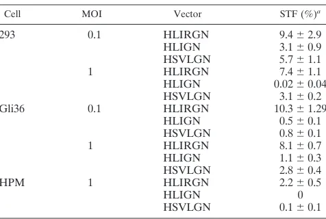
[image:4.587.301.543.84.175.2](ii) Site-specific integration.A variety of vectors with AAV ITRs andrepsequences included have been demonstrated to be able to mediate transgene integration into the AAVS1 site in the transduced human cells (1, 28, 34, 40, 48). To determine the efficiency of site specific integration mediated by the HSV/ AAV hybrid amplicon, stable GFP⫹clones from Gli36 cells infected with HLIRGN, HLIGN, or HSVLGN were expanded, and genomic DNA was extracted and then subjected to South-ern blot analysis. Twenty-seven clones infected with therep⫹
TABLE 1. Dynamics of transgene expression
Cell Vector % of GFP
⫹cells ata:
1 week 2 weeks
293 HLIRGN 11.7⫾0.4 1.9⫾0.4
HLIGN 6.4⫾0.2 0.1⫾0.04
HSVLGN 4.4⫾0.3 0.2⫾0.1
HPMb HLIRGN 15.3⫾2.3 2.0⫾0.4
HLIGN 3.2⫾0.6 0.6⫾0.2
HSVLGN 1.0⫾0.3 0.4⫾0.1
aFACS analysis data.n⫽6 for all of the groups.
bHPM, human primary myoblast.
TABLE 2. STFs
Cell MOI Vector STF (%)a
293 0.1 HLIRGN 9.4⫾2.9
HLIGN 3.1⫾0.9
HSVLGN 5.7⫾1.1
1 HLIRGN 7.4⫾1.1
HLIGN 0.02⫾0.04
HSVLGN 3.1⫾0.2
Gli36 0.1 HLIRGN 10.3⫾1.29
HLIGN 0.5⫾0.1
HSVLGN 0.8⫾0.1
1 HLIRGN 8.1⫾0.7
HLIGN 1.1⫾0.3
HSVLGN 2.8⫾0.4
HPM 1 HLIRGN 2.2⫾0.5
HLIGN 0
HSVLGN 0.1⫾0.1
aSTF⫽Number of stable transduced colonies/number of cells infected⫻
efficiency of colony formation⫻100%.n⫽8 for all of the groups.
on November 8, 2019 by guest
http://jvi.asm.org/
[image:4.587.302.540.551.711.2]vector, 14 clones infected with therep⫺vector, and 18 clones infected with a standard amplicon were examined. Integration at AAVS1 was assessed by hybridizing EcoRI-digested genomic DNA from these cells with a GFP probe followed by an AAVS1 probe. Shifted AAVS1 bands were detected in 26 out of 27 clones infected with therep⫹vector, and 22 of them also hybridized to the GFP probe in the same location. In contrast, there was no shift of AAVS1 bands seen in any of the clones derived fromrep⫺or control vectors (Fig. 2).
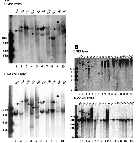
(iii) Integration ofrepgene, amplicon backbone, and AAV-like concatemer.In therep⫹HSV/AAV construct, therepgene is placed outside of the ITRs. Theoretically, transgene integra-tion mediated by Rep proteins should occur primarily at the ITRs, thus excluding the rep gene and other amplicon se-quences from the integration so that the expression and the potential toxicity of Rep proteins would only be temporary. To test this hypothesis, the genomic DNA of Gli36 cells from the clones infected with therep⫹vector was cut with KpnI and analyzed by Southern blotting with a cDNA probe coding for the rep sequence. Six of 27 clones showed a rep⫹ band in various sizes. This indicates thatrep integration is not com-pletely avoided by this vector design but occurs only in a small fraction of the integration events (Fig. 3). The integration of amplicon backbone was also examined. Interestingly, the back-bone sequence was detected in 3 of 27 clones but only 1 of those clones was included an integratedrepsequence (data not shown). In the latent infection of wt AAV and rAAV, integra-tion could occur as concatemers. To examine if this was the case in the integration mediated by therep⫹hybrid vector, the genomic DNA was also cut withStuI, which cuts once inside of transgene, and hybridized with eGFP probe. Putatively, it should generate an extra band with a 4-, 6-, or 0.3-kb size in the case of head-to-tail, head-to-head, or tail-to-tail concatemers, respectively. There was no matched image found (data not shown(.
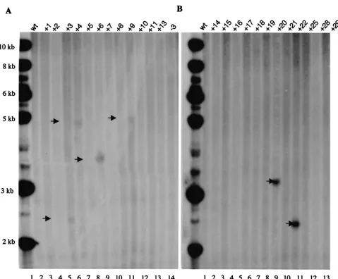
Effects of deconcatenation. (i) Confirmation of Cre/LoxP function.The efficiency of Cre-loxPrecombination for decon-catenation was determined by analyzing the Hirt DNA ex-tracted from both Cre-expressing 293 cells (293 Cre⫹cells) and
wt293 cells infected by the HLIGN vector. Two approaches were utilized. First, Hirt DNA from Cre-expressing 293 Cre⫹ cells or wt 293 cells was used to transform bacteria. Twenty-eight colonies were obtained in the plate transformed by the Hirt DNA from 293 Cre⫹cells and one colony from the wt 293 plate. Ten colonies from the 293 Cre⫹plate and the single colony from the wt 293 plate were analyzed by restriction digestion and agarose gel electrophoresis. Nine out of 10 293 Cre⫹colonies were found to contain only the amplicon back-bone (5 kb) as demonstrated by their size and the fact that all of the enzymes that cut in between theloxPsites (NheI,NotI, and SalI) failed to digest the plasmid, but AscI, which cuts outside of the 5⬘loxP, did linearize the plasmid (Fig. 4B). The remaining Cre⫹ colony as well as the colony from wt 293 contained the intact HLIGN (9-kb) plasmid (Fig. 4B). South-ern blot analysis performed directly on the Hirt DNA digested withNheI that cuts once inside the ITRs of therep⫺amplicon within the transgene should generate a single 9-kb band in monomers and multimers ofrep⫺vector DNA. On the other hand,NheI should generate a 4-kb band in transgene-contain-ing minicircles yielded by Cre-loxP recombination. Identical
amounts of uncut andNheI-cut Hirt DNA from 293 Cre⫹and wt 293 cells were probed with a 0.7-kbPmeI fragment contain-ing the transgene sequence (Fig. 4B). The resulst showed that both of the 4- and 9-kb bands could be found in the lane loaded with 293 Cre⫹ Hirt DNA, and both bands showed similar signal intensity. This indicates that about half of the transgene had been dissociated from the amplicon backbone (Fig. 4C). There was no 4-kb band observed in wt 293 Hirt DNA. In the lanes loaded with uncut Hirt DNA from both Cre⫹and wt 293 cells, no positive signal was observed. This is probably a result of inefficient transfer of circularized DNA. Both of these ex-periments support excision of the ITR-flanked transgene from the amplicon genome in the 293 Cre⫹cells but not in the wt 293 cells.
(ii) Stability of transgene expression after deconcatenation. Next, we examined the effects of deconcatenation by Cre/LoxP
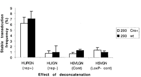
recombination on STF in 293 Cre⫹and wt 293 cells following infection by HLIRGN, HLIGN, HSVLGN, and HSVGN at an MOI of 1. Vector HSVGN is identical to HSVLGN except that it lacks theloxPsignals, and it was added as a control to rule out nonspecific effects of Cre recombinase. GFP-positive cells were FACS sorted 48 h postinfection and plated at cloning density. The sorted cells were grown without G418 selection for 2 weeks, and the GFP⫹colonies were counted. The STF for each vector was calculated using the formula described in the previous section (Fig. 5). In 293 Cre⫹cells, among the four vectors, HLIRGN generated the highest STF. However, when compared to wt 293 cells, deconcatenation did not improve STF for any of the vectors.
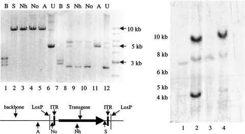
(iii) Site-specific vector integration andrep and amplicon backbone integration after deconcatenation.Site-specific inte-gration after deconcatenation was also examined. A total of 40 clones, 10 each of GFP⫹clones from 293 Cre⫹and wt 293 cells infected by HLIRGN or HLIGN, were expanded and their
EcoRI-digested genomic DNA was analyzed by Southern blot-ting. Site-specific integration was found in 40% of wt 293 and 50% of 293 Cre⫹colonies infected by HLIRGN (Fig. 6). As expected, no AAVS1 band shifts indicative of site-specific in-tegration were detected in any of the colonies infected by HLIGN in either wt 293 or 293 Cre⫹ cells. The effect of deconcatenation on integration of therepgene was also exam-ined in the clones infected by HLIRGN. One of 10 clones from wt 293 cells showed integration of therepgene, and this par-ticular clone integrated into the host genome at multiple sites as shown in the Southern blot probed withrepsequence (Fig. 7). None of clones from 293 Cre⫹cells showed integration of therepgene, which suggests that physically separating therep
gene from the ITR-flanked transgene fragments may help to solve the potential problem ofrep gene integration (Fig. 7). The integration of amplicon backbone was also checked. None of the clones from either cells showed positive band of ampli-con backbone sequence (data not shown).
DISCUSSION
One obstacle that has impeded the application of HSV-1 amplicon vectors in gene therapy is the instability of transgene expression (6, 9, 10, 23, 50). Although some studies observed prolonged physiological changes in experimental animals after gene transfer mediated by HSV-1 amplicon vectors, the
on November 8, 2019 by guest
http://jvi.asm.org/
FIG. 2. Southern blot analysis of site-specific integration in Gli36 cells. Cellular genomic DNA was cut withEcoRI, an enzyme that does not cut the two control vectors, HSVLGN and HLIGN, but cuts inside therepgene of HLIRGN and generates a native 8.2-kb AAVS1 band in the human genome. The blots were probed with GFP sequences first and then with AAVS1 sequences. (A) Colonies from Gli36 cells infected with HLIRGN. (I) The genomic DNA was hybridized with a GFP probe. Lane 1, noninfected cells; lanes 2 to 10, cells infected with HLIRGN. The arrows point out the bands that are coordinated with an AAVS1-positive band in the panel below. (II) The same blot was probed with AAVS1 sequences. The arrows point out the bands that are coordinated with GFP-positive bands in the panel above. (B) Additional samples from Gli36 cells infected with HLIRGN and samples from cells infected with HLIGN and HSVLGN. (I) Hybridization to the GFP probe. Lane 1, noninfected Gli36 cells; lanes 2 to 7, samples infected with HLIRGN; lanes 8 to 12, samples infected with HLIGN; lanes 13 to 18, samples infected with HSVLGN. The arrows point out the GFP⫹bands that are coordinated with AAVS1 bands in the panel below. In lanes 8 to 18, there were no
specific GFP⫹bands observed, but a blurred GFP⫹signal was seen on the top of the gel. (II) The same blot was probed with AAVS1 sequences.
Lanes 2 to 7, shifted AAVS1 bands were detected in every colony. The arrows point out the shifted AAVS1 bands that are coordinated with GFP⫹
bands in I. In lanes 8 to 18 (except for lane 10), no shifted AAVS1 band was detected in samples from HLIGN or HSVLGN-infected cells. Lane 10, clone⫺6, the genomic DNA was only partially digested because of overloaded DNA; the presence of an 8-kb AAVS1 band was proven in two other blots with a reduced amount of DNA.
on November 8, 2019 by guest
http://jvi.asm.org/
ber of cells retaining transgene expression was small (8, 33, 42). It has been believed that the primary cause of this failure in nondividing cells was promoter shut down, and strategies to replace viral promoters by tissue-specific promoters have im-proved the stability of transgene expression to a certain degree (11, 22, 54, 61). However, despite these changes, obtaining a significant fraction of infected cells to sustain expression of transgenes for a considerable therapeutic period has been dif-ficult. Although the factors related to the instability of trans-gene expression mediated by this type of vector remain to be defined, a recent study by Tsai et al. (50) suggested that pe-ripheralization of amplicon vector DNA in the nuclei of trans-duced neurons was correlated with the loss of transgene ex-pression and that at 60 days postinfection the majority of the initially transduced vector DNA (80%) disappeared from transduced nuclei (50). This provides direct evidence that the extrachromosomal state of HSV-1 amplicon DNA is, at least in a large part, responsible for the instability of transgene
[image:7.587.52.531.68.464.2]expres-sion. This aspect can’t be overcome by changing promoters or adding introns and suggests that changing the state of vector DNA in the nuclei of transduced cells could be a solution. Previously, we have constructed an HSV/AAV vector by in-serting an ITR andrepgene of AAV into an HSV-1 amplicon vector containing a LacZ reporter gene under control of a human cytomegalovirus immediate-early 1 (IE1) promoter. This vector was able to sustain transgene expression in cultured glioma cells for 2 weeks, whereas the control amplicon lost the transgene after 10 days postinfection (23). The HSV/AAV hybrid vector was modified subsequently by changing the LacZ gene into GFP and neomycin resistance genes to facilitate further evaluation of the vector. The GFP-neomycin version of HSV/AAV hybrid vector was demonstrated to prolong trans-gene expression in brain and liver (6, 10) but other hybrid characteristics, such as site-specific integration, couldn’t be proven. Consequentially, sequence data demonstrated that there was a construction error in the GFP-neomycin version of FIG. 3. Southern blot analysis of integration ofrepgene in Gli36 cells. The cellular genomic DNA of colonies infected by HLIRGN was cut withKpnI, the enzyme which cuts only inside of therepregion and yields⬃11- and⬃0.5-kb bands. The blot was probed withrepsequence. (A) Lane 1, DNA from noninfected Gli36 cells; lanes 2 to 13, DNA from cells infected with HLIRGN. In lanes 4, 5, 7, and 10,rep⫹bands are seen. Lane
14, DNA from cells infected with HLIGN. (B) Lane 1, DNA from noninfected Gli36 cells; lanes 2 to 13, DNA from Gli36 cells infected with HLIRGN. In lanes 8 and 10, therep⫹band is shown with arrow. Lane 14, DNA from cells infected with HLIGN.
on November 8, 2019 by guest
http://jvi.asm.org/
hybrid vector so that therep sequence was interrupted by a transgene cassette. Furthermore, the ITRs were incomplete, containing only the D element important in replication of AAV genome (52) but not thetrsand RBS sequences needed for site-specific integration (3). This incomplete ITR was the result of a preexisting deletion in the original material that we used to construct the amplicon vector. Nevertheless, these observations suggested that the AAV integration elements, even if they were not intact, could help stabilize transgene expression from HSV-1 amplicon vector, and we presumed
that it should be further improved when their full function was added. In this study, we reconstructed HSV/AAV hybrid vec-tors by using manually synthesized ITRs and modified the design by using arepcassette expressing only Rep68/78 instead of arepcassette expressing all four Rep proteins and by adding
loxPsignals in flanking the ITRs. We examined the new hybrid vectors for their ability to confer stable transgene expression and genomic integration in three cell lines that were undergo-ing active growth. Our data indicate that the addition of the AAV ITRs alone, inrep⫺hybrid vector HLIGN, did not im-FIG. 4. Confirmation of deconcatenation of amplicon vectors via Cre-loxPrecombination. (A). Diagram of an HLIGN concatemer. HLIGN is 9 kb in length and is composed of a 5-kb plasmid backbone and a 4-kbloxP-flanked transgene cassette. Note that when packaged head to tail in the amplicon concatemer, the plasmid backbone, in turn, is also flanked by theloxPsequences. (B) Restriction endonuclease analysis of DNA derived from bacteria transformed with Hirt DNA from HLIGN-infected cells. The 5-kb plasmid derived from 293 Cre⫹cells displayed the pattern
of the amplicon backbone after being digested withBssHII (lane 7). This plasmid no longer contained the unique restriction sites that are located in betweenloxPsequences (lane 8,NotI; lane 9,SalI; lane 10,NheI) but retained the restriction sites (e.g., in lane 11,AscI) that are located outside of theloxPsequences. The 9-kb plasmid derived from a single 293 Cre⫹clone displayed the restriction map of complete pHLIGN amplicon (lanes
1 to 5). (C) Southern blot analysis of Hirt DNA from 293 Cre⫹and wt 293 cells infected with HLIGN. Lanes 1 and 3, uncut Hirt DNA from 293
Cre⫹and wt 293 cells; lane 2, Hirt DNA from 293 Cre⫹cut byNheI. GFP⫹bands were at 4- and 9-kb positions. Lane 4, the Hirt DNA from wt
293 cells cut byNheI. Only the 9-kb GFP⫹band was observed.
on November 8, 2019 by guest
http://jvi.asm.org/
[image:8.587.50.530.239.503.2]prove the stability of transgene expression when compared to a standard amplicon vector HSVLGN, in all three types of human origin cells tested. However, the addition of the AAV ITRs andrep68/78, inrep⫹hybrid vector HLIRGN, substan-tially increased the stability of transgene expression in all cell types studied. Southern blot analysis identified that this in-crease in STF was conferred primarily by site-specific genomic integration of transgene into the AAVS1 site, which is known to require Rep protein mediation.
Like adenovirus, HSV-1 is one of the natural helper viruses of AAV (14, 55). To date, attempts to produce an adeno/AAV hybrid vector that includes both therepgenes and the ITRs of AAV in the same vector have been unsuccessful. It has only been possible to make an adenovirus expressing Rep when the
repgenes are driven by some promoter other than the natural p5 promoter (36, 56). It has also been reported that Rep proteins suppress DNA replication mediated by HSV-1 (16). Thus, it was reasonable to be concerned about achieving ade-quate vector titers with an HSV/AAVrep⫹hybrid system. As expected, when rep68/78 was expressed by amplicon DNA, there was a significant decrease in the production of amplicon vector virions. However, the extent of this reduction could be modulated by the position of therep68/78cassette relative to the position of the OriS/IE 4/5 promoter component of the HSV-1 amplicon vector. VP16, a viral tegument protein that is abundant during viral replication and carried by the virion, activates the I/E45 promoter present upstream of the 5⬘ITR and/or transgene cassette in all amplicons. When therep68and
rep78 genes were placed immediately adjacent to this pro-moter, viral production decreased by 100- to 1,000-fold. How-ever, when the rep68/78 cassette was placed some distance from OriS/IE 4/5 promoter, but in the same orientation, a reasonable titer ofrep⫹ hybrid amplicon vector (107 TU/ml
after concentration) could be achieved. We hypothesize that to recover the full production of amplicon vector, the expression of Rep proteins will have to be suppressed during the packag-ing process. This decrease in titer caused byrep68/78has been confirmed in the companion paper by Heister et al. (17).
There are two distinct stages in the life cycle of wt AAV infection. In the absence of a helper virus, AAV normally enters a latent stage. In this stage, there is a limited expression
of AAV Rep proteins, which prevents further expression of the viral genes and thereby inhibits virus production. However, even the limited expression of Rep proteins is effective in facilitating the genomic integration of the viral genome at a specific site (AAVS1) (5, 15, 24–26, 39). In the presence of a helper virus (e.g., adenol or herpes virus) or cellular stress reagents (e.g., UV irradiation, chemical carcinogens), AAV enters a lytic stage in which infectious AAV particles are pro-duced (4, 19, 58, 59). The first step in the lytic stage is to rescue the integrated AAV DNA from the host genome and then replicate the AAV DNA (29, 38). In the context of the HSV/ AAV amplicon vector, in order to achieve the site-specific integration of the ITR-flanked transgene, the machinery used in both stages of wt AAV infection must have been involved FIG. 5. Effect of deconcatenation. The STFs yielded from 293
Cre⫹and wt 293 cells infected with HLIRGN, HLIGN, HSVLGN, and
HSVGN were compared. HSVGN was identical to HSVLGN except that the former lacksloxPsites. There was no significant difference between 293 Cre⫹cells infected withloxP-containing vectors and the
[image:9.587.46.283.64.194.2]STFs yielded in wt 293 cells.
FIG. 6. Southern blot analysis of Cre⫹and wt 293 cells to detect
site-specific integration into AAVS1 site. Genomic DNA was digested withEcoRI. The blot was first probed with a GFP probe and then reprobed with an AAVS1 probe. (A) Blot was probed with GFP sequences. Lane 1, DNA from wt 293; lanes 2 to 11, DNA from wt 293 cells; lanes 12 to 21, DNA from 293 Cre⫹cells infected by HLIRGN.
The GFP⫹bands coordinated with shifted AAVS1 bands were
indi-cated by arrows. In lane 3, clone⫺/⫹F was not digested because no enzyme was added in this experiment, but it was proved to have site-specific integration in another experiment. (B) The same blot was probed with GFP probe. In lanes 2, 4, 6, 10, 12 to 16, 20, and 21, shifted AAVS1 bands are shown. The shifted AAVS1 bands that coordinate with GFP bands are indicated with arrows.
on November 8, 2019 by guest
http://jvi.asm.org/
but in a reverse order. First, the Rep proteins (Rep68 and -78) needed to rescue the ITR-flanked fragment from amplicon concatemers. Next, the Rep proteins had to replicate the res-cued ITR-flanked DNA and, finally, target them to the AAVS1 site of the host genome. Our data on the dynamic expression of transgene expression at 1 and 2 weeks postinfection (Fig. 3A) revealed that there were much larger percentages of HLIRGN-transduced cells that retained transgene expression at 1 week postinfection than at 2 weeks postinfection, when almost all the nonintegrative expression had vanished. This indicates that additional copies of the transgene produced in cells infected byrep⫹hybrid vector, either by transgene release from the amplicon backbone via the rescue mechanism and/or by ITR-flanked transgene replication mediated by Rep pro-teins. This was more obvious in primary myoblasts in which at 1 week postinfection only 1% of cells infected by a control vector retained transgene expression compared to 15% of cells infected byrep⫹hybrid vector.
The result of Southern blot analysis suggests that the inte-gration that occurred in the cells transduced by therep⫹hybrid amplicon vector underwent a different mechanism of integra-tion than standard or rep⫺ hybrid amplicon vectors. In the latter vectors, the whole⬃150-kb concatemer of the amplicon
vector genome was integrated randomly into the cell genome (data not shown). However, in the case of the rep⫹ hybrid amplicon vector, in most cases only a single copy of the 4-kb transgene cassette appeared to be integrated into the AAVS1 site. In the Gli36 cells, the frequency of integration was 10% of cells infected byrep⫹hybrid vector, and as many as 84% of these integration events were at the AAVS1 site. This gives a positive indication that active and specific Rep functions were involved in the integration events. In 293 cells, the frequency of site-specific integrations was 40% among the stable colonies transduced byrep⫹hybrid vector. This frequency of site-spe-cific integration was somehow lower than the observation with infection by wt AAV (60 to 70%) in the same type of cells (26, 39). This could reflect either the effect of drug selection in their studies and the absence in drug selection in ours or an inherent difference in the efficiency of site-specific integration between the two vectors.
In the HSV/AAVrep⫹hybrid vector construct, therepgene was placed outside of the ITRs. Theoretically, such a design should exclude the repgene from integration. Our Southern blot analysis results showed that this happened in most but not all of the cases. In 23% of Gli36 and 10% of 293 stably trans-duced clones,repwas detected in the cellular genome. Inter-estingly, in these clones, with only one exception, there was no amplicon backbone detected, which raised a question of whether therepgene we detected was intact and functional. It is not clear how therepgene was included in the integrated sequence without the backbone sequence. The general concern regarding the integration of therepgene is Rep-induced tox-icity to the transduced cells and the potential excision of the integrated transgene from cellular genome when the cell is exposed to conditions that render the cell permissive for AAV replication. In addition to that, there are also some concerns about the potential problems that would accompany coinfec-tion with other viruses, such as HSV, adenovirus, and wt AAV. In the cases of coinfection with HSV or adenovirus, it’s un-likely that the presence of therepgene will worsen the infec-tions of HSV or adenovirus, since the role of Rep proteins is to suppress rather than enhance the DNA replication of these two viruses. In the case of coinfection with a latent wt AAV, although it is possible that Rep from the transduced vector could rescue the latent wt AAV genome and elevate the ex-pression of viral genes, it is unlikely that Rep would produce infectious AAV particles since neither Rep proteins nor am-plicon vector can provide helper functions. Nevertheless, the integration of therepgene is an undesirable consequence and needs to be avoided. We sought to minimize this possibility by using the strategy of deconcatenation through Cre-loxP com-bination. When Cre recombinase combines two adjacentloxP
sequences to form an ITR-containing minicircle, therepgene as well as the amplicon backbone are physically excluded from the preintegration unit. It appears that this approach was suc-cessful because when it was used, there was norepintegration detected in any colonies derived from 293 Cre⫹cells in this study.
[image:10.587.51.275.74.391.2]In both 293 and Gli36 cells, HLIRGN was tested at MOIs of both 0.1 and 1. An MOI of 0.1 yielded a slightly higher STF than was seen at the higher MOI, suggesting that the toxicity of Rep proteins and higher level of GFP expression might actu-ally artificiactu-ally decrease the STF. We also noticed that there FIG. 7. Southern blot analysis forrepintegration in Cre⫹and wt
293 cells. Lane 1, DNA from noninfected wt 293; lanes 2 to 11, DNA from wt 293 cells infected by HLIRGN. In lane 4, the clone⫺/⫹G shows multiplerep⫹bands; the same clone showed multiple copies of
integration. Lanes 12 to 21, DNA from 293 Cre⫹ cells infected by
HLIRGN. There was norep⫹band observed in any of these clones.
on November 8, 2019 by guest
http://jvi.asm.org/
were fewer GFP⫹cells seen at 2 weeks by FACS than would be suggested by STF seen using cloning. This might indicate that the stable transduction didn’t always happen in the first gen-eration of infected cells and/or that cytotoxicity of GFP caused cell death in some GFP-expressing cells, since GFP has been shown to be toxic to a number of cell types (30). In fact, GFP⫹ colonies were generally smaller than GFP⫺ colonies in the non-G418-selected group. It was unlikely to be caused by the Rep protein or related to drug selection because a similar phenomenon was observed in the control GFP⫹groups and in therep⫺GFP⫹group and in the group in which drug selection was not used.
To draw a comprehensive conclusion about therep⫹hybrid vector integration mechanism and the factors governing STF, more information, e.g., the sequences of integration junctions and the evidence of rescued ITR-flanked fragments, needs to be analyzed. However, based on the recent work of Fraefel et al. usingrep⫹hybrid amplicon vectors with similar design, such conclusions appear to be justified (17).
In the design of the HSV/AAV hybrid vectors, we had some concerns regarding the efficacy of these vectors based on stud-ies of AAV. In the natural life cycle of AAV, the rescue and replication of the AAV genome depend on increased expres-sion of Rep proteins, which is stimulated by the helper virus or the cellular stress agents (2). Could the HSV/AAV vector express enough Rep proteins to support the rescue and/or replication of the ITR-flanked transgene from the amplicon backbone? Could Rep proteins rescue and/or replicate genes larger than the parent virus, i.e., larger than 4.7 kb? To address these issues,loxPsequences were inserted on either side of the ITR-flanked transgene cassette. Our hypothesis was that in the presence of Cre recombinase, the ITR-flanked transgene cas-sette could be excised from the amplicon background via
Cre-loxPrecombination, and thus, rescue and replication could be avoided. In the present study, we evaluated the STFs yielded in Cre-expressing 293 cells (293 Cre⫹) infected with the three vectors in comparison with those yielded in non-Cre-express-ing 293 cells. The Cre-loxP recombination events were vali-dated by the existence of a circularized 5-kb amplicon back-bone and 4-kb ITR-flanked transgene in the vector-infected 293 Cre⫹cells. Although that there was no improvement on the efficiency of stable integration after the deconcatenation, this experiment does show that it is possible for Rep68/78 to process preformed circularized ITR-containing DNA for genomic integration. This could prove useful for integrating transgene cassettes larger than wt AAV. Our Southern blot analysis results indicated that 50% of the integrations in 293 Cre⫹cells were targeted to the AAVS1 site, and the integra-tion of preformed circles appeared to be less disturbing to the genome of AAVS1 region than integration in wt 293 cells. Three out of five stable colonies had a shift in AAVS1 band comparable to the sum of the AAVS1 band (8.2-kb) plus the 4.3-kb ITR-flanked transgene cassette. In contrast, in the col-onies from wt 293 cells, the sizes of shifted AAVS1 bands were much more diverse (Fig. 7).
We anticipated that the deconcatenation ofrep⫺hybrid am-plicon vector would produce circularized AAV ITR-containing transgene units which resemble the double-stranded circular form of recombinant AAV vectors (rAAV) (7, 32). Presum-ably, this would promote random integration similar to the
mechanism used by rAAV, thereby stabilizing transgene ex-pression. The fact that it did not (Fig. 7) suggests that simply adding AAV ITRs to the amplicon vector will not promote genomic integration either as concatemer or as a monomer but that the presence of Rep proteins is a necessary addition, at least in dividing cells, for both random and site-specific inte-grations. It remains to be determined whether stable transgene expression can be achieved in nondividing cells or in vivo by the deconcatenated or even nondeconcatenatedrep⫺ hybrid amplicon vectors. Studies of gene transfer by rAAV in fully differentiated muscle and liver of mice have all suggested that extrachromosomal double-stranded circular monomers and concatemers of rAAV genomes are important structures for long-term gene expression (7, 32, 60). In a recent investigation of gene transfer by rAAV in liver,⬃90% of long-term trans-gene expression was determined to associate with extrachro-mosmal forms of AAV vector DNA (32). Structurally, the products of deconcatenated HSV/AAVrep⫺ vector should be similar to those of rAAV genomes. Therefore, it is pos-sible that this vector could be used to confer stable trans-gene expression in vivo, potentially with a transtrans-gene larger than 4.7 kb.
A pseudo-loxP site has been shown to exist in the human genome (49), and based on this, Cre-loxP recombination has been proposed as a possible alternative way to promote inte-gration of transgenes into the human genome (41). However, the milieu in the present study could not provide insight into this approach. In this study, the effect of Cre-loxP recombina-tion was evaluated in 293 Cre⫹ cell lines where Cre is con-stantly being expressed. Because the predominant function of Cre recombinase is excision rather than insertion, the final result would not be insertion even if an insertion occurred sometime during the process. To direct a successfulloxP -con-taining transgene insertion into the pseudo-loxPsite, transient expression of Cre recombinase would be desirable and its ef-ficacy in promoting integration would be affected by the level of Cre recombinase expressed.
In summary, inserting the ITRs andrep68/78of AAV into an HSV-1 amplicon vector improved the frequency of stable transgene expression in immortalized human cell lines (293 cells and Gli36 cells) as well as in human primary myoblasts, compared to standard and rep⫺ hybrid amplicon vectors by means of rescue-replication and genomic integration. Forrep⫹ hybrid amplicon, the genomic integration was frequently tar-geted to the AAVS1 site in 84% of stable Gli36 clones and 40% of stable 293 clones. In addition, deconcatenation of HSV/AAVrep⫹amplicon concatemers using Cre-loxP recom-bination did not enhance STF but did improve the percentage of cells that integrated a single copy of the transgene into the AAVS1 site without associatedrepintegration. The HSV/AAV
rep⫹ amplicon vector described here provides an advanced gene delivery tool for ex vivo genetic manipulation of dividing cells through their high infection efficiency, large gene capac-ity, and high frequency of site-specific integration. The HSV/ AAVrep⫹amplicon vector also holds promise for solving the problem of transient transgene expression in vivo through both site-specific integration and the rAAV-like mechanism of ep-isomal retention.
on November 8, 2019 by guest
http://jvi.asm.org/
ACKNOWLEDGMENTS
We thank D. Baltimore, A. Compagni, H. Federoff, C. Fraefel, E. Gussoni, D. Jacoby, P. M. Kotin, P. Leboulch, R. J. Samulski, and R. Sandri-Goldin for their gifts of reagents used in this study. We thank S. Muhkerjee, R. Hirsch, W. Liu, and A. F. Flint for their technical assistance.
The work was supported by grants from MDA to P.D.A. and Y.W., NIAMS grant K01AR02148 to Y.W., and NINDS grants NS-24279 and NCI CA-69246 to X.O.B.
REFERENCES
1.Balague, C., M. Kalla, and W. W. Zhang. 1997. Adeno-associated virus Rep78 protein and terminal repeats enhance integration of DNA sequences
into the cellular genome. J. Virol.71:3299–3306.
2.Berns, K. I., and C. Giraud.1996. Biology of adeno-associated virus. Curr.
Top. Microbiol. Immunol.218:1–23.
3.Brister, J. R., and N. Muzyczka.2000. Mechanism of Rep-mediated
adeno-associated virus origin nicking. J. Virol.74:7762–7771.
4.Buller, R. M., J. E. Janik, E. D. Sebring, and J. A. Rose.1981. Herpes simplex virus types 1 and 2 completely help adenovirus-associated virus
replication. J. Virol.40:241–247.
5.Cheung, A. K., M. D. Hoggan, W. W. Hauswirth, and K. I. Berns.1980. Integration of the adeno-associated virus genome into cellular DNA in
latently infected human Detroit 6 cells. J. Virol.33:739–748.
6.Costantini, L. C., D. R. Jacoby, S. Wang, C. Fraefel, X. O. Breakefield, and O. Isacson.1999. Gene transfer to the nigrostriatal system by hybrid herpes simplex virus/adeno-associated virus amplicon vectors. Hum. Gene Ther.
10:2481–2494.
7.Duan, D., P. Sharma, J. Yang, Y. Yue, L. Dudus, Y. Zhang, K. J. Fisher, and J. F. Engelhardt.1998. Circular intermediates of recombinant adeno-asso-ciated virus have defined structural characteristics responsible for long-term
episomal persistence in muscle tissue. J. Virol.72:8568–8577.
8.During, M. J., J. R. Naegele, K. L. O’Malley, and A. I. Geller.1994. Long-term behavioral recovery in parkinsonian rats by an HSV vector expressing
tyrosine hydroxylase. Science266:1399–1403.
9.Federoff, H. J., A. Brooks, B. Muhkerjee, and T. Corden.1997. Somatic gene transfer approaches to manipulate neural networks. J. Neurosci. Methods
71:133–142.
10.Fraefel, C., D. R. Jacoby, C. Lage, H. Hilderbrand, J. Y. Chou, F. W. Alt, X. O. Breakefield, and J. A. Majzoub.1997. Gene transfer into hepatocytes
mediated by helper virus-free HSV/AAV hybrid vectors. Mol. Med.3:813–
825.
11.Fraefel, C., S. Song, F. Lim, P. Lang, L. Yu, Y. Wang, P. Wild, and A. I. Geller.1996. Helper virus-free transfer of herpes simplex virus type 1
plas-mid vectors into neural cells. J. Virol.70:7190–7197.
12.Geller, A. I., and X. O. Breakefield.1988. A defective HSV-1 vector ex-presses Escherichia coli beta-galactosidase in cultured peripheral neurons.
Science241:1667–1669.
13.Hamilton, D. L., and K. Abremski.1984. Site-specific recombination by the bacteriophage P1 lox-Cre system. Cre-mediated synapsis of two lox sites. J.
Mol. Biol.178:481–486.
14.Handa, H., and B. J. Carter.1979. Adeno-associated virus DNA replication complexes in herpes simplex virus or adenovirus-infected cells. J. Biol.
Chem.254:6603–6610.
15.Handa, H., K. Shiroki, and H. Shimojo.1977. Establishment and character-ization of KB cell lines latently infected with adeno-associated virus type 1.
Virology82:84–92.
16.Heilbronn, R., A. Burkle, S. Stephan, and H. zur Hausen.1990. The
adeno-associated virusrep gene suppresses herpes simplex virus-induced DNA
amplification. J. Virol.64:3012–3018.
17.Heister, T., I. Heid, M. Ackermann, and C. Fraefel.2002. Herpes simplex virus type 1/adeno-associated virus hybrid vectors mediate site-specific inte-gration at the adeno-associated virus preinteinte-gration site, AAVS1, on human
chromosome 19. J. Virol.76:7163–7173.
18.Heller, R. A., K. Song, D. Villaret, R. Margolskee, J. Dunne, H. Hayakawa, and G. M. Ringold.1990. Amplified expression of tumor necrosis factor receptor in cells transfected with Epstein-Barr virus shuttle vector cDNA
libraries. J. Biol. Chem.265:5708–5717.
19.Hoggan, M. D., N. R. Blacklow, and W. P. Rowe.1966. Studies of small DNA viruses found in various adenovirus preparations: physical, biological, and
immunological characteristics. Proc. Natl. Acad. Sci. USA55:1467–1474.
20.Huard, J., G. Acsadi, A. Jani, B. Massie, and G. Karpati.1994. Gene transfer
into skeletal muscles by isogenic myoblasts. Hum. Gene Ther.5:949–958.
21.Huard, J., G. Akkaraju, S. C. Watkins, M. Pike-Cavalcoli, and J. C. Glo-rioso.1997. LacZ gene transfer to skeletal muscle using a
replication-defec-tive herpes simplex virus type 1 mutant vector. Hum. Gene Ther.8:439–452.
22.Jin, B. K., M. Belloni, B. Conti, H. J. Federoff, R. Starr, J. H. Son, H. Baker, and T. H. Joh.1996. Prolonged in vivo gene expression driven by a tyrosine hydroxylase promoter in a defective herpes simplex virus amplicon vector.
Hum. Gene Ther.7:2015–2024.
23.Johnston, K. M., D. Jacoby, P. A. Pechan, C. Fraefel, P. Borghesani, D. Schuback, R. J. Dunn, F. I. Smith, and X. O. Breakefield.1997. HSV/AAV hybrid amplicon vectors extend transgene expression in human glioma cells.
Hum. Gene Ther.8:359–370.
24.Kotin, R. M., R. M. Linden, and K. I. Berns.1992. Characterization of a preferred site on human chromosome 19q for integration of
adeno-associ-ated virus DNA by non-homologous recombination. EMBO J.11:5071–5078.
25.Kotin, R. M., J. C. Menninger, D. C. Ward, and K. I. Berns.1991. Mapping and direct visualization of a region-specific viral DNA integration site on
chromosome 19q13-qter. Genomics10:831–834.
26.Kotin, R. M., M. Siniscalco, R. J. Samulski, X. D. Zhu, L. Hunter, C. A. Laughlin, S. McLaughlin, N. Muzyczka, M. Rocchi, and K. I. Berns.1990. Site-specific integration by adeno-associated virus. Proc. Natl. Acad. Sci.
USA87:2211–2215.
27.Lam, P. Y., and X. O. Breakefield.2000. Hybrid vector designs to control the
delivery, fate and expression of transgenes. J. Gene Med.2:395–408.
28.Lamartina, S., G. Roscilli, D. Rinaudo, P. Delmastro, and C. Toniatti.1998. Lipofection of purified adeno-associated virus Rep68 protein: toward a
chro-mosome-targeting nonviral particle. J. Virol.72:7653–7658.
29.Laughlin, C. A., J. D. Tratschin, H. Coon, and B. J. Carter.1983. Cloning of
infectious adeno-associated virus genomes in bacterial plasmids. Gene23:
65–73.
30.Liu, H. S., M. S. Jan, C. K. Chou, P. H. Chen, and N. J. Ke.1999. Is green fluorescent protein toxic to the living cells? Biochem. Biophys. Res.
Com-mun.260:712–717.
31.Miao, C. H., R. O. Snyder, D. B. Schowalter, G. A. Patijn, B. Donahue, B. Winther, and M. A. Kay.1998. The kinetics of rAAV integration in the liver.
Nat. Genet.19:13–15.
32.Nakai, H., S. R. Yant, T. A. Storm, S. Fuess, L. Meuse, and M. A. Kay.2001. Extrachromosomal recombinant adeno-associated virus vector genomes are
primarily responsible for stable liver transduction in vivo. J. Virol.75:6969–
6976.
33.Neill, J. C., M. R. Sarkisian, Y. Wang, Z. Liu, L. Yu, P. Tandon, G. Zhang, G. L. Holmes, and A. I. Geller.2001. Enhanced auditory reversal learning by genetic activation of protein kinase C in small groups of rat hippocampal
neurons. Brain Res. Mol. Brain Res.93:127–136.
34.Palombo, F., A. Monciotti, A. Recchia, R. Cortese, G. Ciliberto, and N. La Monica.1998. Site-specific integration in mammalian cells mediated by a
new hybrid baculovirus-adeno-associated virus vector. J. Virol.72:5025–
5034.
35.Ponnazhagan, S., D. Erikson, W. G. Kearns, S. Z. Zhou, P. Nahreini, X. S. Wang, and A. Srivastava.1997. Lack of site-specific integration of the re-combinant adeno-associated virus 2 genomes in human cells. Hum. Gene
Ther.8:275–284.
36.Recchia, A., R. J. Parks, S. Lamartina, C. Toniatti, L. Pieroni, F. Palombo, G. Ciliberto, F. L. Graham, R. Cortese, N. La Monica, and S. Colloca.1999. Site-specific integration mediated by a hybrid adenovirus/adeno- associated
virus vector. Proc. Natl. Acad. Sci. USA96:2615–2620.
37.Rutledge, E. A., and D. W. Russell.1997. Adeno-associated virus vector
integration junctions. J. Virol.71:8429–8436.
38.Samulski, R. J., K. I. Berns, M. Tan, and N. Muzyczka.1982. Cloning of adeno-associated virus into pBR322: rescue of intact virus from the
recom-binant plasmid in human cells. Proc. Natl. Acad. Sci. USA79:2077–2081.
39.Samulski, R. J., X. Zhu, X. Xiao, J. D. Brook, D. E. Housman, N. Epstein, and L. A. Hunter.1991. Targeted integration of adeno-associated virus
(AAV) into human chromosome 19. EMBO J.10:3941–3950.
40.Satoh, W., Y. Hirai, K. Tamayose, and T. Shimada.2000. Site-specific inte-gration of an adeno-associated virus vector plasmid mediated by regulated
expression of rep based on Cre-loxP recombination. J. Virol.74:10631–
10638.
41.Sena-Esteves, M., Y. Saeki, C. Fraefel, and X. O. Breakefield.2000. HSV-1
amplicon vectors—simplicity and versatility. Mol. Ther.2:9–15.
42.Song, S., Y. Wang, S. Y. Bak, M. J. During, J. Bryan, O. Ashe, D. B. Ullrey, L. E. Trask, F. D. Grant, K. L. O’Malley, H. Riedel, D. S. Goldstein, K. A. Neve, G. J. LaHoste, J. F. Marshall, J. W. Haycock, R. L. Neve, and A. I. Geller.1998. Modulation of rat rotational behavior by direct gene transfer of constitutively active protein kinase C into nigrostriatal neurons. J. Neurosci.
18:4119–4132.
43.Spaete, R. R., and N. Frenkel.1982. The herpes simplex virus amplicon: a
new eucaryotic defective-virus cloning-amplifying vector. Cell30:295–304.
44.Spaete, R. R., and N. Frenkel.1985. The herpes simplex virus amplicon:
analyses of cis-acting replication functions. Proc. Natl. Acad. Sci. USA82:
694–698.
45.Srivastava, A., E. W. Lusby, and K. I. Berns.1983. Nucleotide sequence and
organization of the adeno-associated virus 2 genome. J. Virol.45:555–564.
46.Starr, P. A., F. Lim, F. D. Grant, L. Trask, P. Lang, L. Yu, and A. I. Geller.
1996. Long-term persistence of defective HSV-1 vectors in the rat brain is
demonstrated by reactivation of vector gene expression. Gene Ther.3:615–
623.
47.Sternberg, N., and D. Hamilton.1981. Bacteriophage P1 site-specific
recom-bination. I. Recombination between loxP sites. J. Mol. Biol.150:467–486.
48.Surosky, R. T., M. Urabe, S. G. Godwin, S. A. McQuiston, G. J. Kurtzman,