Int. J. Electrochem. Sci., 14 (2019) 10093 – 10110, doi: 10.20964/2019.11.48
International Journal of
ELECTROCHEMICAL
SCIENCE
www.electrochemsci.org
Development of a Simple, Selective, Stable and Ultrasensitive
Poly(safranine/nano NiO) Modified Carbon Paste Electrode for
Selective Detection of Rutin in Buckwheat and Green Tea
Samples
Duddukuru Saritha1, Vinod Kumar Gupta2,*, Ambavaram Vijaya Bhaskar Reddy3, Shilpi Agarwal2, Muhammad Moniruzzaman3,4, Kowthalam Anitha5, Gajulapalle Madhavi1,*
1 Department of Chemistry, Sri Venkateswara University, Tirupati 517502, India
2 Center of Excellence for Advanced materials Research King Abdulaziz University, Jeddah, Saudi
Arabia
3 Centre of Research in Ionic Liquids, Universiti Teknologi PETRONAS, Seri Iskandar 32610, Perak,
Malaysia
4 Department of Chemical Engineering, Universiti Teknologi PETRONAS, Seri Iskandar 32610,
Perak, Malaysia
5 Department of Chemistry, Sri Krishnadevaraya University, Ananthapuramu 515003, India *E-mail: [email protected] (G. Madhavi), [email protected] (V. K. Gupta)
Received: 7 August 2019 / Accepted: 10 September 2019 / Published: 7 October 2019
Present study demonstrates the construction of a new electrochemical sensor by electropolymerization of safranine on the surface of modified nano NiO carbon paste electrode. Cyclic and differential pulse voltammetric techniques were applied for the detection of rutin using 0.1 M phosphate buffer solution (pH 5.5) as supporting electrolyte. Specific variables including solution pH, analyte concentration and scan rates were optimized to validate the method. The peak currents were proportional with rutin concentration between 1.61x10⁻⁸ M to 2.30x10⁻⁷ M. The assessed limit of detection and limit of quantification values were found to be 0.54x10⁻⁸ M and 1.61x10⁻⁸ M respectively. The modified poly(safranine/nano NiO)CPE was successfully employed for the selective quantification of rutin in buckwheat and green tea samples in presence of other interfering substances. The recoveries were found consistent between 96.5%-102.8% with the modified poly(safranine/nano NiO)CPE. Hence, the electrochemical response of poly(safranine/nano NiO)CPE for the detection of rutin was greatly enhanced after electropolymerization, which is 3 times higher than the nano NiO modified CPE and 6 times higher than the bare CPE. Overall, the proposed poly(safranine/nano NiO)CPE has showed superior electrocatalytic activity and higher sensitivity for the selective detection of rutin in real samples.
Keywords: Electropolymerization, cyclic voltammetry, buckwheat samples, method validation,
1. INTRODUCTION
[image:2.596.105.491.438.579.2]Glycosides are bioactive compounds and crucial medicines besides a lot of drugs. Rutin is a naturally occurring flavonol glycoside that contains an array of biological activities including anti-inflammatory, anticarcinogenic, antiproliferative and antiallergic properties [1-3]. Although, the classical nutrition values of flavonoids are not significant, they are considered to greater extent as useful dietary components that can prevent a variety of human diseases including cancers, coronary heart diseases and inflammatory bowel diseases [4, 5]. It has also been used therapeutically to lower the blood pressure and to decrease the blood vessel fragility whereby some small molecules will flow [6, 7]. Further, pharmacological studies have described the benefits of rutin to prevent several diseases including the ones related to lipid metabolism. Also, rutin can effectively suppress the oleic acid induced lipid accumulation and increase the AMPK activity in hepatocytes. Biochemical studies of rutin showed increased amounts of colonic glutathione that helps to reduce the levels of oxidative stress in the colon. Scientifically, rutin demonstrated to reduce the symptom scores for nocturnal cramps, feelings of leg heaviness and tiredness, pain and paraesthesia associated with varicosities on oral treatment consuming 300 mg of rutin three times a day in 37 pregnant women for 8 weeks [8]. Importantly, rutin alone or in combination with other substances is used to treat hemorrhages linked to diabetes and hypertension, and to treat functional symptoms of the acute attack of piles [9]. Different foods including apricots, buckwheat, grapes, cherries, plums, oranges and grapefruit are the major source of rutin. The average human intake of rutin by ordinary diet is greater than 1.0 g/day [10]. The chemical structure of rutin i.e., 3′,4′,5,7-tetrahydroxyflavone-3β-D-rutinoside is bestowed in Fig. 1.
Figure 1. Chemical structure of rutin.
analytical facilities at low-budget analytical laboratories. Alternatively, electro-analysis has gained significant importance owing to their higher sensitivity, better selectivity, low-cost instrumentation, rapid and precise analysis with nature friendly detection of essential compounds in biological point of view [23-28]. In addition, electrochemical methods simultaneously provide information regarding the reaction mechanism during the analysis [29-32]. Further, electrochemical methods are best choice for the analysis of rutin owing to its electroactive nature at modest oxidation potential. Nevertheless, the traditional carbon paste (CP) and glassy carbon (GC) electrodes provide insufficient detection and undergo electrode fouling and overvoltage because of inactive electron-transfer kinetics [33]. Therefore, it is undoubtedly a challenging approach to develop economical and long-lasting electrode modifications for the sensitive detection of small molecules, which can be done using polymers, metal oxide nanoparticles or the combination of both. Recently, electropolymerization has been emerged as the trending approach for the preparation of modified electrodes [34].
Surface modification of conventional CPEs with polymeric materials provide substantial benefits compared to the chemical modification of electrodes. Polymeric modifiers are essentially more stable than their corresponding monomeric modifiers [35-37]. Therefore, electrodes composed from such materials are possibly more robust, which is admirable property from the applied prospect. Also, polymers at high surface concentration (up to 10 M) offer 105 molecular layers of electro-active species
on electrode surface compared to conventional monolayer denvatised surfaces [38, 39]. This factor helps the polymer-modified electrodes to respond with analytes in three-dimensional zone rather than the two-dimensional zone at monolayer modified electrodes [40, 41]. Considering these advantages of electropolymerization, a modified poly(safranine/nano NiO)CPE was fabricated for the ultrasensitive detection of rutin in various fruit and pharmaceutical samples. Safranine is a natural polymer consisting noteworthy features like good biodegradability, non-toxicity and better compatibility [42]. Next, the use of metal nanoparticles, particularly NiO nanoparticles is an effective alternative owing to their low-cost and competent electrochemical performance compared to platinum and gold at low concentrations of rutin [43, 44]. The fabricated poly(safranine/nano NiO) CPE has showed superior chemical stability, biocompatibility, higher selectivity and sensitivity, rapid electron-transfer kinetics and nature friendly detection with the electro catalytic activity.
the modification of CPE with NiO nanoparticles followed by its electropolymerization by safranine enhanced the stability and electrocatalytic performance of the fabricated sensor for the quantification of rutin. To the extent of our observation, this is the first report on poly(safranine/nano NiO)CPE for the detection of rutin in buckwheat and green tea samples. Moreover, the deposited NiO nanoparticles covered by a safranine layer formed by the electropolymerization is different from the other studies where nanoparticles were simply mixed with or stacked on the top of the polymer layer. Overall, the proposed method could be applicable for the quantification of rutin in any kind of real samples without the interference.
2. EXPERIMENTAL
2.1 Chemicals and reagents
All received chemicals were in analytical grade and consumed as such without any purification. Rutin hydrate (>94.0% purity), NaH2PO4, Na2HPO4 and silicone oil were procured from S.D. Fine
Chemicals (Mumbai, India). The fine graphite powder (particle size is 20 μm) and nickel (II) nitrate hexahydrate (Ni(NO3)2.6H2O) were received from Fine Labs Pvt. Limited (Mumbai, India). Isopropanol,
polyethylene glycol and ammonium hydroxide (NH4OH) were obtained from Hi Media Labs Pvt.
Limited (Mumbai, India). Millipore water was collected from Millipore-Q system and used in the preparation of buffers and stock standard solutions. All electrochemical measures were accomplished at 25±0.5 oC.
2.2 Synthesis of NiO nanoparticles
Accurately weighed quantity of Ni(NO3)2.6H2O was dissolved in isopropanol-polyethylene
glycol mixture (50:50; v/v) and stirred vigorously on a magnetic stirrer for about 24 hr until the solution gets chemically saturated. Thereafter, the solution pH was adjusted to 11 using NH4OH solution and
stirred continuously by gradually increasing the temperature to 80 oC until the formation of a thick gel.
The gel was separated and dried at 150 oC overnight followed by its calcination at 550 oC for 8 hr. The sample was grinded and dried again prior to the structural and morphological analysis.
2.3 Preparation of bare and modified poly(safranine/nano NiO)CPE
The bare CPE was produced by initially mixing graphite powder and silicone oil (70:30; w/w) in agate mortar to obtain the carbon paste analogue. Later, the homogenous carbon paste was filled into an empty plastic tube comprised with 2.0 mm diameter and polished on weighing paper.
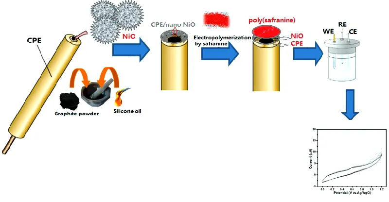
was constructed in a similar way by mixing graphite powder, silicone oil and NiO nanoparticles at an optimized ratio of 75:20:5 by weight. Prior this, the graphite-silicone oil compositions of 45:20, 50:20, 55:20, 60:20, 65:20, 70:20, 75:20 by wt.% and the NiO percentages of 5, 10, 15, 20 were evaluated for the optimization. The derived modified carbon paste analogue was placed in a 1.0 mL plastic syringe into which a copper wire was introduced to acquire the external electric contact. Electropolymerization of safranine at NiO nano CPE was conducted by following Yang method [62]. Accordingly, the working electrode preparation was based on CV method in aqueous solution containing 1.0 mM of safranine and phosphate buffer solution (PBS) at pH 5.5. Electropolymerization or polymer film formation on the electrode surface was achieved in CV mode by applying the potential between 0.0 V to 0.7 V at 50 mVs−1 scan rate for 20 cycles. In electropolymerization reaction, safranine undergone deprotonation and polymerization took place between NiO modified CPE and safranine as presented in Fig. 2.
Figure 2. Schematic representation for the electropolymerization of safranine on the surface of NiO modified CPE.
The resulted poly(safranine/nano NiO)CPE was cleaned with distilled water and stored in a phosphate buffer solution (PBS) of pH 7.0. Then, fabricated poly(safranine/nano NiO)CPE was utilized to evaluate cyclic voltammetric sweeps between potential range from 80 mV to 120 mV at scan rate of 50 mVs-1 in 0.1 M PBS at pH 5.5.
2.4 Characterization of NiO nanoparticles and poly(safranine/nano NiO)CPE
[image:5.596.96.496.277.485.2]
(ZEISS LEO® 906 E TEM) at an acceleration voltage of 20 and 100 kV, respectively. Further, the
functional group analysis of CPE, NiO nanoparticles and poly(safranine/nano NiO)CPE were recorded on Fourier transform infrared spectroscopy (FT-IR) with the help of KBr pelletized method (Shimadzu 8400 S, Japan). Roughly, 1.0 gm of each sample was mixed individually with 100 mg of KBr and made into a disc under the pressure of 7.0 tons, which was subsequently analysed with FT-IR.
2.5 Analytical procedure
The CV and DPV measurements were conducted on CHI610D (USA) Potentiostat consisting a conventional three-electrode cell. Platinum and Ag/AgCl (saturated) wires were employed as the respective counter and reference electrodes. A bare CPE (3.0 mm diameter) and modified poly(safranine/nano NiO)CPE were employed as working electrodes. A buffer solution prepared from 0.1 M NaH2PO4 and Na2HPO4 (pH 5.5) was employed as supporting electrolyte for the analysis of rutin
and its pH was measured using Elico Li 120 pH meter. Initially, rutin stock solution was prepared at 0.01 M concentration and subsequently diluted to prepare the required concentrations of rutin before running the CV scans. Afterwards, the three-electrode system was kept in a plain solution and scanned between 50 mVs-1 to 500 mVs-1 (vs. SCE) for six runs individually prior each measurement and restarted for the modified electrode. Initially, rutin dissolved in ethanol and then subjected to sonication and filtration. The working standards of rutin were freshly prepared by diluting 0.01 M rutin stock solution in supporting electrolyte, and all CV measurements were conducted at room temperature (25±1.0 ºC).
2.6 Real sample analysis
Samples including buckwheat seeds and green tea were procured from the local grocery market in Tirupati (Andhra Pradesh, India). The samples were grinded individually, and then 5.0 g of each sample was mixed with 50 mL of methanol homogeneously using ultrasonic irradiation. Next, the mixture was vortexed for 60 sec and sonicated again for 30 mins. After the ultrasonication, the mixtures were filtered through 0.22 µm syringe filter into a volumetric flask and the liquid phases were kept in a refrigerator (at 4.0 °C) until analysis. Prior to the analysis, the extracted samples were diluted with 0.1 M phosphate buffer (pH 5.5), then 50 mL of this solution was transferred into the electrochemical cell. The samples were analysed by DPV using poly(safranine/nano NiO)CPE to determine the concentrations of rutin.
3. RESULTS AND DISCUSSION
3.1 Characterization of NiO nanoparticles and modified poly(safranine/nano NiO)CPE
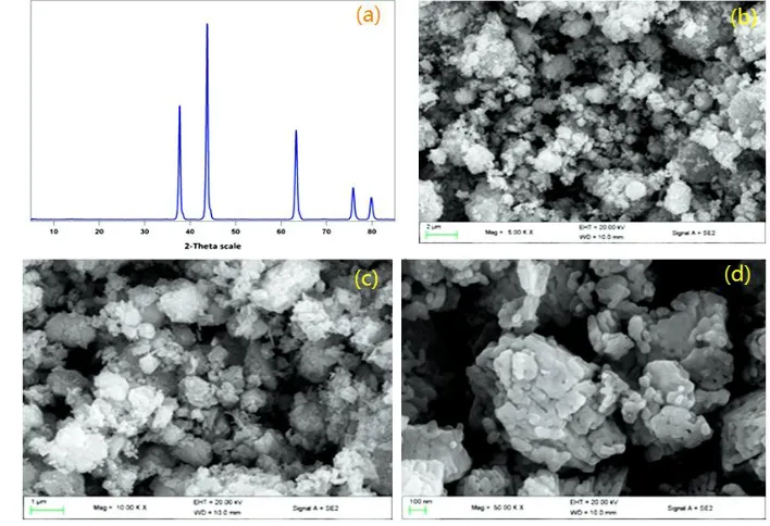
(110), (103), (200), (112) and (201) crystal planes of the NiO, respectively. All these diffraction peaks were perfectly indexed to the face-centered cubic (FCC) crystalline structure of NiO, not only in peak position, but also in their relative intensity of the characteristic peaks, which is in correlation with the standard database JCPDS file No. 98-009-0203. The crystalline phase demonstrated that Ni nanoparticles were readily oxidized to form NiO nanoparticles. The structural analysis endorsed the phase and controlling properties of NiO nanoparticles [63]. The average crystallite sizes of the samples were calculated by Debye-Scherrer's equation (D = k*λ / β cos Ѳ), where D is the crystalline size, λ (1.54178 Å for CuKα) is the wave length of incident X-ray (nm), β is the full width at half maximum, and θ is the diffraction angle. The average crystalline size of the NiO was found greater than 100 nm, which has been calculated from the major diffraction peaks at 37.4º, 43.5º, 63.2º. Further, the synthesized NiO nanoparticles have been characterized using FE-SEM. The morphology of NiO nanoparticles at different magnifications is presented in Fig. 3(b), (c) and (d). In all three SEM micrographs, the NiO appeared as crystalline material with distinct particle sizes. However, the available surface of the NiO seems rough in nature and provides better performance for the electrochemical activity. Particularly at higher magnifications, the NiO nanoparticles in SEM images showed plate like structures (Fig. 3(d)).
Figure 3. (a) XRD image of NiO nanoparticles, and SEM images of NiO nanoparticle at (b) 5.00 kx, (c)
10.00 kx and (d) 50.00 kx magnifications.
[image:7.596.56.513.221.571.2] [image:7.596.117.471.349.591.2]
distributed on the electrode surface [64]. Further, the TEM image of poly(safranine/nano NiO)CPE was also indicated the crystalline nature of NiO nanoparticles (Fig. 4(d)).
Figure 4. SEM morphology for (a) bare CPE, (b) poly(safranine/nano NiO)CPE, and their TEM
morphology for (c) bare CPE and (d) poly(safranine/nano NiO)CPE.
Additionally, the FT-IR spectra of bare CPE, NiO nanoparticles and poly(safranine/nano NiO)CPE were recorded and presented in Fig. 5. The spectra has several significant absorption peaks over the range between 4000-400 cm-1. Fig. 5(a) represents the spectra of bare CPE, where a peak at
2973.54 cm-1 is appeared corresponding to C-H stretching, and the peak at 1285.44 cm-1 is found belongs
to C-H bending. Next, the FT-IR spectra of NiO nanoparticles is presented in Fig 5(b), where a broad peak at 3451.24 cm-1 is assigned to O-H stretching vibration, and another peak at 1628.89 cm-1 is attributed to H-O-H bending vibration mode. These two peaks indicates the presence of traces of water within the sample. Further, a broad peak at 448.23 indicates Ni-O stretching vibration., in which the peak broadness supports nanocrystalline nature of the sample. Moreovr, Fig. 5(c) represents the FT-IR spectra of poly(safranine/ nano NiO)CPE, where two peaks at 2985.52 cm-1 and 2960.24 cm-1 represent
[image:8.596.150.442.106.331.2]
Figure 5. FT-IR spectra recorded for (a) bare CPE, (b) NiO nanoparticles and (c) poly(safranine/ nano
NiO)CPE.
3.2 Electrochemical response of rutin at bare and modified poly(safranine/nano NiO)CPE
NiO nanoparticles possess superparamagnetic features and they can be characterized by high surface area, high magnetic susceptibility with no remanence, coercivity and hysteresis. Therefore, modified poly(safranine/nano NiO)CPE is presumed to exhibit outstanding electrochemical performance owing to its substantial electrochemical windows with excessive mechanical property, better conductivity and stability. The electrochemical behaviour of rutin at bare CPE, NiO modified CPE and poly(safranine/nano NiO)CPE was evaluated by CV and DPV as shown in Fig 6.
Figure 6. Cyclic voltammograms obtained for (a) blank solution at bare CPE, (b) rutin at bare CPE, (c)
[image:9.596.166.426.492.689.2]
The electrochemical oxidation of flavonoids, particularly rutin involves ionization, losing a proton to result monoanionic species followed by a one electron, one proton oxidation of the monoanionic species to form a radical anion. This radical anion then undergoes a second reversible one-electron oxidation to give dehydrorutin. Later, dehydrorutin species rapidly protonates and dehydrates to yield the final product of 3’,4’-diquinone [66]. As conveyed in Fig. 6 (b), a set of redox peak potentials were recorded at 221.7 mV (Epa) and 172.9 mV (Epc) (vs. SCE) at bare CPE. The anodic peak current (Ipa) and cathodic peak current (Ipc) were reported respectively at 9.052 μA and 4.062 μA, from which the redox peak current ratio (Ipa/Ipc) was calculated. Next, the cyclic voltammogram of NiO modified CPE in 0.1 M PBS at pH 5.5 is depicted in Fig. 6 (c), in which the peak potentials were recorded at 227.7 mV (Epa) and 181.2 mV (Epc) (vs. SCE) which shows a slight increase in redox peak currents comparing with bare CPE. By comparison, the peak potentials of rutin at poly(safranine/nano NiO)CPE in 0.1 M PBS at pH 5.5 were dramatically enhanced, indicating the excellent electrical conductivity, improved electron transfer rate and high electrocatalytic ability of poly(safranine/ nano NiO) for the redox of rutin as illustrated in Fig. 6 (d). A couple of well-defined redox peak potentials were reported at 232.7 mV (Epa) and 188.7 mV (Epc) (vs. SCE) and the corresponding anodic (Ipa) and cathodic (Ipc) peak currents were recorded respectively at 3.655 μA and 2.027 μA. The peak current ratio for Ipa/Ipc was found to be 1.8 that indicates a reversible process at the proposed modified electrode. Further, it is well accepted that the improvement in redox peak current and the decline in over potential are inevitable representations of the electrocatalytic reaction. This tremendous increase in peak currents for the modified electrode is due to the increased electrochemical reaction of rutin resulted from the virtue of poly(safranine/nano NiO) composite on CPE surface. The electropolymerization of safranine on NiO modified CPE would have benefited to increase the electrical conductivity, which has been assumed from the enhanced peak currents and background. Overall improvement in the current (6 times higher than bare CPE) is because of the uniform distribution of NiO nanoparticles on bare CPE and electropolymerization of safranine that are provided high surface area and higher electrical conductivity. In accordance with the above results, it is confirmed that modified poly(safranine/nano NiO)CPE is best choice for the electrochemical detection of rutin.
3.3 Optimization of experimental conditions 3.3.1 pH effect of buffer
Figure 7. (a) Cyclic voltammograms of rutin at poly(safranine/nano NiO)CPE over the pH range
[image:10.596.197.398.559.711.2]
The pH effect of the buffer solution on electrochemical response of rutin on the modified poly(safranine/nano NiO)CPE was examined over the range between 5.5-8.0. As depicted in Fig. 7, the highest reduction peak current was found at pH 5.5 and the peak currents were gradually declined with increasing the pH of buffer solution and it was even progressed to negative value with further increasing the buffer pH. These outcomes described the involvement of proton during the electrochemical reaction. Hence, PBS with pH 5.5 was chosen as more appropriate supporting electrolyte for the detection of rutin in subsequent experiments. Also, we have investigated the effect of buffer pH on peak potential (E0) within the pH range from 5.5 to 8.0. The results showed that E0 valuehasshifted towards lower direction with increasing pH of the PBS. A linear regression equation was obtained as E0(V) = 0.061 pH + 0.8078 (n = 5, Y = 0.9984). The slope value 0.061 V is nearly close to the theoretical value of 0.059 V/pH at 25
oC. According to the equation 0.061x/n =0.059, n denotes the number of electrons transferred and x
[image:11.596.140.461.431.515.2]denotes the number of hydrogens involved in the reaction. According to the slope of 0.061 V/pH, it could be deduced that H+ participated in this reaction and the number of electrons and protons transferred was equal in the electrochemical reaction. When the pH was over 5.5, the anodic peak became very small and irreversible. These experimental phenomena were related to the proton involved in the electrochemical reaction. When pH exceeded 5.5, with the increase of negative ions, the electrostatic repulsion occurred between NiO/safranine and rutin, leading to the reduction of current. A study was also reported the similar electrochemical behavior of rutin on Chit/G/GCE whose rate of slope is similar to our own result [67]. Therefore, the electron uptake was steered by an equal number of hydrogens i.e., x = n = 2 (Fig. 8). At the same time, according to the Faraday’s law, the investigated reaction mechanism at this electrode involved two electrons and two protons.
Figure 8. Electrochemical redox process mechanism for rutin.
Figure 9. (a) Differential pulse voltammograms obtained at poly(safranine/nano NiO)CPE for rutin over
[image:11.596.184.413.548.724.2]
The linearity of the formal potential Vs PBS at pH 5.5-8.0 and at scan rate 50 mVs-1 is depicted
in Fig. 9. The results confirmed that the feasible mechanism for electro-oxidation reaction of rutin at modified poly(safranine/nano NiO)CPE was a two electron-two proton process and the electrode reaction is manifested as follows. In the electro-oxidation mechanism of rutin, pre-dissociation of proton takes place initially to produce a monotype anion, which is further oxidized to result a radical anion. The radical anion undergoes a second reversible one electron (1e-1) oxidation and forms dehydro-rutin. The final species were rapidly dehydrated to give final product of 3’,4’-diquinone.
3.3.2 Effect of scan rate
The impact of scan rate between 50 mVs-1 to 500 mVs-1 on the electrochemical response of 0.4 mM rutin at poly(safranine/nano NiO)CPE is presented in Fig. 10. The results described a gradual increase in peak currents with increasing scan rates and exhibited adequate linear relationship between the anodic peak currents and scan rates (ν). Both anodic and cathodic peak currents were raised linearly with increasing scan rates from 50 mVs-1 to 500 mVs-1. The regression equation presents Ipa(μA) = 2.98308E-4 Cm(M) + 4.5433E-5 mA (R1=0.9907), Ipc (mM) = 2.79633E-5 Cm(M/L) + 5.6755E-6 mA
(R2=0.9897) between the redox peak currents and the scan rates.
Figure 10. (a) Cyclic voltammograms for rutin at poly(safranine/nano NiO)CPE for scan rates of 50,
100, 150, 200, 250, 300, 350, 400, 450 and 500 mVs-1, and (b) The plot of peak current vs. scan rate between 50-500 mVs-1 (rutin concentration is 0.4 mM, buffer concentration 0.1 M, pH 5.5).
[image:12.596.112.474.394.544.2]
determined. The electron transfer coefficient (α) was found to be 0.95, and the charge transfer rate constant (ks) was calculated from the following Laviron equation [68].
logks = α log(1 − α) + (1 − α)logα − log (RT
𝑛Fv) − α(1 − α) 𝑛𝐹𝛥𝐸𝑝
2.3 𝑅𝑇 ………Eq (1)
Where, R represents universal gas constant, α presents electron transfer coefficient, ks denotes charge transfer rate constant, T denotes absolute temperature, n denotes number of electrons transferred, F denotes Faraday constant and v shows the scan rate. The calculated ks value of 1.717 describes that the electron transfer between electrode and rutin was complemented by the deposition of modified poly(safranine/nano NiO) CPE. The peak potential difference (Ep) was found to be 29.3 mV that is equal to 59/n mV, suggesting that the equal number of electrons and protons were took part in the reaction mechanism. In Fig. 9 (B), the Ep and current values were plotted, for which the linear regression is recorded as r2 = 0.9951.
3.4 Determination of rutin
The analytical performance and sensitivity of the developed poly(safranine/nano NiO)CPE was ascertained by DPV technique.
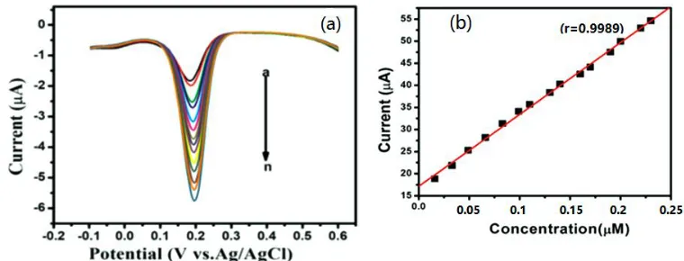
Figure 11. (a)Differential pulse voltammograms obtained for rutin over the linear range between 1.63
x 10-8 – 2.30 x 10⁻⁷ M at poly(safranine/nano NiO)CPE, and (b) The calibration plot of DPV
current response against rutin concentration.
The quantitative determination of rutin is represented in Fig. 11. The reduction peak currents were increased with increasing the concentration of rutin and has showed good linear relationship between 1.63 x 10⁻⁸ - 2.3 x 10⁻⁷ M under the optimal conditions. Corresponding linear regression
equation is expressed as Ipa (μA) = 0. 95940 [Crutin] (M) + 1.9 μA (r = 0.9957). The limit of detection
(LOD) and limit of quantification (LOQ) values of rutin with the modified electrode were calculated using the following equations Eq (2) & (3).
LOD =3 S
m ………Eq (2)
LOQ =10 S
m …….…Eq (3)
[image:13.596.108.488.357.502.2]
showed superior electrocatalytic performance for the detection of rutin over other reported methods, which might be the result of electropolymerization of safranine and uniform distribution of NiO on CPE that provided large surface area and excellent conductivity. Also, the proposed electrode provided superior electrocatalytic activity in addition to its wider linearity range and lower detection limit for the rutin.
3.5 Reproducibility and stability
The reproducibility and stability are the two essential characteristics for any modified electrode to prove its ability over the conventional electrodes and to provide their efficiency in commercial purpose. The electrochemical performance of poly(safranine/nano NiO)CPE for the detection of 0.4 µM rutin has been measured using ten individual determinations. In all the runs, almost similar peak currents were recorded with %RSD value <3.6%. This data clearly proves that the detection of rutin using the modified electrode is highly reproducible. Further, the stability of the modified poly(safranine/nano NiO)CPE was investigated keeping the electrode at room temperature for more than two weeks and measured the peak currents. The results confirmed that there were no significant changes in the recoveries and peak currents were deviated just below 4.0% for the 0.4 μM rutin solution compared to that of initial current response, which indicated sufficient stability of the modified electrode.
3.6 Real sample analysis
In order to test its practical application, the fabricated poly(safranine/nano NiO)CPE was used to determine rutin in buckwheat and green tea samples. Since the electrochemical techniques are capable to determine the analytes without pretreatment, the real samples were directly introduced to the experimental cell.
Table 1. Determination of rutin in selected real samples of buckwheat and green tea using
poly(safranine/nano NiO)CPE.
Sample Spiked Recovered conc.a Recoveryb±RSDc(%) CVd (%) concentration (mmol L-1) (mmol L-1)
Buckwheat
Sample 1 0.1 0.097 97.0±3.28 2.41 Sample 2 0.2 0.196 98.5±2.73 1.83 Sample 3 0.3 0.302 100.8±2.50 3.18
Green tea
Sample 1 0.1 0.965 96.5±4.85 3.91 Sample 2 0.2 0.196 98.0±3.36 1.58 Sample 3 0.3 0.385 102.8±4.95 2.65
a Found (mmol L-1) = Injected Ipa*Added/Synthetic Ipa. b Recovery (%) = Synthetic Ipa*100/Injected Ipa,
c % RSD calculated from triplicate determinations
[image:14.596.64.528.561.719.2]
The samples were diluted with 0.1 M PBS (pH 5.5) and employed the standard addition method to obtain the final concentrations of 0.1, 0.2, and 0.3 mmol L-1 respectively. It is worth to mention that the above concentrations were considered based on the amount of rutin present in buckwheat and green tea samples, which varies between 0.2 and 2.0 mmol. As given in Table 1, the recoveries under optimized conditions were found between 96.5%-102.8% with the maximum %RSD and %CV values respectively as 4.95 and 3.91 at the poly(safranine/nano NiO)CPE for three concentration levels. All experiments were conducted in triplicate and the resulted %RSD values were found below 5.0% demonstrating the capability of poly(safranine/nano NiO)CPE for the accurate voltammetric quantification of rutin in real samples. Further, the common interfering substances of buckwheat and green tea such as tryptophan, threonine, iron, zinc, vitamin C, and β-carotene were added at 100-fold concentrations, and glucose and fructose were added at 20-fold concentration to evaluate their impact during the rutin detection. Nearly, none of the above substances were interfered with rutin according to the relative error < ±5.0%.
Therefore, the recovery of rutin from the selected real samples of is almost quantitative in presence of possible interferences with poly(safranine/nano NiO)CPE. From the recovery data, it is concluded that ZnO/CNS/MCPE is highly selective for the detection of rutin in presence of other interfering substances in real samples. However, very few compounds such as flavonoids and dopamine caused negligible interferences at extremely higher concentrations due to their similar peak potentials with rutin.
3.7 Analytical merits
[image:15.596.84.512.539.701.2]A comparison was made in terms of linearity range and detection limits of the modified poly(safranine/nano NiO) CPE with other reports established with different modified electrodes for the quantification of rutin and is presented in Table 2.
Table 2. Comparison of linearity range and LOD values of poly(safranine/nano NiO) CPE with other
modified electrodes for rutin determination.
nm = not mentioned
The linearity range and detection values of poly(safranine /nano NiO) CPE were better than several reported methods [68-74]. Additionally, this modified poly(safranine /nano NiO) CPE showed
Modifier Linearity range (M) LOD (M) Stability Reference
Chit/G/GCE 5.0x10⁻⁷ -1.04x10⁻⁵ 1.0 x 10⁻⁶ nm [68]
GOMGCE 1.0x10⁻⁷ -1.16x10⁻⁶ 2.0 x 10⁻⁶ nm [69]
SWNTMGE 2.0x10⁻⁸-1.0x10⁻⁸ 1.0 x 10⁻⁸ nm [70]
PABSAg/GCE 2.5x10⁻⁵ - 1.0x10⁻⁶ 7.0 x 10⁻⁷ nm [71]
MWNT/GCE 2.8x10⁻⁷ - 2.1x10⁻⁵ 7.0 x 10⁻⁷ nm [72]
Graphene/GCE 1.0x10⁻⁷ - 2.0x10⁻⁶ 2.3 x 10⁻⁸ nm [73]
exceptional selectivity for the identification and quantification of rutin in real samples even at trace concentrations. Furthermore, the linearity and LOD values noticed for the rutin with poly(safranine/nano NiO) CPE were significantly better than our previous study, where we have determined rutin using Fe3O4@SiO2 microspheres modified CPE i.e., FS-MCPE. This results clearly concludes that, the
electropolymerization of safranine on the surface of NiO modified CPE enhanced the surface area and electrochemical behaviour extensively.
5. CONCLUSIONS
In this study, a modified poly(safranine/nano NiO)CPE is fabricated and successfully examined for the quantification of rutin in real samples of buckwheat and green tea. The results confirmed superior electrochemical performance of poly(safranine/nano NiO)CPE over bare CPE and NiO modified CPE for the detection of rutin. The combined use of safranine and NiO nanoparticles provided tremendous catalytic performance owing to their large surface area, strong electrocatalytic ability and higher conductivity. The developed poly(safranine/nano NiO)CPE in 0.1 M PBS at pH 5.5 has provided exceptional electrochemical response for the oxidation and reduction of rutin and the electrode process was regulated by absorption effect under low scan rates. At the optimized conditions, the anodic peak currents and rutin concentrations were linear with in the dynamic range of 1.63 x 10⁻⁸ - 2.3 x 10⁻⁷ M with
low LOD and LOQ values. Further, the use of modified poly(safranine/nano NiO)CPE is a good choice for the selective and sensitive analysis of rutin in various real samples without the interference of matrix effects. Furthermore, the developed method offers the advantages of simplicity in electrode fabrication, reliability, precision, and low cost.
CONFLICTS OF INTEREST
No conflicts of interest declared by the authors
References
1. K. B. Magalingam, A. Radhakrishnan and H. Nagaraja, Int J Immunopathol Pharmacol., 29 (2016) 30.
2. C. Ganbaatar, M. Gruner, D. Mishig, R. Duger, A. W. Schmidt and H. J. Knolker, The Open Nat Prod J., 8 (2015) 1.
3. W. Ren, Z. Qiao, H. Wang, L. Zhu and L. Zhang, Med Res Rev., 23 (2003) 519. 4. V. Diwan, L. Brown and G. C. Gobe, J Funct Foods, 33 (2017) 85.
5. L. Lv, Y. Yao, G. Zhao and G. Zhu, Exp Ther Med., 15 (2018) 506. 6. M. Gross, Pharm Biol., 42 (2004) 21.
7. M. T. Olaleye, O. O. Crown, A. A. Akinmoladun and A. A. Akindahunsi, Hum Exp Toxicol., 33 (2014) 602.
8. R. Aviva, Botanical medicine for women's health - 2nd Edition, Elsevier (2017) USA.
9. K. Sattanathan, C. K. Dhanapal, R. Umarani and R. Manavalan, J Appl Pharma Sci., 01 (2011) 227.
12.H. Xu, Y. Li, H. W. Tang, C. M. Liu and Q. S. Wu, Anal Lett., 43 (2010) 893. 13.J.W. Kang, X.Q. Lu, H.J. Zeng, H.D. Liu and B.Q. Lu, Anal Lett., 35 (2002) 677. 14.Z. H. Song and S. Hou, Talanta, 57 (2002) 59.
15.J. Dolejsova, M. Polasek, P. Solich, R. Karlicek and L. Tumova, Ceska Slov Farm.,53 (2004) 145. 16.H. N. A. Hassan, B. N. Barsoum and I. H. I. Habib, J Pharm Biomed Anal., 20 (1999) 315.
17.Z. Legnerova, D. Satinsky and P. Solich, Anal Chim Acta, 497 (2003) 165.
18.Y. Peng, M. X. Liao, X. Ma, H. Deng, F. Gao, R. Dai and L. Lu, Int. J. Electrochem. Sci., 14 (2019) 4946.
19.S. R. Benjamin, R. S. Vilela, H. S. de Camargo, M. I. F. Guedes, K. F. Fernandes and F. Colmati, Int J Electrochem Sci., 13 (2018) 563.
20.J. L. He, Y. Yang, X. Yang, Y. L. Liu, Z. H. Liu, G. L. Shen and R. Q. Yu, Sens Actuators B, 114 (2006) 94.
21.I. Baranowska and D. Rarog, Talanta, 55 (2001) 209.
22.C. Y. Guo, J. H. Wang, Y. M. Zhao, L. X. Shen and D. S. Zhang, Pharmacogno Mag, 9 (2013) 192. 23.Y. V. Manohara Reddy, V. Prabhakara Rao, A. V. Bhaskar Reddy, M. Lavanya and G. Madhavi,
Mater Sci Eng C., 57 (2015) 378.
24.V. K. Gupta, A. Nayak, S. Agarwal and B. Singhal, Comb Chem High T Scr., 14 (2011) 284. 25.V. K. Gupta, B. Sethi, R. A. Sharma, S. Agarwal and A. Bhartia, J Mol Liq., 177 (2013) 114. 26.A. K. Jain, V. K. Gupta and L. P. Singh, Anal Proc., 32(1995)263-265.
27.H. Karimi-Maleh, F. Tahernejad-Javazmi, N. Atar, M. Lutfi Yola, V. K. Gupta and A. A. Ensafi, Ind. Eng. Chem. Res., 54 (2015) 3634.
28. V. K. Gupta, H. Karimi-Maleh and R. Sadegh, Int J Electrochem Sci., 10 (2015) 303.
29.M. Noman, A. Sanginario, P. Jagadale, D. Demarchi and A. Tagliaferro, Mater Sci Eng C, 75 (2017) 402.
30.V. K. Gupta, A. K. Singh and L. K. Kumawat, Sens Actuators B, 195 (2014) 98.
31.V. K. Gupta, N. Atar, M. L. Yola, Z. Ustundag, and L. Uzun, Water Res., 48 (2014) 210. 32.M. L. Yola, V. K. Gupta, T. Eren, A. E. Şen, N. Atar, Electrochim Acta.,120 (2014) 204. 33.H. C. Chien and T. C. Chou, Electroanalysis 22 (2010) 688.
34.M. Khadem, F. Faribod, P. Norouzi, A. R. Foroushani, M. R. Ganjili, S. J. Shahtaheri and R. Yarahmadi, Electroanalysis, 29 (2017) 708.
35.V. K. Gupta, L. P. Singh, R. Singh and N. Upadhyay, J Mol Liq., 174 (2011) 11. 36.S. K. Srivastava, V. K. Gupta, M. K. Dwivedi and S. Jain. Anal Proc., 32 (1995) 21. 37.S. K. Srivastava, V. K. Gupta and S. Jain, Analyst, 120 (1995) 495.
38.H. R. Rajabi and A. Zarezadeh A, J Mater Sci: Mater Electron., 27 (2016) 10911. 39.J. Mayahi and H. R. Rajabi, New J Chem., 41 (2017) 14236.
40.D. Saritha, A. R. Koirala, M. Venu, G. Dinneswara Reddy, A. Vijaya Bhaskar Reddy, B. Sitaram, G. Madhavi and K. Aruna, Electrochim Acta, 313 (2019) 523.
41.M. B. Gholivand, M. Shamsipur, S. Dehdashtian and H. R. Rajabi, Mater Sci Eng C, 36 (2014) 102.
42.R. Pauliukaite, A. Selskiene, A. Malinauskas and C. M. A. Brett, Thin Solid Films, 517 (2009) 5435.
43.W. Sun, Y. Maoxia, Y. Li, Q. Jiang, S. Liu and K. Jiao, J Pharma Biomed Anal., 48 (2008) 1326. 44.V. K. Gupta, F. Golestani, S. Ahmadzadeh, H. Karimi-Maleh, G. Fazli and S. Khosravi, Int J
Electrocheem Sci., 10 (2015) 3657.
45.V. K. Gupta, H. Karimi-Maleh, S. Agarwal, F. Karimi and M. Bijad, Sensors 18 (2018) 2817. 46.R. N. Goyal, V. K. Gupta, and S. Chatterjee, Talanta 76 (2008) 662.
47.V. K. Gupta, M. R. Ganjali, P. Norouzi, H. Khani, A. Nayak and S. Agarwal. Crit Rev Anal Chem 41 ( 2011) 282.
48.S. K. Srivastava, V. K. Gupta and S. Jain, Analyst 120 (1995) 495-498.
65.
50.A. Asfaram, M. Ghaedi, S. Agarwal, I. Tyagi and V. K. Gupta, RSC Adv., 5 (2015) 18438.
51.V. K. Gupta, M. Naveen, L. K. Kumawat and A. K. Singh, Sensor Actuat B-Chem., 207 (2015) 216. 52.V. K. Gupta and P. Kumar, Anal Chimic Acta., 389 (1999) 205.
53.A. K. Jain, V. K. Gupta, B. B. Sahoo and L. P. Singh, Analytical Proceedings including Analytical Communications 32(1995)99-101.
54.H. F. Kellen Garcia, A. M. Roberta and F. F. Orlando, Anal Lett., 42 (2009) 881. 55.M. B. Gholivand, L. M. Behzad and H. Hossein, Anal Biochem., 493 (2016) 35. 56.S. Cui, L. Li, Y. Ding, J. J. Zhang, H. Yang and Y. Wang, Talanta 164 (2017) 291.
57.Z. H. Wang, S. Y. Yao, J. F. Xia, F. F. Zhang, X. M. Guo, Y. Z. Xia and Y. H. Li, Adv Mater Res., 531 (2012) 419.
58.X. Liu, L. Li, X. Zhao and X. Lu, Colloid Surf B: Biointer., 81 (2010) 344.
59.S. Li, B. Yang, J. Wang, D. Bin, C. Wang, K. Zhang and Y. Du, Anal Methods, 8 (2016) 5435. 60.L. Tang, X. Li, R. C. Cammarata, C. Friesen and K. Sieradzki, J Am Chem Soc., 132 (2010) 11722. 61.Y. J. Yang, Fuller Nanotub Car N, 24 (2016) 243.
62.F. Cespedes, E. F. Martinez and S. Alegret, Trac Trend Anal Chem., 15 (1996) 296. 63.A. Elzatahry, Int. J. Electrochem. Sci., 9 (2014) 22.
64.E. Laviron, J Electroanal Chem., 101 (1979) 19.
65.H. Filik, A. A. Avan, S. Aydar and B. A. Rabia, Int. J. Electrochem. Sci., 9 (2014) 2775. 66.J. Ana, Y. Y. Bi, C. X. Yang, F. D. Hu and C. M. Wang, J. Pharma. Anal., 3 (2013) 102. 67.S.L. Yang, L.B. Qu and G. Li, J. Electroanal. Chem. 645 (2010) 115.
68.J. An, Y. Y. Bi, C. X. Yang, F. D. Hu, C. M. Wang, J Pharm Anal., 3 (2013) 102.
69.X. Zhu, J. Xu, X. Duan, L. Lu, H. Xing, Y. Gao, S. Hui, L. Dong and T. Yang, Int J Electrochem Sci., 10 (2015) 9192.
70.B. Zeng, S. Wei, F. Xiao and F. Zhao, Sens Actuators B: Chemical, 115 (2006) 240.
71.X. Y. Chen, Z. H. Wang, F. F. Zhang, L. Y. Zhu, Y. H. Li and Y. Z. Xia, Chem Pharm Bull., 58 (2010) 475.
72.G. Ziyatdinova, L. Aytuganova, A. Nizamova, M. Morozov and H. Budnikov, Collect Czech Chem C, 76 (2010) 1619.
73.Y. Lei, D. Du, L. Tang, C. Tan, K. Chen and G. J. Zhang, Anal Lett., 48 (2015) 894.
74.D. Saritha, S. Kiranmai, C. Madhuri, M. Venu, K. G. Rajyalakshmi, G. Madhavi, Anal. Bioanal. Electrochem., 9 (2017) 506.