Int. J. Electrochem. Sci., 14 (2019) 3663 – 3672, doi: 10.20964/2019.04.09
International Journal of
ELECTROCHEMICAL
SCIENCE
www.electrochemsci.org
Electrochemical Determination of Antioxidant Activity of
Different Bee Products
Yuhong Zheng1,2,3, Mengyao Wu4, Yuting Xu4, Xu Peng4, Mingjun Zhang4, Qiao Wang4, Jia Du4*, Huaiwei Zhang1,4,* and Li Fu1,4,*
1 Key Laboratory of Bee Products for Quality and Safety Control, Ministry of Agriculture, Institute of
Apicultural Research, Chinese Academy of Agricultural Sciences Beijing 100093, PR China
2 Institute of Botany, Jiangsu Province and Chinese Academy of Sciences (Nanjing Botanical Garden
Mem. Sun Yat-Sen), Nanjing, PR China
3 The Jiangsu Provincial Platform for Conservation and Utilization of Agricultural
Germplasm, Nanjing, PR China
4 College of Materials and Environmental Engineering, Hangzhou Dianzi University, Hangzhou
310018, PR China
*E-mail: [email protected]; [email protected]; [email protected]
Received: 22 November 2018 / Accepted: 30 December 2018 / Published: 10 March 2019
Bee products are of great benefit to the human body and have a good scavenging effect on free radicals in the human body. These products are rich in flavonoids and terpenes and have good antioxidant activities. Bee products can scavenge free radicals produced by the human body. Therefore, studies of the antioxidant activities of bee products are of great significance to human life and health. In this study, we proposed an electrochemical sensing platform based on the silver ion cross-linked hydrogel for the evaluation of three different bee products. After optimization, the antioxidant performance of the three bee products follows the following order: Royal jelly > honey > honeybee pupa. In addition, the proposed electrochemical sensing platform also exhibits excellent reproducibility and stability.
Keywords: Electrochemistry; Antioxidant activity; Hydrogel; Honey; Metal ions
1. INTRODUCTION
research has found that bee products (mainly consisting of pupae, honey and Royal jelly) have many functions and biological activities, such as rich phenolic compounds, flavonoids and other antioxidant and anticancer effects [3-5]. Free radicals are considered to be an important cause of many diseases. The antioxidant and anticancer effects of bee products are mainly achieved by self-scavenging or promoting the ability of the body to scavenge free radicals. This performance mainly occurs through some antioxidants, such as superoxide dismutase, catalase, flavonoids, phenols and vitamins [6-8].
Honeybee pupae, also known as wasp, are mainly male bee pupa and worker bee pupa. The edible and medicinal use of bee pupae has a long history. As early as 1200 BC, the ancient book “Erya” records of eating bees. The silk book “Fifty-two Cases”, an ancient medical prescription unearthed in Mawangdui, Changsha, contains a prescription for treating diseases with bee embryos and bees. Bee pupae are rich in nutrients. The dry matter of bee pupae contains protein (46.21%), fat (26.09%), carbohydrate (20.34%), total sugar (0.73%), chitin (4.37%), flavonoids, amino acids, fatty acids, vitamins, mineral elements and enzymes, etc. Many studies have shown that honeybee pupae have many functions, such as antioxidant, hypoglycaemic, anti-aging, anti-fatigue and anti-inflammatory effects [9-12]. Honeybee pupa is mainly used in health food, health care wine, and health care granules. Honey is made in the honeycomb by bees from the flowers of plants and is produced by worker bees through the action of amylase in salivary glands. The main components of honey are carbohydrates, proteins, minerals, vitamins, phenols, etc. Honey contains flavonoids, phenolic acid, oxidase, invertase, amylase, catalase, ascorbic acid, tocopherol, superoxide dismutase, glutathione and other substances and has the functions of antimicrobial, antioxidant, hypoglycaemic, anti-inflammatory, anticancer and promoting wound healing [13-16]. Royal jelly is a kind of translucent serum with milky white or light yellow, sweet, sour and astringent taste secreted from the nutrient glands of the head of worker bees. Composition is very complex, and the main components are proteins, a variety of amino acids, fatty acids, sugar, vitamins, etc. Royal jelly has the functions of anti-inflammation, antibacterial, anti-tumour, lowering blood lipid, lowering blood pressure, antifatigue and antioxidation [17-22]. Royal jelly is mainly used in health care products, cosmetics, medicine and feed additives.
The antioxidant enzymes in organisms are mainly superoxide dismutase, peroxidase and catalase, which convert peroxides into non-cellular and tissue-damaging substances. Superoxide dismutase (SOD) is a kind of metal enzyme widely existing in animals, plants and microorganisms [23-25]. SOD catalyses superoxide radicals (O2-) in organisms, disproportionates them to H2 and O2, plays an important role in
scavenging excess O2- and protecting cells from the damage of oxygen free radicals. Glutathione
peroxidase (GSH-Px) is an important enzyme that scavenges hydrogen peroxide (H2) and many organic
hydroperoxides in vivo, protects cells from damage and protects the structural and functional integrity of cell membranes [26-28]. GSH-Px widely exists in tissue cells, red blood cells and plasma and is related to cell damage, hypoxia, poisoning, ageing and other diseases. Catalase (CAT), a terminal oxidase widely existing in aerobic microorganisms, animals and plants, has a high catalytic efficiency. Its main function is to catalyse the decomposition of hydrogen peroxide into water and oxygen, thus preventing membrane lipid peroxidation [29,30].
Ciocalteu colorimetric method [31-33]. These optical detection methods have some insurmountable shortcomings, such as the need for a longer detection time, the need to decolorize the samples in advance, and the lack of anti-interference ability. In contrast, the electrochemical method for antioxidant property detection has become a highly anticipated alternative method because of its simple, highly sensitive, and portable features and other advantages. The direct electrode detection method and the chemically modified electrode detection method are susceptible to the interference of other components in complex samples. The enzyme method and the latest DNA damage method can effectively solve the interference and provide more sensitive signals, but enzymes and DNA are easy to inactivate, expensive and difficult to stably immobilize onto the surface of the electrode [34-38]. Our recent study proposed a screening method for electrochemical oxidants based on polysaccharide-metal ion crosslinked hydrogels [39,40]. This method combines the depolymerization of polysaccharide-metal crosslinked hydrogels by reactive oxygen species (ROS) and the scavenging effect of antioxidants on ROS. The antioxidant properties can be evaluated by determining the difference in the electrochemical behaviour of metal ions before and after hydrogel depolymerization. In this study, we further extend this method for the analysis of the antioxidant properties of honeybee pupa, Royal jelly and honey.
2. EXPERIMENTAL
2.1. Materials
Silver nitrite, chitosan, hydrogen peroxide and potassium ferricyanide were purchased from Aladin Co., Ltd. Honeybee pupa (Zhenwuding Shanzhen, Henan), Royal jelly (Tongren Tang, Beijing) and honey (Tongren Tang, Beijing) were purchased from online shops. Acetic acid has been used as an electrolyte for antioxidant property analysis.
2.2. Hydrogel fabrication
The synthesis of metal ion crosslinked hydrogel was performed according to our previous method, with some modifications [39,40]. Typically, silver ions have been chosen as crosslink ions for hydrogel preparation. First, chitosan was dissolved in acetic acid to form a 1% solution using sonication. Then, silver nitride was dissolved into a chitosan solution to form a 1 mM concentration using sonication. Then, 0.1 M HCl was slowly added dropwise into the above solution until a gelation transformation was initiated.
2.3. Antioxidant activity measurement
Then, a Fenton solution (H2O2:Fe2+ = 1:4) was introduced into the hydrogel to initiate depolymerization.
Honey solution was subsequently added into the hydrogel to scavenge ROS. Cyclic voltammetry (scan rate: 50 mV/s) was used to analyse the signal change of metal ions before and after adding honey to evaluate the antioxidant properties of different bee products.
3. RESULT AND DISCUSSION
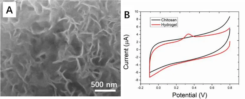
The morphology of the formed hydrogel is shown in Figure 1A. The hydrogel formed in a porous networking structure with uniform distribution, which could hold liquid inside without destroying the structure. The morphology of the silver ion cross-linked hydrogel is very like the chitosan/gelatin/PVA hydrogel [41], genipin-crosslinked catechol-chitosan [42], chitosan/hyaluronan hydrogel [43] and Nanohydroxyapatite-reinforced chitosan hydrogel [44]. The results indicated that the presence of silver ions caused rougher pore walls and open interconnected pores, which were, respectively, necessary for acting as a solid electrolyte [45]. The cyclic voltammetry of the hydrogel was recorded as Figure 1B. A distinct silver redox pair can be found at 0.1 V and 0.4 V, corresponding to the reduction and oxidation of silver, respectively. For comparison, the cyclic voltammetry of the chitosan has been recorded as well. No appreciable redox reaction can be found during the scan range. Since the oxidation peak of metallic silver is much intense than its reduction peak, the peak located at approximately 0.4 V has been used to estimate the antioxidant properties of bee products in this study.
Figure 1. (A) SEM image of the silver ion cross-linked hydrogel. (B) Cyclic voltammetry profile of the chitosan and silver ion cross-linked hydrogel.
[image:4.596.90.506.439.607.2]
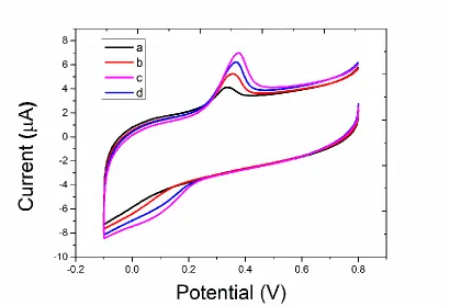
deduced by Fenton solution-produced ROS. The depolymerization process slowly decomposed the interwoven structure of the chitosan chains and silver ions, resulting in low molecular weight chitosan-linked silver ions. These units are more likely to diffuse to the electrode surface when electric potentials are applied. Thus, a higher current response from silver ions could be expected after depolymerization of the hydrogel. Chitosan is constructed by copolymers of glucosamine (GlcN) and N-acetylglucosamine (GlcNAc) linked with b-1,4-glycosidic bonds. Under ideal conditions, depolymerization should release both GlcN and GlcNAc. At 3 min after the introduction of Fenton solution. The cyclic voltammetry profile showed an even higher redox reaction due to the continuous depolymerization. In contrast, 0.1 mM Fenton solution was added to the hydrogel along with 0.1 mM uric acid. Uric acid is a common antioxidant that can scavenge ROS. The cyclic voltammetry profile was recorded at 5 min after the addition. As shown in Figure 2B, the redox profile is much less than the previous uric acid-excluded sample. Based on the above results, the proposed silver ion cross-linked hydrogel showed an excellent antioxidant property evaluation performance. Silver ions has been used as probe in electrochemical device for sensing purpose as well. For example, Wittaya et al. [56] reported a potentiometric glucose biosensor based on silver/silver ions as redox marker. Specifically, the calix[4]arene containing benzothiazole groups, was used as a neutral ionophore and mixed in PVC plasticized with NPOE. Compared with this report, the preparation process of the proposed silver ion cross-linked hydrogel is much convenience than that of the ion selective electrode.
[image:5.596.96.506.384.663.2]
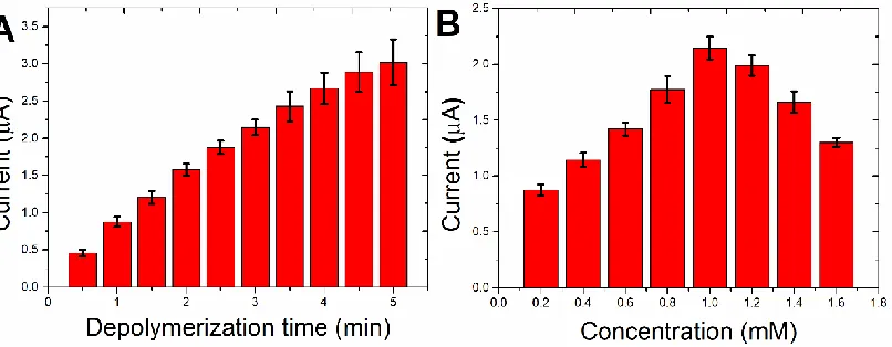
Figure 3. (A) Effect of the depolymerization time on the oxidation current difference. (B) Effect of the silver ions on the depolymerization time.
The optimization process was conducted before the real sample analysis. Figure 3A shows the oxidation difference after Fenton solution addition along with the depolymerization time. The current difference increases with depolymerization. However, a longer depolymerization could result in a high detection error. Therefore, the depolymerization was selected for 3 min. Figure 3B shows the current difference affected by the silver ion concentration. A lower silver concentration could result in a larger current difference before and after the cross-linking process since the excess of silver ions could result in a higher background current, while insufficient silver ions could lead to the unsuccessful formation of a hydrogel [56]. The current difference increased with increasing silver ion concentration. Then, the current difference decreased after further concentration increases due to the excess of silver ions in the hydrogel system.
[image:6.596.99.502.73.230.2]
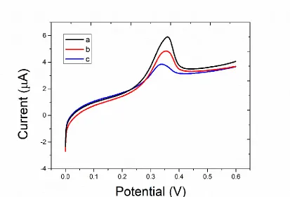
Figure 4. Cyclic voltammetry profiles of silver ion cross-linked hydrogel after introduction of (a) honeybee pupa, (b) honey and (c) Royal jelly.
[image:7.596.92.506.69.354.2] [image:7.596.92.505.407.687.2]
A bulk of the phenolic compounds found in honey is in the form of flavonoids [57]. However, other classes of phenolic compounds are also present in appreciable amount. Figure 4 shows DPV profiles of the antioxidation performance of honeybee pupa, Royal jelly and honey. A clearer difference can be observed compared with the cyclic voltammetry method. More specifically, the Royal jelly exhibited the lowest oxidation peak current, indicating its strong antioxidation performance among the sample. Based on the results, the antioxidant performance of the three bee products follows the following order: Royal jelly > honey > honeybee pupa.
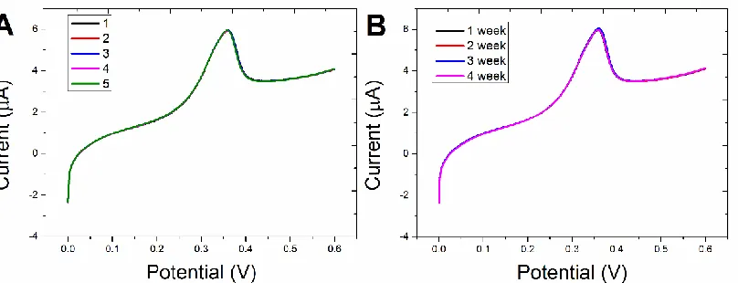
Reproducibility and stability are very important for electrochemical sensing platforms. Figure 6A shows five individual fabricated silver ion cross-linked hydrogels for Royal jelly determination. Very similar results can be obtained during the experiments, suggesting the excellent reproducibility of the hydrogel. Figure 7B shows the DPV curves of one batch hydrogel for Royal jelly determination over a month. The sensing performance could remain more than 90% compared over a month, suggesting that the proposed electrochemical sensing platform has a comparable storage stability.
Figure 6. (A) DPV profiles of five hydrogels after the introduction of Royal jelly. (B) DPV profiles of four hydrogels after the introduction of Royal jelly over 4 weeks.
4. CONCLUSION
In conclusion, measuring the antioxidant activity of bee products plays a practical role in the food industry. In this paper, we propose a cross-linked hydrogel method to measure the antioxidant activity of different bee products. Silver ions in the hydrogel have been used as detection probes for evaluating the antioxidation performance. The results indicate that the Royal jelly purchased from the online shop showed a higher antioxidation performance compared with that of honey and honeybee pupa.
ACKNOWLEDGEMENT
[image:8.596.97.501.315.470.2]
References
1. P. Premratanachai, C. Chanchao, Asian Pac. J Trop. Biomed., 4 (2014) 337.
2. Z. Chen, F. Dong, S. Li, Z. Zheng, Y. Xu, J. Xu, X. Liu, Y. Zheng, J. Chromatogr. A, 1410 (2015) 181.
3. P.M. Kustiawan, S. Puthong, E.T. Arung, C. Chanchao, Asian Pac. J Trop. Biomed., 4 (2014) 549.
4. B. Denisow, M. Denisow‐Pietrzyk, J. Sci. Food Agric., 96 (2016) 4303.
5. J. Ravoet, W. Reybroeck, D.C. de Graaf, Bull. Environ. Contam. Toxicol., 94 (2015) 543. 6. Ż. Bargańska, M. Ślebioda, J. Namieśnik, Crit. Rev. Environ. Sci. Technol., 46 (2016) 235. 7. T. Asama, T.H. Arima, T. Gomi, T. Keishi, H. Tani, Y. Kimura, T. Tatefuji, K. Hashimoto, J. Appl.
Microbiol., 119 (2015) 818.
8. Ö. SARAL, O. Yildiz, R. Aliyazicioğlu, E. Yuluğ, S. Canpolat, F. ÖZTÜRK, S. Kolayli, Turkish J.Med. Sci., 46 (2016) 194.
9. E.V. Ryabov, G.R. Wood, J.M. Fannon, J.D. Moore, J.C. Bull, D. Chandler, A. Mead, N. Burroughs, D.J. Evans, PLoS Path., 10 (2014) e1004230.
10. N.I. Egekwu, F. Posada, D.E. Sonenshine, S. Cook, Exp. Appl. Acarol., 74 (2018) 301.
11. A. Billiet, I. Meeus, F. Van Nieuwerburgh, D. Deforce, F. Wäckers, G. Smagghe, Insect Sci., 24 (2017) 270.
12. A. Yamane, H. Kohno, T. Ikeda, K. Kaneko, A. Ugajin, T. Fujita, T. Kunieda, T. Kubo, PloS one, 12 (2017) e0183522.
13. O. Anjos, M.G. Campos, P.C. Ruiz, P. Antunes, Food Chem., 169 (2015) 218.
14. R.T. Richardson, C.H. Lin, D.B. Sponsler, J.O. Quijia, K. Goodell, R.M. Johnson, App. Plant Sci., 3 (2015) 1400066.
15. A.J. Siddiqui, S.G. Musharraf, M.I. Choudhary, Food Chem., 217 (2017) 687.
16. N. Spano, V. Guccini, M. Ciulu, I. Floris, V.M. Nurchi, A. Panzanelli, M.I. Pilo, G. Sanna, Arabian J. Chem., 11 (2018) 492.
17. G. Martínez-Domínguez, R. Romero-González, A.G. Frenich, Food Chem., 197 (2016) 907. 18. W. Tian, M. Li, H. Guo, W. Peng, X. Xue, Y. Hu, Y. Liu, Y. Zhao, X. Fang, K. Wang, Nature
Communi., 9 (2018) 3373.
19. K. Brudzynski, C. Sjaarda, PLoS One, 10 (2015) e0120238. 20. A. Buttstedt, R.F. Moritz, S. Erler, Bio. Rev., 89 (2014) 255.
21. Y. Zhang, X. Liu, X. Li, J. Zhang, Y. Cao, M. Su, Z. Shi, H. Sun, Food Control, 60 (2016) 667. 22. Y. Jin, J. Zhang, W. Zhao, W. Zhang, L. Wang, J. Zhou, Y. Li, Food Chem., 221 (2017) 1298. 23. H. Zhang, R. Liu, R. Tsao, J. Fun. Foods, 22 (2016) 363.
24. X. Zhao, P. Wei, Z. Liu, B. Yu, H. Shi, Acta Physi. Plant., 39 (2017) 19.
25. P.S. Reddy, G. Jogeswar, G.K. Rasineni, M. Maheswari, A.R. Reddy, R.K. Varshney, P.K. Kishor, Plant Physiol. Biochem., 94 (2015) 104.
26. S. Sarkar, A. Kumari, J. Sarmah, Vegetos-An Int. J. of Plant Res., 31 (2018) 101. 27. T. Wirth, Angew. Chem. Int. Ed., 54 (2015) 10074.
28. B.A. Carlson, R. Tobe, E. Yefremova, P.A. Tsuji, V.J. Hoffmann, U. Schweizer, V.N. Gladyshev, D.L. Hatfield, M. Conrad, Redox Biology, 9 (2016) 22.
29. T. Zhang, J. Fang, K. Ono, W. Islam, H. Tsutsuki, T. Sawa, Free Radical Biol. Med., 128 (2018) S120.
30. F.-K. Shieh, S.C. Wang, C.I. Yen, C.C. Wu, S. Dutta, L.Y. Chou, J.V. Morabito, P. Hu, M.H. Hsu, K.C.W. Wu, J. Am. Chem. Soc., 137 (2015) 4276.
31. J. Taira, E. Tsuchida, M.C. Katoh, M. Uehara, T. Ogi, Food Chem., 166 (2015) 531.
32. S.R. Iora, G.M. Maciel, A.A. Zielinski, M.V. da Silva, P.V.d.A. Pontes, C.W. Haminiuk, D. Granato, Int. J. Food Sci. Tech., 50 (2015) 62.
34. J. Hoyos-Arbeláez, M. Vázquez, J. Contreras-Calderón, Food Chem., 221 (2017) 1371.
35. S.M. Gomes, M.-E. Ghica, I.A. Rodrigues, E. de Souza Gil, A.M. Oliveira-Brett, Talanta, 154 (2016) 284.
36. A.M. Pisoschi, C. Cimpeanu, G. Predoi, Open Chem., 13 (2015)
37. R. Tirawattanakoson, P. Rattanarat, N. Ngamrojanavanich, N. Rodthongkum, O. Chailapakul, J. Electroanal. Chem., 767 (2016) 68.
38. F. Gao, D. Zheng, H. Tanaka, F. Zhan, X. Yuan, F. Gao, Q. Wang, Mater. Sci. Engin: C, 57 (2015) 279.
39. L. Fu, A. Wang, F. Lyu, G. Lai, J. Yu, C.-T. Lin, Z. Liu, A. Yu, W. Su, Sensors Actuators B: Chem., 262 (2018) 326.
40. L. Fu, A. Wang, F. Lyv, G. Lai, H. Zhang, J. Yu, C.-T. Lin, A. Yu, W. Su, Bioelectrochemistry, 121 (2018) 7.
41. L. Fan, H. Yang, J. Yang, M. Peng, J. Hu, Carbohydr. Polym., 146 (2016) 427.
42. J. Xu, S. Strandman, J.X.X. Zhu, J. Barralet, M. Cerruti, Biomaterials, 37 (2015) 395. 43. M. Fan, Y. Ma, J. Mao, Z. Zhang, H. Tan, Acta Biomater., 20 (2015) 60.
44. S. Dhivya, S. Saravanan, T. Sastry, N. Selvamurugan, J. Nanobiotech., 13 (2015) 40.
45. A. Moatary, A. Teimouri, M. Bagherzadeh, A.N. Chermahini, R. Razavizadeh, Ceram. Int., 43 (2017) 1657.
46. Z. Wang, Z. Wang, H. Zhang, X. Duan, J. Xu, Y. Wen, RSC Advances, 5 (2015) 12237. 47. M. Yang, J.-M. Jeong, K.G. Lee, S.J. Lee, B.G. Choi, Biosens. Bioelectron., 89 (2017) 612. 48. L. Li, Y. Wang, L. Pan, Y. Shi, W. Cheng, Y. Shi, G. Yu, Nano Lett., 15 (2015) 1146.
49. E. Peltola, N. Wester, K.B. Holt, L.-S. Johansson, J. Koskinen, V. Myllymäki, T. Laurila, Biosens. Bioelectron., 88 (2017) 273.
50. M.R. Romero, D. Peralta, C.I.A. Igarzabal, A.M. Baruzzi, M.C. Strumia, F. Garay, Sensors Actuators B: Chem., 244 (2017) 577.
51. T. Wang, H. Guo, X. Chen, M. Lu, Mater. Sci. Engin: C, 72 (2017) 62.
52. M. Choudhary, R. Brink, D. Nandi, S. Siwal, K. Mallick, J. Mater. Sc., 52 (2017) 770.
53. A. Fandrich, J. Buller, H. Memczak, W. Stoecklein, K. Hinrichs, E. Wischerhoff, B. Schulz, A. Laschewsky, F. Lisdat, Electrochim. Acta, 229 (2017) 325.
54. M. Hasanzadeh, N. Shadjou, Y. Lin, M. de la Guardia, TrAC, Trends Anal. Chem., 86 (2017) 185. 55. D.I. Torres, M.V. Miranda, V.C. Dall’Orto, Sensors Actuators B: Chem., 239 (2017) 1016. 56. W. Ngeontae, W. Janrungroatsakul, P. Maneewattanapinyo, S. Ekgasit, W. Aeungmaitrepirom, T.
Tuntulani, Sensors Actuators B: Chem., 137 (2009) 320.
57. M. Viuda‐Martos, Y. Ruiz‐Navajas, J. Fernández‐López, J. Pérez‐Álvarez, J. Food Sci., 73 (2008) R117.