Vol.63, No. 10
Fatty
Acid
Acylation of Vaccinia Virus
Proteinst
CHRISTINEA. FRANKE, PAMELAL. REYNOLDS, AND DENNIS E. HRUBY*
Department ofMicrobiology, Center forGeneResearch, Oregon State University, Corvallis, Oregon 97331-3804
Received 27 March 1989/Accepted 9 June 1989
Labeling of vacciniavirus-infectedcells with[3H]myristicacid resulted in the incorporation oflabelintotwo viral proteins withapparentmolecular weights of35,000and25,000(designated M35 and M25, respectively). M35andM25wereexpressed ininfected cells after theonsetof viral DNA replication, andboth proteinswere
present in purified intracellular virus particles. Virion localization experiments determined M25 to be a
constituentofthe virion envelope, while M35 appearedtobeperipherally associatedwith the virioncore.M35
and M25 labeled by [3H]myristic acid were stableto treatment with neutral hydroxylamine, suggesting an
amide-linkedacylation oftheproteins. Chromatographic identificationof the protein-bound fatty acid moieties
liberated after acidmethanolysisof M25, isolated from infected cells labeled duringa4-hpulse,resulted in the recoveryof 25% of the protein-boundfatty acidasmyristate-associated label and 75% aspalmitate, indicating thatinterconversion of myristatetopalmitatehadoccurredduring the labeling period. Similar analyses of M25
and M35, isolatedfrominfectedcells labeled duringa0.5-h pulse, determined that 46 and 43%, respectively,
of theprotein-bound labelhad been elongatedtopalmitateevenduringthisbrieflabeling period. Incontrast,
M25andM35 isolatedfrompurffied intracellular virions labeledcontinuously during 24 h of growth contained 75and70%,respectively, myristate-associated label, suggestinggreaterstabilityofthese proteinsorafavored
interactionof the proteins containing myristate with the maturing orintracellular virion.
Thecovalent attachment of fattyacid residuesto nascent
polypeptideshasbecomewidely recognizedas animportant,
and in some cases essential, protein modification reaction
whichoccursineucaryotic organisms (28; forareview, see
references 29, 33, and 41). Although the functional
signifi-cance of this type of protein modification is not clearly understood, theattachment of the acyl prosthetic groupsis
likely to influence the structure, activity, and possibly the
subcellularlocalization oftheacceptorproteins (29, 33,41).
Several differentfatty acidmoietieshavethusfarbeen found attached to proteins, includingpalmitic acid (for a review, seereferences29 and33),myristicacid(1, 6, 8, 9, 11, 15, 17, 20-24, 26, 34, 35, 37), and, to a lesserextent, stearic acid, linoleic acid, and oleic acid (33). In the more common
palmitation and myristylation modifications, distinct
differ-encesexistwithregardto howtheacylgroupsareattached toproteins,which amino acidsaremodified,and where the
modifiedproteinsaretargeted.Forexample, palmitic acidis
posttranslationally added via an ester or thioester bond to
internalthreonineorcysteine residuesofproteinswhichare
primarily destined to be membrane associated (16-19, 21, 22). Incontrast, myristylationofproteinsoccurs
cotransla-tionally via an amide linkage to the N-terminal glycine
residue of acceptor proteins which may or may not be membraneassociated (21, 23, 33, 41). Little isknown about whatprotein structuralorsequenceparametersinfluencethe selection ofacylationsitesordetermine whichfattyacidwill
be added. Inordertoaddress these questions andto study
what role protein acylation may play in the regulation of
gene expression,theuse of viral systems whichare
amena-bletodirectedgeneticmanipulationswouldseemtoofferan
attractiveexperimental approach.
Acylatedviralproteinshavebeenreported,identified, and
studied inseveral different families ofRNAviruses, includ-ing the Togaviridae, the Retroviridae, and the
Picornaviri-* Correspondingauthor.
tOregon Agricultural Experiment Station technical paper no.
8823.
dae(30, 32, 33, 41). In sharpcontrast, little isknownabout
the status of fattyacid acylation reactions during the
repli-cativecycles ofsomeof the largeDNA viruses, such asthe
Poxviridae. Withregardtovaccinia virus (VV), which is the prototype of thePoxviridae, a single majorpalmitated
pro-tein(P37)has thus far beenreported (12). Thestatusof other types of potential acylation modifications on VV-encoded
proteins hasnotbeen investigated. The function of the P37 protein is unknown, but it is found in the envelope of
extracellular but not intracellular virus particles and has been suggested to be nonessential for virus replication in tissueculture cells (12, 13). Since VVcompletes its
replica-tive cycle within the cytoplasm of infected cells and has already been shown to encode many ofthe cis- and
trans-acting factors necessary to modulate the expression of its
geneticprogram (36), this virusprovides anexcellent model
systemforstudyingtranscriptional and translational regula-torymechanisms.Therefore, aspartofanefforttobeginto
elucidate the various posttranslational regulatory strategies which may be used by VV (and perhaps the host cell), a
studywasundertakentocharacterize the nature, specificity,
andextentoffatty acidacylation of VVproteins.
Wereporthere thatmyristicacid(aswellaspalmitic acid)
is covalently linked to two VV proteins (Mr -25,000 and -35,000) by a hydroxylamine-resistant (i.e., presumably
amide) linkage. Both proteins are expressed at late times duringthe virusreplicative cycleand arepresentinpurified
intracellular virusparticles.Wealsoconfirmed andextended
theprevious observationswithregardto P37(12) by identi-fyingtheattachedacylgroupandexploringthenatureof its protein linkage. Experiments were carried out to examine
the synthesis, maturation, and subvirion localization of the
VV acyl proteins. Theresults obtained arediscussed in the context of the potential biological roles of these modified proteins duringthe viralreplicative cycle.
MATERIALSAND METHODS
Cells,virus,andlabelingconditions.The WR strain of VV
wasgrownin monolayersofBSC40 (African greenmonkey)
4285 JOURNALOFVIROLOGY,OCt. 1989, p.4285-4291
0022-538X/89/104285-07$02.00/0
CopyrightC) 1989, American Society for Microbiology
on November 10, 2019 by guest
http://jvi.asm.org/
cells maintained in Eagle minimal essential medium with Earle salts (MEM-E) supplemented with 10% heat-inacti-vated fetal calf serum, 2 mM L-glutamine, and 10
F.g
ofgentamycin sulfate per mlaspreviously described(14).
For pulse-labeling experiments, BSC40 cell monolayers (60-mm dishes) infected with VV (20 PFU/cell) were incu-bated in MEM-E(1/10thenormalmethionine concentration
wasused when [35S]methionine was usedforlabeling) con-taining 5% delipidated fetal calf serum (7),
L-glutamine
(2mM), and either L-[35S]methionine (25
ljCi/ml,
1,129 Ci/mmol;NewEnglandNuclearCorp.),[9,10-3H]myristic acid (250 pLCi/ml, 22.4 Ci/mmol; New England Nuclear), or
[9,10-3H]palmitic
acid (250pLCi/ml,
60Ci/mmol;
NewEn-gland Nuclear) at 37°C for the times specified in each
experiment. At the end ofthe labeling period, the medium was removed, and the cell monolayer was washed three times with ice-cold phosphate-buffered saline and lysed in
gel sample buffer (68 mM Tris hydrochloride [pH 6.8], 2% sodiumdodecylsulfate[SDS],10 mMdithiothreitol contain-ing 10% glycerol and 0.1% bromophenol blue). DNA was
sheared by repeated pipetting through a 22-gauge needle,
and lysates were boiled for 3 to 5 min priorto analysis on SDS-polyacrylamide gels.
Radiolabeled virus was isolatedfrom two150-mmdishes of BSC40 cells infected with 10 PFU of virus per cell and
incubated in the presence ofeither
L-[35S]methionine
(12.5paCi/ml), [3H]myristic
acid(125pLCi/ml),
or[3H]palmiticacid(125
V.Ci/ml)
for24h at37°Candpurified bysucrosegradient sedimentationasdescribedpreviously
(14).Separation of viral envelope and core fractions. Purified
radiolabeledvirions were separated into NonidetP-40 (NP-40)-soluble (envelope) and NP-40-insoluble (core) fractions asdescribed previously (42). SDS-polyacrylamide gel
elec-trophoresis and fluorography were used to analyze the
protein composition of bothfractions of each labeled virus
preparation.
Gel electrophoresis and protein isolation. Radiolabeled
proteins were analyzed on 10% polyacrylamide slab gels containing SDS (39). The gels were then impregnated with
2,5-diphenyloxazole andfluorographed at -70°C on Kodak XAR-5 film(5).
Toisolate individual protein species,radiolabeledproteins wereseparated by SDS-polyacrylamide gel electrophoresis,
and theregion ofthegel that containedthe 3H-labeledacyl
protein was located by interpolation from adjoining lanes thatcontained
[35S]methionine-labeled
virion proteins. The labeledproteinswererecoveredfromthe excised gelstrip byelectrodialysis in avolatile buffer system (3).
Analysis ofprotein-bound fatty acid. For hydroxylamine
treatmentoflabeledproteins following SDS-polyacrylamide gel electrophoresis, duplicate sets ofprotein extracts were
separatedon a 10% polyacrylamide gel, and thegel was cut in half and soaked in either 1.0Mhydroxylamine (pH 7.0) or 1.0 M Trishydrochloride(pH 7.0) for 2 h at room
tempera-ture (18, 22). Gels were then fluorographed as described above.
Identification of protein-bound lipids was carried out
essentially as describedpreviously (22). Briefly, individual
3H-labeled
VVacyl proteins were subjected to acidmetha-nolysis
asfollows. Labeledprotein (20p.l)
was treated with1mlof2 N
HCI-83%
methanol in vacuo at 95°C for 60 h. Thereaction products were extracted three times with 1 ml of
petroleum ether at4°C. The combined ether extracts were
evaporated to dryness under a stream of nitrogen and
redissolvedin 150
p.l
of high-pressureliquid chromatography(HPLC)-grade
methanolcontaining
6.25 mg eachofmyristicMet Myr Pal
MWUIU E L tUl E L Ul E L
97.4 -68.0
-43.0- _
25.7--- " =
..s .ss
A
-.a
vv
Met Myr Pal
U-am
o--.
S
3*o
. P37 4M35
4IM25
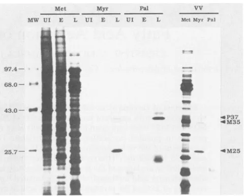
FIG. 1. SDS-polyacrylamide gel electrophoresis ofmetabolically
labeled proteins. Uninfected (UI) andvaccinia virus-infected cell monolayers were labeledwith [35S]methionine (Met), [3H]myristic
acid (Myr), or[3H]palmiticacid(Pal)atearly (E,0.5 to2.5h)orlate
(L, 4 to 8h)timesafterinfection. Purified intracellularvirus(VV)
waspreparedfrominfected cells labeledcontinuously for24hafter infection. Labeled proteins from total-cell lysates and purified intracellular virions were separated by SDS-polyacrylamide gel electrophoresis on a 10% polyacrylamide gel and visualized by fluorography. Arrows indicate the positions ofthe major protein labeled with[3H]palmitic acid(P37) and the major (M25)andminor
(M35)proteins labeledwith[3H]myristicacid.Radioactive
molecu-larsize markers(MW) arephosphorylaseB(97.4 kilodaltons[kDa]), bovine serum albumin (68 kDa), ovalbumin (43 kDa), and
a-chymotrypsin (25.7 kDa).
acid, palmitic acid, myristicacidmethylester, andpalmitic
acidmethylester(SigmaChemicalCo.)permlasstandards. The ether-extractable radioactivity recovered after acid methanolysiswas separated and identified by reverse-phase HPLC with a ,uBondapak C18 column (3.9 mm by 30 cm; WatersAssociates)elutedisocraticallywith 80% acetonitrile
asthe mobile phase at a flowrate of 1.0 ml/min. Fractions elutingfrom the columnwere collected in0.5-min intervals and counted in 3 ml of 3a70 scintillation fluor (Research ProductsInternational, Inc.).Theabsorbanceelutionprofile
(A254) of the internal standards was compared with the
elution oflabeled speciesto identifyeachlipid.
RESULTS
Acylated proteins of VV. In order to identify VV acyl
proteins, uninfected orVV-infectedBSC40 cells were pulse-labeled with either [35S]methionine, [3H]palmitic acid, or
[3H]myristic
acidatearly (0.5 to 2.5 h) or late (4 to 8 h) timespostinfection, and total-cell lysates were prepared.
Simi-larly,intracellularvirus particles were purified from infected cells labeled continuously for 24 h. The pattern of proteins which were labeled with the fatty acids was examined by
electrophoresis on 10% polyacrylamide gels, followed by
fluorography. Equivalent amounts of fatty
acid-abeled
ex-tracts were analyzed along with 0.02% of the amount of
[35S]methionine-labeled
extracts.Theradiolabeled palmitateandmyristatewereincorporated into distinctsetsof proteins which comigrated with methionine-labeled polypeptides
on November 10, 2019 by guest
http://jvi.asm.org/
[image:2.612.319.560.77.270.2]VV PROTEIN ACYLATION 4287 from VV-infected cell lysates (Fig. 1). The protein which
was most highly labeled with palmitate had an apparent Mr of -37,000 (designated protein P37). P37 has been reported previously by other investigators to be the major antigen present on the envelope of extracellular VV particles, but absent from intracellular virus particles (12). P37 has been included in this study as a control and to extend the information known about the nature of the fatty acid moiety and its linkage to this acyl protein. The protein which was most highly labeled with myristate had an apparent Mr of -25,000 (designated M25) and did not comigrate electro-phoretically with any of the palmitate-labeled VV proteins. In addition to M25, a minor myristate-labeled proteinof Mr -35,000 (designated M35) was also detected. In some ex-tracts, an additional minor myristate-labeled protein was seen that had a slightly slower electrophoretic mobility than M35 and appeared to comigrate with P37. The relative amounts of the two species varied from almost equivalent (lane Myr/L) to exclusively the faster-migrating M35 (lane VV/Myr). It is unclear at this time whether the myristate-labeled polypeptide which comigrated with P37 is a myristy-lated protein distinct from M35, a modified form of M35, or perhaps P37 which has become labeled by metabolic inter-conversion of the myristate label.
The data shown in Fig. 1 also established that the myris-tate-labeled proteins M25 and M35 were present in purified intracellular VV particles (lane VV/Myr). None of the pro-teins observed in infected cells labeled with palmitate were detected in an equivalent amount of protein from purified intracellular virus particles (lane VV/Pal). Other investiga-tors have demonstrated that the major palmitate-labeled protein, P37, was present in extracellular virus particles (but not intracellular virus particles) and that the gene encoding this protein maps within the viral genome (12, 13). Although the major myristate- and palmitate-labeled proteins differed in their apparent Mr and presence in or absence from intracellular virus particles, both sets of labeled proteins belonged to the late temporal class of virus gene products, since they were expressed in infected cells at times after the onset of viral DNA replication. Little or no evidence of these proteins was observed when pulse-labeling with the fatty acids was performed at early times during infection (lanes Myr/E andPal/E). This kinetic class was assigned to P37 in the characterization of this protein described previously (12).
The subvirion location of M35 and M25 was investigated by experiments in which purified [35S]methionine- or
[3H]myristic acid-labeled intracellular virus particles were treated with 0.5% NP-40 and 50 mM dithiothreitol and then separated into viral envelope (detergent soluble) and core (detergent insoluble) fractions prior to SDS-polyacrylamide gel electrophoresis and fluorography. M25 was present en-tirely in the NP-40-soluble fraction, suggesting that it resides within the virus envelope, while the majority of M35 was seen in the NP-40-insolublefraction, with a lesser amount in the envelope fraction, indicating aperipheral or weak asso-ciation of M35 with the virion core (data not shown). The biological function of the major (M25) and minor (M35) proteins labeled withmyristate has not yet beendetermined. The covalent attachment of myristate andpalmitate tothe proteins was investigated by experiments in which fatty acid-labeled cell extracts weredelipidated byextraction with chloroform-methanol (2:1) prior to SDS-polyacrylamide gel
electrophoresis. The pattern oflabeled proteins was unaf-fected by this treatment except that the non-protein-bound
radioacti-e lipid which migrated to the dye front during
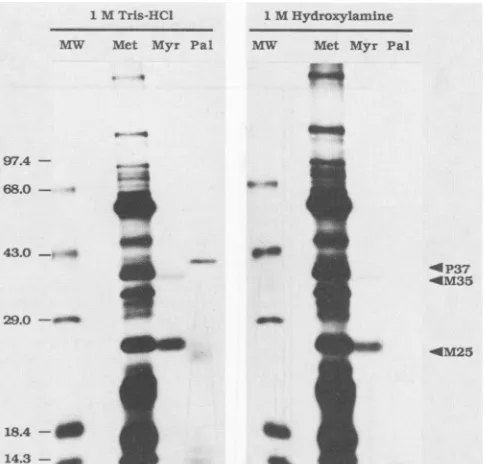
1MTris-HCI 1 MHydroxylamine M`W Met Myr Pal AM Met Myr Pal
on"
97.4
-68.0
-43.0 - sow .4
<~~~~- P37
_
~~~~~~~~~OM35
29.0 -a_
__ _ * ~~~~~~~~0M25
18.4-d f u
14.3
-FIG. 2. Sensitivity of acylatedproteins tohydroxylamine. Vac-ciniavirus-infected cell monolayerswere labeled with [3H]myristic
acid (Myr) or [3H]palmitic acid (Pal) at late (4 to8 h) timesafter
infection. Purified intracellular virus (Met) was prepared from infected cells labeled continuously with [35S]methionine for 24 h after infection. After separation of labeled proteins by SDS-poly-acrylamide gelelectrophoresis on a10% gel, thegelwas cutin half
and treated for 2 h at room temperature with either 1 M Tris hydrochloride(pH 7.0) or 1 Mhydroxylamine(pH 7.0), afterwhich they were treated forfluorography. Arrows indicatethepositions of
the major proteinlabeled with[3H]palmiticacid(P37)and themajor
(M25) and minor (M35) proteins labeled with [3H]myristic acid.
Radioactive molecular size markers (MW) are phosphorylase B (97.4 kDa), bovine serum albumin (68 kDa), ovalbumin (43 kDa),
carbonic anhydrase(29kDa), 3-lactoglobulin(18.4 kDa),lysozyme
(14.3kDa).
electrophoresis was absent after treatment(datanotshown).
The possibility that the fatty acids are metabolically
inter-converted cannot be ruled out from these experiments.
However, the specificity of fatty acid labeling of these proteins appears to minimize the possibility that the
fatty
acid labeling of the proteins was due solely to metabolic
conversion offattyacids toamino acidswhichwere reincor-porated into protein. Likewise, the
fatty
acid-labeling
pro-files were substantially distinct from each other and from those ofproteins labeled with methionine.Analysis of protein-bound fatty acids. Fatty acids have been shown to be attached to proteins via both ester and amide linkages, and in general, some
fatty
acyl
chain spec-ificity of protein-lipid linkages has been observed. The linkage ofthe palmitic acidmoiety
to themajority
ofacyl
proteins studied has been identified as an ester or
highly
reactive thioester bond from itslabilitytoalkaline methanol-ysis andsensitivity to thenucleophile
hydroxylamine
(4,
16,
19, 22, 43). Incontrast, the bond
through
whichmyristate
is linked to proteins is resistant to treatment withhydroxy-lamine and alkaline methanolysis,
suggesting
amidelinkage
of this fatty acid to the
acyl
protein (22).
In order toinvestigate the nature ofthe
fatty
acidlinkage
toP37, M25,
and M35, thesensitivity
of themyristate-
andpalmitate-labeled proteins to cleavage
by
hydroxylamine
wasexam-ined. As shown in Fig. 2, treatment of the
proteins
with VOL.63, 1989on November 10, 2019 by guest
http://jvi.asm.org/
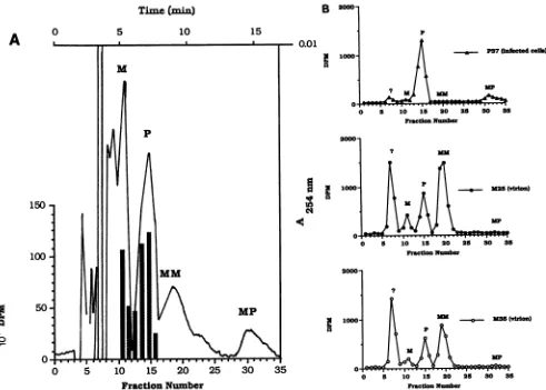
[image:3.612.320.562.74.306.2]0.01
1000-
A
-s-P37(infecftdcehb)0 0 15 20 25 30 35
FractionNumber 200(
1004
0 P
0- p =~~-M5(viuion)
N
up
o .
0 5 10 15 20 25 30 35
Fraction Number
2000-1000- NM
P
M
--O.- MS (vddon)
MP
D 5 0 15V 20 2 O 3
Fraction Number
FIG. 3. Analysisoffattyacids.(A)HPLC ofunlabeledfattyacid andfattyacidmethylesterstandards andlabeledfattyacid standards. HPLCseparationswereperformedasdescribed in Materials and Methods. The elution ofradioactivityafterchromatographyof[3H]myristic
acid(solid bars)or[3H]palmiticacid(stippledbars)issuperimposedonatracingof the absorbance elutionprofileof internalfattyacid and
fattyacidmethylesterstandards(M,myristic acid; P,palmiticacid; MM,myristicacidmethylester; andMP, palmiticacidmethylester). (B)HPLC of labeledprotein-bound fattyacids. Purified labeledproteinsweresubjectedtoacidmethanolysis,andthe releasedfattyacids and
fattyacidmethylesterswereseparated byHPLCasdescribed in Materials and Methods. The distribution ofradioactivityis comparedwith theelutionoffattyacid andfattyacidmethylesterinternal standards(see above)toidentifytheradioactive acidmethanolysis products. Top panel, Fatty acids released fromgel-isolated P37(infected cells); centerpanel, fattyacids released from gel-isolated M25 (virion)protein;
bottompanel,fatty acids released fromgel-isolated M35(virion) protein.
hydroxylamine resulted in the removal of label from the
palmitate-labeledproteins,butdidnotsubstantially alter the
myristateormethioninelabelingpatterns.Thereleaseof the
palmitate-bound label from P37by this treatment is
consis-tentwithanesterorthioesterlinkageof thepalmitatetothe
protein. Conversely, the stability of the protein-bound
my-ristate label to hydroxylamine suggests that the fatty acid
linked to these proteins (M25 and M35) is attached via an
amide bond. However,thistypeofexperimentstill doesnot
ruleoutlabelingof theproteins bymetabolicinterconversion
of the labeledfatty acids into the amino acid pool.
To determine whether the label associated with the
pro-teinswaspresent asfattyacid rather thanlipidmetabolites,
andto identifythemoiety covalentlyboundto theproteins,
individualfatty acid-labeled proteins wereisolatedby
SDS-polyacrylamide gelelectrophoresisasdescribed in Materials
and Methods. The identity and purity of the recovered
proteins were confirmed by gel electrophoresis (data not shown). The chromatographic separation of unlabeled fatty
acid and fatty acid methyl ester standards which were
cochromatographed with the labeled fatty acids
([PHipal-mitic acid and [3H]myristic acid) used for labelingin these
studies is shown inFig. 3A. Fromthese data itcanbe seen
that the fatty acid (myristic acid [M] andpalmitic acid [PI)
andfattyacidmethylester(myristicacidmethylester[MM]
and palmitic acid methyl ester [MP]) standards were well
separated bythischromatographic systemintopeaks(A254)
which eluted at 5.2 (M), 7.4 (P), 9.5 (MM), and 15.5 (MP)
min. The purity of the [3H]palmitic acid and [3H]myristic
acid used forlabelingin thesestudieswasalso demonstrated
by the coelution of the labeled fatty acids with thepeak of
absorbance for either myristic acid or palmitic acid
stan-dards.
Inthe experiment shown in Fig. 3B, M25 and M35 (from
myristate-labeled purified intracellular virus particles) and P37 (from virus-infected cell lysates labeled with palmitic
acid from 4 to 8 h postinfection) were isolated and then
subjectedtoacidmethanolysisfollowedby etherextraction
as described in Materials and Methods, and the liberated
fattyacidswere identified afterseparation by reverse-phase
0
Time
(min)
5 10
B 2000" 15
A
V
10
.4
0
cq)
Fraction Number
AI
I
I-W .I-W .. .U-t
on November 10, 2019 by guest
http://jvi.asm.org/
[image:4.612.66.557.77.428.2]VV PROTEIN ACYLATION 4289 TABLE 1. Fattyacid composition of acylated proteins'
%of totalprotein-bound
Protein source Radioactivityas fattyacid'
and protein fatty acid(%)b
Myristate Palmitate Virion (24-h pulse)
M25 72 75 25
M35 60 70 30
Infected cells 4-hpulse
P37 96 2 98
M25 92 25 75
30-min pulse
M25 99 54 46
M35 97 57 43
"The compositionof the isolated, labeled protein-bound fatty acids was
determined after acid methanolysisfollowed by HPLC.
bThe percentage of the totalradioactivityrecovered that coeluted with the
fatty acid and fatty acidmethylesterstandards upon separation byHPLC.
c The percentage of the totalprotein-boundradioactivity that coeluted with
eithermyristic acid and myristic acidmethyl ester (myristate) or palmitic acid and palmitic acid methyl ester (palmitate) standards upon separation by
HPLC.
HPLC. In each case, an unidentified peak of radioactivity elutedearly in the chromatography (fraction 7, 3.2 min) that wasminor in some instances (top panel) and considerable in others (center and bottom panels). The magnitude of this peak correlated directly with the length of the labeling
period, suggesting that thispeak represents amino acids or other metabolites which had become labeledby
interconver-sion of the radioactive fatty acids during long labeling
periods. Wheneither unlabeled (20 amino acids) or labeled amino acids,
[3H]lysine
or [35S]methionine, were subjectedto the same chromatographic analysis, greater than 97% of the peak of either absorbance or radioactivity eluted at 3.2 min (in fractions 7 and 8), corroborating the suspected identity of this peak (data not shown). Acid methanolysis of thepalmitate-labeled proteinP37 resulted in the recovery of
palmitic acid and methyl palmitate (top panel). Essentially nomyristicacid ormethyl myristatewasdetected, and less than 5% ofthe label associated with the protein existed as
labeled amino acids(Fig. 3B and Table 1). Themajority(70 to75%) of the labeled lipid bound to M25 and M35(isolated
from purified intracellular virus particles) wasfoundtoexist
asmyristate moieties (Fig. 3B and Table1). These analyses indicatesomeelongation ofmyristatetopalmitate duringthe prolonged labeling period (24 h), as well as substantial
interconversion ofthe labelinto amino acids (28 to
40%).
Inan effort to circumvent the extensive metabolysis of the labeled myristate during prolonged labeling periods, M25 was isolated from infected cells that had been labeled with
[3H]myristic
acid during a 4-h pulse (from 4 to 8 h postin-fection). Analysis of the M25-bound radioactive species byHPLC after acid methanolysis revealed very little conver-sion oflabelintothe amino acid component of the
protein
(4 to8%)
(Table 1). Strikingly, 75%of theacylgroupsboundtoM25had been elongatedfrom
myristate
topalmitate during
the 4-hpulse-labelingperiod.Reductionof thepulse-labeling period to 30 min (6to 6.5 hpostinfection)
still resulted in conversion of 43 to 46% of the labeledmyristic
acid topalmitic acid when the fatty acid content of M35 and M25
(isolatedfrom infectedcells) was
analyzed (Table
1).DISCUSSION
Fatty acidacylationhasbeenreportedfora
variety
of viral and cellularproteins; however, thespecificity
of thismodi-fication and thefunction ofthe attachmentoffatty acidsto
these proteins are only beginning to be examined. The present study demonstrates that at least two types offatty acylation reactions (palmitylation and myristylation) occur on distinct late
protein
species of VV. Two intracellular virion-associated proteins, designated M35 and M25, werefoundto containprimarily myristicacid. However,analysis
of the fatty acid content of M25 and M35 isolated from
infectedcells revealedsignificantinterconversion ofmyristic
acidto
palmitic acid,
theamountdepending
onthelengthofthe labeling period. The finding thatfatty acids are
readily
converted to their
longer-chain
derivatives has been noted before by Schmidt (31), and the metabolic fate of myristic acidhasbeen showntodiffer in different cells(22). Whetherthe
myristic
acid is metabolized before orafter it isutilizedas the acyl donor, whethermyristic acid and palmitic acid
areattachedtothesameordifferent acceptorsites,and what role the host cell plays with respectto the interconversion
have not yetbeen determined forM25 and M35. However,
since
N-myristyl
transferase has been shown to behighly
specificformyristate,
this impliesthatelongation of myris-tate to palmitate is most likely occurring afterprotein
acylation.
There have beenprevious
reports ofprotein-bound palmitate present in an amide
linkage
(40). The observation that normal cellular enzymes arefattyacylated
indicates thatacylation
can beperformed by
cellular en-zymes;however,
thereis still thepossibility
ofparticipation
by
viral enzymes orregulatory
factors. In contrast, thepalmitate-labeled
protein
isolated from infectedcells, P37,
was demonstrated to contain almost exclusively palmiticacid bound by a
hydroxylamine-sensitive,
presumably
thioester
linkage.
Thelabeledfatty
acids boundtoM25and M35 were resistant to treatmentwithhydroxylamine,
sug-gesting that they are attached to the
proteins by
an amide bond. With respect to thelinkage
of thelipid
to M25 andM35, the
fatty
acid could be attached to the N-terminalamino acidofthe
protein
or tothe E-NH2group ofalysine
residue. In mostinstances ofmyristylation
of viralproteins,
the
fatty
acid has been attached to an N-terminalglycine
residue(8, 24).Further studies will be neededto
identify
theresidue(s)
modifiedby
thelipids
in M35 and M25.Several features involved in
characterizing
andidentifying
M25and M35should be noted. Both M25 and
M35,
likeP37,
can be
provisionally
classified aslateproteins,
in thatthey
are
expressed
in cells after the onset of viral DNAreplica-tion.
They
appear to berelatively
abundantproteins.
Pre-suming that,
asusual,
myristylation
occurs atonly
1mol permolof
protein
andconsidering
thelowspecific activity
ofthe[3H]myristic
acid(22.4
Ci/mmol)
used in theseexperiments,
fluorography
hadtobe doneforonly
2to3days
tovisualizethelabeled viral
proteins. Again assuming
onefatty
acylated
residue per
protein,
M25wasmuchmoreabundantorstable than M35. Bothmyristylated
proteins
were associated with the mature intracellular virusparticle
and were present inproportions
similar to those seen in infected cells. In thecells
commonly
used for thepropagation
ofVV,
mostof the progeny virus remain cell associated(intracellular
virus),
and
only
small amounts ofantigenically
distinct virus arenaturallyreleased from the cell
(extracellular
virus) (2).
Both intracellular and extracellular virusesareinfectious,
butthey
differ
markedly
intheir mode ofadsorption
andpenetration
in cell cultures
(25).
It will be of interest to determinewhether M25 and M35 are present in
purified
extracellular virus particles or their destination is intracellular virusspecifically.
In contrast, distinct subvirion locations havebeenobserved for each
protein;
M25 is releasedby
NP-40 VOL.63, 1989on November 10, 2019 by guest
http://jvi.asm.org/
with the virion envelope, whereas M35 remains associated with the virion core, although somewhat loosely.
The finding that M35 and M25 are myristylated raises the
question of the functional significance of the modification. A
scaffolding role for the formation of virus particles has been proposed for the myristylated VP2 proteins of simian virus 40 and polyomaviruses (38) and for the myristylated VP4 proteins of the picornaviruses (8, 24). Mutations in either these proteins or in the glycine acceptor residue have
resulted in either poor replication (10) or immature,
nonin-fectious particles (27). It has also been suggested that
myristate could be important in the early stagesofinfection, during theadsorption and penetration of virus particles into
the cell, by directing the modified protein to the cell or
vesicle membrane, where it could function inthe passage of
thevirusacrossthemembrane (8). In view ofthedifferences
observed in this regard between intracellular and extracellu-lar VV noted above, this might provide a clue toward a
possible biological function of the modification of one or
both of these proteins. Directed genetic studies will be
necessary to clarify the exact function of the myristate moiety of M35 and M25 of VV. To that end, genomic and
peptide mapping procedures are currently inprogress.
ACKNOWLEDGMENTS
We thankW. G. Dougherty for helpful comments onthe
manu-script.
This researchwassupported byaPublic Health Service research
grant(AI-21335) and a Research CareerDevelopment Award
(Al-00666) from theNational Institutes of Health. LITERATURE CITED
1. Aitkin, A., P. Cohen, S. Santikarn, D. H. Williams, A. G. Calder, A. Smith, and C. B. Klee. 1982. Identification of the NH2-terminal blockinggroupofcalcineurin B asmyristicacid.
FEBS Lett. 150:314-318.
2. Appleyard, G.,A.Hapel,andE. A. Boulter. 1971. Anantigenic difference between intracellular and extracellular rabbit-pox virus.J.Gen. Virol. 13:9-17.
3. Bhown, A. S.,J. E. Mole, F. Hunter, and J. C. Bennett. 1980. High-sensitivity sequence determination of proteins
quantita-tively recovered from sodium dodecyl sulfate gels using an
improved electrodialysis procedure. Anal. Biochem. 103:184-190.
4. Bolanowski, M. A.,B. J. Earles, and W. J. Lennarz. 1984. Fatty acylation of proteins during development of sea urchin
em-bryos. J. Biol. Chem. 259:4934-4940.
5. Bonner, W. M., and R. A. Laskey. 1974. A film detection method for tritium-labeled proteins and nucleic acids in poly-acrylamide gels. Eur. J. Biochem. 46:83-91.
6. Carr, S. A., K. Biemann, S. Shagi, D. C. Parmelee, and K.
Titani.1982. n-Tetradecanyl istheNH2-terminalblockinggroup
ofthecatalyticsubunit of cyclic AMP-dependentprotein kinase from bovine cardiac muscle. Proc. Nat]. Acad. Sci. USA 79:6128-6131.
7. Cham, B. E., and B. R. Knowles. 1976. A solvent systemfor delipidation of plasmaor serumwithout protein precipitation.J.
Lipid Res. 17:176-181.
8. Chow, M., J. E. Newman, D. Filman, J. M. Hogle, D. J.
Rowlans, and F. Brown. 1987. Myristylation of picornavirus
capsid protein VP4anditsstructural significance. Nature
(Lon-don)327:482-486.
9. Clark, B.,and U. Desselberger. 1988. Myristylationof rotavirus
proteins. J. Gen. Virol.69:2681-2686.
10. Cole, C.N., T. Landers, S. P.Goff, S.Manteuil-Brutlag,and P.
Berg. 1977. Physical and genetic characterization of deletion mutants of simian virus 40 constructed in vitro. J. Virol.
24:277-294.
11. Henderson, L. E., H. C. Krutzsch, and S. Oroszlan. 1983.
Myristyl amino-terminal acylation of murine retrovirus
pro-teins: an unusual post-translational protein modification. Proc.
Natl. Acad. Sci. USA 80:339-343.
12. Hiller, G., and K. Weber. 1985. Golgi-derived membranes that
contain an acylatedviral polypeptide areusedfor vacciniavirus
envelopment. J. Virol. 55:651-659.
13. Hirt, P., G. Hiller, and R. Wittek. 1986. Localization and fine structure of a vacciniavirusgene encodinganenvelopeantigen.
J. Virol. 58:757-764.
14. Hruby, D. E., L. A. Guarino, and J. R. Kates. 1979. Vaccinia virus replication. I. Requirement for a host cell nucleus. J. Virol. 29:705-715.
15. Jorgensen, E. C., N. 0. Kjeldgaard, F. S. Pedersen, and P.
Jorgensen. 1988. A nucleotide substitution in thegag N
termi-nusof the endogenous ecotropic DBA/2 virus preventsPr659'
myristylation and virus replication. J. Virol. 62:3217-3223. 16. Kaufman, J. F., M. S. Krangel, and J. L. Strominger. 1984.
Cysteines in the transmembrane region of major histocompati-bility complex antigens are fatty acylatedviathioesterbonds. J.
Biol. Chem. 259:7230-7238.
17. Magee, A. I., and S. A. Courtneidge. 1985. Two classes offatty acid acylated proteins exist in eukaryotic cells. EMBO J.
4:1137-1144.
18. Magee, A. I., A. H. Koyama, C. Malfer, D. Wen, and M. J. Schlesinger. 1984. Release offatty acids from virus glycopro-teins by hydroxylamine. Biochim. Biophys. Acta798:156-166. 19. Magee, A. I., and M. J. Schlesinger. 1982. Fatty acidacylationof
eucakyotic cell membrane proteins. Biochim. Biophys. Acta
694:279-289.
20. Marchildon, G. A., J. E. Casnelli, K. A. Walsh, and E. G. Krebs.
1984. Covalently bound myristate in a lymphoma tyrosine
protein kinase. Proc. Natl. Acad. Sci. USA 81:7679-7682.
21. Olson, E. N., and G. Spizz. 1986. Fatty acylation of cellular
proteins. J. Biol. Chem. 261:2458-2466.
22. Olson, E. N., D. A. Towler, and L. Glaser. 1985. Specificity of fatty acid acylation of cellular proteins. J. Biol. Chem. 260:
3784-3790.
23. Ozols, J., S. A.Carr, and P. Strittmatter. 1984. Identification of
the NH2-terminal blocking group of NADH cytochrome b5
reductase as myristic acid and the complete amino acid
se-quence of the membrane-binding domain. J. Biol. Chem. 259:
13349-13354.
24. Paul, A. V., A. Schultz, S. E. Pincus, S. Oroszlan, and E. Wimmer. 1987. Capsid protein VP4 of poliovirus is N-myrist-ylated. Proc. NatI. Acad. Sci. USA 84:7827-7831.
25. Payne, L. G., and E. Norrby. 1978.Adsorption and penetration
of enveloped and naked vaccinia virus particles. J. Virol.
27:19-27.
26. Persing, D. H., H. E. Varmus, and D. Ganem. 1987. Thepre-Sl
protein of hepatitis B virus is acylated at its amino terminus with myristic acid. J. Virol. 61:1672-1677.
27. Rhee, S. S., and E. Hunter. 1987. Myristylation is required for
intracellular transport but not for assemblyof D-type retrovirus
capsids.J. Virol. 61:1045-1053.
28. Schlesinger, M. J., A. I. Magee, and M. F. Schmidt. 1980. Fatty acid acylation of proteins in cultured cells. J. Biol. Chem.
255:10021-10024.
29. Schmidt, M. F. 1983. Fatty acid binding: a new kind of post-translational modification of membrane proteins. Curr. Top.
Microbiol. Immunol. 102:101-129.
30. Schmidt, M. F., M. Bracha, and M. J. Schlesinger. 1979.
Evidence for covalent attachment of fatty acids to Sindbis virus glycoproteins. Proc. Natl. Acad. Sci. USA 76:1687-1691. 31. Schmidt, M. F. G. 1984. The transfer of myristic and other fatty
acids on lipid and viral protein acceptors in cultured cells
infected with Semliki Forest and influenza virus. EMBO J. 3:2295-2300.
32. Schmidt, M. F. G., and M. J. Schlesinger. 1979. Fatty acid binding to vesicular stomatitis virus glycoprotein: a new type of
posttranslational modification of the viral glycoprotein. Cell
17:813-819.
33. Schultz, A. M., L. E. Henderson, and S. Oroszlan. 1988. Fatty acylation of proteins. Annu. Rev. Cell Biol.4:611-647.
34. Schultz, A. M., and S. Oroszlan. 1983. In vivo modification of
on November 10, 2019 by guest
http://jvi.asm.org/
VV PROTEIN ACYLATION retroviralgaggene-encoded polyproteins by myristic acids.J.
Virol. 46:355-361.
35. Sefton, B. M., and J. E. Buss. 1987. The covalent modification of eukaryotic proteins with lipids. J.Cell Biol. 104:1449-1453. 36. Shuman,S., and B. Moss.1988. Factor-dependent transcription
termination by vaccinia RNA polymerase: evidence that the cis-acting termination signal is innascentRNA.J. Biol. Chem. 263:6220-6225.
37. Smith, A., and C. B. Klee. 1982. Identification of the NH2-terminalblockinggroupof calcineurin Basmyristic acid. FEBS Lett. 150:314-318.
38. Streuli,C. H., and B. E. Griffen.1987. Myristic acid is coupled
to a structural protein of polyoma virus and SV40. Nature (London) 326:619-622.
39. Studier, F. W.1973. Analysis of bacteriophage T7 early RNAs and proteinsonslab gels. J. Mol. Biol. 79:237-248.
40. Towler, D., andL.Glaser. 1986. Acylation of cellular proteins with endogenously synthesized fatty acids. Biochemistry 25: 878-884.
41. Towler, D. A., and J. I. Gordon. 1988. The biology and
enzymologyofeukaryotic protein acylation. Annu. Rev. Bio-chem. 57:69-99.
42. Weir, J. P.,andB. Moss. 1985. Use ofabacterialexpression
vector to identify the gene encoding a major core protein of
vaccinia virus. J. Virol. 56:534-540.
43. Wen, D., and M. J. Schlesinger. 1984. Fatty acid-acylated proteins in secretory mutants of Saccharomyces cerevisiae. Mol. Cell. Biol. 4:688-694.
VOL. 63, 1989 4291