Copyright © 1989, AmericanSocietyforMicrobiology
Mutational Analysis of the Conserved Basic Domain
of Human
Immunodeficiency Virus
tat
Protein
JOACHIMHAUBER, MICHAEL H. MALIM, AND BRYAN R. CULLEN*
Howard Hughes MedicalInstituteandDepartmentof Medicine, Box3025, Duke University Medical Center, Durham, North Carolina 27710
Received 8September 1988/Accepted 16 November 1988
Thetattrans-activators encoded by the known strains of primate immunodeficiency virus shareaconserved, highly basicprotein domain. Mutagenesis of thissequence in thetatgeneof human immunodeficiency virus
type1 isshown here toreduce, butnoteliminate, thetrans-activation ofhumanimmunodeficiency virustype 1-specificgeneexpression.The degreeofinhibition is showntovaryinadose-dependentmannerand ismost markedatlow levelsoftatexpression. Multiple mutations of the basic domain oftatwerefoundtoimpair both theinvivo stabilityand the nuclear localization of thetatprotein. It is proposed that this protein domainserves
toefficientlytargetthetatgeneproducttoits appropriate siteorsubstrate within the nucleus of expressingcells.
The pathogenic human retrovirus human immunodefi-ciency virustype1 (HIV-1) encodesanonstructural protein,
termed tat, which is able to activate viralgene expression
whenpresent intrans (2, 5, 26, 34, 36). Functional
expres-sionofthetatgeneis required for HIV-1 replication in vitro (7, 9). The tat protein is localized within the nucleus of expressing cells andacts, atleast inpart, by enhancing the rateof HIV-1 long terminalrepeat(LTR)-specific transcrip-tion (17, 20). This transcriptranscrip-tional activatranscrip-tion is inturn medi-ated by a sequence, termed the trans-activation response
element, which has been mappedtothe HIV-1 LTR (16, 29). Thetatgeneof theHXB-3 strain of HIV-1 consists oftwo coding exons which together define a small protein of 86
amino acids (2). The N-terminal 72 amino acids of the tat
protein, encoded by the firstexon, appear sufficient for full
trans-activation of HIV-1 LTR-specific gene expression (5,
34). Sequence comparisons between isolates ofHIV-1 and themoredistantly related primate immunodeficiency viruses HIV-2 and simianimmunodeficiency virus (11, 14, 18) reveal twohighlyconservedprotein sequence elements within the
firstexonoftat.The first isa15-amino-acid stretch,
extend-ing from amino acids22to37withinHXB-3, which contains several conserved cysteine residues. The second element, extending from amino acids 49to57 inHXB-3, consists ofa
highly charged, basic domain. Mutational analysis of the cysteine-rich element has demonstrated that the targeted replacement of the individual cysteines is, in all but one case, sufficient to completely ablate tat-mediated trans-activation (12, 30, 31). This result is consistent with data suggestinganessential role for thesecysteinemoieties in the bindingof metal ionsbythetatprotein (10). Incontrast, the targeted replacement of several of the conserved basic amino acids by uncharged or acidic amino acids has been
reported tohave little orno effecton thebiological activity oftatintransfected cells(12, 31).
In this report, we presentdata demonstrating that muta-tions of the basic domain can significantly impairboth the
biological activityand the in vivo stabilityofthe HIV-1 tat
protein. It isproposedthat this inhibition isdue, atleastin part,tointerference withthetargetingof thetatproteintoits appropriate subcellular location.
*Correspondingauthor.
MATERIALSANDMETHODS
Construction of molecular clones.Oligonucleotide-directed mutagenesis (35), using a bacteriophage M13 mutagenesis
system(Amersham Corp., Arlington Heights, Ill.),wasused to introduce targeted amino acid substitutions into the full-length, wild-typetatgeneencoded by theexpressionvector pgTAT (23) (Fig. 1).Themutationsintroduced into pAK and pAR each modified four contiguous nucleotides. Inthecase ofpAK, the mutation generated a novel Asp718 restriction enzyme site (agg. aag- aag-> agg. TAC Cag), while the mutation introduced into pAR introduceda novel PvuII site
(cag- cga- cga-+ cag. cTG ACa). MutantpAKRwas
ob-tained by cleaving pAK with Asp718, blunt-ending the resultant linear plasmid with Klenow DNA polymerase I, and thencleavingthe DNAata5'SaII site. Ligationof the
resultant tat DNA fragment with a vector DNA fragment obtainedbySalland PvuII cleavageofpARresulted in the construction of a tat gene lacking the sequences which
encode aminoacids 51 to54 (Fig. 1). The sequences of the
mutanttat geneexpressionvectorswereconfirmed byDNA sequence analysis. The secreted alkaline phosphatase (SEAP)indicatorgeneexpressionvectorpBC12/HIV/SEAP containsageneencodingasecretedform of humanplacental alkalinephosphataseunder thecontrol ofafully tat-respon-sive HIV-1 LTR, aspreviously described (3).
Cell culture and transfection. COS cells were maintained as previously described and were transfected by using DEAE-dextran andchloroquine (5, 6).
Assayof SEAP indicatorgeneexpressionlevels. The SEAP assays were performed as previously described (3) at60 h posttransfection. Briefly, 200-pld portions of supernatant mediumwereremoved from the transfectedcultures,heated at 65°C for 5 min, and clarified by centrifugation in a
microfugeat 14,000 x gfor 2 min. A 100-pdl portionof the sampled medium was then added to 100 of 2x SEAP
assaybuffer(2M diethanolamine[pH 9.8], 1mMMgCl2,20 mM L-homoarginine) in a 96-well flat-bottom culture plate (Falcon)andpreincubatedat37°Cfor 10min.A20-pdl sample of prewarmed 120 mM p-nitrophenyl phosphate (Sigma ChemicalCo.,St.Louis, Mo.)in 1X SEAPassaybufferwas then added, and the reaction was incubatedwith mixingat
37°C. Therateof increaseinA405wasmeasuredwitha Vmax 1181
on November 10, 2019 by guest
http://jvi.asm.org/
HIV-1WT
pAK
pAR
pAKR
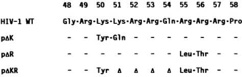
48 49 50 51 52 53 54 55 56 57 58
Gly-Arg-Lys-Lys-Arg-Arg-G1lnArg-Arg-Arg-Pro
- - Tyr-Gln - - -
-- - - Leu-Thr -
-- Tyr A Leu-Thr
-FIG. 1. Predicted amino acid sequence of tat mutants. The wild-type(WT)tatprotein sequenceis derivedfromthe
replication-competent HXB-3 strain of HIV-1 used in the parental vector
pgTAT (23). The mutations were introduced by oligonucleotide-directedmutagenesis, asdescribed in thetext, andwereconfirmed
by sequence analysis. Symbols: -, conserved amino acid; A,
deleted aminoacid.
Kinetic Microplate reader (Molecular Devices Co., Palo Alto, Calif.).
Immunoprecipitationanalysis andsubcellularfractionation. At 60 h posttransfection, cultures were washed with
cys-teine-free Dulbecco modified Eagle medium and then incu-bated in cysteine-free Dulbecco modified Eagle medium containing 10% dialyzed fetal calf serum for 1 h. The cells
were then pulse-labeled with [35S]cysteine (300
,uCi/ml
in cysteine-free Dulbecco modified Eagle medium) for 2 h. In some experiments, this[35S]cysteine pulsewasfollowed bya chase with excess unlabeled cysteine. Labeled cultures
were harvested by using radioimmunoprecipitation assay
(RIPA)buffer(6)andwerethen subjectedto immunoprecip-itation analysis with a rabbit polyclonal antipeptide
antise-rumdirectedagainstthefirst61coding aminoacids oftat,as
previously described (6, 23). Precipitated proteins were
resolved on a discontinuous 14% sodium dodecyl
sulfate-acrylamidegel and visualized by autoradiography.
Subcellular fractionation of transfected, [35S]cysteine-la-beled COS cells was performed essentially as described by
Slamonetal. (33). At 60 hposttransfection,60-mmCOS cell culture disheswerewashedthreetimes with cold
phosphate-bufferedsaline, scraped into phosphate-buffered saline, and pelletedby centrifugationat500 x gfor10min at
4°C.
The cell pellet was gently suspended in 1.3 ml of ice-cold lysis buffer(5 mM sodium phosphate [pH7.4], 50mM NaCl, 150mMsucrose, 5mMKCI,2mmdithiothreitol, 1 mMMgCl2,
0.5 mMCaC12, 0.1 mMphenylmethylsulfonylfluoride, 0.1% Tergitol 15-S-9 [Union Carbide Corp., New York, N.Y.]). The resultantsuspensionwas spunthrougha
300-,lI
cushion(consistingof 30% sucrose, 2.5mM Trishydrochloride [pH
7.4], 10 mM NaCI) at 1,000 x g to pellet the nuclei. The supernatant solution was removed and spun again in a
microfuge at 14,000 x g for 5 min. The supernatant
cyto-plasmic fraction was then removed and adjusted to a 1x
RIPAbuffer concentration.
The nuclear pellet was washed three times (with cold
20 mM HEPES [N-2-hydroxyethylpiperazine-N'-2-ethane-sulfonicacid;pH7.5], 5 mMKCl, 0.5 mM dithiothreitol, 0.1 mMphenylmethylsulfonyl fluoride, 0.2M sucrose) and was
then lysed by incubation in RIPA bufferatambient
temper-ature for 15 min. The nuclear lysate was then cleared by
centrifugation at 14,000 x g for 15 min. Samples of the
cytoplasmic and nuclear lysate fractions were subjected to
trichloroacetic acid precipitation (6) to determine
incorpo-rated radioactivity, and equal amounts of labeled protein were then used for immunoprecipitation with the anti-tat
antibody, as described above.
Indirectimmunofluorescence. Theindirect
immunofluores-cence analysis of the transfected COS cultures was
per-formed as previously described (6, 17). The primary rabbit anti-tat antibody was used at a 1:800 dilution, while the second antibody (rhodamine-conjugated goatanti-rabbit
im-munoglobulin
G;
Boehringer-Mannheim Biochemicals,Indi-anapolis,
Ind.)
was used at a 1:50dilution.RESULTS
The conserved basic domain of tat is required for full biological activity. To address therole of the conservedbasic domain of the tat protein in the trans-activation of HIV-1 LTR-specific gene expression, we used oligonucleotide-directed mutagenesis (35) to alter this region in a wild-type tat expression vector, pgTAT (Fig. 1). In the pAK mutant, lysine residues at amino acid positions 50 and 51 were replaced by tyrosine and glutamine, respectively. In the pAR mutant, arginine residues at positions 55 and 56 were simi-larly replaced by leucine and threonine. Each of these mutations therefore replaced two of the eight charged amino acids in the basic region with uncharged amino acids. The more extensive mutation present in pAKR included the substitution mutations at positions 50, 55, and 56described for pAK and pAR and the deletion of amino acids present between positions 51 and 54 of the tat protein. The pAKR mutant therefore lacks six of the eight basic residues present in the wild-type tat protein (Fig. 1).
The phenotypes of the tat mutants were examined by using a transient expression assay based on the SEAP indicator gene (3). A previously described (3) construction in which the SEAP gene is linked in cis to the HIV-1 LTR (pBC12/HIV/SEAP) was transfected into the HIV-1 replica-tion-competent cell line COS (22) together with a range of concentrations of the tat expression plasmids (Fig. 2). The level of expression of the SEAP protein in the supernatant media of the transfected cultures was determined at 60 h posttransfection byusing a simple, highlyquantitative spec-trophotometric assay for alkaline phosphatase (3). The ob-served levels of tat-mediated trans-activation for each con-centration of tat vector
used,
expressed as a multiple of the basal level encoded by the HIV-1 LTR construction alone, are shown in Fig. 2. An intermediate level of transfection of the wild-type tat gene encoded by pgTAT was observed to yield the maximal observed trans-activation of -90 fold. This is comparable to the maximal level of tat-mediated trans-activation previously observed by us and others using the COS cell line (6, 20). Introduction of higher or lower levels of pgTAT yielded a slightly but consistently lower level of indicator gene activity. The mutant tat vectorspA&R
andpAKdisplayed comparable levels of HIV-1 LTR trans-activation over the entire concentration range used; how-ever, this activity increased dramatically with increasing levels of the transfected tat vector (Fig. 2). The net effect of the different dose responses observed with these mutant tat constructions was that the pAK and pAR clones displayed an essentially wild-type phenotype at the highest level of tat vector used (31-fold trans-activation versus 57-fold for pgTAT), and yet they showed a marked inhibition of trans-activation when the pBC12/HIV/SEAP vector was present at a 100-fold excess (2-fold versus 32-fold for pgTAT). The double mutant pAKRdisplayed a similar dose dependence, yielding no detectable trans-activation at low levels of tat vector transfection but giving rise to a readily detectable -five-fold trans-activation of the HIV-1 LTR at the highest level of tat vector tested (Fig. 2).
One possible explanation for the mutant phenotypes shown in Fig. 2 is that little or no tat mutant protein was
on November 10, 2019 by guest
http://jvi.asm.org/
[image:2.612.59.296.74.150.2]°oor
03) 0
C
0
0
I-4A
>
1o
LTR 1250 1250 1250 1250 1250 1250
TAT 0 12.5 46 140 410 1250
DNA
Tronsfected (ng)
FIG. 2. Relative trans-activation of HIV-1 LTR-specific gene expression mediated by various levels ofthe mutant orwild-typetat geneproduct. A constantlevel(1,250 ng)of the indicator construc-tion pBC12/HIV/SEAP (LTR) was cotransfected with increasing levels (0 to 1,250 ng)ofthe varioustatgene expression plasmids (TAT) as indicated. The total amount of DNA transfected per culture was maintained at 2,500 ng by supplementation with a negative controlvector,pBC12/CMV/IL-2 (5). Thelevels ofSEAP expression in the supernatant media were determined at 60 h posttransfection,aspreviouslydescribed (3), and are expressed as a multiple of the basal level of HIV-1 LTR-specific SEAP gene expression.
being synthesized in the transfected cells. To test this
hypothesis, we performed quantitative immunoprecipita-tions of
[35S]cysteine-labeled
transfected COS cultures with apreviously described rabbit polyclonal anti-tat antiserum (17). As previously noted, the wild-type 86-amino-acid tat protein migrates in sodium dodecyl sulfate-acrylamide gelsat an unexpectedly high relative molecular weight (Mr) of -15,500 (Fig. 3A) (23, 25). Interestingly, both the pAK and pAR mutants, which are ofthe same predicted size as the
wild-type tat protein, migrate at a somewhat lower Mr of -14,500. This suggests that the highly basic nature of the HIV-1 tat protein is at least partially responsible for the aberrant migration of the tat protein observed in sodium dodecyl sulfate-acrylamide gels. This possibility is sup-portedby the migration behavior of thetatprotein encoded by pAKR, which has suffered amoreextensive mutation of the conserved basic region. This protein is predicted to be only 4 amino acids shorter than the pAK and pARmutants, andyetit migratessignificantlymorerapidly (Mrs of-13,000 versus-14,500). In total, therateofsynthesis of each of the tat mutants in the transfected cultures was comparable, although the culture transfected with pAKR did appear to yield aslightly reduced signal (Fig. 3A).
Mutations in theconserved basic domain affectthe subcel-lularlocalization of the tat protein. Because the basic region of the tatprotein possesses amino acid homology to nuclear transportsignals previously noted forother, predominantly nuclearproteins (see below), we nextexamined the subcel-lular localization of the mutant tatproteins. In this experi-ment, transfected cells were labeled with [35S]cysteine as described in the legendtoFig. 3A andwerethen subjectedto a1-h chase with excessunlabeledcysteinetopermitlabeled proteins to fully localize to the appropriate subcellular compartment. Total cytoplasmic and nuclear proteins were thenseparatedby usingthefractionation protocoldeveloped by Slamonetal. (33).Theincorporated radioactivityineach fractionwasdetermined, and equal amountswerethenused for immunoprecipitation with anti-tat antiserum. The tat protein encoded by the parental vectorpgTAT,aswellasthe mutant tatmolecules encodedby the pAK and pAR vectors, waslocalized to the nucleus within 1 hof being labeled with [35S]cysteine (Fig.3B).Insharp contrast, the -13-kilodalton tatprotein encoded by pAKRappeared to remain predomi-nantlywithinthecytoplasm ofexpressing cells. Thisresult therefore indicatesthat themutation presentinpAKR must perturb aprotein sequence necessary for the nuclear local-ization of the encoded tatprotein.
A-..
A,V-;
k- jl7m ( ( (( (
AQN
Q1.
Q i7
QV ql QV
... >ou
C.
40aar
.c.'.en. q. .n
QiQSF.t:
43-- *
-25.7 -~ e~
1 8.4- _..
14.3- ...f
6.2..<fi.SS,... aS.
.--43 ---25.7 -18.4 14.3
- 6.2
43-
25.7- 18.4-
14.3-
6.2-N C N C N C N C
FIG. 3. Analysis oftatprotein expression byimmunoprecipitation analysis.COS cellsweretransfected(6)withequallevels of the various tatexpressionvectors andwere thenmetabolically labeled with[35S]cysteinefor2 h at 60 h posttransfection. Labeledtatproteinswere
isolated by immunoprecipitation with a polyclonal rabbit anti-tat antiserum (6), resolved on a discontinuous 14% sodium dodecyl
sulfate-acrylamide gel,and visualizedby autoradiography.The vector transfected into each culture is indicatedovertheappropriate gellane. Thenegativecontrol(NEG)usedwasthevectorpBC12/CMV/IL-2(5). 14C-labeledproteinmolecular sizemarkers(M)wereobtainedfrom
Bethesda Research Laboratories, Inc., Gaithersburg, Md.,andareindicated in kilodaltons. (A)Cultureswere harvesteddirectlyafterthe
[35S]cysteine labeling pulsetodetermine the rate oftatproteinsynthesis. (B)Cultureswerechasedwith mediacontainingexcessunlabeled
cysteinefor 1 h aftercompletionof the[35S]cysteine pulse.Transfected cellswerethenseparated (33)intonuclear(N)andcytoplasmic(C)
fractions beforeimmunoprecipitation. (C)Thestabilityofthe mutanttatproteinswasexaminedbyexposingthe transfected COS culturesto
excess unlabeled cysteinefor 6haftercompletion of the[35S]cysteine pulsebeforeharvest andimmunoprecipitation. Theexperimentfor
panelCwasperformedinparallel tothat forpanelA andwasexposedtoautoradiographyforthesame periodoftime.
A.
I
B.
on November 10, 2019 by guest
http://jvi.asm.org/
[image:3.612.93.270.67.295.2] [image:3.612.107.509.495.620.2]The immunoprecipitation analysis presented in Fig. 3B examined the subcellulartransportofnewly synthesized tat
protein. To extend this analysis to an examination of the
subcellularlocalization of the various tatproteinsat steady state, we also performed an indirect immunofluorescence
analysis of transfected COS cell cultures using the rabbit anti-tat antiserum(17). These results confirmedourprevious
observation that the wild-type tat protein is concentrated within the nucleus of expressing cells (Fig. 4A). The tat
protein encoded by pAK yielded the samesubcellular
distri-bution pattern as the wild-type tat protein, including the previously described nucleolar concentration (17) (Fig. 4B). Neither the pgTAT- northe pAK-transfected cells yielded any detectable cytoplasmic fluorescence. Analysis of cells
transfected with pAR yielded a fluorescence signal which was consistently fainter than the signal observed for the
wild-type pgTATvectororthepAKvector(Fig. 4CversusA
andB). Compensation for this weaker signal by optimization of thephotographicexposuretime demonstratedthat thetat
protein encoded by pARwasconcentrated within the nuclei
and nucleoli ofexpressing cells but also revealed the pres-enceofalow level ofcytoplasmic tat protein (Fig. 4D). In contrast, the immunofluorescence analysis of COS cell cul-tures transfected with the extensively mutated tat
expres-sionvectorpAKR yieldedno detectable fluorescencesignal above background level in either subcellular compartment (Fig. 4E and F).
Mutagenesis of the conserved basic region affectsthe stabil-ity of thetat protein. Ourinability to detect the mutant tat
protein encoded by pAKR using an assay which measures the steady-state level of protein expression (Fig. 4)
sug-gested the possibility that mutagenesis of the conserved basic domainmight affecttatprotein stabilityinvivo. Totest thishypothesis, transfected COS cultureswerelabeledwith
[35S]cysteine asdescribed forFig. 3A and thenexposedfor 6 htomediacontaining largeexcessesofunlabeledcysteine. This assaypermitted the detection of significant differences inthe in vivo half-livesof the varioustatproteins. Thus,the wild-type tatprotein encoded by pgTAT and the mutant tat
protein encoded by pAK showed littleifanydeclineinsignal intensity during the chase period, suggesting that these proteinsarefairly stable in vivo(Fig. 3C). ThepARmutant exhibitedaslight,-two- tothree-folddropinsignal intensity
overthesame period, suggestinga somewhat reduced
half-life. Incontrast,thetatprotein encoded by pAKR appeared
to be rapidly turned over during the 6-h chase period,
resulting inavery weak residualsignal (Fig. 3Aversus C). These results therefore correlate closely with the data pre-sented in Fig. 4 in that the culture transfected with pA&R
presented areduced tat-specific immunofluorescence signal relativetothe signals produced by pgTAT- and pAK-trans-fected cultures, while the pAKR-transfected culture in that
caseyieldedno detectable tat-specific signal.
DISCUSSION
The tat trans-activators encoded by the known primate
immunodeficiency viruses display extensive amino acid
se-quence heterogeneity and yet demonstrate the ability to
cross-trans-activate virus-specific gene expression (1, 11, 14). This observedsequenceheterogeneitymaytherefore be
viewedas anexperiment bynatureandfocusesattentionon
those sequencedomainswhich have been conservedacross theknownprimate virus isolates. The first of these,ahighly
conserved cysteine-rich domain, has been proposed to be important in the binding of metal ions by tat and in a
subsequent protein dimerization event (10). The targeted mutagenesis ofthis domain wasrecently shownto result in the loss of all trans-activationactivity by tat(12, 30, 31). In this study, we have examined the functional consequences of mutations within the second conserved tat domain, a
highlybasicregioncenteredatapproximately position53 in the tatamino acid protein sequence. We demonstrate that these sequence changes result in an inhibition of tat-medi-ated trans-activation which is partially alleviated by high
levels oftatexpression.Wealso demonstrate that mutagen-esisofthis domaincanresult intwodetectable biochemical
lesions, i.e., interference with the nuclear localization oftat and the destabilization of the tatprotein.
Therapid accumulationof thewild-typetatproteinwithin thenucleus ofexpressing cells (Fig. 3B) suggests the pres-enceofanuclear localizationsignalin theprimarysequence
of this
protein.
Ruben et al. (30) have in fact recently demonstratedthata5-amino-acid sequence derived from thebasic domain oftat
(i.e.,
amino acids48to52)
is sufficientto induce the nuclearlocalizationof thenormally cytoplasmic
P-galactosidase
geneproduct
whenintroducedatthe amino terminus. Acomparison
of the conserved basic domain with thesequences ofknown nuclearlocalizationsignals
reveals ahigh
degree ofhomology
inseveralinstances(Fig. 5A).
Ofinterest is the observation that this homology appears to extend over
only
part of the basic domain in each case. Thus, the well-defined nuclear localizationsignal
of the simianvirus 40large
Tantigen
(19)displays
aclose homol-ogy to amino acids 48 to 53 of tat, while the homologybetween one ofthe
polyomavirus large
Tantigen
nuclearlocalization
signals
(28)andtatisconfinedtoaminoacids 54 to 60. Oneinterpretation
of these sequencecomparisons,
which would be fully consistent withthe data
presented
in this report, is that the basicregion actually
consists oftwoadjacent
orpartially overlapping
nuclear localizationsignals.
Mutagenesis
ofeither partalone,
i.e., pAK
orpAR,
wouldthereforehave little effectonthefinalnuclearlocalizationof tat, andonlyadouble mutant,
i.e., pAKR,
would thenyield
the
predicted
nonnuclearphenotype. Multiple
nuclearlocal-ization
signals
have been observed in several nuclear pro-teinswhichhave beenexaminedindetail(13, 15, 27,28)
and thisredundancy has beenproposed
to enhance the rate ofnuclear
protein
transport(21).An
interesting
alternativehypothesis, suggested
by the recentworkofSiomi etal. (32), is thatthetatbasicregion
might
represent a subnuclear localizationsignal.
We and others havepreviously
noted that the tatprotein
isprefer-entially,
and sometimespredominantly,
localized to the nucleolus ofexpressing
cells (17, 30).Further,
the basic domainoftatdisplays
remarkable sequencehomology
totheproposed nucleolar localization signal ofthe human T-cell leukemia virus type 1rexgeneproduct (Fig. 5B), which has
also been
proposed
to consist of two adjacent nuclearlocalization
signals
(32). However,theobservationthatboth the pAK and, to a lesser extent, thepAR
tat proteinscontinue to
display preferential
nucleolar localization (Fig. 4) may argue againstthis attractive hypothesis.Asecondeffectofmutationswithinthetatbasic domain,
observedto aslight degreein
pAR
and to amarkeddegreeinpAKR,
isareductionin the in vivostability ofthe encodedprotein. It is of interest that the stabilities of the various encoded tat proteins in fact appear to correlate with their subcellularlocalization as derived fromdata shown in Fig.
3B and 4. Thus, the predominantly nuclear
pAK
mutant appearstoshare thelonghalf-lifeoftheparentaltatprotein,while thepredominantly cytoplasmictatprotein encoded by
on November 10, 2019 by guest
http://jvi.asm.org/
> Cb-1
C,
c
0r
I-'
o ^0 O
m °
o . o
o'
~C,o oo
co
CD
CD
CL#
cor CD
C, 05
0
CD
gCo
0O
8Co
coV pcog
CZo
cnQ Co
o5
-a
ONs- co
on November 10, 2019 by guest
http://jvi.asm.org/
HISTONE 2B NUCLEOPLASMIN SV40 large-T HIV-1 TAT
GLUCOCORTICOID RECEPTOR POLY0MA large-T
G
P K K K R K V
G R K KR R QR R R P P Q T K K K I K; G V S R K R P RP
therefore favorthe hypothesisthat the basicdomain serves a second, as yet undefined, targeting function within the nucleus oftheexpressing cell. Clearly, a resolution of this question will require a fuller understanding of the mecha-nismofaction oftatand, inparticular,the reconstitutionof tat-mediatedtrans-activation ina definedin vitro system.
ACKNOWLEDGMENTS
We thank SharonGoodwin for excellent secretarial assistance and CraigRosenforcommunication of results beforepublication.
Bm
HIV-1 TAT
Y
RKK R QIRRRP P [image:6.612.75.295.68.228.2]HTLV-1 REX R[ K RPP
FIG. 5. Homology oftheconserved basicregionoftattoknown nuclear (A) and nucleolar (B) localization signals. (A) Nuclear localization signals in general containastretch of the basic amino
acids lysine and arginine (boxed) flanked by one or more
helix-breaking amino acids (proline orglycine) and may form a short,
highly basica-helixstructurein vivo (4). This is also the secondary
structurepredicted for the conservedbasicdomain of thetatprotein
(2). (The indicated nuclear localization signals werederived from
references 4, 19, 24, 27, and 28). SV40, Simian virus 40. (B) The basicregion ofthe HIV-1 tat protein displays extensivesequence homologytothe recently definedhumanT-cell leukemiavirustype
1 (HTLV-1) rex protein nucleolar localization signal (32). Both
contain eightbasic aminoacids(boxed)andcontainglutamineand proline residuesatequivalent positions.
thepAKRmutantappears tobe significantly more labile in vivo. The tatprotein encoded by pAR, which is predomi-nantlylocalizedwithin the cellnucleus yetdisplays atleast some cytoplasmic expression (Fig. 4D), in turn demon-stratedaslightlybutsignificantlyreducedhalf-life relativeto that ofthe wild-type tat protein. These observations are
thereforeconsistent with the hypothesisthat the tatprotein isstable whenlocalized tothe cell nucleus but significantly
more labile when inappropriately expressed in the cell cytoplasm. Clearly, however, thecorrelationbetweenthese twotatmutantphenotypes doesnotprove acause-and-effect
relationship.
Themutations introduced intothetatproteininthiswork
were observed to result in a complex trans-activation phe-notype. Thus, the activity of the mutated tat proteins is markedly reducedatlowlevelsofexpression butrecovers,
intwocasestoanalmostwild-type level,athigh levels oftat
protein expression. This suggests that the conserved basic domain,unlikethe cysteine domain, is notan intrinsic part oftheactivesiteof thetatprotein andmayinstead indicate thatthissequencefunctionstoenhancethe affinity oftatfor its appropriate site or substrate within the cell nucleus.
Thus,mutagenesis of this domain would result inareduced trans-activation activity which could be significantly
re-stored by increasing the concentration oftat. It has been reported that only very low levels oftat are actually
ex-pressedwithinHIV-1-infected cells (8), sothat the targeting oftattoitsappropriate cellular locationmaybe essential for
optimum biological activity in vivo. In this context, the hypothesis that the conserved basic domain oftatserves to enhancethenucleartransport, and hencethein vivo stabil-ity, of this proteinappears only partly sufficient to explain thetrans-activation phenotypesdisplayed by the mutatedtat
proteins. Thus, the pAKmutant,which isnotdistinguishable fromthewildtypeby thesecriteria (Fig.3 and4), neverthe-less fails to demonstrate full trans-activation (Fig. 2). We
LITERATURE CITED
1. Arya,S. K., B.Beaver, L.Jagodzinski,B.Ensoli,P.J. Kanki, J. Albert, E.-M. Fenyo, G. Biberfeld, J. F. Zagury,F. Laure, M.
Essex, E. Norrby, F. Wong-Staal,and R. C. Gallo. 1987. New
human and simian HIV-related retrovirusespossessfunctional transactivator(tat)gene. Nature(London)328:548-550. 2. Arya, S. K.,C. Guo, S. F. Josephs, and F. Wong-Staal. 1985.
Trans-activator gene of human T-lymphotropic virus type III (HTLV-III). Science 229:69-73.
3. Berger, J.,J. Hauber, R. Hauber,R.Geiger, andB. R.Cullen. 1988. Secreted placentalalkalinephosphatase: apowerful new
quantitative indicator ofgene expression in eukaryotic cells. Gene 66:1-10.
4. Burglin, T. R., and E. M. De Robertis. 1987. The nuclear
migration signal of Xenopus laevis nucleoplasmin. EMBO J.
6:2617-2625.
5. Cullen, B. R. 1986. Trans-activation of human
immunodefi-ciency virusoccursviaabimodalmechanism. Cell46:973-982.
6. Cullen, B. R. 1987. Use of eukaryotic expression technologyin
the functional analysis of cloned genes. Methods Enzymol. 152:684-703.
7. Dayton, A. I., J. G. Sodroski, C. A. Rosen, W. C. Goh, and W. A. Haseltine. 1986. Thetrans-activatorgeneof thehuman T
celllymphotropic virustypeIII isrequired for replication. Cell 44:941-947.
8. Feinberg, M.B.,R.F.Jarrett, A. Aldovini,R.C.Gallo, andF.
Wong-Staal. 1986. HTLV-III expression and production involve complex regulation at the level ofsplicing and translation of
viral RNA. Cell 46:807-817.
9. Fisher, A. G., M. B. Feinberg, S. F. Josephs, M. E. Harper, L. M. Marselle, G. Reyes, M.A. Gonda, A. Aldovini, C.
De-bouk,R. C.Gallo,and F.Wong-Staal. 1986.Thetrans-activator
gene of HTLV-III is essential for virus replication. Nature
(London) 320:367-371.
10. Frankel, A. D.,D. S.Bredt, and C. 0.Pabo. 1988. Tatprotein
from human immunodeficiency virus forms ametal-linked
di-mer.Science 240:70-73.
11. Fukasawa,M., T. Miura,A.Hasegawa, S. Morikawa, H.
Tsuji-moto,K.Miki, T. Kitamura, and M. Hayami.1988. Sequence of
simianimmunodeficiency virusfrom African greenmonkey, a
new member of the HIV/SIV group. Nature (London) 333:
457-461.
12. Garcia, J. A.,D.Harrich, L. Pearson, R. Mitsuyasu, and R. B. Gaynor. 1988. Functional domains required for tat-induced transcriptional activation of the HIV-1 long terminal repeat.
EMBO J. 7:3143-3147.
13. Greenspan, D., P. Palese, and M. Krystal. 1988. Two nuclear
locationsignals in the influenza virus NS1 nonstructuralprotein.
J. Virol. 62:3020-3026.
14. Guyader, M.,M.Emerman, P.Sonigo, F. Clavel, L. Montagnier,
and M. Alizon. 1987. Genomeorganization and transactivation
of the humanimmunodeficiency virustype2. Nature(London)
326:662-669.
15. Hall,M.N., L. Hereford, andI.Herskowitz. 1984. Targeting of
E. coli P-galactosidase tothe nucleus in yeast. Cell
36:1057-1065.
16. Hauber, J., andB. R.Cullen. 1988. Mutationalanalysis of the trans-activation-responsive region of the human immunodefi-ciency virustype Ilong terminal repeat.J. Virol.62:673-679.
A
on November 10, 2019 by guest
http://jvi.asm.org/
17. Hauber, J., A. Perkins, E. P.Heimer, and B. R. Cullen. 1987. Trans-activation of human immunodeficiency virus gene expres-sion is mediated by nuclear events. Proc. Natl. Acad. Sci. USA 84:6364-6368.
18. Hirsch, V., N. Riedel, and J. I. Mullins. 1987. The genome organization of STLV-3 is similar to that of the AIDS virus exceptfor a truncatedtransmembrane protein. Cell 49:307-319. 19. Kalderon, D., B. L. Roberts, W. D. Richardson, and A. E. Smith. 1984. A short amino acid sequence able to specify nuclear location. Cell39:499-509.
20. Kao,S.-Y., A. F. Calman, P. A. Luciw, and B. M. Peterlin. 1987. Anti-termination of transcription within the long terminal repeat ofHIV-1bytatgeneproduct. Nature (London) 330:489-493. 21. Lanford, R. E., P. Kanda, and R. C. Kennedy. 1986. Induction
of nuclear transport with a synthetic peptide homologous to the SV40Tantigen transport signal. Cell 46:575-582.
22. Levy, J. A., C. Cheng-Mayer, D. Dina, and P. A. Luciw. 1986. AIDS retrovirus (ARV-2) clone replicates intransfected human and animal fibroblasts. Science 232:998-1001.
23. Malim, M. H., J. Hauber, R. Fenrick, and B. R. Cullen. 1988. Immunodeficiency virus rev trans-activator modulates the expression of the viral regulatory genes. Nature (London) 335:181-183.
24. Moreland, R. B., G. L. Langevin, R.H. Singer, and R. L. Garcea,and L. M. Hereford. 1987. Amino acidsequences that determine the nuclear localization of yeast histone 2B. Mol. Cell. Biol. 7:4048-4057.
25. Muesing,M.A.,D. H.Smith,and D.J. Capon.1987.Regulation ofmRNA accumulation by a human immunodeficiency virus trans-activator protein. Cell48:691-701.
26. Peterlin, B. M.,P. A. Luciw, P. J. Barr, and M. D. Walker. 1986. Elevated levels of mRNA can account for the trans-activationof humanimmunodeficiency virus.Proc.Natl.Acad. Sci. USA 83:9734-9738.
27. Picard, D., and K. R. Yamamoto. 1987. Twosignals mediate hormone-dependent nuclearlocalization of the glucocorticoid
receptor. EMBO J. 6:3333-3340.
28. Richardson, W. D., B. L. Roberts, and A. E. Smith. 1986. Nuclear location signals in polyoma virus large-T. Cell 44: 77-85.
29. Rosen, C. A., J. G. Sodroski, and W. A. Haseltine. 1985. The locationof cis-acting regulatory sequences in the humanTcell lymphotropic virus type III (HTLV-III/LAV) long terminal repeat.Cell 41:813-823.
30. Ruben,S.,A.Perkins, R. Purcell, K. Joung, R.Sia, R. Burghoff, W. A.Haseltine, and C. A. Rosen. 1989. Structural and func-tional characterization of human immunodeficiency virus tat protein.J. Virol.63:1-8.
31. Sadaie,M.R., J.Rappaport,T.Benter, S.F.Josephs, R.Willis, and F. Wong-Staal. 1988. Missense mutations inan infectious HIVgenome: functionalmapping oftatanddemonstration ofa novel trs splice acceptor. Proc. Natl. Acad. Sci. USA 85: 9224-9228.
32. Siomi, H.,H.Shida, S. H. Nam,T.Nosaka,M. Maki,and M. Hatanaka. 1988. Sequencerequirements for nucleolar localiza-tionof human Tcell leukemia virus type 1 pXprotein, which regulates viralRNAprocessing. Cell 55:197-209.
33. Slamon, D. J., W. J. Boyle, D. E. Keith, M. F. Press, D. W. Golde, and L. M. Soura. 1988. Subnuclear localization ofthe trans-activatingproteinof thehumanT-cellleukemiavirustype I. J. Virol.62:680-686.
34. Sodroski, J., R.Patarca, and C. Rosen. 1985. Location of the trans-activatingregiononthegenomeof human T-cell lympho-tropic virus typeIII. Science229:74-77.
35. Taylor, J. W., J. Ott,andF.Eckstein. 1985.Therapid genera-tion of oligonucleotide-directed mutations at high frequency using phosphorothioate-modified DNA. Nucleic Acids Res. 13:8765-8785.
36. Wright, C. M.,B. K. Felber,H. Paskalis, andG. N. Pavlakis. 1986. Expressionandcharacterization of the trans-activator of HTLV-III/LAVvirus. Science 234:988-992.
![FIG. 3.isolatedThetatfractionssulfate-acrylamide[35S]cysteineexcessBethesdacysteinepanel Analysis of tat protein expression by immunoprecipitation analysis](https://thumb-us.123doks.com/thumbv2/123dok_us/1326639.86543/3.612.93.270.67.295/isolatedthetatfractionssulfate-acrylamide-cysteineexcessbethesdacysteinepanel-analysis-protein-expression-immunoprecipitation-analysis.webp)
