0022-538X/92/063514-08$02.00/0
CopyrightX 1992,AmericanSocietyforMicrobiology
Role of
N-Linked
Oligosaccharides
in
Processing
and
Intracellular
Transport
of E2
Glycoprotein
of Rubella
Virus
ZHIYONG QIU, TOM C. HOBMAN,t HELEN L. McDONALD, NINA0. L. SETO4
AND SHIRLEY GILLAM*
Departmentof Pathology, Research Centre, University of British Columbia, 950 West 28th Avenue, Vancouver, British Columbia, Canada VSZ 4H4
Received 6September1991/Accepted 22 February 1992
Therole of N-linked glycosylationinprocessingandintracellulartransportofrubella virusglycoproteinE2 has beenstudied by expressingglycosylationmutantsof E2 in COS cells.ApanelofE2glycosylationmutants weregenerated by oligonucleotide-directed mutagenesis.Each of the threepotentialN-linkedglycosylationsites was eliminated separately as well as in combination with the othertwo sites. Expression of the E2 mutant
proteins in COScells indicated that in rubella virus M33 strain,all three sites are used for the addition of N-linkedoligosaccharides. Removal ofanyof theglycosylationsites resulted in slowerglycanprocessing,lower stability, andaberrantdisulfidebondingof the mutantproteins,with theseverityofdefectdependingonthe numberof deleted carbohydratesites.Themutantproteinsweretransportedtotheendoplasmicreticulum and
Golgi complexbutwerenotdetectedonthe cell surface.However,the secretion of theanchor-free form of E2 into themediumwasnotcompletelyblockedbythe removal ofanyoneof itsglycosylationsites. This effectwas dependentontheposition of thedeletedglycosylation site.
Rubella virus (RV) is the sole member of the genus Rubivirus in thefamily Togaviridae (23) and contains three major structuralproteins,thecapsid protein (C;33kDa)and twoenvelope glycoproteins (El [58 kDa] and E2 [42 to 47
kDa]) (30). The Cprotein is argininerich and is thought to
interact with the genomic RNAto form the nucleocapsid,
which is surrounded bya lipid bilayer containing the spike complex of the El and E2glycoproteins (30). The strategy forexpression of structural proteins during RV replication is similar tothat ofalphavirus, in whichapolyprotein
precur-sor(pllO) is translated fromasubgenomicRNA in theorder
NH2-C-E2-E1-COOH (29, 31). The precursor is then prote-olytically processedtogenerateeach structuralprotein (30).
El and E2aretransportedoutof theendoplasmicreticulum
(ER) totheGolgi cisternae and the cell surface (13, 14, 16), where virus maturation can occur on either membrane dependingonthecell type (3, 43).
RV E2isatype Imembraneglycoprotein, and its biolog-ical function remains unclear, although the protein appears
tocontainatleastoneviralneutralizationepitope (11) anda strain-specific epitope (6). Expression of an RV E2 cDNA
constructinCOS cells showed that translocation of E2 into the lumen of the rough ER is mediated by a signal peptide residing in the C terminus of the capsid protein (13).
Follow-ing translocation, asparagine-linked (N-linked) glycosylation of E2 takesplace. Processing ofRV E2glycans involvesat least two stable intermediates, a 39-kDa high-mannose-containing precursor and a 42-kDa form bearing some com-plex sugars (13, 14). The transport of E2 to the plasma membrane is inefficient, as only a small fraction of E2 is destined for the cell surface, while the majority of E2 accumulates in the ER and the Golgi cisternae (13). E2 from RV virions migrates as a diffuse band on sodium dodecyl
*Corresponding author.
tPresent address: Divisionof Cellular and MolecularMedicine,
UniversityofCaliforniaatSanDiego,LaJolla,CA 92093-0651. t Present address: Institute for Biological Sciences, National ResearchCouncilofCanada, Ottawa, Ontario, Canada KlA OR6.
sulfate(SDS)-polyacrylamide gelelectrophoresis (PAGE)at
42 to 47 kDa because ofheterogeneous glycosylation (30).
Furthermore, E2 is known to contain 0-linked
carbohy-drates (20). In the presence of tunicamycin, Vero cells infected with RVand COS cells transfected with RV cDNAs produce E2 with amolecularweight of 31 kDa (5, 30). The amino acid sequence predicted from E2 cDNAs reveals a
protein of 281 residues including three potential N-linked
glycosylation sites in M33 (4) and HPV77 (44) strains as
opposedtofourin Therien(9, 41) and RA27/3(26)strains. So far, the importance of N-linked oligosaccharides on E2 in virionassembly and infectivity is unknown.
N-linked glycosylation is one of the most common post-translational modifications ofproteins in the exocytic
path-way of eukaryotic cells. Animal viruses utilize host cell glycosylationmachinerytosynthesize and process oligosac-charides attached to viralglycoproteins. Thus, the expres-sion of viral antigens in cells has proved to be a useful systemforstudying the stepwiseeventsinglycan processing and intracellular transport along theexocytic pathway. Sev-eralapproaches have been usedtodefine the functional roles of N-linkedcarbohydrateaddition in cells. These include the
useof agents that interfere withglycosylation, elimination of eachN-linkedglycan addition siteonthe cDNA by oligonu-cleotide-directedmutagenesis,and use of glycan-processing-deficient cell lines. Avarietyof cellularand viral glycopro-teins have been analyzed by these approaches. It appears that oligosaccharides on glycoproteins play a role in initia-tion and maintenance of folding into biologically active conformation, protecting polypeptides from proteolytic at-tackandinfluencing the antigenicity and immunogenicity of glycoproteins (reviewed in references 8, 17, and 32).
Al-though the functional contribution of thecarbohydrate var-ies with the glycoprotein in question, it has been generally accepted that glycosylation is necessary to aid proteins in attaining and maintaining correcttertiary structure and
un-dergoingintracellular transport.
In this report, we analyze the role of N-linked glycosyl-ationintheprocessingandintracellular transport of RV E2 3514
on November 9, 2019 by guest
http://jvi.asm.org/
ROLE OF GLYCOSYLATION ON RV E2 3515 glycoprotein from M33 strain. Oligonucleotide-directed
mu-tagenesis was used to construct a panel of E2 glycosylation mutants that were subsequently analyzed by transient expression in COS cells. Our data show that all three potential sites are used for N-linked carbohydrate addition in the E2 glycoprotein. E2 glycosylation mutants were proc-essed to obtain some endoglycosidase H (endo H)-resistant sugarmoieties, indicating that they reached the Golgi appa-ratus. Although none of the mutants were detected on the cell surface by indirect immunofluorescence, the single glycosylation mutants were secreted when expressed in a soluble form.
MATERUILSAND METHODS
Oligonucleotide-directed mutagenesis. The cDNA insert
encoding RV E2 protein (14) in the expression vector pCMV5(2)wassubclonedinto the EcoRI and HindIII sites ofM13mpl8 (25). Oligonucleotide-directed mutagenesis was performed on single-stranded uracil-containing templates by the method of Kunkel (19). The oligonucleotides used to eliminate each consensus sequence for N-linked glycan addition were
pGTCGCTGGCTJflTCGGTG,
which intro-duced two nucleotide changes (underlined), resulting in a single amino acid change of asparagine 53 to glutamine;pCCAGTCGCCCAGGTTGTA,whichdestroyeda glycosyl-ation site by changing serine 73 to glycine; and pGCTG
CTAATGAGGCC,whichchangedasparagine 115to isoleu-cine. Double and triple mutants were generated by ligating the restrictionfragment containing the appropriate wild-type
or mutantsite(s). All the mutants were confirmed by DNA sequence analysis (36) and subcloned into the expression vectorpCMV5 (2).
Transfection, metabolic labeling,andimmunoprecipitation. COS cells were transfected with recombinant plasmids
ac-cordingto the method of Adams and Rose (1) with modifi-cations (16). Metabolic labeling and immunoprecipitation
werecarriedout asdescribedpreviously (13, 16).
N-Glycanase and endo H digestions. N-Glycanase
(Gen-zyme) treatment was performed as recommended by the manufacturers. Immunoprecipitates to be digested with N-glycanase were adjusted to 100 mM sodium phosphate
(pH 8.6)-1%Nonidet P-40-100mM EDTA-0.5%
3-mercap-toethanol-0.1% SDS and incubated with different amounts
of enzyme for atleast 8 h(for complete digestion) orfor10 min (for partial digestion) at 37°C. Endo H glycosidase digestion wasperformedaspreviouslydescribed(37).
Indirect immunofluorescence. Transfected COS cells grown on polylysine-coated 9-mm glass coverslips were
washed three times with phosphate-buffered saline (PBS)
with 0.7mM CaCl2 and 0.3 mMMgCl2, fixed for 20 minat room temperature in 2% formaldehyde-PBS, and washed with PBS. Some cellswerepermeabilizedwith0.1% Nonidet P-40-PBS for 30 min and incubated with
rhodamine-conju-gated wheat germ agglutinin (WGA) or concanavalin A (ConA) priortobeingblockedby1% bovineserumalbumin (BSA)-PBS. Coverslips were overlaid with diluted human
serum (1:200), incubated for 60 min at room temperature, and then washed with BSA-PBS. Incubation withsecondary antibody, i.e., fluorescein-conjugated goat anti-human
im-munoglobulinG(1:100;KirkegaardandPerry
Laboratories),
was for 60 min. Coverslips werewashed, mounted, exam-ined byepifluorescence, and photographed.
1
2
3
4 5 6
43-
29-FIG. 1. Determination of the number of N-linked glycans on RV E2. [35S]methionine-labeledE2 was incubated with no (lane 1), 10 mU(lane 2), 20 mU (lane 3), 50 mU (lane 4), 100 mU (lane 5), and 300 mU(lane 6) of N-glycanase (Boehringer Mannheim) for 10 min at37°C, separated by SDS-PAGE, and fluorographed. Positions of molecularsize markers are shown on the left (in kilodaltons).
RESULTS
Determination of functional N-linked glycosylation sites in
E2
glycoprotein.
The predicted amino acid sequenceindi-cates that E2 from RV M33 strain contains three putative N-linked glycosylation sites (4). However, it remains un-known how manysites are actually used. To characterize the number of oligosaccharide side chains on wild-type E2, recombinant plasmid pCMV5-E2 (14), which contains E2 cDNA in amammalianexpressionvectorpCMV5 (2)
down-stream from the human cytomegalovirus immediate early
gene promoter, wasusedtotransfect COS cells.Transfected COS cells (40 to 50 h posttransfection) were metabolically labeled with 100 ,uCi of [35S]methionine for 30 minat37°C
andlysed. E2 proteinwas immunoprecipitatedwith human anti-RV serum,digestedwithaserially dilutedN-glycanase,
andsubjectedtoSDS-PAGE andfluorography.N-Glycanase hydrolyzes theglucosylamine linkage of all types of N-linked
oligosaccharides on glycoproteins to give free
oligosaccha-rides andpolypeptides(33).Thispartial digestiongenerated
fourspecieswith molecularsizes of39, 36,33.5,and 31 kDa
(Fig. 1). It is likely that these species corresponded to E2 with three, two, one, and no carbohydrate side chain(s), suggestingthatwild-typeE2glycoprotein normallyhasthree N-linkedoligosaccharidechains. It has beenreported
previ-ously that digestion of E2 from RVvirion byN-glycanase changed the apparent molecular weight dramatically and resulted inasmeared appearance on SDS-PAGE(20).
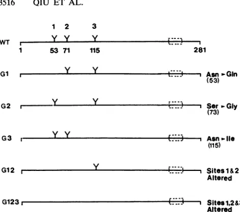
Construction and expression of RV E2 glycosylation mu-tants.Toconfirm the above-mentionedobservation,apanel
of E2glycosylation mutants wasconstructed andexpressed transientlyin COS cells. Oligonucleotide-directed mutagen-esis was employed to introduce one or two nucleotide
changesin the codonsencodingasparagineorserine,
result-ing in a single amino acid substitution at each potential glycosylationsite.Theaddition of N-linkedoligosaccharides was prevented by changing the Asn-X-Ser consensus
se-quenceat asparagine residues53, 71, and 115to
Gln-X-Ser,
Asn-X-Gly, and Ile-X-Ser, respectively. The mutants in whichconsensussequenceswerealtered singlyarereferred
to as G1, G2, and G3; the double mutant is referred to as
G12;and the
triple
mutantisreferredto asG123(Fig. 2).
The positions arenumberedsequentiallyfromtheNterminusofE2.
The cDNAinserts ofRVE2mutants weresubcloned into thepCMV5vector(2) andusedto transfect COS cells. The
expression of mutant
proteins
wasanalyzed by
metaboliclabeling,
radioimmunoprecipitation,
andSDS-PAGE as de-scribedpreviously (13,16).
Wild-typeE2expressed
a prom-inent 39-kDa glycoprotein anda 42-kDaglycoprotein
(Fig.
3A), which correspond to isoforms of E2
containing
high
VOL.66, 1992
on November 9, 2019 by guest
http://jvi.asm.org/
[image:2.612.367.500.82.146.2]3516 QIU ET AL.
1 2 3
WT , Y Y Y
1 53 71 115
Gl
r-G2
r-Y Y
y Y
Y Y
G3
y G12
r-+ B-Me - B-Me
f. r-l,
281
e -,1 Asn-Gin
(53)
i.-Y--t Ser-Gly 97
(73)97
68--t:.:J Asn,-lie
(115)
43
j- -,- Sites 1 &2
Altered
29
_
...
W_ s
G1231 SHOItM1,
[image:3.612.67.303.58.267.2]Altered FIG. 2. Schematic representation ofwild-type(WT) and
glyco-sylationmutantsof RV E2.TheE2protein contains three N-linked glycosylation sites at residues 53, 71, and 115, as indicated by branched structures (Y). The putative transmembrane region is locatednearthe C terminusofE2
(;
).Thefirst residue ofmatureE2 isglycine1,and theC-terminal residue ofE2beforeElisglycine
281.
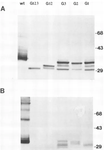
mannose and complex sugars, respectively (13). The elec-trophoretic mobilities of themutant proteinsincreased
pro-portionally with the number of inactivated glycosylation
sites (Fig. 3A). Removal of anysingle
glycosylation
site atposition 1, 2, or 3 resulted in synthesis ofa
major
36-kDaglycoprotein, while the double mutant G12 and the triple mutant G123 directed the synthesis of proteins which
mi-gratedat33.5 and 31 kDa,respectively
(Fig.
3A).Toverify that the differences in electrophoretic
mobility
betweenwild-type andmutant E2wereduetothe numbers of N-linked oligosaccharide side chains attached, some
transfected cellswere treatedwithtunicamycin.
Tunicamy-cin at a low concentration efficiently inhibits N-linked gly-cosylationwithoutinterfering withprotein synthesisincells
(8).Inthe presence of 3 ,ug oftunicamycinperml, all the E2
polypeptides synthesizedin cells transfected withwild-type ordifferentglycosylation mutantcDNAconstructs had the
samemolecularweightasthetriple mutant, G123 (Fig. 3A,
+Tm). Tunicamycin did not affect the apparent molecular
[image:3.612.327.563.78.251.2]A
FIG. 4. Formationof aberrantdisulfidebondingin E2 glycosyl-ationmutants.Transfectedcellswerepulse-labeledwith 100,Ciof [35S]methioninefor 30 min and chased withexcessmethionine for 2 h.RV-specific proteinswereanalyzed byimmunoprecipitationwith human anti-RV serum, separated on 11% SDS-PAGE with or
without1-mercaptoethanol(13-Me),andfluorographed.Positions of molecularsize markersareshown ontheleft(in kilodaltons), and thearrowindicatesthestartof theseparatinggel.
weight of G123 from transfected cells (Fig. 3) nor did
digestionwithN-glycanase (Fig. 3B). These results suggest that all three potential N-linked glycosylation sites are normally used and that the difference in molecular
weight
between mutant and wild-type E2is due to the number of
carbohydrate chains attached.
Formation ofaberrant disulfidebonds in the
glycosylation
mutants.Thepossibleformation of aberrantdisulfide bonds in E2glycosylation mutants was examined by pulse-chase
analysis. COS cellsweretransfectedwith recombinant
plas-mids containing E2 glycosylation mutant cDNAs, labeled
with
[35S]methionine
for 30min, and incubated withachasemedium (with excess unlabeled methionine) for 2 h. Cells were lysed, and E2polypeptideswere immunoprecipitated
with human anti-RV serum and separated by SDS-PAGE underreducing and nonreducingconditions. Wild-type and mutantE2proteinsmigrated slightlyfaster in the absence of
13-mercaptoethanol
than in itspresence(Fig. 4), implying
theexistence of intramolecular disulfide bonds in E2 that have
B
Gi G2 G3 G12 G123 wt
- + - + - + -+ - + - +Tm
43
a,
wt E2
- +
-- - +
E2G123
- + - Tm
glycarnase
43-
29-29
FIG. 3. Expressionofwild-type(wt)andglycosylation mutants of E2 in COS cells. (A) Transfected cells were labeled with[35S]methionine for30 min in the presence(+)orthe absence(-)of 3 ,ug oftunicamycin(Tm)perml. RV-specificproteinswereimmunoprecipitatedwith humananti-RVserumandseparated by11%SDS-PAGE.(B) Some immunoprecipitated E2 proteinsweretreated with 100 mU ofN-glycanase at37'Covernight (+glycanase)andsubjected to SDS-PAGE andautoradiography. Positions of molecular size markersareshownonthe left (inkilodaltons).
J. VIROL.
Fl",
" :.., :..,
G
""'(.5 LI (.5 3.,
Aw--.0
on November 9, 2019 by guest
http://jvi.asm.org/
[image:3.612.102.530.564.676.2]ROLE OF GLYCOSYLATION ON RV E2 3517
wt G123 G12 G3 G2 GI
WT
43--68
-43
29-Gi
43-
29-0 1 2 4 chase(hrs)
- + - + - + - + endoH
4~~~~~~~~~~~~~~
-~~~~~~~~~fI-,
'a4*
--R
-S
G2
43-:.:
.f
_
.X
_ *'Wf'
29-B
-68
-43
G3
43-29- No 4
GI
-G12 4
U_
-29 FIG. 5. Western blot(immunoblot) analysis of steady-state
wild-type(wt) andmutantE2proteins in transfected cells under reducing
andnonreducing conditions.Transfected COS cellswerelysed (40 h
posttransfection)with RIPAbuffer(50 mM Tris-HCl [pH 7.5], 1% Triton X-100, 10mMEDTA, 0.15 MNaCl, 0.1% SDS, 1% sodium deoxycholate) containing 10 mM iodoacetamide. Cytoplasmic ex-tractswereelectrophoresed on11% reducing (A) and nonreducing (B) gels. The proteinswere transferred to cellulose nitrate
mem-branes. Membraneswereblocked in4% milk powder in TBS (0.15
M NaCl, 0.02 M Tris-HCl [pH 7.5]) and incubated with human
anti-RV serum (1:200 dilution). The proteinswerevisualized with
alkaline phosphatase-conjugated anti-human immunoglobulin G. Positions of molecular size markers are shown on the right (in kilodaltons).
also been observed inmanyotherglycoproteins (21, 22, 40).
TheG12proteinran as adiffuseband,andtheG123 protein wasnotdetectableonthe gel under nonreducing conditions, althoughin thepresenceof 3-mercaptoethanol,thereexisted clearsharpbands for thesemutant proteins (Fig. 4). These results suggest the formation of aberrant intermolecular disulfide bonds thatcausetheproteinsto migrateasdiffuse smearswhendisulfide bonds arenotdisrupted.
The possible formation of aberrant intermolecular disul-fide bonds in E2mutantswas further analyzedby immuno-blots (39). COS cells were transfected with wild-type and mutantrecombinantplasmids.Celllysateswereprepared48 h after transfection and analyzed directly by SDS-PAGE under reducing and nonreducing conditions. Transferred
proteins were probed with human anti-RV sera. Under reducing conditions, single glycosylation mutants had a predominant 36-kDa and minor 34.5- and 32.5-kDa high-mannoseglycoprotein species (Fig.5A).Twohigh-mannose glycoprotein speciesat33.5 and 32 kDaweredetectedinthe doublemutant(Fig. 5A). Onlythe 31-kDaunglycosylatedE2 proteinwasobserved in the triple mutant (Fig. 5A). Under
nonreducing conditions, thesamples migrated slightlyfaster because ofthe presence of intramolecular disulfide bonds (Fig.5B).Deletion ofanyglycosylationsite from E2 seemed
29-FIG. 6. Timecoursefor glycanprocessing of wild-type (WT) and
mutantE2proteins. Cellswerepulse-labeled with [35S]methionine
for30 min and chased for various timesasindicated. Some immu-noprecipitated samplesweredigested with endo H for atleast 8 h (+endo H). Endo H-resistant(R) and -sensitive (S) oligosaccharide-containing proteinsareindicated. Positions ofmolecular size
mark-ers areshownonthe left(inkilodaltons).
eithertoabolish thebindingofantibodiestoE2ortoreduce
theamountofmonomericforms, especially in G12 and G123 (Fig. SB). It is possible thatthese mutant proteins existas alternatively folded structures that are not recognized by
anti-RVserumand thattheantigenic sites inG12 andG123
formsaredetectableonlyafterunfoldingof theseproteins by cleavage of intramolecular disulfide bonds. These findings suggestthatthepatternofdisulfidebondingfor E2
glycosyl-ation mutants is heterogeneous and that glycosylation may be important in preventing aberrant disulfide bond forma-tion.
Glycanprocessingandintracellularstabilityof E2proteins.
The kinetics ofprocessing and the turnover rate of the E2 mutantproteinswereexaminedby pulse-chase experiments followed by densitometric analysis of processed proteins.
TransfectedCOScellswerepulse-labeledwith
[35S]methio-nine for30minand chased for various times. The celllysates were immunoprecipitated with human anti-RV serum, and one half of eachsamplewas digestedwithendo H glycosi-dase. After a 30-min pulse-label, wild-type E2 was found
predominantly in the 39-kDa form, and removal of high-mannoseglycans by digestionwithendoH(38) reducedthe molecular sizeto32 kDa(Fig. 6). Approximately 25, 40,and 50% of wild-type E2 was found to possess complex-type
sugar after 1-, 2-, and4-h chaseperiods, respectively (Fig. 6). In contrast, Gl, G2, and G3 mutant proteins containing complex-type glycans represented only 17, 14, and 10% of the total amount of each mutant protein after a 2-h chase (Fig.6). No endo H resistancewasobserved for the double mutant,G12(Fig. 6).As theacquisitionof endo Hresistance is believedtobe indicativeoftransportof theglycoproteins through the medial Golgi apparatus, it is evident that
re-A
=R
-S
=R
-S VOL. 66, 1992
-29
':-.. :,-ijia
0. "
A
4!".
a0 I".' 4.","' =iRs
on November 9, 2019 by guest
http://jvi.asm.org/
[image:4.612.86.269.77.340.2] [image:4.612.332.526.77.324.2]120QPERCENTAGE
0 2 3 4
HOURS
FIG. 7. Intracellular stability ofwild-type and mutant E2 pro-teins.Cellswerepulse-labeledwith[35S]methionine for 30 minand chased for various times as indicated. RV-specific proteinswere immunoprecipitated withhuman anti-RV serum. Rates of degrada-tion ofwild-type and mutant E2proteinswere quantified by scan-ningdensitometryof theX-ray filmsfrom threetosixindependent experiments,asshowninFig.6.Differentchase timesareindicated. Symbols: A,wild type; *,Gl; *,G2; A,G3;*,G12;*,G123.
moval of glycosyl moieties impairs the transport of E2
mutantproteins. This effect isdependent onboth the posi-tion and the numberofglycosylation sitesaltered.
To determine the turnover rate ofwild-type and mutant
E2, immunoprecipitates from transfected COS cells were
fractionatedby SDS-PAGE and quantitatedby densitomet-ric analysis of the autoradiographs (Fig. 7). Wild-type E2 wasrelatively stable in COS cells, with 70% of E2remaining
aftera4-h chase.By contrast, themutantsexhibitedahigher
turnover rate. The half-lives formutantproteinsin thecells were3h forGl, G2, andG3; 2h forG12; and30to60min for G123. It could be that the mutant proteins were not
properly folded and transported because of an altered
gly-cosylation pattern and were rapidly degraded, as has been
reported forsome otherglycoproteins (24).
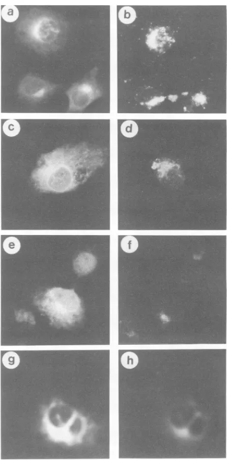
Intracellular localization of mutant E2 proteins. The sub-cellular localization of E2mutantproteinswasexaminedby
indirectimmunofluorescence. Cellsexpressingwild-type E2 exhibited staining throughout the cytoplasmicreticulum as
well as in the juxtanuclear region (Fig. 8a). The single,
double, andtripleglycosylationmutantproteinsdisplayeda predominantlyreticularstainingpatternaswell asGolgi-like
staining (Fig.8c, e, andg).Tovisualize thedistributionof E2 protein in the ER and the Golgi, fluorescent-conjugated
WGA and ConA were used as markers for the
compart-ments.WGA has been showntolabel transGolgi cisternae,
associatedvesicles, and the cell surface (38) bybinding to
clustered terminalN-acetylneuraminic acid residues as well
as N-acetylglucosamine-containing oligosaccharide chains
on glycoproteins (42). Costaining of transfected COS cells with human anti-RV serum andfluorescein-conjugated WGA revealed that wild-type E2 was concentrated in the Golgi region (Fig. 8b) while the mutant E2 proteins were distrib-utedthroughout the reticulum network and the Golgi region
(Fig.8dandf). A strong reticular staining, which colocalized with ConA, was observed in COS cells transfected with
[image:5.612.72.304.73.235.2]glycosylationmutants(Fig. 8h).Inaddition, unlike wild-type E2, which has been shown to exhibit a limited amount of cell surface expression (14), the glycosylation mutants had no detectablecellsurfacesignals (data not shown). Elimination
FIG. 8. Indirectimmunofluorescenceofwild-type andmutant E2 proteinsinCOS cells.Cellswerepermeabilizedpriortoadditionof rhodamine-conjugated WGAorConAandanti-RVserum.Afterthe cells were washed, a secondary antibody (fluorescein-conjugated goat anti-human immunoglobulin G) was added. (a) Wild type, anti-RV; (b) wild type, TRITC-WGA; (c) G2, anti-RV; (d) G2, TRITC-WGA; (e) G12, anti-RV; (f) G12, TRITC-WGA; (g)G123, anti-RV; (h) G123,TRITC-ConA.
ofanyoftheglycosylation sites in E2seemed toimpair the intracellular transport and block the cellsurface expression of E2.
Secretion of an anchor-free form ofwild-typeand mutant E2 proteins. Toanalyzethetransport behavior ofE2 mutants
in the secretory pathway, we constructed a panel of
trun-catedE2glycosylation mutants,each ofwhichhad68 amino acids deleted from the hydrophobic C terminus (4). The
truncated formofwild-type E2 was previously observedto be secreted into the culture medium as a 37-kDa endo H-resistant glycoprotein (15a). The truncated forms of E2
on November 9, 2019 by guest
http://jvi.asm.org/
[image:5.612.333.556.74.525.2]ROLE OF GLYCOSYLATION ON RV E2 3519
wt Gi G2 G3 G12 G123
- + - + - + - + - + - + endoH
B
GI G2 G3 G12 G12. wt
- + +- + - + - + - + endo H
97- 68-
43-29
29P-FIG. 9. Intracellular processing and secretion of a soluble form of wild-type (wt) and mutant E2 proteins. Cells were labeled with [35S]methionine for 30 min and chased for 4 h. (A) Immunoprecipitated samples from culture media of cells transfected with anchorless wild-type and mutant E2 cDNA constructs. (B) Intracellular forms of each anchorless E2 protein of wild-type and glycosylation mutants. Equal volumes of eachsample were incubated at 37°C for at least 8 h with (+) or without (-) endo H and separated by11% SDS-PAGE. Positions of molecular size markers are shown on the left (in kilodaltons).
singleglycosylation mutants (G1, G2, and G3) but not double
(G12) andtriple(G123) mutants were also secreted into the culture medium, although not as efficiently as the anchorless wild-type E2 (Fig. 9A). In addition, the efficiency of secre-tion appeared to depend on the position of the deleted
glycosylation site. The deletion of the glycosylation site proximal to the C terminus (G3) had a more profound
inhibitoryeffectonsecretion than did deletion of the central site(G2)and thatproximal to the N terminus (G1) (Fig. 9A). The intercellular forms of anchorless wild-type E2 and E2
glycosylationmutants werefoundtobesensitive to endo H
digestion (Fig. 9B), whereas the secreted E2 was endo H resistant (Fig. 9A). However, expression of the truncated triple mutant was not detected intracellularly (Fig. 9B). A 31-kDa endo H-sensitive protein species was found in the culture medium of the G2-transfected cells (Fig. 9A). This probably was due to somecell lysis during the chase period that released the intracellular G2 mutant protein into the
medium. Taken together, it was evident that the single
glycosylationmutantsG1, G2, and G3 weretransported out of the ERthrough theGolgitothecell surface and that they then exited the cell into the culturemedium, althoughnotas
efficientlyasthe otherwise unaltered anchorless E2. DISCUSSION
Protein movement from the ER to the medial Golgi apparatus has been identifiedastherate-limitingstepin the exocytic pathway (35), asmeasuredby the acquisition of a
varietyoforganelle-specific posttranslational modifications. Regarding the intracellular transport rate, several viral
gly-coproteins thathave been extensively investigated fall into
twocategories.Thefirst group includes the vesicular
stoma-titisvirus G protein (34) and influenza virus hemagglutinin (10), whichmovequickly alongtheexocytic pathway.After
a 15-min chase, 50% of the oligosaccharides on vesicular stomatitis virus Gprotein and 25%on influenza virus hem-agglutinin acquire endo H resistance (10, 34). The second groupcontains humanimmunodeficiency virus type 1
enve-lope protein (Env) (7) and simian virus 5
hemagglutinin-neuraminidase (28), for which acquisition of endo H
resis-tance was observed within 80 and 60 min
postlabeling,
respectively. Wefoundthat thecarbohydrates onwild-type RVE2wereconvertedtocomplex-typesugarmoietiesby1
hpostlabeling. However, the conversionwas notcomplete
even after an 8-h chase (data not shown), reflecting a slow movement of RV E2 from the ER to the Golgi apparatus.
Our data showed that E2 contains threepotential oligosac-charide addition sites and that all three potential N-linked glycosylation sites were utilized (Fig. 1 and 3). Inactivation
ofthese functional sitesimpaired theprocessing as well as
the intracellular stability ofE2 proteins (Fig. 6 and 7), the severity of the defectdependingonboth thenumber and the position of the glycosylation site deleted. Deletion of one N-linkedglycosylation siteon RVE2considerably reduced the rate of transport, as determined by the fraction of
proteins thatacquiredendo H-resistantcarbohydrates(Fig. 6). The glycosylation site proximalto the N terminus (G1) seems tobe less important than the siteproximal to the C terminus
(03),
asjudged by the fraction of molecules con-taining endo H-resistant carbohydrates for the membrane-bound form and by the secretion ratios of their anchorless counterparts (Fig. 6 and9). Oligosaccharide at each glyco-sylation site plays a different functional role, as has been notedpreviouslyin otherglycoproteins(24, 27).Inaddition,studies on other glycoproteins by the same approachhave shown thatglycosylation on all the predicted sites isnot a
prerequisite for folding, assembly, and transport of the
protein (12). It has been suggested that the contribution of eachcarbohydratechain varies dependingon itslocation in
adifferent conformational circumstance ofaparticular pro-tein. RVE2is rich incysteineandundergoesintramolecular disulfide bonding (Fig. 5). Inspection of the amino acid sequenceofRVE2 reveals thattheG2 and G3glycosylation
sites areflankedbytwocysteineresidues (4). Itispossible
that theoligosaccharidesattachedtotheG2 and G3 sitesare important in preventing improper intramolecular disulfide
bondformation,whereasglycosylationattheG1site hasless effect on proper folding and transport. The diffuse or
smeared appearances ofnonreduced mutant G12 and G123
proteinsonanimmunoblotprobablyreflecttheformation of aberrant intermolecular disulfide bonds. Thus, it appears thatoligosaccharideaddition isrequiredfor proper intramo-lecular disulfidebonding topromote correct
folding,
which in turnisessential forefficient transport(40).
Removal ofaglycosylationsite leadstoformation ofimproper intramolec-ulardisulfide bonds and
protein
misfolding.
Dramatic alter-ation in protein conformation could be the consequence whenglycosylation sites are inactivated. ThismayaccountA
68b-43b- O VOL.66, 1992
on November 9, 2019 by guest
http://jvi.asm.org/
[image:6.612.101.517.78.211.2]for diminished antibody binding by G12 and G123 proteins
under nonreducingconditions (Fig. SB).
Proteinsthataretransported slowlyin cellsdisplay heter-ogeneity in endo H resistance. Forexample, this has been observed for simian virus 5 hemagglutinin-neuraminidase (28), influenza virus neuraminidase (18), and human immu-nodeficiencyvirus gpl20 (7). Pulse-chase experiments have demonstrated that endo H-sensitive, partiallyendo
H-resis-tant,and endo H-resistantE2 formsrepresenttheER, Golgi,
and cell surface isoforms ofRV E2. Immunofluorescenceof transfected COS cells showed that the majority of the glycosylationmutantproteinswerelocalized in the ER(Fig. 8). A small fractionwerefound in the Golgi region (Fig. 8). Transport of the E2 single glycosylation mutants into the Golgi compartment was evidenced by the presenceof
par-tiallyendo H-resistant bands after the chase period (Fig. 6) aswellas bythe secretion of the C-terminal-truncatedform of E2 single glycosylation mutants (Fig. 9A). Thus, the transport of E2 to the Golgi apparatus appeared to be significantly affected but not completely blocked by the absenceofany oneof the N-linked oligosaccharides.
The anchorless E2 single glycosylation mutants were secreted into the culture medium, although less efficiently.
Theoligosaccharidesonthe secreted forms ofwild-typeand mutant E2 were completely endo H resistant, as was E2 from the RV virion (20), suggesting that carbohydrates
attached to these proteins are modified by Golgi enzymes. This finding indicates that the soluble forms of E2 single glycosylation mutants are transported through the normal exocytic route. Inabilityto detect cellsurface expressionof E2 single glycosylation mutants could be due either to the lowsensitivityof indirect immunofluorescenceinour
exper-iments or to the fact that mutant E2 proteins were quickly and extensively internalized from theplasma membrane, as has been observed for other glycoproteins (28).
The role of glycosylation generally varies for any given protein, and no concrete rules exist for predicting the phenotype ofa particular glycosylation mutant. Analysis of this type is further complicated by the fact that when glycosylationsitesarechanged bysite-directedmutagenesis, targeting and/or processing kinetics may differ as a
conse-quence of the amino acid substitution itself rather than of lack of glycosylation per se. Glycosylation may also be important for expression of immunogenic epitopes.
How-ever,inthiscase,human anti-RVseraseemedtorecognizes
the glycosylation mutantsjust as well as the wild-type RV antigens did. Studies are in progress to determine whether RV E2 is internalized anddegraded rapidly and to examine the capacities of the E2 glycosylation mutant proteins to function in RV virions.
ACKNOWLEDGMENTS
We thank F. Tufaro for critical readingof themanuscript.
This work was supported jointly by grants from the Medical Research Council of Canada and British Columbia Health Care
ResearchFoundation. S.G. isaninvestigator of the British
Colum-bia Children's Research Foundation. T.C.H. was a predoctoral
trainee receiving support from the Medical Research Council of
Canada in thebiotechnology trainingprogram.
REFERENCES
1. Adams,G.A.,andJ. K. Rose. 1985. Structure requirements of
a membrane-spanning domain for protein anchoring and cell surface transport. Cell 41:1007-1015.
2. Andersson, S.,D. L.Davis,H.Dahlback,H.Jornvall, andD. W. Russell. 1989. Cloning, structure, and expression of the mito-chondrialcytochrome P-450 sterol 26-hydroxylase, abile acid
biosyntheticenzyme. J.Biol. Chem.264:8222-8229.
3. Bardeletli,G.,J.Tektoff,andD.Gautheron. 1979.Rubella virus maturationandproductionintwohostcellsystems. Intervirol-ogy11:97-103.
4. Clarke, D., T. Loo, I. Hui, P. Chong, and S. Gillam. 1987. Nucleotidesequenceand in vitroexpressionof rubella virus 24S subgenomicmRNAencodingthestructural
proteins
El,E2and C. NucleicAcids Res. 15:3041-3057.5. Clarke, D.,T.Loo,H.McDonald,and S.Gillam. 1988.
Expres-sion of rubella virus cDNAcodingfor the structural
proteins.
Gene65:23-30.
6. Dorsett,P.H.,D.C.Miller,K. Y.Green,and F. I.Byrd.1985. Structureand function of the rubella virusproteins.Rev.Infect. Dis. 7(Suppl. 1):S150-S157.
7. Earl,P.L.,B.Moss,and R. W. Doms.1991.Folding,interaction withGRP78-BiP, assembly, and transportof the human immu-nodeficiencyvirus type Ienvelopeproteins. J. Virol. 65:2047-2055.
8. Elbein,A. D.1987. Inhibitorsof thebiosynthesisand
processing
of N-linked oligosaccharide chains. Annu. Rev. Biochem. 56: 497-534.
9. Frey, T. K., and L. D. Marr. 1988. Sequence of the
region
codingfor virionprotein Cand E2 and the
carboxyterminus
of the non-structural proteins of rubella virus:comparison
withalphaviruses. Gene62:85-99.
10. Gallagher, P.,J.Henneberry,I.Wilson,J.Sambrook,andM.J.
Gething. 1988. Addition ofcarbohydrateside chains at novel sitesoninfluenza virushemagglutinincanmodulatethe
folding,
transport, andactivityof the molecule. J. Cell Biol. 107:2059-2073.
11. Green,K.Y., and P. H. Dorsett. 1986. Rubellavirusantigens:
localization ofepitopesinvolved inhemagglutination and neu-tralizationby usingmonoclonal antibodies. J. Virol.57:893-898. 12. Guan, J.-L.,H.Cao, andJ.K. Rose.1988.Cell-surface expres-sion of a membrane-anchored form of the human chorionic
gonadotropina-subunit. J. Biol. Chem.253:5306-5311. 13. Hobman, T. C., and S. Gillam. 1989. In vitro and in vivo
expressionofrubella virusglycoproteinE2: thesignal
peptide
is contained in theC-terminal region ofcapsidprotein. Virology
173:241-250.
14. Hobman, T.C., M. L. Lundstrom, and S.Gillam. 1990. Proc-essing and intracellular transport of rubella virus structural proteins in COS cells. Virology 178:122-133.
15. Hobman,T.C.,Z.Qiu,H.Chaye,andS. Gillam. 1991.
Analysis
of rubella virus El glycosylation mutants expressed in COS cells. Virology 181:768-772.
15a.Hobman,T.C.,N.0.L.Seto,andS. Gillam.Unpublisheddata. 16. Hobman, T.C., R. Shukin,andS. Gillam. 1988. Translocation of rubella virusglycoproteinElinto the
endoplasmic
reticulum. J. Virol. 62:4259-4264.17. Klenk,H.-D.1989. Influence ofglycosylationonantigenicityof viral proteins, p. 25-37. In M. H. V. Van Regenmortel and A. R. Neurath (ed.), Immunochemistry of viruses, vol. 2. Elsevier,Amsterdam.
18. Kunda,A.,M. A.Jabbar,and D. P.Nayak. 1991.Cell surface transport,oligomerization,andendocytosisof chimerictypeII
glycoproteins: role of cytoplasmic and anchor domains. Mol. Cell. Biol. 11:2675-2685.
19. Kunkel,T.A.1985.Rapidandefficientsite-specificmutagenesis
without phenotypic selection. Proc. Natl. Acad. Sci. USA 82:4753-4757.
20. Lundstrom,M. L., C. A. Mauracher, and A. J.Tingle. 1991. Characterizationofcarbohydrateslinkedtorubella virus
glyco-proteinE2. J. Gen. Virol. 72:843-850.
21. Machamer, C. E., and J. K. Rose. 1988. Influence of new glycosylationsitesonexpressionof the vesicular stomatic virus G protein at the plasma membrane. J. Biol. Chem. 263:5948-5954.
22. Machamer,C.E.,andJ.K.Rose.1988. Vesicularstomatic virus Gproteinswithalteredglycosylationsitesdisplay temperature-sensitive intracellular transport and are subject to aberrant intermolecular disulfidebonding.J.Biol. Chem. 263:5955-5960. 23. Matthews, R. E. F. 1982. Classification and nomenclature of
on November 9, 2019 by guest
http://jvi.asm.org/
ROLE OF GLYCOSYLATION ON RV E2 3521 viruses. Fourth report of the International Committee on
Tax-onomyof Viruses. Intervirology17:1-199.
24. Matzuk, M. M., andI.Boime. 1988. The role of the asparagine-linked oligosaccharide of the alpha-subunit in the secretion and assembly of human chorionic gonadotropin. J. Cell Biol. 106: 1049-1059.
25. Messing, J. 1983. New M13 vectors for cloning. Methods Enzymol. 101:20-78.
26. Nakhasi, H. L., D. Thomas, D. Zheng, and T. Y. Liu. 1989. Nucleotide sequence of capsid, E2 and El protein genes of rubella virus vaccine strain RA27/3. Nucleic Acids Res. 17: 4393-4394.
27. Ng, D. P., S. W. Hiebert, and R. A. Lamb. 1990. Different roles ofindividual N-linkedoligosaccharide chains in folding, assem-bly,and transport of the simian virus 5 hemagglutinin-neuramin-idase. Mol., Cell. Biol. 10:1989-2001.
28. Ng, D. P., R. E. Randall, and R. A.Lamb. 1989. Intracellular maturation and transport of the SV5 type II glycoprotein hemagglutinin-neuraminidase: specificand transientassociation with GRP78-Bip in the endoplasmic reticulum and extensive internalization from thecellsurface. J.CellBiol.109:3273-3289. 29. Oker-Blom, C. 1984. The gene order for rubellavirus structural
proteinsisNH2-C-E2-E1-COOH. J. Virol.51:354-358. 30. Oker-Blom, C., N. Kalkkinen, L.Kiarifiinen,and R. F.
Petters-son. 1983. Rubella virus containsonecapsid proteinand three envelope glycoproteins,El, E2a,and E2b. J. Virol. 46:964-973. 31. Oker-Blom, C.,I.Ulmanen, L.Kiarifinen,and R. F. Pettersson. 1984. Rubellavirus 40S genome RNAspecifies a 24S subge-nomic mRNAthat codes foraprecursor to structuralproteins. J.Virol. 49:403-408.
32. Pfeffer, S. R., and J. E. Rothman. 1987. Biosynthetic protein transport and sorting by the endoplasmic reticulum andGolgi. Annu. Rev.Biochem. 56:829-852.
33. Plummer, T. H., J. H. Elder, S. Alexander, A. W. Phelan, and A. L. Tarentino.1984. Demonstration ofpeptide N-glucosidesF activity in
endo-Io-N-acetylglucosaminidase
F preparations. J. Biol. Chem.259:10700-10704.34. Rose, J. K., and J. E. Bergmann. 1983. Altered cytoplasmic domainsaffect intracellular transportof the vesicularstomatitis virus glycoprotein. Cell 34:513-524.
35. Rose, J. K., and R. W. Doms. 1988.Regulation of protein export from the endoplasmic reticulum. Annu. Rev. Cell Biol. 4:257-288.
36. Sanger, F., S. Nicklen, and A. R.Coulson. 1977. DNA sequenc-ing with chain-terminating inhibitors. Proc. Natl. Acad. Sci. USA74:5463-5467.
37. Tarentino, A. L., and F. Maley. 1974. Purification and properties
ofan
endo-o-N-acetylglucosaminidase
fromStreptomycesgni-seus. J. Biol. Chem. 249:811-817.
38. Tartakoff, A., and P. Vassalli. 1983. Lectin-binding sites as markers of Golgi subcompartment: proximal-to-distal matura-tion ofoligosaccharides. J. Cell Biol.97:1243-1248.
39. Towbin, H., T. Staehelin, and J. Gordon. 1979. Electrophoretic transfer ofproteins frompolyacrylamide gels tonitrocellulose sheets:procedureand someapplications.Proc.Natl. Acad.Sci. USA76:4350-4354.
40. Vidal,S.,G. Mottet, D. Kolakofsky, and L. Roux. 1989.Addition ofhigh-mannosesugars mustprecede disulfidebond formation for proper folding of Sendai virus glycoproteins. J. Virol. 63:892-900.
41. Vidgren, G. K., N. Takkinen, L.Kaariainen, and R. F. Petters-son. 1987. Nucleotide sequence of the genes coding for the membrane glycoproteins El and E2 of rubella virus. J. Gen. Virol. 68:2347-2357.
42. Virtanen, I., P. Ekblom, and P. Laurila. 1980. Subcellular compartmentalizationofsaccharide moieties incultured normal andmalignant cells.J. Cell Biol.85:429-434.
43. von Bonsdorff, C.-H., and A. Vaheri. 1969. Growthof rubella virus in BHK21 cells: electronmicroscopyofmorphogenesis.J. Gen.Virol. 5:47-51.
44. Zheng, D., L. Dickens, T. Y. Liu, and H. L. Nakhasi. 1989. Nucleotidesequence of the24SsubgenomicmRNAofavaccine strain (HPV77) of rubella virus: comparison with a wild-type strain(M33).Gene82:343-349.
VOL.66, 1992
![FIG.1.300molecularE2.mUat 37°C, Determination of the number of N-linked glycans on RV [35S]methionine-labeled E2 was incubated with no (lane 1), 10 (lane 2), 20 mU (lane 3), 50 mU (lane 4), 100 mU (lane 5), and mU (lane 6) of N-glycanase (Boehringer Mannhe](https://thumb-us.123doks.com/thumbv2/123dok_us/1307807.84043/2.612.367.500.82.146/moleculare-determination-methionine-labeled-incubated-glycanase-boehringer-mannhe.webp)
![FIG. 9.wild-typeEqualPositions[35S]methionine Intracellular processing and secretion of a soluble form of wild-type (wt) and mutant E2 proteins](https://thumb-us.123doks.com/thumbv2/123dok_us/1307807.84043/6.612.101.517.78.211/typeequalpositions-methionine-intracellular-processing-secretion-soluble-mutant-proteins.webp)
