0022-538X/88/051647-06$02.00/0
Copyright ©D 1988, American SocietyforMicrobiology
A
Monoclonal
Antibody Specific for
the
Cellular
Receptor
for the
Group
B
Coxsackieviruses
KUO-HOM LEE HSU, KARL LONBERG-HOLM, BARBARA ALSTEIN, AND RICHARD L. CROWELL* Departmentof Microbiology and Immunology, Hahnemann University School of Medicine,
Philadelphia,
Pennsylvania
19102-1192Received24September 1987/Accepted27January 1988
A 50-kilodalton receptor protein (Rp-a) for the group B coxsackieviruses (CB) was isolated in a virus-receptorcomplex from detergent-solubilized HeLa cells (J. E. Mapoles, D. L. Krah, and R. L. Crowell, J. Virol. 55:560-566, 1985). It was used as an immunogen for preparation of a mouse monoclonal antibody
(RmcB) which protected HeLacellsand Buffalogreenmonkey kidneycellsfrom infectionby all six serotypes of CB.RmcBdid not protect HeLa cells frominfection bypoliovirus, echovirus 6,orcoxsackievirusA18.This
monoclonalantibody differed inreceptorepitope specificityfromapreviously isolated antibody (RmcA) (R.L. Crowell, A. K. Field, W. A.Schleif, W. L. Long, R.J. Colonno, J. E. Mapoles,and E. A.Emini,J. Virol. 57:438-445, 1986)whichblocked receptorsonlyfor type 1 CB(CB1),CB3,CB5,andechovirus 6. RmcA and RmcB recognized two distinct saturable receptors on HeLa cells, designated HR2 and HR1, respectively.
Humanrhabdomyosarcoma (RD) cells have the HR2receptorfor CB3-RD (avariant ofCB3),but lacktheHR1 receptorforCB3.Therefore, RD cellswereresistanttoinfection by CB3. Although bindingof CB3-RDtothe HR2 receptoronRDcellscanleadtoinfection, bindingofCB3-RDtothe HR2 receptoronHeLacellsdidnot
lead toinfection. Apparently, both CB3 and CB3-RDuseonly theHR1 receptorfor infection ofHeLa cells.
Thus,agivenvirusmay usetwodistinctreceptors tobindtocells when onlyonevirus-receptor interaction leads toinfection.
Picornaviruses are among the smallest and simplest
hu-manpathogens, andyetthey produceabewilderingarrayof
diseases. It is clear that among the factors contributing to
diseasediversityarethespecific requirementsof viruses for host cell receptors. During the past twodecades there has been a continuous interest in receptors and in the early
eventsofpicornavirusinfection (forreviews, seereferences 4-6, 12, and 13). However, relatively little is known about the structures of these receptors ortheirroles in initiating virus infection. Receptor specificity was firstdemonstrated
by binding competition betweentwovirus serotypes which sharedthesamereceptor(1, 2,9, 11, 16). Different HeLa cell
receptorsforpolioviruses, human rhinoviruses oftwo
sepa-ratefamilies, andgroupB coxsackieviruses (CB)have been discovered (11).
More recently, it was discovered (17) that a variant ofa type 3 CB virus (CB3-RD) that was selected by growth in
humanrhabdomyosarcoma (RD) cells recognized botha new humancellularreceptor(designatedHR2) foundonRD cells and the receptor (HR1) recognized by parental virus on
HeLa cells. Itwaspostulated thatHeLacells have bothHR1 and HR2 and that RD cells lack HR1. Partial evidence for
tworeceptors on HeLacells was provided by the observa-tion of separate specificities during the screening of mono-clonal antibodies (3). Here we report a new monoclonal antibody (RmcB) prepared against the HeLa cell receptor
HR1 which protects HeLa cells and Buffalogreen monkey
kidney cells (BGM) from infection by all six serotypes of CB. This monoclonal antibody shows a different receptor
epitope specificitythan anantibody obtained previously (3), now designated RmcA, which did not protect HeLa cells against the even-numbered CB serotypes. Our data reveal that RmcA andRmcB recognize twodistinct and saturable
*Correspondingauthor.
receptors (HR2 and HR1) for CB3-RD on HeLa cells and thatonly RmcBconsistentlyprotectsthe cellsagainst infec-tionbyall six CB and their RD variants.
MATERIALSAND METHODS
Cells andviruses. HeLa cells(Mandel strain)and RDcells were cultured in suspension and in monolayers,
respec-tively, as described previously (7, 18). The BGM cell line was obtained from Marilyn Menegus (15), and a mouse
T-lymphomacellline,YAC-1,wasobtainedfrom the Amer-icanTypeCulture Collection.
Theprototypestrainsof CB(serotypesCB1through CB6) and echovirus 6 have been described previously (2). The variantcoxsackievirusesCB1-RD andCB3-RDwerederived by Reagan et al. (17) by serial passages of the parental viruses on RD cells. Coxsackievirus A18 (CA18) was ob-tained originally from the University of Minnesota stock collection (8). Poliovirus type 1 was purchased from the AmericanTypeCultureCollection, Rockville,Md.Methods for the growth and purification of unlabeled, [3H]uridine-labeled, and [35S]methionine-labeled viruses have been de-tailed previously (3, 14). The concentration ofvirions was estimated from the optical density at 260 nm, at which a valueof 1.0correspondstoapproximately 1013particlesper
ml.
Purification of VRC. Virus-receptor complex (VRC) was formed byusing detergent-solubilized HeLa cells and puri-fied by previously described methods (14) with modifica-tions. Inbrief, HeLacellswere washedtwicein phosphate-buffered saline (PBS) and suspended in TD1 buffer (1% Triton X-100 and 0.5% sodium deoxycholate in PBS with
protease inhibitors, 2 mM phenylmethylsulfonyl fluoride, 2
mMN-ethylmaleimide,and 5 mM EDTA)ataconcentration of 4 x 10⁷cellspermlat4°Cfor15min.Thepreparationwas
centrifugedat1,000 x gfor5mintoremovenucleiandthen 1647
on November 10, 2019 by guest
http://jvi.asm.org/
ultracentrifuged at 55,000 x g for 4.5 h. Purified CB3 and
[3HJCB3 wereaddedtothe supernatant fluidata
concentra-tion of 4 x 10¹¹particles per ml (5 ,ug/ml). After incubation
overnightat4°C, the VRC preparationwas warmedtoroom
temperature,and sodium dodecyl sulfatewasaddedtoafinal
concentration of 1%. The VRC preparation (25 ml) was
overlaid onto a step gradient containing 1.5 ml of 54% metrizamide and 5 ml of 25% sucrose, both of which were
prepared in TD1 buffer. The preparationwascentrifuged in
an SW27 rotor for 4.5 h at 24,000 rpm. The gradient was
fractionated from the bottom of the tube. The VRC peak fractions(atthemetrizamide-sucrose interface, identified by liquid scintillation counting)werepooled and dialyzed
over-nightat4°C against TD2 buffer (0.2% Triton X-100 and 0.2% sodium deoxycholate in PBS with 0.4 mM
phenylmethylsul-fonyl fluoride, 0.4mMN-ethylmaleimide, and1 mMEDTA). TheVRC dialysate (4 ml)wasoverlaidontoa30-ml5to35%
sucrose gradient in TD2 buffer and centrifuged for 4 h at
24,000rpminanSW27 rotor. TheVRC peak fractionswere
pooled and dialyzed overnight against TD2 buffer. The VRC
was pelleted by centrifugation at 200,000 x g for 45 min
through a25%sucrose cushion in TD2 buffer, suspended in
a small volume of TD2 buffer, and stored at -70°C until used.
Preparation of polyclonal antibodies. Polyclonal antiserum
to VRC was prepared in a New Zealand White rabbit by
subcutaneous immunization with approximately 40 ,ug of purified VRC in complete Freund adjuvant. Two booster injections with 40 ,ug of VRC in incomplete Freund adjuvant
weregiven at monthly intervals.
Preparation of monoclonal antibodies. A/J mice were
in-jected intraperitoneally at monthly intervals with 20 ,ug of VRC which had been heat treated (60°C for 30 min) to
inactivate the virus. The first injection was of VRC in
complete Freund adjuvant, and the second and thirdwereof
VRC in incomplete Freund adjuvant. Following the second injection, mice were bled and sera were examined for
evidence of polyclonal antibodies by the cell protection
assay (see below). The titers of the antisera ranged from
1:100 to 1:300. The mice were given a final boost of VRC
without Freund adjuvant 5 daysbefore the cell fusion. The spleen cells from immunized micewerecollected and fused
with sp2/o myeloma cells as described previously (10).
Supernatants from clones with approximately 40%
hybrid-oma cell confluency were assayed by the cell protection assay 10 to 14 days after cell fusion. The clones producing antibody that protected cells from infectionweresubcloned
by limiting dilution.
Purification of monoclonal antibody. Fluids from subcloned RmcB hybridoma cell cultures were found to contain the
mouse immunoglobulin Gl isotype as determined by gel
diffusion assays with isotype-specific antiserum. The
anti-body was concentrated from culture fluids by 45%
(NH4)2SO4 precipitation. The precipitatewas dissolved ina
smallvolumeand dialyzed overnight against PBS. Antibody
was then purified on a protein A-Sepharose (Pharmacia,
Inc., Piscataway, N.J.) column. The dialysate was mixed
with an equal volume of loading buffer (3.0M NaCI, 1.0 M
glycine [pH 8.8]) and passed over the column, which was
thenwashedwithloading buffer untilA280wasequaltozero.
The immunoglobulin Gl was eluted with 0.2 M NaH2PO4
(pH 6.0) and then dialyzed overnight against PBS. The purified antibody was iodinated by a procedure using low
concentrations ofchloramine-T at 0°C (14).
Cell protection assay. Cells were seeded in 96-well,
flat-bottom microdilution plates at 4 x 10⁴ cells per well and
incubatedovernight at 37°C in a humidified incubator with 5% CO2.Thecell monolayers werepretreatedwith 50 to 100 ,ulofhybridomaculture fluid for 30 min at room temperature before addition of the virus. Aminimumamountof viruswas added toyield a cytopathic effect within 24 to48 h. Cyto-pathic effect was detected by crystal violet staining ofthe cells.
Virus or antibody binding. Cells grown in suspension or
removed frommonolayersby trypsinwerewashed twiceand
resuspended in PBS with 3% fetal calf serum-20 mM
HEPES
(N-2-hydroxyethylpiperazine-N'-2-ethanesulfonic
acid)(pH 7.4). ReceptorsforCB3 and CB3-RDwere
insen-sitive to the trypsin treatment. The cells were mixed with
radioactivevirusorantibodytoafinalconcentration of2 x
107
cells per ml and were maintained insuspension
with areciprocatingshaker.Directlyfollowing addition of radioac-tive ligand, a
50-ll
sample wasremoved and diluted with 2 ml of the ice-cold buffer described above. Other portionswere removed and diluted at intervalsand then centrifuged
to remove unattached virus orantibody. Thesampleswere
dissolved in0.5 ml of0.5 M NH40H,and radioactivitywas determined by liquid scintillation counting. When
125I-la-beled antibody was employed as the ligand, samples were counted directly in a gamma counter. Samples ofthe
sus-pension ofradioactive ligand plus cells were also counted
withoutwashing. Results were expressed asthe fraction of
total counts per minute found in the washed cells. In the
binding inhibitionassay,cellswereincubated with
nonradio-active competing ligands for 30 min
prior
to addition ofradioactivevirusorantibody.Portions were then removedat intervals andassayed as described above.
Fortitrationtodetermine thenumberofbinding sitesper
cell at
ligand
saturation, samples
wereassayed
at a fixedtime(30to60min) with increasing amountsoflabeled virus or antibody. The percentage of cell-associated label was used tocalculatethe numberof moleculesor
particles
boundpercell.This numberwasthen
plotted against
thenumberoflabeledmoleculesorparticlesadded percell.Wedidnotuse the scatchard plot to linearize the binding data, since that methodassumesthatbindingismonovalent, whichclearlyis not the case for virions or bivalent antibodies. Also, the
binding
ofCB3 to HeLa cells has a very lowreversibility
(14). Thus,we have assumed that theadsorption isotherms forantibodies orcoxsackieviruses oncellsare
composed
oftwocomponents. The first isa
specific
saturableattachment;superimposed
upon this is anonspecific
attachment, whichappears not to be saturable butto be
proportional
toinput
multiplicity.
The intercept of the slope ofthenonspecific
bindingatzeroinputwasconsidered tobe the
approximate
numberof
specific
sites per cell. RESULTSPolyclonal anti-VRC. A rabbit was immunized with
puri-fied VRC.Toobtainlargeramountsof the VRCforuseasan
immunogen, themethod reported
previously
(14) was mod-ified as detailed in Materials and Methods. Although the antiserum obtained was notspecific
for Rp-a, itrecognized
Rp-a and protected HeLa cells
against
CB3 infection. The antiserumimmunoprecipitated
fourpolypeptides
fromdeter-gent-solubilized
3H-labeled HeLa cells as measured by so-dium dodecyl sulfate-polyacrylamide gelelectrophoresis
(Fig.
1). The protein at 50 kilodaltons is Rp-a; the others remaintobeidentified. Incellprotection
assays, this rabbitantiserum hadatiterof1:100.
Selection of monoclonal antibody.
Hybridomas
werepre-pared by
immunizing
mice with VRC, and the antibodieson November 10, 2019 by guest
http://jvi.asm.org/
8-o 6D
2-cm
FIG. 1. Patternofimmunoprecipitation of
3H-amino-acid-label-ed HeLa cellproteins byrabbit anti-VRCantibody. 3H-amino-acid-labeled HeLacellsweresolubilized with TD buffer (1% Triton X-100
and 0.5%deoxycholateinPBS)andimmunoprecipitated withrabbit
anti-VRCfollowed bygoatanti-rabbitimmunoglobulin.The immu-noprecipitates were solubilized and assayed on sodium dodecyl
sulfate-polyacrylamide gel electrophoresis (10% gel). Thegel was
sliced formeasurementofradioactivity (givenascountsperminute onthe ordinate). Positions of molecular weight markers are
indi-cated.
produced were screened for blocking of CB3 infection of
HeLa cells. From 1,200 cultures, a single positive culture wasfound and subcloned. The antibody wasdetermined to
beamurineimmunoglobulinGlisotypeandwasdesignated
RmcB to distinguish it from the antibody obtained previ-ously(3), which hasnow beenredesignated RmcA.
Receptor specificity of monoclonal antibodies. RmcB
pro-tected HeLa cellsagainst infection by all sixserotypesof CB
aswellasby theCB-RDvariants(Table 1). It didnotprotect
HeLacells against echovirus 6, poliovirus type 1, or
cox-sackievirusA18. Thus, RmcB showedadifferent specificity
thanRmcA, which protected only against infection by CB1,
CB3, and CB5 aswellas by echovirus 6 (Table 1).
To further characterize the receptor specificity, binding studieswith HeLa cellswereperformed using iodine-labeled
RmcAand RmcB. No competitionwasfound between these
two monoclonal antibodies (Fig. 2A). Interestingly, RmcB blocked onlya low percentage of the binding of
[35S]CB3-RD, whereas RmcA showed extensive blockade of CB3-RD
binding (Fig. 2B). These results are consistent with the
numberofreceptors onHeLacells for thesetwoantibodies
(see below). Pretreatment of HeLa cells with both RmcA
ac
.5
0
co 6t
0 0
D
0
X 2(
.0 ]l
u
A
RmcA
(+or-RmcB)
| RmcB
(+or-RmcA)
DZ
n I.
20 40 60
min
TABLE 1. RmcAandRmcBprotection of HeLa cells from virusinfection"
Protectionbyc:
Challengevirusb
RmcA RmcB
CB1, CB3,CB5 + +
CB2, CB4,CB6 +
CB1-RD, CB3-RD +
Echovirus6 +
PoliovirusTi CA18
"Cell protectionassayswereasdescribed inMaterialsand Methods. Aminimumamountof viruswasaddedtoyieldacytopathic effect within 24to48 h.
' +,Protectedagainstinfection; -,didnotprotectagainstinfection.
and RmcB totally blocked binding ofCB3-RD to the cells. These results confirmed thehypothesis,basedonpreviously
reportedviruscompetitionstudies(17),thatCB3-RD
recog-nizestwoseparatereceptorsonHeLacells.Thefindingthat
RmcB protected HeLa cells against CB3-RD infection
(Table 1) indicated that the attachment site which RmcA identified doesnotleadtoproductive infectionby this virus. Both RmcAand RmcB inhibited attachment of the parental virus CB3 to HeLa cells (Fig. 2C). However, we do not
know why RmcA wasslightly moreeffective than RmcB in
inhibitingthe attachment ofCB3.
Cell specificity of monoclonal antibodies. The differences between RmcA and RmcB in receptor specificities and in protection against infection by CB3 and CB3-RD were
furtherinvestigated with different cells. In addition to the RD cells, which cannot be infected by the parental
CB,
BGM, a monkey kidney cell line, and YAC-1, a mouse
T-lymphoma cell line,werestudied. The lattertwocellscan
beinfectedbyall members ofCBaswellasbythe RD virus
variants. RmcA protected RD cells against CB3-RD infec-tion (Table 2), but did not protect BGM or YAC-1 cells.
RmcB did not protect RD or YAC-1 cells, but protected
BGM against both CB3 and CB3-RD.
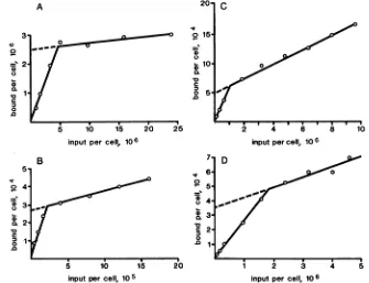
Numberofantibody and virus binding sitesoncells.
Exper-iments with labeled monoclonal antibodieswere performed to determine the number ofantibody binding sites on
dif-ferent cell types (3). Titrations to determine the numberof binding sites per cell were done (Fig. 3). As described in
Materials andMethods, the number of molecules boundper
cell was determined and plotted versus the number of
labeledmolecules addedpercell. Theintercept of the slope
%O 20 40 60 % 20
B min C min
FIG. 2. Attachmentoflabeled antibodies and virusestoHeLacells.(A)Bindingof RmcAandRmcBtodifferentsitesonHeLacells.(B)
RmcAblocks thebindingof CB3-RDtothe HR2receptorsonHeLacells,whereas RmcBblocks thebindingof CB3-RDtothe HR1receptors. Together,RmcA and RmcBtotally blockbindingofCB3-RDtoHeLa cells. (C) RmcA and RmcBblockCB3bindingtoHeLa cells.
92.5 66 45 31 21.5 14.4
1 2 3 4 5 6 7 8 99
0 9
D-
3-0-I
on November 10, 2019 by guest
http://jvi.asm.org/
[image:3.612.58.298.74.207.2] [image:3.612.311.554.97.186.2] [image:3.612.116.490.569.697.2]TABLE 2. Summary of RmcA and RmcB protection of different cell typesagainstinfection by CB3 and CB3-RD'
RmcA RmcB
Celltype
CB3 CB3-RD CB3 CB3-RD
HeLa + - + +
RD NA" + NA
-BGM - - + +
YAC-1 - - -
-"'Protection assays were as described in Materials and Methods. +,
Protectedagainst infection;-,didnot protect against infection.
bNA,Notapplicable; virus producednocytopathic effect incontrol cells.
of the nonspecific binding at zero input was used as a measureofthe numberof specific binding sites per cell. In
experiments of similardesign, the number of virion binding
sitespercell also wasdetermined (2). The number of binding
sites for CB3,CB3-RD, RmcA, and RmcB on different cells wasdetermined (Table 3). HeLa cells were found to be rich
in receptorsfor CB3-RD (5.6 x 10⁵percell) compared with
RDcells (3.5 x 10³per cell)and BGM cells(3.3 X 10³per
cell). The monoclonal antibodies consistently bound to be-tween 3- to 10-fold more sites than did the virions. This
observation is in keeping with the different sizes and va-lences of the ligands.
DISCUSSION
The HeLa cell plasma membrane receptor for CB was
solubilizedby detergents, purifiedasaVRC,and used as an
immunogento prepare amurine hybridoma.The monoclonal
antibodythus obtained, RmcB, wasfound to protect HeLa cells and BGM cells from infection by CB1 through CB6.
TABLE 3. ComparativenumberofbindingsitesforCB3,
RmcA,and RmcB ondifferentcells
Number ofbinding siteson:
Ligand
HeLa RD BGM
CB3 1.8 x 10⁴ _a 2.7 x 10³
CB3-RD 5.6 x 10⁵ 3.5 x 10³ 3.3 x 10³
RmcA 2.5 x 10⁶ 2.7 x 10⁴
RmcB 5.0 x 10⁴ _ 3.6 x 10⁴
Less than 100 per cell (limit of detection of this assay; binding considered absent).
Cell protection correlated with blockage of specific recep-tors. It is remarkable that the receptorspecificity of RmcB is distinct from that of another receptor-specific monoclonal antibody (RmcA) described previously (3). RmcA protected HeLacells against infection by only the odd-numbered CB serotypes (CB1, CB3, and CB5) and by echovirus 6 and CA21. It alsoprotected RD cells against infection by CB1-RD, CB3-RD, and CB5-RD virus variants, but not by 16 otherpicornaviruses. RmcA did notprotectthe BGM cells of simian origin, even though these cells are highly suscep-tible to infectionby all six CB serotypes (15).
ThefindingsthatCB3-RD virussaturated the receptors on HeLa cells and blocked binding ofCB3, while saturating amountsof CB3 didnotblockbinding of CB3-RD, indicated that a second receptor site was utilized for binding the
CB3-RD variant (17). The results of binding studies with RmcA and RmcB are compatible with the existence of two
distinct receptors on HeLa cells which bind the CB3-RD variant virus.
Tohelpclarify discussion of the receptors for coxsackie-viruses, we have developed a system of nomenclature in
201 C
o
15-
1-0
. 10-D0
c
o 5.
.0
5 10 1*5
inputpercell, 106
20 2
7.
6
0
o, 5.
e
o4-0
c 3.
c 2
=1
5 0b 15 20
inputpercell,105
;
. linputpercell, 10 6
D_
1 2 3 4 5
inputpercell, 10 6
FIG. 3. Titrationofbinding sites for monoclonal antibodiesoncells. Dilutions of'25I-labeled monoclonal antibodieswereincubated with
cellsuspensions for 60minatroomtemperature.Thecellswerewashedand counted for radioactivity. The number of molecules boundper cellwasdeterminedand plottedversusinput virus multiplicity. Shownarebinding of RmcAto(A)HeLa cells (B) RDcells andbinding of
RmcB to(C)HeLacells and(D) BGM cells.
A
37
0so~
so2
co
0
cx
0
.0m
z
0
.0
1*
5,
4.
3.
2' B
"S
00
on November 10, 2019 by guest
http://jvi.asm.org/
[image:4.612.67.305.94.168.2] [image:4.612.143.478.428.685.2]A
x
B
r
C
y~~~~
N
y~~~~~~
mqlrmc
A P RmcA RmcAFIG. 4. Schematic model for CBreceptors andeffects ofRmcAoninfection. Symbols: HR2(functional); t. HR2 (nonfunc-tional); 9.HR1(functional);P,parentalCB3virus.(A)RDcells with CB3-RDvir'us;RmcA blocks infection. (B)HeLacellswithparental CB3 virus; RmcAblocks attachment and infection. (C) HeLa cells with CB3-RD virus; RmcA blocks most ofattachment, but pe'rmits infection. Seetextfor- details.
ligand via endosomes.Thelimitedfunction of HR2onHeLa cells is in sharp contrast to its role on RDcells, wherethe HR2 receptors arefully functional infacilitating infection by CB3-RD.
The separate functional specificities of RmcA and RmcB areevident,since RmcB protects BGM cellsagainstparental virusinfectionandattachment and RmcA protects RDcells. However, the paradoxical effects of RmcA and RmcB on HeLa cells require explanation. It is possible that RmcA prevents attachment of CB3 to the HR1 receptor by a secondary steric blockade (Fig. 4B). This requiresthat the HR2 receptors (presentinroughly 50-fold excessoverHR1) be associated with HR1 molecules on the cell surface. A possible explanation for the ability of RmcA to inhibit
attachment of CB3-RD to HeLa cells without preventing
infection is that the affinity ofRD virus for the cell may be sufficient to compete with binding ofRmcA to HR2 sites,
since RD virus can bind to both HR1 and HR2 (Fig. 4C). This could lead to interactions promoting the binding of infecting virus to the HR1 receptors andthento internaliza-tionofthe virusandinfection.
Moreinformation, such asbindingconstantsofvirus and antibodies to HR1 and HR2, will be needed before our models for the interactions of viruses and antibodies with HeLa cells can be accepted. The results presented here, however, illustrate the complexity which can be encoun-tered in investigations ofcellular receptors for viruses with monoclonal antibodies. Furtherinvestigationsinthis system
using adenovirus type 2 fiberproteinas aprobe (11) for the HR1 receptor willbereported elsewhere.
ACKNOWLEDGMENTS
This investigation was supported by Public.Health Service
re-searchsupport grantAI-03771 fromthe NationalInstituteofAllergy
and Infectious Diseases and by our institutional Biomedical Re-searchSupportgrant2S07RR05413.
LITERATURE CITED
1. Crowell, R. L. 1963. Specific viral interference in HeLa cell cultureschronicallyinfected with coxsackie B5virus.J. Bacte-riol. 86:517-526.
2. Crowell, R.L. 1966. Specific cell-surfacealteration by
entero-viruses as reflectedby viral-attachment interference. J. Bacte-riol. 91:198-204.
whichthefirstletterreferstothe species of origin of thecell, (e.g., H for human), R indicates receptor, and the arabic numerals indicate distinct, saturable receptors in their chronological order ofdiscovery. Forexample, HR1 refers
to the HeLa cell receptor which binds the parental CB viruses, and HR2 refers to the receptoronRD cells which binds the CB3-RD variant virus. HeLa cells appear to possessboth HR1 andHR2,whereas BGMcells have simian
receptor1(SRi),a receptorwhichclosely resembles HR1in both virus and monoclonal antibodyspecificity.
The virus and cell specificity of monoclonal antibodies RmcAandRmcB have been compared(Tables 1and 2;Fig. 2). We have concluded that RmcA specifically combines only with HR2 andRmcB combines onlywith HR1. Byuse
of radioiodinated antibodies it was found that there are
approximately 3 x 10⁶ and 5 x 10⁴ binding siteson HeLa
cells for RmcA and RmcB, respectively (Table 3). These
values may be increased by afactor of 2in estimating the number ofreceptorsitesperHeLacells, since the antibodies
are divalent. Thus, there are approximately 10 times more
receptor sites for RmcA than for CB3-RD. Likewise there are approximately five times morereceptor sites for RmcB thanfor CB3. These values areconsistentwith the concept
thatvirions havemultiple bindingsites for cellularreceptors
(12, 13). Also, the results which showed no competition
betweenRmcA and RmcB inbindingtoHeLacells did show additive inhibition of attachment of CB3-RD when the two
antibodieswere used incombination (Fig. 2).
Theseparatespecificitiesof RmcA andRmcBareevident,
since RD cells bind neitherRmcB norCB3, andBGM cells
bindCB3 butnotRmcA. Inaddition,RmcA and RmcBwere foundto differ significantlyin their abilities to protect cells against infection, and this has led us to some interesting
conclusions. RmcA saturated a receptor on HeLa cells
(HR2) but did not protect the cells against infection by CB3-RD, although it caused significant inhibitionof
attach-ment (Tables 1 and 3; Fig. 2). Incontrast, RmcB saturated HR1andprotected HeLacellsagainstinfection by CB3 and CB3-RD.Thus, HR1 onHeLacells serves asthefunctional
receptor for binding, internalizing, and uncoating of both CB3 and CB3-RD, whereas HR2 canattach CB3-RD but is unable to facilitate the succeeding early events in virus infection. It is possible that the HeLa cell HR2 lacks a
cytoplasmic domain required for internalization of bound
on November 10, 2019 by guest
http://jvi.asm.org/
[image:5.612.136.471.77.229.2]3. Crowell, R. L., A. K. Field, W. A. Schleif, W. L. Long, R. J. Colonno, J. E. Mapoles, and E. A. Emini. 1986. Monoclonal antibody that inhibits infection of HeLa and rhabdomyosarcoma cells by selected enteroviruses through receptor blockade. J. Virol. 57:438-445.
4. Crowell, R. L., K.-H. L. Hsu, M. Schultz, and B. J. Landau. 1987. Cellular receptors in coxsackievirus infections, p.
453-466. In M. A. Brinton and R. R. Rueckert(ed.), Positive strand RNA viruses. Alan R. Liss,Inc.,New York.
5. Crowell, R. L., and B. J. Landau. 1983. Receptors in the initiation of picornavirus infections, p. 1-42. In H. Fraenkel-Conrat and R. R. Wagner (ed.), Comprehensive Virology, vol. 18.Virus-host interaction:receptors,persistence, and neurolog-ical diseases. Plenum Publishing Corp., New York.
6. Crowell, R. L., and K. Lonberg-Holm (ed.). 1986. Virus attach-mentandentryinto cells. American Society for Microbiology, Washington, D.C.
7. Crowell, R. L., and L. Philipson. 1971. Specific alterations of coxsackievirus eluted from HeLa cells. J. Virol. 8:509-515. 8. Crowell, R. L., and J. T. Syverton. 1955.The viralrangeinvitro
ofamalignant, human epithelial cell (strain HeLa, GEY). IV.
Thecytopathogenicity of C viruses.J. Immunol. 74:169-177. 9. Crowell, R. L., and J. T. Syverton. 1961. The mammalian
cell-virusrelationship. VI. Sustained infectionofHeLa cellsby coxsackie B3 virusand effect onsuperinfection. J. Exp. Med.
113:419-435.
10. Hughes, J. V., L. W.Stanton, J. E. Tomassini, W. J. Long, and
E. M. Scolnick. 1984. Neutralizing monoclonal antibodies to
hepatitisAvirus: partiallocalizationofa neutralizingantigenic site.J. Virol. 52:465-473.
11. Lonberg-Holm, K., R. L. Crowell, and L. Philipson. 1976.
Unrelatedanimalviruses sharereceptors. Nature(London)259: 679-681.
12. Lonberg-Holm, K., and L. Philipson. 1974. Early interaction between animal viruses andcells. Monogr.Virol. 9:1-148.
13. Lonberg-Holm, K., and L. Philipson (ed.). 1981. Receptorsand
recognition series B,.Vol. 8. Virusreceptors, part 2. Animal viruses. Chapman andHall, Ltd., London.
14. Mapoles, J. E., D. L. Krah, and R. L.Crowell. 1985.Purification ofa HeLa cell receptor protein for the group B
coxsackievi-ruses. J.Virol. 55:560-566.
15. Menegus, M. A., and G. E. Hollick.1982.Increasedefficiency of
group Bcoxsackievirus isolation from clinicalspecimens byuse
of BGMcells.J. Clin. Microbiol.15:945-948.
16. Quersin-Thiry, L., and E. Nihoul. 1961. Interaction between cellularextracts andanimal viruses. II. Evidenceforthe pres-enceof different inactivatorscorrespondingtodifferent viruses.
ActaVirol. 5:282-293.
17. Reagan, K. J., B. Goldberg, and R. L. Crowell. 1984. Altered receptor specificity of coxsackievirus B3 after growth in rhab-domyosarcoma cells. J. Virol.49:635-640.
18. Schultz, M., and R. L. Crowell.1983. Eclipse of coxsackievirus infectivity: therestrictiveeventforanon-fusing myogenic cell line. J.Gen. Virol.64:1725-1734.