0022-538X/90/031233-08$02.00/0
Copyright©) 1990, American Society for Microbiology
Fusion Function of the Semliki
Forest
Virus Spike
Is
Activated by
Proteolytic Cleavage of the Envelope
Glycoprotein Precursor
p62
MARIOLOBIGS*AND HENRIK GAROFF
Department of Molecular Biology, Center for Biotechnology, Karolinska Institute, Blickagangen 6, 5-141 52 Huddinge, Sweden
Received 20September 1989/Accepted 30 November 1989
The precursor protein p62 of the prototype alphavirus Semliki Forest virus (SFV) undergoes during
transport tothe cellsurfaceaproteolyticcleavagetoform thematureenvelopeglycoproteinE2. Toinvestigate thebiologicalsignificance ofthiscleavageevent,single aminoacidsubstitutionswereintroducedatthecleavage sitethrough mutagenesisofcDNAcorrespondingtothestructural regionoftheSFVgenome.Thephenotypes of the cleavage site mutants were studied in BHK cells by using recombinant vaccinia virus vectors. Nonconservative substitutionscompletelyabolishedp62cleavage. Uncleavedp62wastransported with normal
kinetics to the cell surface, where it became accessible to low concentrations of exogenous trypsin. The
proteolytic cleavage of envelope glycoprotein precursors has been shown to activate the membrane fusion potentialofviral spikes in several virusfamilies.Herewedemonstrate thatthefusionfunction oftheSFV spike is activated by the cleavage ofp62. Cleavage-deficient p62 expressed atthe cell surface did not function in low-pH-triggered (pH 5.5)cell-cell membranefusion; however, cleavage ofthe mutated p62 withexogenous
trypsinrestored thefusion function.Wediscussamodel forSFVassemblyandfusionwherep62 cleavage plays a crucialroleinthe stability ofthe multimericassociation ofthe viralenvelopeglycoproteins.
The infection cycle of enveloped animal viruses requires during maturation the envelopment of the nucleocapsid with
alipid membrane and duringentryof the newlyinfected cell the release of the nucleocapsid into the cytoplasm. An attractive model suggeststhatthedisassembly of the envel-oped particle is initiated viavirus-host cell membrane fusion. Insomevirusfamilies, this fusioneventappearstobe tightly regulatedbytwoactivationsteps(11, 33,59): (i) the
proteo-lytic cleavage of a spike protein precursor during virus
maturationwhich induces a low-pH-sensitive conformation
of the fusion domain and(ii) anacid-inducedconformational
change of the spike, which triggers the fusion of the virus and host membranes andthereby the release of the nucleo-capsid into the cytoplasm.
The prototype ofa cleavage-activated, low-pH-triggered
fusion glycoprotein is the hemagglutinin of the influenza virus (61). The spikeprecursorprotein is cleaved late during
virus assembly byatrypsinlike hostenzyme, which cleaves
afterapair of basic amino acids and is normally responsible
fortheprocessing of prohormones (14, 49). Virusentryisby receptor-mediated endocytosis. The cleaved hemagglutinin undergoes aconformationalchange in the acidic endosomal
compartments, releasing the hydrophobic fusion domains which mediate the virus-cell membrane fusion and the
re-lease of the nucleocapsid into the cytoplasm. The cleavage ofthe hemagglutinin is essential for productive virus infec-tionbutnotparticle formation. Other examples of cleavage-activatedfusionglycoproteinsarefound in the
paramyxovi-ruses(37,41), retroviruses (34), and coronaviruses (51, 52).
Semliki Forest virus (SFV) is amember of the
Togaviri-dae, a family of small, enveloped RNA viruses. The
enve-lope is modified by two virally encoded transmembrane glycoproteins, El and E2, which remain associated as het-erodimers during virus assembly (17, 44). In the mature
particle, three copies of the E1-E2 heterodimerarethought
to form the hexameric spike (16). Alphaviruses enter by
*Corresponding author.
receptor-mediatedendocytosis into newly infected cells(25,
32). The penetration of thenucleocapsid intothecytoplasm takes place in the endosomal compartments via low-pH-induced virus-host membrane fusion (57-60). The virus
receptororthefusion domain hasnotbeenclearly assigned
to any of the two spike glycoproteins, but there is good indirect evidence that the El protein is the fusogen. (i) El particlesessentially free of E2 generated by protease diges-tion are fusogenic and infectious (39). (ii) Sindbis virus
variants which differ in their optimal fusion pHs have amino acidchangesin El(4). (iii) UponexposuretolowpH, the El protein undergoesaconformational change, whichpromotes the protease resistance of this protein (24) and exposes
previously buried disulfide bonds at the surface of the molecule (40).
The mature E2glycoprotein originates from the cleavage of the precursor protein p62 lateduring transport from the endoplasmic reticulumtotheplasmamembrane. Itprobably
occurs during or after exit of the viral protein from the
trans-Golgi network but before arrivalatthe cell surface(9). Thecleavage is mediated byatrypsinlike hostenzymewith specificityfor dibasic residues (14,49). The conservationof the p62 cleavage site(8, 50) and the efficientprocessing of the spike precursor among alphaviruses suggest that the conversion ofp62 to E2 is crucial for virus maturation or
infectivity. Thus, it has been proposed that p62 cleavage triggers budding possibly by promoting the lateral interac-tion between the mature spike heterodimers at the cell surface(7, 21,46).
Inthisstudy, wehave addressed whethercleavageofthe
spikeprecursor protein activates thefusion function of the alphavirus spike similarlytothecleavageactivation offusion proteins in other virus families (as described above). We have employed in vitro mutagenesis to introduce single amino acid substitutions at the p62 cleavage site. Here we
demonstrate that cleavage-deficient p62 does not mediate cell-cell membrane fusion after low-pH treatment. The
fu-1233
on November 10, 2019 by guest
http://jvi.asm.org/
1234 LOBIGS AND GAROFF
sionpotentialcanbe restored after mild trypsin digestion of
cellsurface-expressed p62.
MATERIALS AND METHODS
Cellsand virus. BHK-21 cellsweregrowninBHKmedium
(GIBCO Laboratories) supplemented with 5% fetal calf
serum. Human TK-143 cells were grownin Eagle minimal
essential medium (EMEM) containing 10% fetal calfserum.
Wild-type (wt) vaccinia virus (strain WR) andthe
tempera-ture-sensitive mutant ts7 (10) were agift fromH.
Stunnen-berg, European Molecular Biology Laboratory, Heidelberg, Federal Republic of Germany, andwerepropagatedonBHK cell monolayers.
Plasmids. The vaccinia virus recombination plasmid p7.5K-HBsAgwasprovided by H. Stunnenberg. The hepa-titis B surface antigenwas excised with XhoI andSall. The
religated plasmid p7.5K was cut with BglII, and the
com-plete SFV structural genome, excised as a 4,004-base-pair
BamHI fragment from pL2-SFV (36),wasinserted under the control of the 7.5Kvacciniavirus early-latepromoter.
For the construction of the p62 cleavage site mutants, a
362-base-pair XhoI-NcoI fragment encompassing the p62 cleavagesitewas replacedin thevaccinia virus recombina-tion plasmid p7.5KSFV with the corresponding mutant fragment. The same approach was taken to subclone the cleavage site mutations into the simian virus 40-based expressionvectorpSVSSFV (27).
Preparation of recombinant vaccinia virus. Homologous recombination using the vaccinia virus temperature-sensi-tivemutantts7 and subsequentbromodeoxyuridine selection
wereperformed accordingto theprocedure of Kieny et al. (26) and as described elsewhere (20). Recombinants were
screened for the expression of the SFV structural proteins bysodiumdodecylsulfate-polyacrylamide gel electrophore-sis (SDS-PAGE) of pulse-labeled infected-cell lysates (as describedbelow),twiceplaque purifiedonTK-143cells, and
amplifiedonBHK cells. Crude,high-titervirus stockswere
preparedasdescribed previously (30).
Oligonucleotide site-directed mutagenesis. A 1,355-base-pairEcoRI fragment containing the SFV p62 cleavage site
was subcloned from plasmid pSVSSFV (27) into M13mp9. The mutating oligonucleotides were 5'-CGACACGCTCTC
CCGGTGTC (for mutE), 5'-CGACACGCTGAGCCGGTG TC(formutL), and5'-CGACACGCTCTTCCGGTGTC (for mutK). In vitro mutagenesis was by the gapped-duplex
approach (29), using the site-directed mutagenesis kit
man-ufactured by Boehringer GmbH. Mutantswerescreened by
single-track dideoxy sequencing by usingthe M13
sequenc-ing primer (Boehringer), and positives were resequenced
entirelyintheregiondelineated by theNcoI and XhoI sites, which contains the p62 cleavage site and which was sub-cloned into theexpressionvectors.Dideoxy sequencingwas
with Sequenase (United States Biochemical Corp.) as
out-linedin theprotocol ofthe manufacturer.
Metabolic labeling. BHK cell monolayers were infected withrecombinant vacciniavirus atamultiplicity of 10. At 8 h after infection, the cells were washed twice with
phos-phate-buffered saline (PBS) and starved with methionine-free EMEM for 0.5 h. The cells were then pulsed with methionine-free EMEM containing [35S]methionine (1,000 Ci/mmol; Amersham Corp.) ata final concentration of100
,uCi/ml. Following two washes with PBS, the label was chased by the addition ofEMEM containing 10 times the normalconcentration ofmethionine. Pulse and chase
peri-odswere as describedbelow. The monolayerswere
solubi-lized in Nonidet P-40 buffer(1%Nonidet P-40, 50 mM Tris hydrochloride [pH 7.4], 150 mM NaCl, 2 mM EDTA), and theSFV spikeglycoproteins wereimmunoprecipitatedwith monoclonal antibodies anti-El 8.139 and anti-E2 5.1 (3). Immunoprecipitation, SDS-PAGE, and fluorography were performed asdescribedpreviously(56).
Treatment of cellswithexogenous trypsin.After the
pulse-chase treatment, the cell monolayers were washed twice with PBS, cooled on ice for 5 min, and incubated with trypsin(Boehringer)in PBS(15
iLg/ml)
for 0.5 honice. After the protease digestion, the monolayers were washed with PBS and incubated with soy bean trypsin inhibitor (SBTI; Boehringer) in PBS (100j.g/ml)
onice for 10min. Thecells were then lysed in Nonidet P-40 buffercontaining 20 ,ug of phenylmethylsulfonyl fluoride (Sigma Chemical Co.) per ml.Trypsin treatment of microinjected cells was as follows: cellmonolayerswerewashed twice with PBSandcooledon icefor5min;ice-coldtrypsinin PBS(0.5
jig/ml)
wasadded,and the cells were incubated on ice for 10 min; and the monolayers were then washed withice-cold BHK medium
containing 5% fetal calf serum and incubated with fresh BHK medium in a 37°C incubator for 20 min before being processed foracid-induced fusion and immunofluorescence
staining.
Microinjection, fusion, and immunofluorescence staining. Circular plasmid DNA at a concentration of 1
j.g/ml
was injected into the nuclei of subconfluent BHK cells grownon glasscoverslips essentiallyasdescribedpreviously (27, 28,53).AZeiss automated injection systemwasused,andglass capillaries were from Eppendorf. Injected cell monolayers
wereincubatedovernight before being tested for viralspike protein-mediatedlow-pH-inducedcell-cellfusion.Following two washes with PBS, fusion medium (EMEM without
bicarbonate,containing10mMsodiumsuccinate, pH 5.5)at 37°C was addedfor 60 s. The fusion mediumwas replaced
with BHK medium containing 5%fetalcalfserum, and the cells were returned to a 37°C incubator for 2 h to allow
polykaryon formation. Immunofluorescence staining was
essentially as described previously (53). A mixture of two monoclonal antibodies (anti-El 8.139 and anti-E2 5.1) was used for surface staining with either sheep anti-mouse
im-munoglobulinGfluoresceinorgoatanti-mouse
immunoglob-ulin G rhodamine (Biosys, Compiegne, France) as second antibodies.
RESULTS
Mutagenesis of the proteolytic cleavage site of the spike precursor p62.Duringvirusmaturation, thespikeprecursor p62 isproteolyticallycleaved toE2and E3. At thecleavage
site of p62, the consensus sequence R-X-R/K-R
I
(with cleavageattheposition ofthearrow),which is alsopresent atthecleavage sites ofspikeprecursorsofanumberof other virusfamilies (50), is found (22). To inhibitcleavage of p62, we introduced a conservative (R-*K in mutK) or noncon-servative(R-*L
in mutLandR->EinmutE) substitutionat the -1 position of the cleavage site consensus sequence (Fig. 1). Site-directed mutagenesis using syntheticoligonu-cleotides was performed on Ml3mp9 DNA containing the structural genes of SFV (as described in Materials and
Methods). Mutant fragments were subcloned into vaccinia virusrecombinantvectors or asimianvirus 40-based expres-sion vectorfor the phenotypic analysis ofthe cleavage site mutants.
Expression of the structural proteins of SFV in BHK cells via recombinant vaccinia virus vectors. The vaccinia virus J. VIROL.
on November 10, 2019 by guest
http://jvi.asm.org/
E3
wild type mutK mutL mutE
E2
G T R H R R t S V S N H F
K L E
FIG. 1. Spikeprecursor cleavage site mutants. The SFV spike precursor p62, whichis proteolytically processed to E3 and E2, is drawn schematically at the top. The amino acid sequence at the p62 cleavage site region of the wt virus is shown below, with the vertical arrowindicating the cleavage site (22). Theamino acid substitutions atthe -1 positioninthe threecleavage site mutants are listed. The single-letter amino acid code is used.
expression system wasused to study the phenotypes of the
p62 cleavage site mutants in vivo. Vaccinia virus vectors which had the wt or mutant SFV subgenomic cDNA inserted under the control of the 7.5K vaccinia virus early-late promoter were constructed. In pulse-chase experiments (15-min pulse, chase as indicated in Fig. 2), the correct
synthesis and processing of the wt construct was ascer-tained. The spike proteins El and p62/E2 were
immunopre-cipitated with monoclonal anti-El and anti-E2 antibodies from recombinant vaccinia virus-infected cell lysates and analyzedby SDS-PAGE (Fig. 2, lanes 1 to 3). The two spike proteins El andp62/E2aresimilar, if not identical, in size to those synthesized in SFV-infected cells and are seen to undergo a number of well-characterized maturation events (19). Thus, El is converted to a higher-molecular-weight form because ofthe addition ofa complex oligosaccharide
(5). Aftera 10-minchase(Fig. 2, lane 1), only the immature
form ofElwasseen;bothformswerepresentaftera45-min chase (Fig. 2, lane 2); and only the mature form remained aftera 100-min chase(Fig. 2, lane 3). The spike precursor
p62appearedas adoubletshortly aftersynthesis (Fig. 2, lane 1). Thelower band disappeared after longer chase intervals, withaslightly diffusehigher-molecular-weight band
remain-ing. Aftera45-minchase(Fig. 2, lane 2), processing of p62 toE2wasclearlyvisible,and the ratioof E2top62 increased withlonger chase intervals. Themature E2 alsomigratedas
wt mutK
mutL
mutE10 45 100 10 45 100 10 45 100 10 45 100
NWp,Up62
E 2
NMI~~~*Ei1
[image:3.612.324.555.70.364.2]1 23 4 5 7
8910112
FIG. 2. Spike precursorcleavage phenotypesof the wtandp62
mutants. BHKcellswere infected with recombinant vaccinia virus andpulse-labeledfor 15min,and thelabelwaschased for10,45,or
100 min. The spike glycoproteins were immunoprecipitated and resolvedby electrophoresison a 10%SDS-polyacrylamide gel.
70
cm 60
CD
a. o
50
CD C)
co
co 40
C)
30
20
20
40
60
80
100
Time
(min)
FIG. 3. Cleavage kinetics of p62 in wt andmutK recombinant vacciniavirus-infectedcells. Pulse-chase experiments in wt(U) or mutK (*) recombinant vaccinia virus-infected BHK cells were
performedasdescribedin the legendtoFig. 2. Afterfluorography, theradiolabeled bands correspondingto p62and E2 were excised from dried gels and solubilized in Protosol (DuPont Co.). The radioactivity was measured by liquid scintillation counting. After adjusting forthe numberof methionines in p62 and E2,thecleavage of p62 was calculated as the counts per minute in the E2 band dividedbythe sumofthe counts perminuteinthep62and E2bands and wasexpressedas apercentage. Error bars represent theresults fromtwoseparateexperiments from whichtheaveragewasplotted. a slightly diffuse band. The second cleavage product E3 is notvisibleonthesegels. Aclusterof bandswas commonly seen in the 100,000-molecular-weight range (Fig. 2). These bands probably correspond to heterodimers of the spike glycoproteins, which can be converted totheir monomeric
forms if SDS-PAGE isperformed underreducing conditions
(eventhough these conditionsdid notresolveEl and E2).
Cleavage phenotypes ofthep62cleavagesite mutants.BHK cells were infected with recombinant vaccinia virus and
pulse-labeled for 15 min, and the label was chased for intervals of10 to 100 min. The spike proteins were
immu-noprecipitated and analyzed by SDS-PAGE (Fig. 2). In mutL and mutE, the cleavage ofp62was completely
abol-ished. Even overexposuresofthefluorograms didnotreveal anyE2. Theuncleaved p62waschased toadiffuse,
higher-molecular-weight molecule, which probably represents the sialylatedform ofp62. In contrast, the conservative amino acid substitution in mutK did not inhibit the
processing
of p62. E2became visible aftera45-min chase(Fig.
2,lane5)
and accumulated withlongerchase intervals (Fig. 2,lane
6).
However, reducedp62cleavage kineticsofmutK
compared
with thatof thewt canbe noted. Quantitation shows 38 and 51% conversion, respectively, of p62 to E2 after 45- and 100-minchases inmutK,comparedwith 59 and
69%,
on November 10, 2019 by guest
http://jvi.asm.org/
[image:3.612.66.296.71.165.2] [image:3.612.66.299.533.678.2]1236 LOBIGS AND GAROFF
mutL T
Inh
5 25 45 65 85 105105 5 2545
i
-6
la-is isI.naS_
o1
am*1|
fftsow
_m
s
tg
dm m1 2 34 5 67 8 9 10
FIG. 4. Cleavage of mutL and mutE wit] BHK cells wereinfected with recombinant %
mutLormutE, pulse-labeled for 10min,andcl as indicated(min). After the chase intervals,
incubated with trypsin (15 ,ug/ml) for 0.5 h inactivation of theproteasewithSBTI (as desc Methods). In lanes 7 and 14 (marked by Inm during the trypsintreatment. The spike glycc noprecipitated from cell lysatesandresolved
a10%SDS-polyacrylamide gel.
tively, in the wt (Fig. 3). These result! dibasic residues at the p62 cleavage 4
necessaryforthe maturationofp62to E Cleavage-deficient p62 can be cleav4
trypsin. In mutL and mutE, the consens
p62cleavagesitewaschangedatthe -1p
cleavage inhibition. However, twobasic ing potential trypsin cleavage sites rei
cleavage region at the -2 and -4 posi accessibility of these to exogenous tryp Recombinant vacciniavirus-infectedcells (10-min pulse, chase asindicated in Fig. layerswere incubatedonicefor 30min i low concentrationoftrypsin (15 ,ug/ml), vationof theproteasewith SBTI. The flu( the cleavage of p62 can be restored w
exogenoustrypsin. Aftera45- to60-min 4, lanes 3, 4, 10, and 11), p62 first be cleavage by exogenous trypsin. It was I
molecule withanidenticalelectrophoreti( thewtform(datanotshown)but whichs
oneadditionalamino acidattheamino-te
mutLor anE in mutE). Withlongercha:
ingamounts ofp62were cleaved. The tr established thatthecleavage-deficient p(
thecell surface. Aftera 100-min chase, of the pulse-labeled p62 was susceptit
exogenoustrypsin. The residual p62 rep lularpool whichbecomes susceptible to after detergent solubilization of the mo
shown). The addition of SBTI concomit teaseinhibited the enzymeactivity, shov
ageofp62atthecell surfacewasentirely
oftrypsin (Fig. 4, lanes 7 and 14). Cleavage of the mutL and mutE p(
trypsinwasfirstseenafterasomewhat lc thancleavageof thewtp62 bytheendog 2). This is consistent with the idea th
lutE enzyme is active in an intracellular compartment at a late stage of the exocytotic pathway but before arrival of the Inh glycoprotein at the cell surface.
65 85 105 105 Fusion phenotypes of p62 cleavage site mutants. The fusion of the virus and host cell membranes constitutes an
impor-tantbiologicalfunction in SFV entry. It is acid activated and occurs in the early endosomal compartmentat a pHbelow 6.2 (57-60). To test whetherp62 cleavage is importantfor
fusion, we examined the fusion after low-pH treatment US _ m
~p
betweenneighboring
cellsexpressing
the SFVspike
pro-teins. Because of the early cytopathic effect observed in E2 recombinant vaccinia virus-infected cells, the distinction * " _ _ El betweenpolykaryons
andaggregates
of rounded cells could not be made accurately. Therefore, we chose a simian virus 40-based expression system with the vector pSVSSFV,which upon injection into the nucleus expresses the SFV 11 12 13 14 structural proteins and induces polykaryon formation after
low-pH treatment (27). The three cleavage site mutations h exogenous trypsin. were subcloned into pSVSSFV, and the plasmid DNAwas vaccinia virus mutant
microinjected
into the nuclei ofneighboring
BHKcells.
At hiasedfor timeperiodsthe monolayers were 16 h after injection, the monolayers were treated with or on ice, followed by without low concentrations oftrypsin at 0°C and fusionwas ribed in Materials and triggered by a 60-s incubation with pH 5.5 medium. The
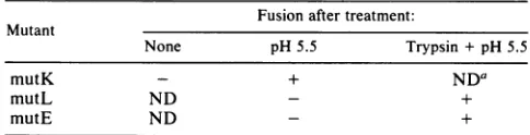
h), SBTI was present expression of the spike proteins was confirmed by double )proteins were immu- immunofluorescence staining, and areas of positive cells by electrophoresis on were examined for polykaryon formation (Fig. 5). Cell-cell fusion assays of the cleavage site mutants were repeatedat least three times.
s demonstrate that Thefusionphenotypes aresummarized inTable 1.Fusion site are absolutely correlated strictly with p62 cleavage. In mutK, where cor-2. rect cleavage of p62 took place (Fig. 2), polykaryon forma-ed with exogenous tion was observed after acid treatment (Fig. Sc and d). As ,us sequence at the expected, fusion was not seenprior to acid treatment (Fig. osition, resulting in 5a and b). In the p62 cleavage-deficient mutants mutL and residues constitut- mutE, low-pH treatment failed to activate the fusion func-mained in the p62 tion (Fig. 5e, f, i, and j), although the spike proteins were itions (Fig. 1). The expressed on the plasma membrane as shown by surface sin was examined. staining. However, when the microinjected monolayers
swerepulse-chased were treated with trypsin prior to pH 5.5 treatment, fusion 4), and the mono- activity was restored (Fig. 5g, h, k, and 1), as demonstrated in the presence of a by large areas of immunofluorescence corresponding to followed by inacti- polynucleated cells. This clearly shows that cleavage activa-orogram shows that tion of p62 is important for the acid-induced fusion activity ,ith the addition of of the SFV spike heterodimer.
chase interval (Fig.
came accessible to DISCUSSION
processed
to an E2 We have geneticallyengineered and expressed proteolyticcmobility to that of cleavage site mutantsof the SFV spike glycoprotein precur-should have at least sor p62 to address the function of p62 cleavage in virus :rminal end (an L in assembly and disassembly. One conservative (K) and two se periods, increas- nonconservative (E and L) substitutions were introduced at ^ypsin assay clearly the -1 position of the ubiquitous cleavage site consensus 62 is transported to sequence. We have used the vaccinia virus expression approximately 50% system to analyze the phenotypes of the p62 cleavage site
Mle to cleavage by mutants. Similar to a previous report on the expression of tresents an intracel- the Sindbis virus structural proteins via a recombinant
itrypsin processing vaccinia virus vector (43), we have confirmed that the size,
)nolayers (data not processing events, and intracellular transport of the SFV -antly with the pro- spike proteins were similar, if not identical, to those in wing that the cleav- virus-infected cells. We have confirmed the strict require-due to the presence ment for dibasic residues at the p62 cleavage site for proc-essing by the trypsinlike host enzyme. As noted by others 52 with exogenous (15, 38), an R->K substitution can somewhat reduce the nger chase interval efficiency of cleavage by the protease. Uncharged oracidic enous enzyme (Fig. residues at the cleavage site completelyabolishedprocessing
vat the endogenous of p62.
J.VIROL.
on November 10, 2019 by guest
http://jvi.asm.org/
[image:4.612.65.294.73.241.2]mutK
pH5.5 TRYPSIN
p-
pH5.
mutL
mutE
a t..
'-M..iti-- "
FIG. 5. Fusionphenotypesofp62 cleavagesitemutants. NeighboringBHKcellsweremicroinjectedwithplasmidDNAand incubatedat
370Cfor 16 h. Fusionwastriggeredbya60-sincubation withpH5.5medium withorwithoutpriortreatmentof themonolayerswithtrypsin
(0.5
R~gIml)
for 15 minonice (asdescribed in Materials and Methods). Afterafurther 2-h incubation at 37C,double immunofluorescencestainingwasperformed.Areas showingsurfaceexpressionof thespike glycoproteins were photographedbyfluorescence(toppanelin each pair) andphase-contrast (bottompanelin each pair) microscopy.
1237
on November 10, 2019 by guest
http://jvi.asm.org/
[image:5.612.135.465.33.670.2]1238 LOBIGS AND GAROFF
TABLE 1. Fusion phenotypes of p62 cleavage site mutants Fusion after treatment:
Mutant
None pH 5.5 Trypsin +pH5.5
mutK - + NDa
mutL ND - +
mutE ND - +
aND,Notdetermined.
Surface expression of the spike precursor p62 was
dem-onstrated with exogenous trypsin, which mimicked the
ac-tivity of the host enzyme to generate the mature spike protein E2. The cleavage probably occurred at one of the
two remaining basic residues at the mutated p62 cleavage
site. The resultant E2 was relatively resistant to further digestion with trypsin at an at-least-10-fold-higher enzyme
concentration (datanotshown). Thus, thecleavage region of p62appears tobe exposedduring and aftertransport tothe cellsurface, giving access totrypsin orthetrypsinlike host
enzyme. In many strains of ortho- and paramyxoviruses,
uncleaved fusion proteinprecursors are exposed atthe cell surfaces and can be activated with exogenous trypsin (18,
37). Spike precursor cleavage site mutants of retroviruses (34, 42) and ortho- and paramyxoviruses (23, 41) display a
similar sensitivity toexogenous trypsin ofsurface-exposed
uncleavedprecursors.Interestingly,arequirement for cleav-age in theactivation of the biological functions of the spike,
but not in particleformation, canbe noted.
Our resultssuggest thatp62 cleavage playsarole in virus
entry. The spikeprotein-induced and acid-triggered cell-cell membranefusion in transient expression studies wasclearly
dependent on p62 cleavage. No polykaryonformation was
observed afterlow-pHtreatment whenuncleaved p62 from mutLormutEwas expressed,but it could be induced when
the mutant p62 was cleaved with exogenous trypsin. The SFV spike is therefore composed of cleavage-activated fusion glycoproteinheterodimers.This conclusion contrasts thesuggestion of Brown and co-workers that p62cleavage is not essential for fusion (31).Their interpretationwas based
on cell-cell fusion experiments with the p62
cleavage-defi-cient Sindbis virus ts2Omutantandpretreatment of Sindbis virus-infected cells with trypsin at 3 h after infection (31), which apparently blocks spike precursor cleavage (1). In
fusion assays with the ts2O mutant at a nonpermissive
temperature, thefusion activitywasreduced relativetothat in wt virus-infected cells. Nevertheless, a residual fusion
activityremained, which could be correlated with the leak-iness of thismutant,permittingsomeprocessing of the spike
precursor. Whenfusion medium was added 7 hafter
infec-tionto thetrypsin-treated cells, efficient acid-induced poly-karyon formation was noted. However, the investigators
have not excluded the possibility that residual, normally processedglycoproteins aresynthesized priortothetrypsin treatment and that these can mediate fusion. Moderate
amounts of Sindbis virus glycoproteins can be seen at the cell surface asearlyas2 h afterinfection,which is sufficient
forthe induction of cell-cell membrane fusion (13).
In a recent report (45), an interesting mutant of Sindbis virus withachange attheamino-terminal amino acid of E2, which introduced a novel glycosylation site and,
impor-tantly, abolished cleavage of p62, was described. The
un-cleaved spike precursor was incorporated into mature
viri-ons with normal growth characteristics in tissue culture
cells. The fusion phenotype of this mutant has not been reported, but assuming that virus-cell membrane fusion is
neededforvirus entry, itwould conflict withourresults. An
explanationwhich may reconcile their data with ours could
be that the additional carbohydrate moiety in the Sindbis virus mutant, exactly at thecleavagesite, substitutesforthe
structural change normally caused by the cleavage, which would be important for fusion activation (as described be-low).
Ourpresentwork suggests that alphavirusesare partofa growingnumberof virus families which regulate their disas-sembly via cleavage activation of their oligomeric fusion proteins (33). Spike precursor cleavage activation at a late stage during the surface transport appears to be a
wide-spread mechanism to circumvent a fusion-inducing
confor-mational rearrangement of the spike fromoccurring in the acidic compartments of the exocytotic pathway (2, 6). How-ever,inSFV, thefusion function probably residesontheEl
protein (as described above) and the cleavage activation is mediated via p62 cleavage, thus involving differentpartners
of the spike heterodimer. We therefore predict that the
cleavage ofp62 exerts aconformational effect on El viaan oligomerization-controlled mechanism. According to our model, the matureEl-E2 spike proteinheterodimer becomes sensitive to acid-induced dissociation after cleavage ofp62,
which allows the putative fusion domain on El to become exposed. Support for this comes from
coimmunoprecipita-tion and cosedimentation analyses of the SFV spike het-erodimer in buffers of decreasing pHs (56). A marked resistance to dissociation of the El-p62 complex was ob-served, in contrast to the mature El-E2 complex, which dissociated in mildly acidicbuffers. The fact that the
disso-ciation was occurringat ahigher pH than that required for
optimal fusion suggests that subsequent to dissociation a second low-pH-dependent change is needed. This may be related toadditional changesinspike subunitconformation, resulting in the exposureoftheputativefusion domain of El (12, 24). The cleavage-activated, low-pH-triggered fusion mechanism of influenza virus also envisages acid-induced
conformational rearrangements in the tertiary and quater-nary structures of the hemagglutinin homotrimer (61). A
pH-dependent weakening of the trimeric structure as well as a rearrangement of the hemagglutinin monomers, exposing the well-characterized fusion domain, have been described previously (33, 61).
Severalfunctions in theassembly and disassemblyof SFV cannow be associated with the p62/E2 spikeprotein. (i)The interaction between the cytoplasmic tail of p62 and the nucleocapsid in virus budding has been inferred(16, 48,54).
(ii) p62 is responsible for the transport of the spike het-erodimer from theendoplasmicreticulum to the cell surface. Expressed alone, p62 is routed to the cell surface (27),
whereas Elexpressedfrom a single coding unit is retained in the endoplasmic reticulum (35). Expression of p62 and El from separate coding units in the same cell results in
heterodimerization and surface transport (M. Lobigsand H. Garoff, unpublished results). (iii) The cleavageofp62 to E2 may play a role in the lateralinteraction between the spike heterodimers at the cell surface and regulation of the bud-ding event (16, 21, 47, 55). (iv) The oligomerization of the spike heterodimer is controlled via p62 cleavage, which assures a stable E1-p62 dimer for transport via the acidic compartments of the exocytotic route and a much more acid-labile oligomer which can undergo fusion in the acidic
compartments of the endocytotic pathway (56; our data).
Thus, a picture emerges in whichp62 plays a crucial role in theassembly andactivation ofdisassembly of SFV,whereas J. VIROL.
on November 10, 2019 by guest
http://jvi.asm.org/
[image:6.576.38.280.80.142.2]the El protein carries the critical signals for infection and fusion.
ACKNOWLEDGMENTS
We thank H. Stunnenberg and J. Schmitt for teaching us the
techniques for vaccinia virus recombination, P. Liljestrom for helpfuladviceonsite-directedinvitromutagenesis,M.Ekstromfor excellent assistance with microinjection and cell culture, and I.
Sigurdsonfortyping.WeacknowledgeH. Stunnenbergfor
provid-ing plasmidsandvirus strainsand W. A. M. Boereforprovidingthe monoclonal antibodies.
The work was supported by the Swedish Medical Research Council(B88-12X-0872-01A),Swedish NationalBoard for Technical Development,and SwedishNational ScienceResearch Council.
LITERATURE CITED
1. Adams, R. H., and D. T. Brown. 1982. Inhibition ofSindbis virus maturation aftertreatmentof infectedcells withtrypsin.J. Virol. 41:692-702.
2. Anderson, R. G. W., and L. Orci. 1988. A view of acidic intracellularcompartments. J. Cell. Biol. 106:539-543. 3. Boere, W. A. M.,T. Harmsen,J. Vinje, B. J.Benaissa-Trouw,
C. A.Kraaijeveld,and H.Snippe.1984.Identification of distinct antigenicdeterminantson SemlikiForest virusby using
mono-clonal antibodies with different antiviral activities. J. Virol. 52:575-582.
4. Boggs, W. M., C. S.Hahn, E. G. Strauss, J. H. Strauss, and D. E. Griffin. 1989. LowpH-dependent Sindbis virus-induced fusion of BHK cells: differences between strains correlate with amino acid changes in the El glycoprotein. Virology 169: 485-488.
5. Bonatti, S.,G.Migliaccio,and K.Simons. 1989.Palmitylationof viral membrane glycoproteins takes place after exit from the
endoplasmicreticulum.J. Biol. Chem.264:12590-12595. 6. Boulay, F., R. W.Doms,I. Wilson,and A. Helenius. 1987. The
influenzahemagglutininprecursoras an acid-sensitiveprobeof thebiosynthetic pathway. EMBOJ. 6:2643-2650.
7. Bracha, M., and M. J. Schlesinger. 1976. Defects in RNA+
temperature-sensitivemutantsof Sindbisvirus andevidencefor
acomplexofpE2-E1viral glycoproteins. Virology74:441-449. 8. Dalgarno,L., C. M. Rice, andJ. H. Strauss. 1983. Rossriver virus 26 S RNA: complete nucleotide sequence and deduced sequence of the encoded structural proteins. Virology 129: 170-187.
9. deCurtis, I.,and K.Simons. 1988.Dissectionof SemlikiForest virusglycoproteindeliveryfrom thetrans-Golginetworktothe cellsurface inpermeabilizedBHKcells. Proc. Natl. Acad. Sci. USA85:8052-8056.
10. Drillien, R.,and D.Spehner.1983.Physical mappingof vaccinia virustemperature sensitive mutations.Virology 131:385-393. 11. Dubois-Dalcq, M.,K. V.Holmes,and B.Rentier. 1984.
Assem-blyofenvelopedRNAviruses. Springer-Verlag,New York. 12. Edwards, J.,E. Mann,and D. T. Brown. 1983. Conformational
changesinSindbis virusenvelopeproteins accompanying
expo-suretolowpH.J. Virol. 45:1090-1097.
13. Erwin, C., and D. T. Brown. 1980. Intracellulardistributionof Sindbis virusmembraneproteinsin BHK-21 cellsinfectedwith
wild-type virus and maturation-defective mutants. J. Virol. 36:775-786.
14. Fischer,J.M.,and R. H.Scheller. 1988. Prohormoneprocessing and thesecretorypathway. J. Biol.Chem.32:16515-16518. 15. Freed, E. O., and R. Risser. 1987. The role of envelope
glycoproteinprocessingin murine leukemia virus infection. J. Virol. 61:2852-2856.
16. Fuller, S. D. 1987. The T=4 envelope of Sindbis virus is
organized by interactions with a complementary T=3 capsid. Cell48:923-934.
17. Garoff,H.,C.Kondor-Koch,and H.Riedel. 1982.Structure and
assembly of alphaviruses. Curr. Top. Microbiol. Immunol. 99:1-50.
18. Garten, W.,F. X. Bosch, D.Linder,R. Rott,andH.-D. Klenk. 1981. Proteolytic activation of the influenza virus
hemaggluti-nin: the structure ofthe cleavage site andthe enzymesinvolved incleavage. Virology 115:361-374.
19. Green, J.,G. Griffiths, D. Louvard, P. Quinn, and G. Warren. 1981. Passage of viral membrane proteins through the Golgi complex. J. Mol. Biol. 152:663-698.
20. Hanggi, M., W. Bannwarth, and H. G. Stunnenberg. 1986. Conserved TAAAT motif in vaccinia virus late promoters: overlapping TATA box and site of transcription initiation. EMBO J. 5:1071-1076.
21. Harrison, S. C. 1986. Alphavirus structure, p. 21-34. In S. Schlesinger and M. J. Schlesinger(ed.),The Togaviridae and Flaviviridae. Plenum Publishing Corp., New York.
22. Kalkkinen,N., H. Jornvall, H. Soderlund, and L. Kaariainen. 1980. Analysis of Semliki-Forest-virus structural proteins to illustrate polyprotein processingof alpha viruses. Eur. J. Bio-chem. 108:31-37.
23. Kawaoka, Y., and R.G. Webster. 1988.Sequencerequirements for cleavage activation of influenza virus hemagglutinin ex-pressed in mammalian cells. Proc. NatI. Acad. Sci. USA 85:324-328.
24. Kielian, M., and A. Helenius. 1985. pH-induced alterations in thefusogenic spike protein of Semliki Forest virus. J. Cell Biol. 101:2284-2291.
25. Kielian, M., and A. Helenius. 1986. Entry of alphaviruses, p. 91-120. In S. Schlesinger and M. J. Schlesinger (ed.), The Togaviridae and Flaviviridae. Plenum Publishing Corp., New York.
26. Kieny, M. P., R. Lathe, R. Drillien, D. Spehner, S. Skory, D. Schmitt, T. Wiktor, H. Koprowski, and J. P. Lecocq. 1984. Expression of rabies virus glycoprotein from a recombinant vacciniavirus. Nature (London) 312:163-166.
27. Kondor-Koch, C., B. Burke, and H. Garoff. 1983. Expression of Semliki Forest virus proteins from cloned complementary DNA. I. The fusion activity of the spike glycoprotein. J. Cell Biol. 97:644-651.
28. Kondor-Koch, C., H. Riedel, K. Soderberg, and H. Garoff. 1982. Expression of the structural proteins of Semliki Forest virus from cloned cDNA microinjected into the nucleus of baby hamsterkidney cells. Proc. Natl. Acad. Sci. USA 79:4525-4529. 29. Kramer, W., V. Drutsa, H. W. Jansen, B. Kramer, M. Pflugfelder, and H.-J. Fritz. 1984. The gapped duplex DNA approach to oligonucleotide-directed mutation construction. NucleicAcids Res. 12:9441-9456.
30. Mackett, M., G.L.Smith, and B. Moss. 1984. The construction and characterization of vaccinia virus recombinants expressing foreigngenes, p.191-212. In D. M. Gover(ed.), DNA cloning, vol. 2. IRL Press,Oxford.
31. Mann, E., J. Edwards, and D. T. Brown. 1983. Polycaryocyte formation mediated by Sindbis virus glycoproteins. J. Virol. 45:1083-1089.
32. Marsh, M. 1984. The entry ofenveloped viruses into cells by endocytosis.Biochem. J. 218:1-10.
33. Marsh, M., and A. Helenius. 1989. Virus entryintoanimal cells. Adv. Virus Res. 36:107-151.
34. McCune,J. M., andL. B. Rabin, M. B. Feinberg, M. Lieber-man, J. C. Kosek, G. R. Reyes, and I. L. Weissman. 1988. Endoproteolytic cleavageofgpl60 isrequired forthe activation of humanimmunodeficiencyvirus. Cell 53:55-67.
35. Melancon, P., and H. Garoff. 1986. Reinitiationoftranslocation in theSemlikiForestvirus structuralpolyprotein:identification of thesignalfor the El glycoprotein.EMBO J. 5:1551-1560. 36. Melancon, P., and H. Garoff. 1987. Processingofthe Semliki
Forestvirus structuralpolyprotein:roleof thecapsidprotease. J.Virology61:1301-1309.
37. Morrison, T. G. 1988. Structure, function, and intracellular processing ofparamyxovirus membrane proteins. Virus Res. 10:113-136.
38. Ohuchi,M.,M.Orlich,R.Ohuchi,B. E.J.Simpson,W.Garten, H.-E.Klenk,and R. Rott.1989.Mutationsatthecleavagesite of the hemagglutinin alter the pathogenicity of influenza virus A/Chick/Penn/83(HSN2). Virology168:274-280.
39. Omar, A., andH. Koblet. 1988. Semliki Forest virusparticles containing onlytheElenvelope glycoproteinareinfectious and
on November 10, 2019 by guest
http://jvi.asm.org/
1240 LOBIGS AND GAROFF
caninduce cell-cell fusion.Virology 166:17-23.
40. Omar, A.,and H. Koblet. 1989. The use of sulfite to study the mechanism of membrane fusioninduced byE1of Semliki Forest virus. Virology 168:177-179.
41. Paterson, R. G., M. A. Shaughnessy, and R. A. Lamb. 1989. Analysis of the relationshipbetween cleavability of a paramyx-ovirus fusion protein and length of the connecting peptide. J. Virol. 63:1293-1301.
42. Perez, L.G., and E. Hunter. 1987. Mutations within the proteo-lytic cleavage site of the Rous sarcoma virus glycoprotein that blockprocessing to gp85 and gp37. J. Virol. 61:1609-1614. 43. Rice, C.M., C. A. Franke, J. H. Strauss, and D. E. Hruby. 1985.
Expressionof Sindbis virus structural proteins via recombinant vaccinia virus: synthesis, processing, and incorporation into matureSindbisvirions. J. Virol. 56:227-239.
44. Rice, C. M., and J. H. Strauss. 1982. Association of Sindbis virion glycoproteins and their precursors. J. Mol. Biol. 154: 325-348.
45. Russell, D. L., J. M. Dalrymple, and R. E. Johnston. 1989. Sindbisvirus mutations whichcoordinately affect glycoprotein processing, penetration, and virulence in mice. J. Virol. 63: 1619-1629.
46. Scheefers, H., U. Scheefers-Borchel, J. Edwards, and D. T. Brown. 1980. Distribution of virus structural proteins and pro-tein-protein interactions inplasma membrane of baby hamster kidneycellsinfected with Sindbis or vesicularstomatitis virus. Proc. Nati.Acad. Sci. USA77:7277-7281.
47. Schlesinger,M.J., and S.Schlesinger (ed.). 1986. The Togavir-idae and Flaviviridae, p. 121-148. Plenum Publishing Corp., New York.
48. Simons, K., and H. Garoff. 1980. The budding mechanisms of envelopedanimalviruses. J.Gen. Virol. 50:1-21.
49. Steiner, D. F., K. Docherty, and R. Carroll. 1984.Golgi/granule processingofpeptide hormone and neuropeptide precursors: a minireview.J. Cell.Biochem. 24:121-130.
50. Strauss,J.H., E. G. Strauss, C. S.Hahn, Y.S.Hahn, R.Galler, W.R.Hardy, and C. M. Rice. 1987.Replicationof alphaviruses andflaviviruses:proteolyticprocessing of polyproteins,p.
209-225.In M. A.Brinton and R. R. Rueckert(ed.),Positive strand RNAviruses. Alan R.Liss, Inc., NewYork.
51. Sturman, L.S., and K. V. Holmes. 1984. Proteolytic cleavage of peplomeric glycoprotein E2 of MHV yields two90K subunits and activates cell fusion. Adv. Exp. Med. Biol. 171:25-35. 52. Sturman, L. S., C. S. Ricard, and K. V. Holmes. 1985.
Proteo-lytic cleavage of the E2glycoprotein of murine coronavirus: activation of cell-fusing activity of virions by trypsin and separation of twodifferent 90K cleavage fragments. J. Virol. 56:904-911.
53. Timm, B., C. Kondor-Koch, H. Lehrach, H. Riedel, J.-E. Edstrom, and H. Garoff. 1983. Expression of viral membrane proteins from cloned cDNA by mircoinjection into eukaryotic cell nuclei. MethodsEnzymol. 96:496-511.
54. Vaux, D. J. T., A. Helenius, and I. Mellman. 1988. Spike-nucleocapsid interaction in Semliki Forest virus reconstructed using network antibodies. Nature(London) 336:36-42. 55. Vogel, R. H., S. W. Provencher, C.-H. von Bonsdorff, M.
Adrian, and J. Dubochet. 1986. Envelope structure of Semliki Forest virus reconstructed from cryo-electron micrographs. Nature(London) 320:533-535.
56. Wahlberg, J., W. A. M. Boere, and H. Garoff. 1989. The heterodimeric association between the membrane proteins of Semliki Forest viruschanges its sensitivity to low pH during virus maturation. J. Virol. 63:4991-4997.
57. White, J., and A. Helenius. 1980.pH-dependent fusion between the Semliki Forestvirusmembrane andliposomes.Proc. Natl. Acad. Sci. USA 77:3273-3277.
58. White, J., J. Kartenbeck, and A. Helenius. 1980. Fusion of Semliki Forest virus with theplasmamembrane can be induced by low pH. J. Cell Biol. 87:264-272.
59. White, J.,M.Kielian,andA. Helenius.1983. Membrane fusion proteins ofenvelopedvirus. Q.Rev.Biophys. 16:151-195. 60. White, J., K. Matlin, and A. Helenius. 1981. Cell fusion by
Semliki Forest, influenza, and vesicular stomatitis viruses. J. Cell Biol. 89:674-679.
61. Wiley, D.C., and J. J. Skehel. 1987. The structure and function of thehemagglutininmembraneglycoprotein of influenza virus. Annu. Rev. Biochem. 56:365-394.
J. VIROL.

![FIG. CleavagehBHK 4. of mutL and mutE wit] cells were infected with recombinant%vaccinia](https://thumb-us.123doks.com/thumbv2/123dok_us/1322041.85915/4.612.65.294.73.241/fig-cleavagehbhk-mutl-mute-cells-infected-recombinant-vaccinia.webp)