0022-538X/88/093281-07$02.00/0
Copyright © 1988, AmericanSocietyforMicrobiology
Expression of
Herpes
Simplex Virus
Type 1
(HSV-1)
Latency-Associated Transcripts and
Transcripts
Affected
by
the
Deletion in
Avirulent
Mutant HFEM:
Evidence for
a
New
Class of HSV-1 Genes
JORDAN G. SPIVACK* AND NIGEL W. FRASER
The Wistar Institute, 36th Street at Spruce,Philadelphia, Pennsylvania 19104
Received 22 March 1988/Accepted 2 June 1988
During latent herpes simplex virus type 1 (HSV-1) infection in the trigeminal ganglia of mice, three virus-specific transcripts, 2.0, 1.5, and 1.45 kilobases (kb), are detectable byNorthern(RNA) blotanalysis,but only the 2.0-kbtranscript can be detected in HSV-1-infected tissueculture cells (J. G. Spivack and N. W. Fraser, J. Virol. 61:3842-3847, 1987). Since these latency-associated genes map to a diploid region ofthe genome,transcription from the deletionmutantHFEM, which contains onlyonecomplete copyof thesegenes, wasinvestigated todetermine theeffect of gene dosage. The4.1-kb HFEM deletion is locatedbetweenthea genesICPOandICP27. ICPO mRNA and the 2.0-kb latency-associated transcriptwerepresentatnormal levels during HFEMinfection, but ICP27mRNA and0.9- and 1.1-kbtranscripts that mapnearthedeletionwere not readily detectable. The levels of expression of one or more of these genes might beanimportant determinant of HSV-1 virulence inanimal hosts. ICP27 mRNA accumulated whenprotein synthesis wasinhibited before HFEM infection, implying that the deletion may affect ICP27 regulatory rather than coding elements. Expression of the 2.0-kblatency-associated transcript was characterized in infected CV-1 cells with metabolic inhibitors andstrand-specific probes. On the basis of metabolic inhibitor studies, the gene encoding the 2.0-kb latency-associated transcript is not anagene. During HSV-1 replicationininfected tissue culture cells, the ,1 and y genes require the prior expression of a gene products. However, the latency-associated RNAs are expressed in the absence of detectable levels ofa transcripts in latently infected mice. Thus, this latency-associated genefamilyappeartoberegulated quitedifferentlythana,I,or ygenes. For these reasons, and because thelatency-associated genes may perform latent rather than replicativefunctions,weproposethat they should beconsideredmembersofa newHSV-1 gene class, theA genes.
After a primary infection, herpes simplex virus type 1
(HSV-1) can remain in a latent state for the life of the
individual (2, 16, 46). Virus-specific transcripts have been detectedby in situhybridization during HSV-1latency in the central(10, 12,48) andperipheral (9, 47)nervoussystemsof mice and rabbits (33), in human trigeminal ganglia (8, 45), andduring HSV-2latency inguinea pig(49) and human (13, 14) sensory ganglia. The HSV-1 transcripts present in the sensoryganglia of latently infected animals originate from the repeat regions (9, 10, 12, 32, 40, 41, 47), which are present intwo copies per genome. Three viral transcripts,
2.0, 1.5, and 1.45 kilobases (kb), which are presentduring HSV-1 latency in mice map to a 3.0-kb region contained withinBamHI-B and -E and the long repeat region (Fig. 1)
(41). These RNAspartially overlap the 3' terminus ofICPO (41) andaretranscribedin theopposite direction(33, 41, 47).
Only the 2.0-kb transcript has been detected, atlow levels, in HSV-1-infected cells (41).
Bydefinition, the a genes,
ICPO,
-4, -22,-27, and -47, aretranscribed in HSV-1-infectedcellsinthe absenceofprotein
synthesis (17, 22, 34, 51); e and some
-Yl
genes require protein synthesis but not viral DNAsynthesis, and _Y2 genetranscription is strictly dependent upon both protein and
viral DNA synthesis (17, 34). During HSV-1 replication in
tissue culture, ,Band ry genesrequire the prior expression of a genes (17, 34). The latency-associated transcripts are
expressedinthe absenceofdetectable levels of a or 1 RNAs
*Correspondingauthor.
during latentinfectioninmice (8, 35, 41, 47) and in humans (45). In this study, the 2.0-kb latency-associated transcript
was characterized in infected CV-1 cells with metabolic
inhibitorsand strand-specific probes. The dataindicate that the latency-associated RNAs are not transcribed from a genes. Since they are expressed with characteristics
dif-ferentfromthoseencodedbya,
13,
or -ygenes, wepropose that theybe termed A(lambda)genes.The deletion mutant HFEM, which encodes only one completecopyofthelatency-associated genes, wasstudied toexaminetheeffects ofgenedosage. HSV-1strainHFEM, whichwasderived fromHF (52),has a4.1-kb deletion within BamHI-B and the long internal repeat (of the prototype
configuration of the genome) that is between the a genes
ICPO and ICP27 (Fig. 1) (36). The termini of the deletion
have been precisely located by sequence analysis (19). HFEMreplicatesat37°Cintissueculture (26)and isvirulent
in mice inoculated intracerebrally but avirulent in mice inoculated intraperitoneally, subcutaneously, or
intrave-nously (1). HFEM can establish a latent infection in mice
after peripheral inoculation (26, 41). Since replacement of the deleted region by BamHI-B or a 3.8-kb HpaI-HpaI subfragment ofB from strain F leads to virulent recombi-nants (1, 37) the gene(s) affected by the HFEM deletion is
likely to be important in the pathogenesis of HSV-1 infec-tions in experimental animal models.
We have shown that the viraltranscriptspresent inmice
latently infected with strain F are also present with strain
HFEM (41). One copy of the latency-associated genes is
3281
on November 10, 2019 by guest
http://jvi.asm.org/
A
HSV-1
GENOME
B
BAM HI
FRAGMENTS
C
BAM HI B
D
BAM HI E
TRLLIRL IR8 U TR
TL UL L S US S
L I 3
E B SP
I
I~~~~~~~~~~
""w...~~~~~~~-
11I 1111111 1111 I Hill .1TTn~~~~~~~~~~~~~~~~~~~~~~~~~~~~~~~~~~~~~~~~~~~~~~~~~~~~~~~~~~~~~~~~~~~~~~_
t 1, IIC~P27 I 4- 3 Latent
I
_ lb
-ICP27 - 0
20k
*.t. unique
2.4kb
:x4
:a:
I*e
:
.e
T1
2kb
'U
RNAB
HFEM deletion ICPO
0. 33.3kb *3.Okb
X
l
*a
.r
l
...
i
to
Iunique
GAICPO
3 Latent RNAs
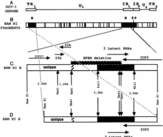
FIG. 1. Mapof HSV-1 genome and transcripts within BamHI-B. (A) The HSV-1 genome, illustrating the unique long and short (UL and Us)regions of the genome
(-)
bounded by theinternal(IR) andterminalrepeat(TR) regions (O). (B) The positions of theBamHIrestriction sites of HSV-1 straini F (29). The fragments that are positive by in situ hybridization during latency are labeled and shaded (9, 10). (C) Detailed mapofBamHIB. Thesizes of the HpaIfragments (McGeoch et al.,unpublished
data) andpositions of other restrictionsites used in this study areshown (1, 22,28, 38). The approximate locations ofICPO,ICP27 (6, 22,28), 0.9-kb (20k), and 1.1-kb (22k) (23, 41) mRNAs aremarked by arrows that indicate the direction of transcription. The shaded areabetween the PstI and MluI restriction sites hybridized to three overlapping HSV-1 latency-associated transcripts, which arerepresented by a single arrow, byNorthern blotanalysis (41). The hatched region is the boundary between the unique long region and longinternalrepeat(19).The 4.1-kbdeletion in strain HFEM is marked by a shaded boxaboveBamHI-B(36). (D) Detailed map of BamHI-E.BamHI-Eis shown inreverseorientationtoemphasize that theregion between the boundaryof thelong repeat and theBamHIsite on theright is also contained within BamHI-B.deleted inHFEM,while thesecond copyispresentintactin BamHI-E(Fig. 1).Inthisreport,evidence ispresented that
ICP27 mRNA and 0.9- and 1.1-kb RNAs that map to
BamHI-B (41) are not present athigh levels in CV-1 cells
infected with HFEM. The 1.1- and 0.9-kb transcripts
par-tially overlap each other and are transcribed in opposite directions.The 0.9-and
1.1-kb
transcriptsaresynthesizedinHSV-1(F) infected cells when viral
DNA
synthesis is inhib-ited butnotwhenproteinsynthesis is blocked,implyingthatthey are, or
-Yl
genes.Theimplications ofthese resultsforHSV-1replication in tissue culture
cells
and the pathogene-sis of infections in experimental animal models aredis-cussed.
MATERIALSANDMETHODS
Cell cultureandHSV-1 growth and titration. Subconfluent monolayersof CV-1 cells grown in Eagle minimum essential mediumwith5% fetalcalfserum at37°C with5%CO2were
iifectedwith HSV-1(strain Ffrom B.Roizman, University
ofChicago;HFEMfrom G. Cohen, University of Pennsyl-vania) at 1 PFU per cell, as previously described (9,
42).HSV-1
titers were determined on CV-1 cells, and theplaqueswerestainedwith 1%methyleneblue and counted 2
dayslater(43, 44).
RNAextraction. Toisolate HSV-1 RNA, CV-1 cellswere infectedat5PFU percell. The cells were trypsinizedat5to
6 hpostinfection,and afterlow-speedcentrifugation,the cell pellets were homogenized (Polytron, setting 5, 20 s) in a solution containing 4 M guanidinium thiocyanate, 0.5%
sodium-N-lauroylsarcosine, 100 mM
P-mercaptoethanol,
25 mM sodium citrate (pH 7.0), and 0.1% antifoam A (Sigma ChemicalCo.) (4).The RNAwaspelletedthroughacushion of5.7 M CsCl-0.1 M EDTA (pH 7.0) by centrifugation at 150,000xginaBeckmanSW40.1rotorfor20to24 hat18°C (4, 40, 41). Aftercentrifugationthe RNAwas suspendedinH20 andstoredas anethanol
precipitate
at-20°C.
A260
was measured.Agarose gelelectrophoresis, Northern (RNA) blot transfer ofRNA, and hybridization and washing of Northern blots. RNAwas denaturedwithglyoxal, electrophoresed through 1.2%agarose,and
capillary
blottedtoGene Screen Plus(DuPont Co.), aspreviously described (40, 41). RNA markers werepurchasedfrom Bethesda Research
Laboratories,
Inc. Afterblottingandairdrying,theglyoxylationwasreversed, aspreviouslydescribed(40, 41).The filterswereprehybrid-ized, and a 32P-labeled nick-translated or
single-strand-labeled probe was added to hybridize
overnight (40, 41).
After hybridization, the filters were washed in decreasing
concentrations of SSCto0.1x (1x SSC is 0.15MNaCl
plus
0.015 M sodiumcitrate)with 1% sodiumdodecyl sulfateat 65°Cfor two 30-min washes. The filters were covered with
plastic wrap and autoradiographed with XAR-5 film
Nk-l
on November 10, 2019 by guest
http://jvi.asm.org/
[image:2.612.141.470.71.348.2](Eastman Kodak Co.) and an intensifying screen (Lightning Plus; Du Pont) at
-70°C.
Preparation of
32P-labeled
probes. The cloned BamHI-B fragment of HSV-1(F) was obtained from B. Roizman (29). Subfragments of BamHI-B were prepared by gel electropho-resis of restriction digests and electroelution into dialysis tubing (24) or onto DEAE paper (NA-45; Schleicher & Schuell, Inc.), as recommended by the manufacturer. DNA restriction enzymes were purchased from Boehringer Mann-heim or Bethesda Research Laboratories and used as rec-ommended. DNA probes were nick translated by standard procedures (24). Single-strand-labeled probes were synthe-sized from HSV-1 DNA cloned into M13 mpl8 and mpl9 vectors, by the method of Hu and Messing (18). Theprobes were separated from unincorporated nucleotides bypassage through Sephadex G-50 mini-spin columns (Boehringer) and ethanol precipitation. Specific activities of the nick-trans-lated probes were 1 x 10⁸to 5 x 10⁸cpm/,Lg
of DNA.Inhibitors. Stocks of phosphonoacetic acid (PAA) and cycloheximide (Sigma), 10 and 1 mg/ml, respectively, were made in phosphate-buffered saline, adjusted to pH 7.0,filter sterilized, and stored in 1-mlportions at -20°C. PAA (400
,ug/ml)
and cycloheximide (50 ,ug/ml) were added to cell cultures 30 to 60 min before infection and were presentcontinuously untilRNAextraction.
RESULTS
Determination of the kinetic class of the 2.0-kb latency-associated transcript. Of the three latency-associated tran-scripts present in latently infected mice (40, 41), only the 2.0-kb RNA is detectable during the acute stage ofinfection in mice (40, 41) and in infected tissue culture cells(41). This transcript was characterized in infected CV-1 cells in the presence and absence of PAA, which blocks viral DNA synthesis (25), and cycloheximide, which inhibits protein synthesis (Fig. 2). The 2.0-kb latency-associated transcript
was detected in HSV-1-infected cells with a
single-strand-labeled HpaI-MluI probe (Fig. 2A, lane 2). Inthepresenceof PAA (Fig. 2A, lane 3) or cycloheximide (lane 4), this RNA could not be detected. Because of the long exposure time, there was some cross-hybridization with the 2.7-kb ICPO mRNA (6, 28, 51), especially in the cycloheximide-treated
sample (lane 4). For comparison, the HpaI-MluI single-strand probe labeled in the opposite orientation hybridized
to ICPOmRNA in HSV-1-infectedcellsintheabsence(Fig. 2B, lane 2) orpresence of PAA (lane 3) or cycloheximide
(lane 4). These resultsdemonstrate that the 2.0-kb
latency-associated transcript isnotsynthesized ininfectedcells with characteristics ofan aor
PI
gene, incontradistinction tothe a geneICPO(17, 34), which partially overlapsits 3'endand is transcribed in the opposite direction(41).Detection of HSV-1 transcripts that map to BamHI-B in strains F and HFEM. Onegoalof this study was toanalyze
viral transcription in the region surrounding the HFEM deletion, which is locatedcompletelywithinBamHI-B(Fig. 1). To localize the HSV-1transcripts,BamHI-B was cutwith
HpaI to yield four major subfragments (Fig. 1). The 2.4-kb BamHI-HpaI probe hybridized toICP27 mRNA(6, 22, 51) and a 0.9-kb transcript inCV-1 cells infected with strain F but did not hybridize to either transcript in cells infected with strainHFEM(Fig. 3A), except in very longexposures
(data not shown).The1.2-kbHpaI-HpaIprobehybridizedto the 0.9- and1.1-kbtranscripts incellsinfectedwith F but not in cells infected with HFEM(Fig. 3B). In very long expo-sures, the 0.9-kb RNA, but not the 1.1-kb RNA, was just
A 1 2 3 4 B 1 2 3 4
9.5-
7.5-4.4
'2.4
-2.0 LAT - *
1.4-9
4t0
0.3-
-FIG. 2. Synthesis ofthe 2.0-kblatency-associated
transcript
in thepresenceand absenceofPAAandcycloheximide.Lanes:1,5,ug ofRNAfrom uninfected CV-1cells; 2,5 ,ug ofRNAfrom HSV-1-infected CV-1 cells(multiplicityofinfection, 5),5to6hpostinfec-tion; 3,5,gofRNAfrom HSV-1-infected CV-1 cells with400
.g
of PAAperml;4,5 ,ug ofRNAfrom HSV-1-infected CV-1 cells with 50,ugofcycloheximide per ml. Thedrugswerepresentcontinuously
from 1 h before infection until the RNA was isolatedat5 to 6 h postinfection. (A) 1.2-kb HpaI-MluI single-strand-labeled
probe
(mpl8), 6-day exposure; (B) single-strand-labeled
probe
(mpl9),
16-h exposure. Thepositionsof the 2.0-kb
latency-associated
tran-script (2.0LAT) andICPO(0)mRNAareindicatedbyarrows.The positions of RNA markers(kilobases)arelabeledontheleft,andthe positions of 28S and 18SrRNAsare indicatedbyasterisks.
A B c
1 2 3 2' I 1 2 3 1 2 3
9.5
-75
-4...
a
4
---.-t+
24
27
1.4-
0.3-FIG. 3. Detection of HSV-1 RNAs that map to the HpaI
frag-ments of BamHI-B in strains F and HFEM
by
Northern blotanalysis.Lanes:1,2.5 jig ofRNAfromuninfectedCV-1cells;2,2.5 ,ug ofRNAfrom HSV-1(F)-infected CV-1cells; 3, 2.5
ptg
ofRNA fromHSV-1(HFEM)-infected CV-1 cells. (A) 2.4-kbBamHI-HpaI
probe; (B) 1.2-kb HpaI-HpaI probe; (C) 3.3-kb
HpaI-HpaI
probe;
(D) 3.0-kb HpaI-BamHI probe (see
Fig.
1 for locations). The positions of ICPO andICP27mRNAsareindicatedby
arrows. ThepositionsofRNAmarkers(kilobases)arelabeledontheleft,and the positions of28S and 18S rRNAsareindicated
by
asterisks.1-ko 1.0
o0
on November 10, 2019 by guest
http://jvi.asm.org/
[image:3.612.355.537.72.258.2] [image:3.612.340.547.431.631.2]A B C D
1 1 2 1 2 1 2
-...
.5.
..-I*
"-4*tp _ '.1'..0
.X)"I
* ---1 1Dn
>9-In
0
FIG. 4. Synthesis of ICP27 and the 0.9- and1.1-kbtranscriptsin thepresenceand absenceofPAAandcycloheximide. Lanes: 1,2.5 ,ug of RNA from uninfected CV-1 cells; 2, 2.5 ,ug ofRNA from HSV-1(F)-infected CV-1 cells; 3, 2.5 ±g of RNA from HSV-1-infected CV-1cellswith400,ug ofPAAperml; 4, 2.5,g ofRNA
fromHSV-1-infected CV-1 cells with50,ug ofcycloheximideperml. (A) strainF, 2.4-kbBamHI-HpaI probe;(B)strainHFEM, 2.4-kb BamHI-HpaI probe; (C) strain F, 1.2-kb HpaI-HpaI probe. The positionsofICP27 andthe0.9-kb (900)and 1.1-kb(1100)RNAsare
indicatedbyarrows.Thepositions of RNAmarkers(kilobases)are
labeled onthe left, and the positions of 28S and 18S rRNAs are
indicatedby asterisks.
barely detectable (datanot shown). The 3.3-kb HpaI-HpaI probe hybridized faintly to a 2.0-kb transcript in cells in-fectedwith either ForHFEM(Fig. 3C).This isoneof three latency-associated transcripts thatarepresent athigh levels during latent infection (41) and low levels during theacute stageof infection in the trigeminal ganglia of mice (40, 41). The 3.0-kb HpaI-BamHI probe hybridized to ICPO mRNA (6, 22, 28, 31, 51) and the 2.0-kb latency-associated tran-script(Fig. 3D). Thus, in HFEM-infected CV-1 cells, ICPO mRNAand the 2.0-kb latency-associated RNAare
synthe-sized,butthe levelsofICP27 mRNA and the 0.9-and 1.1-kb RNAsaresignificantly reducedincomparisonwith levels in HSV-1(F)-infected cells.
Transcription ofICP27 in HFEM under a conditions. It
was surprising that ICP27 mRNA was difficult to detect during HFEM infection, since the deletion in HFEM is approximately 1.5kbdownstream from the5'end ofICP27 mRNA (54). We hypothesized that the region deleted in HFEM might contain ICP27 regulatory elements or that there are additional mutations within ICP27. To determine whether the a gene ICP27 could be transcribed, cells were
treated with cycloheximide before HFEM infection.
Be-causeofthe absenceof feedbackinhibition from HSV-1gene
products (17), the aRNAsaccumulate tohigh levels when
protein synthesis is blocked in HSV-1-infected cells (34). ICP27wastranscribed in cells infected with strain F in the
absence (Fig. 4A, lane 2) and presence (lane 3) ofPAA.
ICP27 mRNA accumulated when protein synthesis was
blocked by cycloheximide (Fig. 4A,lane 4). Incontrast, in cellsinfected withstrainHFEM, ICP27mRNAwasvirtually undetectable (Fig. 4B, lane 2).ICP27mRNAwaspresent at lowlevelswhen viral DNAsynthesiswasinhibited (Fig. 4B,
lane 3) and at higher levels when protein synthesis was
inhibited (lane 4). These results indicate that (i) ICP27
FIG. 5. Direction oftranscriptionofthe 0.9- and 1.1-kb HSV-1 RNAs. Lanes:1,2.5 p.gof RNA fromuninfectedCV-1cells; 2,2.5 ,ug of RNA from HSV-1(F)-infected CV-1 cells (multiplicity of infection, 5), 5to6hpostinfection. (A) 2.4-kb BamHI-HpaI probe, mpl9 single-strand labeled (15); (B) 2.4-kb BamHI-HpaI probe (mpl8), single-strand labeled (15); (C) 1.2-kb HpaI-HpaI probe (mpl9), single-strand labeled (15); (D) 1.2-kb HpaI-HpaI probe (mpl8), single-strand labeled(15).Thepositionsof the0.9-kb(900) and 1.1-kb(1100)RNAsareindicatedbyarrows.The positionsof RNA markers (kilobases) arelabeledontheleft,and the 28S and 18SrRNAsareindicatedbyasterisks.
mRNAcan be transcribedin HFEMinfected cells, (ii) low
levels ofICP27 mRNAmaybesufficient for HFEM replica-tion (38), and (iii) themutation(s) inHFEM mayaffect the
regulationofICP27 expression.
Determination of the kinetic class of the 0.9- and 1.1-kb transcripts. The0.9- and 1.1-kb transcripts thatmapwithin BamHI-B instrain F(41)weresynthesizedatreducedlevels, ifat all, in cells infected with HFEM (Fig. 3). PAA and cycloheximidewereusedtodeterminewhether their
expres-sionmightbeaprerequisitefor thesynthesisofICP27RNA. The 0.9- and 1.1-kb transcripts were synthesized when HSV-1(F)DNAsynthesis wasblockedwith PAAbutwere
not produced when protein synthesis was inhibited with
cycloheximide (Fig. 4C). These results imply that the 0.9-and 1.1-kb RNAsareencodedbyPorylgenes.SinceICP27 is synthesized in cells infected with HFEM treated with cycloheximide and the 0.9- and 1.1-kb transcripts are not, they are notrequiredfor ICP27 expression. However, it is possible that they playa role in maintaining ICP27mRNA levels.
Directionoftranscriptionof the0.9-and 1.1-kbRNAs. To establish the direction oftranscriptionof the 0.9- and 1.1-kb RNAs,the2.4-kbBamHI-HpaIand 1.2-kbHpaI-HpaI sub-fragmentsof BamHI-B(Fig. 1)werecloned into M13 in both
orientationsto synthesize single-strand-labeled probes (18). The 2.4-kb BamHI-HpaI mpl9probe hybridized to ICP27 RNAand the 0.9-kb transcript in HSV-1(F)-infected cells, while intheopposite orientation(mpl8)it didnothybridize stronglytoeither RNA(Fig.5A andB).SinceICP27is read rightward (6, 22, 51), the 0.9-kb RNA is also transcribed from lefttoright.The 1.2-kbHpaI-HpaI probe hybridizedto the 0.9-kbRNA inonedirection(mpl9) (Fig. SC)andtothe 1.1-kb transcript in theoppositedirection(mpl8) (Fig. SD). Thus, these RNAs are transcribed from different DNA
strands. The directions oftranscriptionof the 0.9- and 1.1-kb
A B
4 t i '. 4
c
..)
on November 10, 2019 by guest
http://jvi.asm.org/
[image:4.612.338.522.71.258.2] [image:4.612.71.278.71.262.2]RNAs are indicated in Fig. 1, consistent with the prediction
ofDNA sequence analysis of this region (McGeoch et al.,
unpublished data).
DISCUSSION
The genes encoding the HSV-1 latency-associated tran-scripts, 2.0, 1.5, and 1.45 kb, are present in two copies per genome in F and one complete copy inHFEM. The 2.0-kb latency-associated RNA is the only one that has been detected in HSV-1-infected tissue culture cells (41). While it is possible that the virulence of HFEM in experimental animal models is influenced by a gene dosage effect, the current work demonstrates that the 2.0-kb latency-associ-ated transcript is expressed at similar levels in F- and HFEM-infected cells (Fig. 3D). In addition, during latent infection in mice with HFEM (26, 41), strain F, or the virulent KOS strain (39, 50), thelevels oflatency-associated transcripts are comparable (41). Thus, the dataindicate that although HFEM contains only one complete copy of the latency-associated genes, the levels of their expression in infected CV-1 cells and during latency in mice are not significantly diminished compared withexpression levels of more virulent HSV-1 strains.
The 2.0-kb latency-associated transcript was synthesized in infected tissue culture cells but was not present when either viral DNA or proteinsynthesis wasinhibited (Fig. 2). However, before assignment of thislatency-associated tran-script or the 1.45- and 1.5-kb latency-associated transcripts to a kinetic class, the data concerning their expression during the pathogenesis of infection in mice should be considered. In previous work, the following observations have been made. (i) During latent infection in mice, these RNAs appear to be expressed in the absence ofdetectable
levels of the a genes (41, 47). (ii) The 2.0-kb latency-associated transcript is first detectable at the peak of the acute phase ofinfection (40) and is present at much higher levels during HSV-1latency in thetrigeminalganglia of mice than during the acute phase of infection (40, 41). (iii) The 1.45- and 1.5-kb latency-associated transcripts are not de-tectable in HSV-1-infected cells (41) and are not detectable
until after theacute phase ofinfection in mice (40). (iv) All three RNAs accumulate steadily from the time they first appear until at least60days postinfection (40).
Although the2.0-kblatency-associated transcriptdoes not appear to be an a gene, on the basis ofinhibitor studies in infected cells, the data obtained with infected miceindicate
notonly that the latency-associated transcripts can be ex-pressed in the absence ofdetectable levels ofother HSV-1 messages (40, 41) but also that they are present at much higher levels during latency than during the acute stage of infection (40, 41). Thus, the data suggest that the
latency-associatedgenesmay beregulatedina differentmannerthan genes that are involved in the lytic replication cycle. It is possible thatthe latency-associatedgenes are undercellular
ratherthanviraltranscriptional controland that theyencode viralfunctions involvedinthe establishmentormaintenance
oflatentinfections ratherthanviralreplication.Therefore, it may beinappropriate to categorizethem withrespect tothe replicative HSV-1 gene classes. Hence, we propose that the
latency-associated genes be considered members of a new class of HSV-1 genes represented by the Greek letter X (lambda).
It is also possible that the A genes are under viral
tran-scriptional control. Intissueculture, the host cell islytically
destroyed duringHSV-1 infection, sothat the Xgenesdo not
have the opportunity to be expressed in infected cells in the
absence ofviral replication. While it is known that HSV-1 replicates in the nervous systems of mice during the acute phase of infection (11, 16, 32, 35, 39, 50), it has not been resolved whether or not viral replication actually has
oc-curred in neurons that harbor the latent infection (7, 26). Estimates ofthe numberofHSV-1 genomeequivalents per
cell in the trigeminal gangliaoflatentlyinfected mice suggest that there may be multiple DNA copies in each latently
infected cell (30, 32, 35, 40).This may be the result of HSV-1 DNA replication orof a high-input multiplicity of infection. It is theoretically possible that some neurons survive the initialinfection, that the 2.0-kb latency-associated transcript is first expressed late in infection, and that after the acute infection subsides the levels of all three latency-associated
transcripts increase. Sincethe latency-associatedtranscripts are at least partially overlapping and colinear (Fig. 1) (37), and only the 2.0-kb latency-associated transcript is detect-able in infected cells (Fig. 2 and 4) (37), an attractive hypothesis is that the replicative and latenttranscription and RNA-processing patterns of the X genes may be distinct.
The severity of acute diseaseproduced by HSV-1variants in animal models varies considerably (1, 3, 11, 27, 37, 39, 50). In some studies, genetic determinants of HSV-1 viru-lence have been localized to a region in the vicinity of the long internal repeat (1, 3, 37, 50). In the present study, HSV-1 transcription within the BamHI-B restriction frag-ments of strains F and HFEM, which is less virulent than F in mice and tree shrews (1, 36), was compared. Until recently no HSV-1 transcripts had been mapped to the region of the HFEM deletion (41). This deletion is located between ICPO and ICP27 (Fig. 1) (36). The 0.9- and 1.1-kb RNAs, which map near the left end of the HFEM deletion, and ICP27 mRNA (Fig. 1) are not synthesized at high levels in HFEM-infected cells (Fig. 3). Thus, the decreased expres-sion of one or more of these genes might be factors that affect the virulence of HFEM in mice and tree shrews.
Although it was not part of the initial rationale for the study, it was observed that there are low levels of ICP27 transcription during HFEM infection (Fig. 3A), eventhough the deletion is 1.5 kb from the 3' endof the message (54). A similar effect on ICP27 expression has been observed by MacLean and Brown with HSV-1deletion mutants that have termini 500 basesdownstream from the 3' end of ICP27 (23). However, theICP27 gene in HFEM can be expressedunder a conditions (Fig. 4B), which is evidence that ICP27 is transcriptionally functional in HFEM. Although the herpes-virus a gene enhancer sequences that have been identified
are 5' elements (21, 53), it is also possible that there are 3' enhancers located at a considerable distance from the poly(A)signal, as in
P-globin
(5, 15, 20). Genetic analyses of several temperature-sensitive ICP27 mutants indicate that ICP27 may be an essential aregulatory gene (38). Thus, we favor the hypothesis that although ICP27 may be essential for viral replication, low levelsof ICP27 expression maybe sufficient.Genetic analysis has not been particularly successful in identifying HSV-1 functions involved in latentinfections, in part because of thecomplexity of the virus-hostinteractions (for a review, see reference 35). However, because HFEM contains only one complete copy of the A genes that encode the latency-associated transcripts (41), it may be a suitable starting pointfor engineering deletion mutants to determine whetherkgeneexpressionisessential in thepathogenesis of latent infections. If an HSV-1 variant is obtained that is negative in explant reactivation assays (2, 16, 42, 46), the
on November 10, 2019 by guest
http://jvi.asm.org/
following intermediate steps in the pathogenesis of latent infections should be studied independently. (i) Infectious viruscanbe measured during theacute stageof infectionand during reactivation by plaque assay (2, 32, 42). (ii) HSV-1 DNA can be detected by dot blot andSouthern blot analyses (32, 42). (iii) HSV-1 RNA can be detected by in situ hybridization (9, 10, 12, 33, 47, 48) and Northern blot analysis (33, 40, 41, 47). The abilities of HSV-1 mutantsto multiply, maintain the stability of their genomes, and ex-pressviral transcripts during the acute, latent, and reactiva-tion phases of infecreactiva-tion can be monitored withacombination of thesetechniques andshouldprovide valuable insights into the mechanism of HSV-1 latency.
ACKNOWLEDGMENTS
Wethank Robert Carroll andWayne Eisenbergforexpert tech-nicalassistance and D. McGeoch, University ofGlasgow, for the unpublishedsequenceofBamHl-B from HSV-1(17).
This work was supported by Public Health Service program projectgrant AI-23968from theNational Institutes ofHealth.
LITERATURECITED
1. Becker, Y., J. Hadar, E. Tabor, T. Ben-Hur, I. Raibstein, A. Rosen, andG. Darai. 1986.A sequenceinHpaI-Pfragment of herpes simplex virus-1 DNA determines intraperitoneal viru-lence in mice. Virology 149:255-259.
2. Blyth, W. A., and T. J.Hill. 1984.Establishment,maintenance, and controlof herpes simplex virus latency, p. 9-32. In B. T. Rouse and C. Lopez (ed.), Immunobiology of herpes simplex virus infection. CRC Press, Inc., Boca Raton,Fla.
3. Centifanto-Fitzgerald,Y.M.,T.Yamaguchi,H. E.Kaufman,M. Tognon, and B. Roizman. 1982. Oculardiseasepattern induced by herpes simplex virusisgenetically determinedbyaspecific region of viral DNA. J. Exp. Med. 155:475-489.
4. Chirgwin, J. M., A. E.Przybyla, R. J. MacDonald,and W.J. Rutter. 1979. Isolation of biologically active ribonucleic acid fromsources enriched in ribonuclease. Biochemistry 18:5294-5299.
5. Choi, O.-R.,andJ. D.Engel. 1986. A 3'enhancerisrequired for temporal and tissue-specific transcriptional activation of the chicken adult f-globingene. Nature(London)323:731-734. 6. Clements, J. B., J. McLauchlan, and D. J. McGeoch. 1979.
Orientation of herpes simplex virus type 1 immediate early mRNAs.Nucleic AcidsRes.7:77-91.
7. Cook,M.L., V. B. Bastone, andJ. G.Stevens. 1974.Evidence thatneuronsharbor latent herpessimplex virus. Infect.Immun. 9:946-951.
8. Croen,K.D., J. M.Ostrove,L.J.Dragovic, J.E.Smialek,and S. E. Straus. 1987. Latent herpes simplex virus in human trigeminal ganglia: detection ofanimmediateearlygene "anti-sense"transcript by in situhybridization.N.Engl.J.Med.317: 1427-1432.
9. Deatly, A.M., J. G.Spivack,E.Lavi,andN.W. Fraser. 1987. RNAfromanimmediate earlyregion of the HSV-1genome is presentin thetrigeminal ganglia of latently infected mice.Proc. Natl.Acad. Sci. USA84:3204-3208.
10. Deatly, A. M., J. G. Spivack, E. Lavi, D. R. O'Boyle H, and
N.W. Fraser. 1988. Latentherpes simplex virus type 1 tran-scriptsinperipheral and centralnervoussystemtissues of mice map tosimilarregions oftheviralgenome. J.Virol.62:749-756. 11. Dix, R. D., R. R. McKendall, and J. R. Baringer. 1983. Com-parative neurovirulence of herpes simplex virustype 1 strains after peripheral or intracerebral inoculation ofBALB/c mice. Infect. Immun. 40:103-112.
12. Fraser, N. W., A. M. Deatly, D. M. Mellerick, M. I.Muggeridge, andJ. G. Spivack. 1986. Molecularbiology of latent HSV-1, p. 39-54. In C.Lopez and B. Roizman(ed.), Humanherpesvirus infections: pathogenesis, diagnosis, and treatment. Raven Press, NewYork.
13. Galloway, D. A., C. Fenoglio, M. Shevchuk, and J. K. McDou-gall.1978.Detection of herpes simplexRNAin humansensory
ganglia. Virology 95:265-268.
14. Galloway, D. A., C. M. Fenoglio,andJ. K. McDougall. 1982. Limitedtranscriptionof theherpessimplexvirusgenomewhen latent in humansensoryganglia.J.Virol. 41:686-691. 15. Hesse, J.E., J.M.Nickol,M. R.Lieber,andG. Felsenfeld. 1986.
Regulated gene expression in transfected primary chicken erythrocytes. Proc.Natl.Acad. Sci. USA 83:4312-4316. 16. Hill,T.J. 1985.Herpessimplexviruslatency,p.175-240.InB.
Roizman (ed.), The herpesviruses, vol. 4. Plenum Publishing Corp.,NewYork.
17. Honess,R.W.,andB. Roizman.1974.Regulationofherpesvirus macromolecularsynthesis. I.Cascaderegulationof the synthe-sis of threegroupsof viralproteins. J.Virol. 14:8-19. 18. Hu,N.-T.,andJ. Messing.1982.Themakingofstrand-specific
M13probes. Gene17:271-277.
19. Koch, H.-G.,A.Rosen,F.Ernst,Y.Becker,andG.Darai.1987. Determination ofthenucleotidesequenceflankingthedeletion (0.762to0.789mapunits)in thegenomeofanintraperitoneally avirulent HSV-1 strainHFEM.VirusRes.7:105-115. 20. Kollias, G., J. Hurst, E. deBoer, and F. Grosveld. 1987. The
human P-globin gene contains a downstream developmental specificenhancer. Nucleic AcidsRes. 15:5739-5747.
21. Lang, J. C.,D. A. Spandidos, and N. W. Wilkie. 1984. Tran-scription regulationofaherpessimplex virus immediateearly geneis mediatedthroughanenhancer-typesequence. EMBO J. 3:389-395.
22. Mackem,S., and B. Roizman.1980. Regulationofherpesvirus macromolecule synthesis: transcription-initiationsites and do-mains ofagenes. Proc.Natl. Acad. Sci. USA 77:7122-7126. 23. MacLean,A.R.,andS. M. Brown.1987. Aherpessimplexvirus
type 1 variant which fails tosynthesizeimmediateearly Poly-peptideVmwIE63. J. Gen. Virol. 68:1339-1350.
24. Maniatis, Y., E.F.Fritsch, andJ. Sambrook. 1982. Molecular cloning:alaboratorymanual. ColdSpringHarborLaboratory, ColdSpring Harbor, N.Y.
25. Mao, J. C.-H., E. E. Robishaw, and L. R. Overby. 1975. Inhibition of DNA polymerase from herpes simplex virus-infected Wi-38 cellsbyphosphonoaceticacid.J.Virol. 15:1281-1283.
26. McLennan, J. L., and G. Darby. 1980. Herpes simplex virus latency: the cellular location of virus in dorsalrootgangliaand the fate oftheinfected cell followingvirusactivation. J. Gen. Virol. 51:233-243.
27. Meignier, B.,R.Longnecker,P.Mavromara-Nazos,A. E.Sears, and B.Roizman. 1988.Virulenceof and establishment oflatency by genetically engineered deletion mutants of herpes simplex virus I.Virology162:251-254.
28. Perry,L.J.,F.J.Rixon,R.D.Everett,M.G.Frame,and D.J. McGeoch. 1986. Characterization of the IE110gene ofherpes simplexvirus type 1.J.Gen. Virol. 67:2365-2380.
29. Post,L.E.,A.J.Conley,E.S.Mocarski,and B. Roizman.1980. Cloningof reiteratedand nonreiteratedherpes simplexvirus 1 sequencesasBamHIfragments.Proc.Natl. Acad. Sci. USA77: 4201-4205.
30. Puga, A., J. D. Rosenthal, H. Openshaw, and A. L. Notkins. 1978. Herpes simplex virus DNA and mRNA sequences in acutely and chronically infected trigeminal gangliaof infected mice. Virology89:102-111.
31. Rixon, F. J., M. E. Campbell, and J. B. Clements. 1984. A tandemlyreiteratedDNAsequence in thelongrepeatregionof herpes simplexvirus type 1foundincloseproximity to imme-diate-earlymRNA 1. J. Virol. 52:715-718.
32. Rock, D. L., and N. W. Fraser. 1983. Detection of HSV-1 genome in the centralnervoussystemoflatentlyinfected mice. Nature(London)302:523-525.
33. Rock, D. L., A. B. Nesburn, H. Ghiasi, J. Ong, T. L. Lewis, J. R.Lokensgard,and S. L. Wechsler.1987. Detectionof laten-cy-related viral RNAsin trigeminal ganglia of rabbits latently infected with herpes simplex virus type 1. J. Virol. 61:3820-3826.
34. Roizman, B.,and W. Batterson. 1985. Herpesvirusesand their replication, p. 497-526. In B. N. Fields etal. (ed.), Virology. RavenPress, NewYork.
on November 10, 2019 by guest
http://jvi.asm.org/
35. Roizman, B., and A. E. Sears. 1987. An inquiry into the mechanisms of herpes simplex virus latency. Annu. Rev. Mi-crobiol.41:543-571.
36. Rosen, A., and G.Darai. 1985. Mapping of the deletion in the genome ofHSV-1 HFEMresponsible for its avirulent pheno-type. Med.Microbiol. Immunol. 173:329-343.
37. Rosen, A., F. Ernst, H.-G. Koch, H.Gelderblom, G. Darai, J. Hadar, E. Tabor, T. Ben-Hur, and Y. Becker. 1986. Replace-ment of thedeletion in the genome (0.762-0.789 mu) of avirulent HSV-1 HFEM using cloned MIuI DNA fragment (0.7615-0.76 mu) of virulent HSV-1 F leads togeneration of virulent intraty-picrecombinant. Virus Res. 5:157-175.
38. Sacks, W. R., C. C. Greene, D. P.Aschman, and P. A. Schaffer. 1985. Herpessimplex virus type 1 ICP27 is anessential regula-toryprotein. J. Virol. 55:796-805.
39. Sedarati, F., and J. G. Stevens. 1987. Biological basis for virulenceofthree strains ofherpes simplex virus type 1. J. Gen. Virol. 68:2389-2395.
40. Spivack, J. G., and N. W. Fraser. 1988. Expression of herpes simplex virus type 1 latency-associated transcripts inthe trige-minalganglia of mice during acute infection and reactivation of latent infection. J. Virol. 62:1479-1485.
41. Spivack, J. G., and N. W. Fraser. 1987. Detection of herpes simplex virus type1transcripts during latent infection in mice. J. Virol. 61:3841-3847.
42. Spivack, J. G., D. R. O'Boyle H, and N. W. Fraser. 1987. Novobiocin and coumermycin A1 inhibit viral replication and thereactivation of herpes simplex type 1 from the trigeminal ganglia of latently infected mice. J. Virol. 61:3288-3291. 43. Spivack, J. G.,W. H.Prusoff,and T. R.Tritton.1982.Inhibition
of herpes simplex virus by methyl daunosamine. Antimicrob. AgentsChemother.22:176-179.
44. Spivack, J. G.,W. H.Prusoff, and T. R. Tritton. 1982. Disso-ciation of the inhibitory effects of2-deoxy-D-glucose onVero cellgrowth and thereplication of herpessimplex virus. Antimi-crob.AgentsChemother. 22:284-288.
45. Steiner,I.,J. G. Spivack,D. R.O'BoyleII,E.Lavi,and N. W.
Fraser. 1988. Latentherpes simplex virus type 1 transcription in humantrigeminal ganglia. J. Virol. 62:3493-3496.
46. Stevens, J. G. 1980.Herpetic latency and reactivation, p. 1-12. In F. Rapp(ed.), Oncogenic herpesviruses, vol. 2. CRC Press, Inc., Boca Raton, Fla.
47. Stevens, J. G., E. K. Wagner, G. B. Devi-Rao, M. L. Cook, and L. T.Feldman. 1987. RNAcomplementary to aherpesvirus a genemRNAisprominent in latently infectedneurons. Science 235:1056-1059.
48. Stroop,W.G.,D. L.Rock,and N. W.Fraser.1984.Localization of herpessimplex virus in thetrigeminal and olfactory systems of the mouse central nervous system during acute and latent infections byinsituhybridization. Lab. Invest. 51:27-38. 49. Tenser, R. B., M. Dawson, S. J. Ressel, and M. E. Dunstan.
1982. Detection of herpes simplex virus mRNA in latently infected trigeminal ganglion neurons by in situ hybridization. Ann. Neurol. 11:285-291.
50. Thompson, R. L., E. K. Wagner, and J. G. Stevens. 1983. Physical location ofa herpes simplex virus type-1 gene func-tion(s) specifically associated witha10million-fold increasein HSVneurovirulence.Virology 131:180-192.
51. Watson, D. H., W. I. H. Shedden, A. Elliot, T. Tetsuka, P. Wildy,D.Bourgaux-Ramoisy,and E.Gold. 1966.Virusspecific antigensinmammalian cellsinfected with herpessimplex virus. Immunology 11:339-408.
52. Watson, R. J., C. M. Preston, and J. B. Clements. 1979. Separation and characterization of herpes simplex virus type1 immediate-early mRNA's. J. Virol. 31:42-52.
53. Weston, K. 1988. An enhancer element in the short unique region of humancytomegalovirus regulatestheproductionofa
groupof immediateearlytranscripts. Virology 162:406-416. 54. Whitton, J. L.,F. J. Rixon,A.J. Easton,andJ.B. Clements.
1983. Immediate-early mRNA-2 of herpessimplexvirus type 1 and2is unspliced: conserved sequences aroundthe 5' and 3' terminicorrespond to transcriptionregulation sequences. Nu-cleicAcidsRes. 11:6271-6287.