Vol.65, No. 12 JOURNALOFVIROLOGY, Dec. 1991,p. 6447-6453
0022-538X/91/126447-07$02.00/0
Copyright © 1991,American Society for Microbiology
The Ability of Large
T
Antigen
To
Complex
with
p53 Is
Necessary
for the Increased Life
Span and Partial Transformation of Human
Cells
by Simian Virus 40
JIA-YUH LIN ANDDANIEL T. SIMMONS*
Schoolof Life and Health Sciences, University of Delaware, Newark, Delaware 19716 Received 8 May1991/Accepted22 August 1991
Simian virus 40 (SV40) T antigen bindsto the tumor suppressor p53 protein, and this association may contribute to oncogenic transformation by the virus. We investigated the importance of this binding on transformation by examining three replication-competent mutants of SV40 (402DE, 402DN, and 402DH). ThesemutantsexpressTantigens defectiveinbindingtohuman and monkeypS3sbutretainsomebinding with mouse p53. All showed significant reduction intheir ability to induce transformed cell foci oftwo normal
human cell lines as well as a slight reduction with mouse embryo cells. Other comparable mutants which
expressTantigensretainingtheabilitytocomplex withp53wereabletoinducefociatwild-type levelsinboth humanandmousecells. Further studieswereperformed with fiveT-antigen-positiveclones isolatedfromthe few human cell foci thatappearedafter transfectionwith402mutantDNAs. Allfiveclonesreachedsenescence atabout thesamepointasdid the parental untransformed cells. However, six other human cell clonesobtained after transfectionwith DNAfrom nondefectivemutantsorwild-type viruswerestillgrowing wellatmorethan
10passagesbeyond their expectedlifespan. Theseresultssuggestthat theabilityof T antigen toform stable
complexeswithp53isnecessaryforSV40toextend thelifespanand partially transformhumancellsin culture.
Simian virus 40 (SV40) can transform both primary and establishedcells andinduce tumors in laboratory animals (4, 24,33, 37, 53). Large T antigen ofSV40plays amajor role in transformation (4, 21, 37), but the mechanism bywhich it transforms cellsislargelyunknown. Although T antigen has several biochemical activities, including ATPase (14, 52), helicase (45), and binding tothe viral DNA origin (43, 51),
none isrequired foroncogenictransformation in culture (1, 22, 32, 39). Intransformed cells, T antigen complexes with twoknown tumor suppressorproteins, p53 (25, 29) and Rb, the product of the retinoblastoma susceptibility gene (8), suggesting that these complexes are involved in oncogenic transformation by the virus. Although wild-type p53 is a
tumor suppressor protein (2, 6, 9, 12), various mutations activateitto atransformingprotein (11, 19). Recent studies from our laboratory (27) showed that transformation of
mouse cells by SV40 does not depend on the mutational activation ofp53to anoncogenicform. Onepossible model forcellulartransformation by SV40, therefore, involves, at
the minimum, the inactivation or alteration of the normal functions of
p53
and Rb by forming complexes with T antigen. Binding of T antigen to these two proteins may preventtheir normal suppressor functions and thereby con-tribute tothe process ofoncogenic transformation. Recent studies have shown that binding of Rb by T antigen is essential for transformation of established rodent cells but isnotnecessaryfor immortalizationof primary rodent cells (7, 50). However, binding to Rb is by itself not sufficient to transform and immortalize human or mouse cells in culture (31,41). Binding of
p53
byTantigenhasalso beensuggested byTevethia etal. (47-49)to also be necessaryfor transfor-mation by SV40. However, others (44) have demonstrated that in some cases, binding top53
is not required for the transformationofsecondaryratembryo cells.* Correspondingauthor.
Since the role of these complexes in SV40-mediated transformation is unclear, we examined the transforming activity of three replication-competent mutants of SV40 which express T antigens defective in p53 binding (28). Although theformation of stable complexes is notrequired for virus replication (28), we show here that it is necessary for SV40 toinduce transformed foci of human cells and to
extend the life spanof these cells in culture.
MATERIALSANDMETHODS
Plasmids and cells. pBS-SV40 consists of a Bluescript vector (Stratagene) and the entire genome of SV40 (30). Mutations corresponding to single amino acidsubstitutions in T antigen were generated in pBS-SV40 as previously described(28). Detroit 551(D.551; ATCC CCL 110) isaline of normal diploid skin fibroblasts, and WI-38 cells (ATCC CCL 75) are normal human diploid lung fibroblasts (17). Mouse embryo cells were prepared from 14-day-old mouse
embryos. All cells weremaintained in Dulbecco's modified
Eagle'smedium(DMEM)containing10% fetal bovineserum
(GIBCO or Whittaker) plus penicillin and streptomycin (GIBCO).
Transfection and focusformation assays. For dense focus
assays,secondarymouseembryocells and human cellswere seededat adensitiesof 5 x 10⁵and2 x 105cells per 60-mm petri dish, respectively. DNAtransfectionswerecarriedout 1 day later by usinga modified calcium phosphate precipi-tation method (5). Five micrograms ofwild-type (WT) or mutant pBS-SV40 DNA and 5 p,g of salmon sperm carrier DNAwereused per dish. After 16to20hof exposuretothe
precipitate, the medium was removed from the dishes and replaced with DMEM containing 10% fetal bovine serum. Threedays aftertransfection, the serumconcentration was reducedto2 or3%,and the medium in thedishwas
changed
every 3 days. Transformed colonies were stained with
Gi-emsa and counted4 to 6weeks aftertransfection. 6447
on November 10, 2019 by guest
http://jvi.asm.org/
1 2 3 4 5 6 7 8
TAg
---p
53--FIG. 1. Association of WT or mutant T antigen with p53 in infected human D.551 cells. Cellswereinfectedwith WTormutant
SV40 and labeled withL-[35S]methionine for 16h.Celllysateswere
incubated with either pAb416 (16), a monoclonal antibody to T
antigen (lanes 1, 3, 5, and 7),orpAb421 (16),amonoclonalantibody
to p53 (lanes 2, 4, 6, and 8). Labeled immunoprecipitated proteins
were detected by autoradiography after electrophoresis on 13% SDS-polyacrylamide gels. Lanes: 1 and 2, WT SV40; 3 and 4, 402DE; 5 and6,402DN; 7 and 8,402DH.
Isolation of cloned cell lines and growth assays. Trans-formed fociof D.551 cellswereisolatedat4to7weeks after
transfection, and the cells were propagated in culture. Clonedcellswerethenchecked forT-antigen expression by indirect immunofluorescence. For growth curve assays, D.551 cells were seeded at a density of5 x 104 cells per
60-mm dish. Cell numbersweredeterminedevery dayfora 6-day period. Forestimatingthe lifespanof cloned cell lines inculture, cellsweregrownin DMEMcontaining 10% fetal bovine serum and routinely subcultured at a 1:5 ratio (36)
when confluent. Senescence was recognized by the inhibi-tion of cell growthas describedby others (18).
Immunoprecipitation and gel electrophoresis. Cloned cells orcells infected with WTormutantSV40werelabeledwith L-[35S]methionine (200 to 500 pCi/ml) for 16 h. Cells were lysed with Nonidet P-40 lysis buffer (26), and labeled T
antigen and associated proteins were immunoprecipitated with anti-T monoclonal antibody pAb416 (16) or pAb419 (16). p53wasimmunoprecipitatedwithp53-specific antibody pAb240 (13), pAb246 (56),orpAb421 (16). Labeled Rb was immunoprecipitatedwithanti-Rbmonoclonalantibody (kind-ly provided by Ed Harlow). Labeled immunoprecipitated proteinswereanalyzed by sodium dodecyl sulfate(SDS)-gel electrophoresis andautoradiography.
RESULTS
Complexing of mutant T antigens with p53. We have
previously demonstrated that residue Asp-402 in SV40 T
antigen is important for the binding to the antioncoprotein p53 (28). Three different mutations atthis site (Aspto Glu, Asn, or His) totally prevented the formation of stable complexes with the cellular p53 protein in monkey BSC-1
cells but hadnoeffectonvirusmultiplication (28). Thesame three mutations reduced but did not abolish the ability to bind to mouse p53 (28). In this study, we examined the bindingof themutantTantigenstohumanp53. Figures1and 3 show that stablecomplexeswere undetectable,aswasthe case with monkey p53 (28). Levels of wild-type T-p53 complexeswerelow,however(Fig. 1,lanes1and2),andwe
cannot
totally
exclude thepossibility
that very small amountsof stablecomplexes
betweenmutantTantigens
andp53
formed in human cells.Focus formation assays on human cells. SV40
carrying
mutationsat
T-antigen
codon 402replicate
atWTlevels(28),
indicating
that thereplication
activities of the mutant Tantigens
must berelatively
normal. It appears,therefore,
that the
only
defectof thesemutantsis theirreducedbinding
to p53.
Accordingly,
the mutants were used toinvestigate
the involvementof stable
T-p53
complexes
intransformationby
SV40. Two normal human cell lines were firsttested,
D.551 and WI-38. D.551 is a line of normal
diploid
skin fibroblasts with alimited lifetime ofapproximately
25 pas-sages from the tissue oforigin.
Thep53
in this cell line is similar to the WTprotein,
since it is notrecognized
by
pAb240
and itcomplexes
with SV40 Tantigen (data
notshown).
WI-38cellsarenormaldiploid lung
fibroblasts with afinite life span of 50 + 10population doublings (17).
Cellsweretransfectedatpassage14with WTormutant
pBS-SV40
DNA
(28)
by
using
a modifiedCaPO4
protocol
(5).
DNAtransfectionsratherthan virusinfectionswereusedbecause
(i)
moreprecise
estimateswereobtainedofDNA concentra-tions than of virus titers and(ii)
thisapproach
allowedusto includeanonviablemutant(398LV)
in thestudy.
Threedays
after
transfection,
theserumconcentrationwasreducedto2 or 3% to eliminate the appearance ofspontaneously
trans-formedcell foci andtoprevent thepeeling
of themonolayer.
Transformedcell fociwere scoredat4weeks after transfec-tion
(Table 1).
All three 402 mutants weresignificantly
reduced in their
ability
toinduce foci of both human cell linescompared
with WT DNA.Importantly,
other mutant Tantigens
which harbor mutations atneighboring
sites but which bind top53
normally (28)
induced about as many transformed cell focias didWTantigen (Table 1;
Fig. 2A).
Similar resultswereobtained withhumanWI-38 cells
(Table
1;
Fig. 2B).
Thereduced numbersoffocigenerated
with 402 mutant DNAs were not due to a lower transfection fre-quency because we observed that all DNAs tested hadapproximately
the same transfectionfrequency
in human D.551cells(Table 2).
Theseresults correlate theinability
of mutantTantigens
tocomplex
with humanp53
withamajor
loss in
transforming activity
onhumancells.Focus formation assays on mouse cells. Since the 402 mutantT
antigens
retained somebinding activity
withmousep53
(28),
we nextinvestigated
whether thesemutants trans-formedsecondary
mouseembryo
fibroblasts. Thep53
in these cells is similartotheWTprotein
in that itreacts withpAb246
but not withpAb240
andcomplexes
well with Tantigen (data
notshown).
Table 1 shows thatthesemutants transformed mouse cells less than half as well as did WTDNA,
with the 402DN mutantshowing
the leastactivity.
Other mutants with
changes
at sites other than residue 402 transformed at WT levels. There was no apparent correla-tion betweentransforming
activity
and the relative percent-age of mousep53
bound to mutant Tantigens
in infected cellscompared
withthat boundto WT Tantigen (28)
(43%
for
402DE,
12% for402DN,
and3% for402DH).
Rather,
thetransforming
activity
of these mutants on mouse cells wasmore
closely
correlated with the relative percentage of mutantTantigens
bound top53
(28)
(21%
for402DE,
8%for402DN,
and 19% for402DH).
Onepossible
explanation
of these results is thatp53 binding
is not involved in the transformation of mouse cellsby
SV40. This would be consistent with the results ofSompayrac
and Danna(44),
who
investigated
the transformation of rat cellsby
anNH2-terminal
147-amino-acidfragment
of Tantigen.
How-?i
On.
40
i",
:..
_6
Z.
4. 2;. .;
.s :.:1
io 11
A.-* 60
on November 10, 2019 by guest
http://jvi.asm.org/
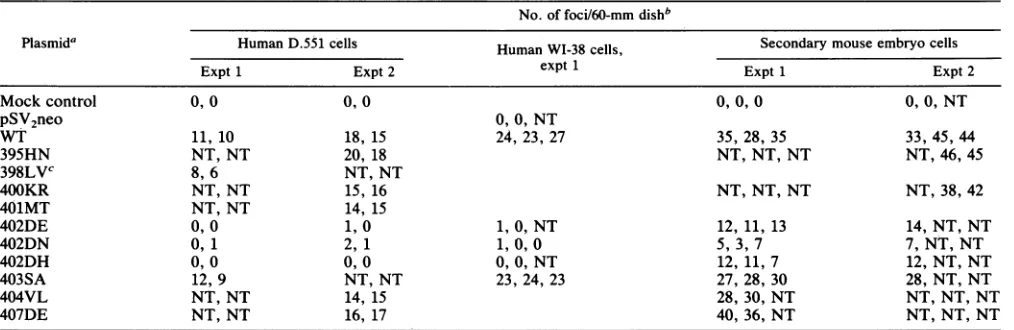
[image:2.612.69.273.76.225.2]T ANTIGEN-p53 COMPLEXES IN TRANSFORMATION BY SV40 6449 TABLE 1. Transformingactivities of WT and mutantSV40DNAs inhumanandmousecellS
No. of foci/60-mmdishb
Plasmida HumanD.551 cells Human WI-38 cells, Secondary mouse embryo cells
Expt 1 Expt 2 expt 1 Expt 1 Expt 2
Mockcontrol 0, 0 0, 0 0, 0, 0 0, 0, NT
PSV2neo
0, 0,NTWT 11, 10 18, 15 24,23, 27 35, 28, 35 33, 45, 44
395HN NT,NT 20, 18 NT, NT,NT NT, 46,45
398LVC 8,6 NT, NT
400KR NT, NT 15, 16 NT, NT, NT NT, 38, 42
401MT NT, NT 14, 15
402DE 0,0 1, 0 1, 0, NT 12, 11, 13 14, NT,NT
402DN 0, 1 2, 1 1, 0,0 5, 3,7 7, NT, NT
402DH 0, 0 0, 0 0, 0, NT 12, 11, 7 12, NT, NT
403SA 12, 9 NT, NT 23, 24, 23 27, 28, 30 28, NT, NT
404VL NT, NT 14, 15 28, 30, NT NT, NT, NT
407DE NT, NT 16, 17 40, 36, NT NT, NT, NT
aMutants aredesignated by the amino acid residue number which is mutated followed by thesubstitution,using single-letter codes. The two letters indicate theamino acid before and after the mutational change.
bScoredat 4weeksposttransfection on plates which contained 2x
10'
cells. NT, not tested.cMutantvirus unable to replicate in monkey BSC-1 cells. All others replicate like WTSV40.
ever, because mutations at residue 402 did have a slight effectontransformation ofmousecells (Table 1),webelieve that a better interpretation is that T-p53 complexes are involved, but that these mutants are able to transform
because somecomplexes still form.
Characteristics of transformed human cellfoci and cloned
lines. Since the effect of mutations atresidue 402was more pronounced with human cells than with mouse cells, we further investigatedthe transformation of human cells by T antigen. In this analysis, we characterized the few human cellcolonies thatappearedaftertransfectionwith402mutant DNAs. At 4weeks posttransfection, there were few or no humancell foci in the plates that received 402mutantDNAs
(Table 1; Fig. 2).Thefociweregenerally smaller than those appearingaftertransfectionwith WTorwithmutantDNAs encodingTantigensthat bindtop53 normally. One to three more foci per plate slowly formed over the next several weeks (by then the foci in all other plateswereconfluent and uncountable) (Fig. 2A). Foci ofD.551 cellswere recovered
eitherat6to7 weeksposttransfection (forthe402mutants) orat4 to5weeks posttransfection (for all others). The foci recovered at these two different times were of about the same size, indicating that these cells had undergone about
the same number ofdoublings after transfection. An early
observationwasthatcells derived fromtransfectionwith402 mutantDNAsgrew moreslowly than all other cloned cells.
Cellswere propagated in culture andchecked forT-antigen expression byindirectimmunofluorescence. Fewer than half
(5 of 12) of the cloned cells derived by transfection with mutant402 DNAswere Tantigen positive, whereas all nine of the clones obtained after transfection with control WT, 398LV,or403SA DNAwereTantigen positive.Some of the
T-antigen-positive cell clones were then testedby immuno-precipitation for the presence of stable T-p53 complexes.
Table 3 shows that whereasa WT, a398LV-derived, anda 403SA-derived clone contained stable complexes, a 402DE and a 402DN-derived clone contained no detectable T-p53 complexes,aspredicted bythe effect of mutationsatresidue 402 (28). Immunoprecipitation results for the 402-derived clonesareshown in Fig. 3.One clone(402DE(5)] expressed normal levels of T antigen and p53 but no detectable com-plexes (Fig. 3, lanes 3 and 4). The other clone (402DN(6)]
expressed lowlevels of Tantigenandundetectablelevels of p53 and complexes(Fig. 3, lanes 5 and 6). The other three T-antigen-positive 402-derived clones [402DE(2), 402DE(3), and 402DH(3)]reachedsenescence atthethirdpassageafter isolation(inaT-75flask) (Table 3) and couldnotbe tested. Determinationofthelife span ofclonedcell lines. Immor-talization of human cells by SV40 is quite rare (15, 20), However, SV40can extend the life span ofhuman diploid cells by about 20 to 40 population doublings (21, 45). To
assessthe effect ofT-antigen mutations onthe life span of human cells, we continuously passaged cloned cells (at a
split ratio of 1:5) (36) until some ofthe cell lines reached
senescence. Wecalculated thetotal number ofpassagesfor eachclone,starting fromtheoriginal isolation ofD.551 cells (Table 3). Since cells were transfected at passage 14, we
calculated from cell numbers that cells recovered from a
typical focus were atthe equivalence ofabout passage 22. Based on these calculations, all T-antigen-positive clones generatedwithmutant402DNAsreachedsenescence at or nearpassage25,correspondingtothelifespanoftheoriginal D.551 cells(Table 3).In contrast, cellstransformed withWT
DNA, mutant 398 DNA, or mutant 403 DNA were still growingwellbeyond passage 35. Wepropagatedtwoclones
to passage 47 or 48 and one to passage 55 before the experimentwasterminated (Table 3).
Figure4showsgrowthcurvesforsome402DNA-derived clonesasthey approached senescence.TwoclonesofD.551 cells derived by transfection with 402DE were no longer growing at passage 26 (Fig. 4). One clone generated with 402DNwasgrowing slowlyatpassage26andatroughlythe
same rate asthe parentalcellline atpassage 18. This clone stoppedgrowingatthenextpassage (Table 3).WT
transfor-mantsand cells transformed with 403SAweregrowingwell atpassage 26or27anddividingfaster than theparentalcells. These results indicate that the presence of stable T-p53 complexesin these cloned cells contributedtothe extension of their life spans.
DISCUSSION
In this report, we showed that three mutants of SV40 codingfor T antigens with various substitutions at residue VOL.65,1991
on November 10, 2019 by guest
http://jvi.asm.org/
6450 LIN AND SIMMONS
A
B
i9'SkN
401M 40101
402n
A. pSV2 NEO
/
D. 402DN
_
.:
r..
'It
B. WTSV40
t ip
.;;@ 4.e
[image:4.612.54.552.78.425.2]-^
%A..
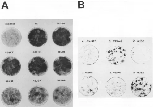
,. _ E. 402DHFIG. 2. Morphology ofmutantorWTSV40-transformed foci. Cellsweretransfectedasdescribed in Materials andMethods,fixedwith
methanol, and stained with 5% Giemsa stain 6 weeks after transfection. (A) Background monolayer of human D.551 cells after mock
transfection(control)orfoci from cells transfected with WTormutantSV40DNA,asindicated.(B) Background monolayerof human WI-38
cells after pSV2neotransfection(control)orfoci fromcells transfected with WTormutantSV40DNA,asindicated. Theplateswerestained
with Giemsa 4 weeksaftertransfection.
402 and defective in human p53 binding are significantly reduced in their ability to induce transformed foci oftwo
normal human cell lines. This defect is not due to the inability of the mutant T antigens to complex with the product ofthe Rb gene, asjudged by immunoprecipitation assays using anti-Rb antibodies (data not shown). Our
re-sults indicate that stableT-p53 complexes playanimportant role in the formation of human cell foci.
TABLE 2. Transfectionfrequency of human cells by WT or
mutantpBS-SV40DNA
Transfected DNA %of
T-antigen-positivecellsa
WTSV40... 9
402DE... 12
402DN... 9
402DH... 12
403SA... 13
aHuman D.551cells were transfected with wt or mutantpBS-SV40DNA. At48 hposttransfection,cells werechecked for theexpressionof T antigen by
indirect immunofluorescencetodeterminethepercentageof T-antigen-posi-tivecells.
The nature of the few human cell foci that formed after transfection with 402 mutantDNAsis notknown. Some of the foci could have consisted of cells that
expressed
Tantigen early on and then lost that
expression.
TheT-anti-TABLE 3. ComparisonoflifespanofWTor mutant SV40-derived cell lines
Cloned line T-p53complexes Approx life span(no. ofpassages)
Parental D.551 - 25
WTSV40(2) NTa >38
WTSV40(4) + >35
398LV(1) + >39
398LV(2) NT >48
402DE(2) NT 25
402DE(3) NT 25
402DE(5) 27
402DN(6) 27
402DH(3) NT 25
403SA(1) + >55
403SA(4) NT >47
a NT,nottested. 4088K1
C. 402DE
"I0?0 40UBN
F. 403SA
A
- V
J.VIROL.
aWl
on November 10, 2019 by guest
http://jvi.asm.org/
[image:4.612.311.551.578.715.2] [image:4.612.54.294.622.692.2]T ANTIGEN-p53 COMPLEXES IN TRANSFORMATION BY SV40 6451 1 2 3 4 5 6
TAg
p53
[image:5.612.124.239.82.225.2]* ..7a _...
FIG. 3. Association of WT or mutant T antigen with p53 in human D.551 cloned cells. Cells were labeled with L-[35S]methio-nine for 16 h. Cell lysateswereincubated with eitherpAb416(16),a
monoclonal antibodytoTantigen (lanes1, 3,and 5),orpAb421(16), amonoclonal antibodytop53(lanes2, 4, and6). Labeled
immuno-precipitated proteins were detected by autoradiography after
elec-trophoresison13% SDS-polyacrylamide gels. Lanes: 1and 2, WT
SV40; 3 and 4, 402DE(5); 5 and6,402DN(6).
gen-positive clones could have formed either because tran-sient T-p53 complexes may have existed or because the
othertransformation-related activities ofthemutant T anti-gens (such asbindingtoRb) are intact.
Therole of Tantigen-p53 complexes inthetransformation of rodent cells is less clear.Previous studies by others have
shownthatmutantTantigens missingsome orallportions of thep53 binding region (amino acid residues 272to 517 [42]) donotbind p53 and failtoimmortalizemousecellsefficiently (47-49).Theseresultsareconsistent with the findingthat the
E6protein of human papillomavirus 16, whichbinds top53
(54), can immortalize primary rat cells (23). On the other
hand, Sompayrac and Danna (44) have shown that an
NH2-terminal 147-amino-acid fragment of T antigen,
ex-pressed under the control of a retrovirus promoter,
trans-07
403SA(1)
WTSV40 (4)
6
U)10
/ / ,=WTSV40(2)
E 403SA(4)
E
6 parentalD.551
(D ~~~~~~~~~~~~~402DN(6)
~~~~~~~~~~~~~402DE(5)
402DE(2)
4
10
0 1 2 3 4 5 6
Days
FIG. 4. Growth curves of WT or mutant SV40 DNA-derived clones. Parentalorcloned D.551 cellswereplatedatadensity of 5
x 104 cells per60-mm petri dish. Cell numbers were determined
everyday fora6-dayperiod. Passage numbers for the clones used:
403SA(1),26; WTSV40(4), 27;WTSV40(2), 26; 403SA(4), 26;
paren-talD.551, 18; 402DN(6), 26; 402DE(5), 26; 402DE(2), 25.
formssecondary rat cells as efficiently asdoes full-length T antigen.
Our results do notclearly demonstrate the involvement of complexes in the transformation ofmousecells becausethe 402 mutant T antigens retain a small amount of binding activity to mousep53. However, thesemutations had some
effect on focus formation (Table 1), which suggests that in the contextoffull-length Tantigen,and under thecontrol of theSV40early promoter, T-p53 complexesarenecessaryfor the transformation of mouse cells as they are for human cells.Onepossible explanationfor the discrepancy between
ourresults and those ofSompayrac and Danna (44) is that mouse and rat cells are transformed by SV40 by slightly differentmechanisms. However, we think it is more likely that the expression ofa truncated T antigen (residues 1 to
147) under the control ofadifferent promotermightabrogate therequirementforp53 binding.
Although SV40 T antigen can immortalize rodent cells, it failstoimmortalizehumancellsasefficiently (15,20,38, 46). Wright et al. (55) have suggested that human cells are immortalized only aftertwostages(mortality stagesMl and M2) arebypassed. Theexpression ofSV40T antigen over-comes or bypasses the Ml but not the M2 stage (55). Asa
consequence,thelife spanofthesecells is extended and they continue to divideuntil M2 is reached, at which point they undergo crisis. Because T antigen can immortalize rodent cells, the bypass ofonly one stage (correspondingtoMl in human cells) may be required in this case. Our results indicate that stable T-p53 complexes are necessary for the extension in the life span of human cells. This would suggest that the ability ofT antigento form stable complexes with
p53
is necessarytobypass orovercometheMl mechanism in human cells. Furthermore, it also suggests that stableT-p53
complexes may be requiredat the same stage in the immortalization of rodent cells.One way in which T-p53 complexes could contribute to theextension of the life span of human cells is through the inactivation oralteration of thenormal function of WTp53. Fields and Jang (10) and Raycroft et al. (40) have recently shown that p53 is an activator oftranscription. The target genes for
p53
are unknown, but conceivably one of them couldhavegrowthsuppressorfunction. Bybindingtop53,T antigenwould inhibit thisactivityand promote cell prolifer-ation. Alternatively,p53
could normally be a suppressorof certaingeneswhich function in cellgrowth, and thisactivity could be inhibitedormodifiedbyTantigen.Inhibitionofp53 function could also come about through protein-protein interactions rather than at the levelof gene regulation. WTp53
islooselyassociated with thep34cdc2
kinase(34,35), and humanp53
is phosphorylated by cdc2 kinase in vitro (3). This interaction may be functional in the control of cell growth (3). When Tantigenbinds to p53, it may inactivate this function and promote the extension of the life span of the cells.Finally, ourresults suggest yet anothermodel. The
trans-formation ofmouse cells by various 402 mutants correlates
best with the relativepercentage ofTantigen incomplexes ratherthanwith the relative percentageofWTp53 boundto mutant T antigen (28) (Table 1). This offers the possibility that
T-p53
complexes function directly in promoting cell growth and that theinactivation ofWTp53
is notofprime importance in transformation by SV40. The presence of large amounts ofuncomplexed (andpresumably functional) p53 in these mousecells supports thishypothesis.
In conclusion, we have provided evidence that stable T-p53 complexes are necessary for SV40 to induce foci of VOL.65, 1991
on November 10, 2019 by guest
http://jvi.asm.org/
[image:5.612.83.286.493.674.2]transformed human cells in culture. Furthermore, these
complexesappear tobe required for theextensionofthelife
spanof these cells.
ACKNOWLEDGMENTS
We thank D. Lane for monoclonal antibody pAb240 and Ed Harlow and Q. Hu for anti-Rb monoclonal antibodies. We also thank P. DeLeon for the preparation of primary mouse embryo cells, F. Schmieg for careful readingof themanuscript,Huey-JenL. Lin for help with the figures, and William Young for technical assistance.
This work was supported by grant CA36118 from the National Cancer Institute.
REFERENCES
1. Auborn, K., M. Guo, and C. Prives. 1989. Helicase, DNA-binding, and immunological properties ofreplication-defective simian virus 40 mutant Tantigens. J. Virol. 63:912-918. 2. Baker, S. J., S. Markowitz,E. R.Fearon, J. K. V. Willson, and
B.Vogelstein.1990.Suppression ofhumancolorectalcarcinoma cell growth by wild-typep53. Science 249:912-915.
3. Bischoff, J. R., P. N. Friedman, D. R. Marshak, C. Prives, and D. Beach. 1990. Human p53 isphosphorylated by p60-cdc2 and cyclin B-cdc2. Proc. Natl. Acad. Sci. USA 87:4766-4770. 4. Brugge, J.S., and J. S.Butel. 1975. Role of simian virus 40 gene
A function in maintenance oftransformation. J. Virol. 15:619-635.
5. Chen, C., and H. Okayama. 1987. High-efficiency transforma-tion of mammalian cells by plasmid DNA. Mol. Cell. Biol. 7:2745-2752.
6. Chen, P.-L., Y. Chen, R. Bookstein, and W.-H. Lee. 1990. Genetic mechanisms of tumor suppression by the human p53 gene. Science250:1576-1580.
7. Chen, S., and E. Paucha. 1990. Identification of a region of simian virus 40 large Tantigenrequired for celltransformation. J. Virol. 64:3350-3357.
8. DeCaprio, J. A., J. W. Ludlow, J. Figge, J.-Y. Shew, C.-M. Huang, W.-H. Lee, E.Marsilio, E. Paucha, and D. M. Living-ston. 1988. SV40 large tumor antigen forms a specificcomplex withtheproductof theretinoblastoma susceptibilitygene. Cell 54:275-283.
9. Eliyahu, D., D. Michalovitz, S. Eliyahu, 0. Pinhasi-Kimhi, and M. Oren. 1989. Wild-type p53 can inhibit oncogene-mediated focusformation. Proc. Natl. Acad. Sci. USA86:8763-8767. 10. Fields, S., and S. K. Jang. 1990. Presenceof apotent
transcrip-tion activating sequence in the p53 protein. Science 249:1047-1049.
11. Finlay, C.A., P. W. Hinds, T.-H. Tan, D. Eliyahu, M. Oren, and A. J. Levine. 1988. Activating mutationsfor transformation by p53 produce agene product that forms an hsc7o-p53 complex with an alteredhalf-life. Mol. Cell. Biol. 8:531-539.
12. Finlay, C. A., W. Hinds, and A. J. Levine. 1989. The p53 protooncogene can act as asuppressor oftransformation. Cell 57:1083-1093.
13. Gannon, J. V., R. Greaves, R. Iggo, and D. P. Lane. 1990. Activating mutations in p53produceacommonconformational effect. A monoclonal antibody specific for the mutant form. EMBO J. 9:1595-1602.
14. Giacherio, D., and L. P. Hager. 1979. A poly(dT)-stimulated ATPaseactivityassociatedwith simian virus 40 large T antigen. J. Biol.Chem. 254:8113-8116.
15. Gotoh, S., L. Gelb, and D.Schlessinger. 1979.SV40-transformed human diploid cells that remain transformed throughout their limited lifespan. J. Gen. Virol. 42:409-414.
16. Harlow,E.,L. V. Crawford, D. C. Pim, and N. M.Williamson. 1981. Monoclonal antibodies specific for simian virus 40 tumor antigen.J. Virol. 39:861-869.
17. Hayflick,L.1965. Thelimitedinvitrolifetimeofhumandiploid cell strains. Exp. Cell Res. 37:614-636.
18. Hayflick,L., andP.S. Moorhead. 1961. Theserial cultivation of humandiploidcell strains. Exp. Cell Res. 25:861-869. 19. Hinds, P., C. Finlay, and A. J. Levine. 1989. Mutation is
required toactivate the p53 gene for cooperation with theras oncogeneandtransformation. J. Virol. 63:739-746.
20. Huschtscha, L. I., and R. Holliday.1983. Limited and unlimited growth of SV40-transformed cells for human diploid MRC-5 fibroblasts.J. Cell Sci. 63:77-99.
21. Ide, T. Y., Y. Tsuji, T. Nakashima, and S. Ishibashi. 1975. Initiation and maintenance of cell transformation by simian virus 40: a viral genetic property. Proc. Natl. Acad. Sci. USA 72:673-677.
22. Kalderon,D., and A. E.Smith. 1984. Invitromutagenesisof a putative DNAbinding domain ofSV40 large-T. Virology 139: 109-137.
23. Kanda, T., S. Watanabe, and K. Yoshike.1988.Immortalization of primary rat cells by human papillomavirus type 16 subge-nomic DNA fragmentscontrolled by theSV40promoter. Virol-ogy 165:321-325.
24. Kimura, G., and A. Itagaki.1975.Initiation and maintenance of celltransformationbysimian virus 40:aviral genetic property. Proc. Natl. Acad. Sci. USA 72:673-677.
25. Lane, D. P., and L. V.Crawford.1979. T-antigenis boundtoa host protein in SV40 transformed cells. Nature(London)278: 261-263.
26. Levitsky, K., K. Chandrasekaran, P. T. Mora, and D. T. Simmons. 1983. Immunoaffinity chromatography ofa cellular tumor antigenfrom mouse neuroblastoma cells. Int. J. Cancer 32:597-602.
27. Lin, J.-Y., and D. T.Simmons. 1990. Transformationby simian virus 40 does not involvethe mutationalactivation of p53toan oncogenic form. Virology 176:302-305.
28. Lin, J.-Y., and D. T. Simmons. 1991. Stable T-p53 complexes are notrequiredforreplication of simian virus 40 in culture or for enhanced phosphorylation of T antigen and p53. J. Virol. 65:2066-2072.
29. Linzer, D. I. H., W. Maltzman,and A. J. Levine. 1979. Charac-terization of a 54K dalton cellular SV40 tumorantigenpresentin SV40-transformed cells and uninfected embryonal carcinoma cells. Cell 17:43-52.
30. Loeber, G., R. Parsons, and P. Tegtmeyer. 1989. Thezincfinger regionof simian virus 40large T antigen. J. Virol. 63:94-100. 31. Manfredi, J.J.,andC.Prives. 1990.Binding of p53andp105-RB
is not sufficient for oncogenic transformation by a hybrid polyomavirus-simian virus 40 large T antigen. J. Virol. 64:5250-5259.
32. Manos, M.M., andY. Gluzman. 1985. Genetic and biochemical analysis of transformation-competent, replication-defective simian virus 40 large Tantigen mutants. J. Virol. 53:120-127. 33. Martin,R. G., and J. Y. Chou. 1975. Simian virus40functions
required for the establishment and maintenance ofmalignant transformation.J. Virol. 15:599-612.
34. Milner,J., A. Cook, andJ. Mason. 1990. p53 isassociatedwith p34cdc2in transformedcells. EMBOJ. 9:2885-2889.
35. Milner, J., J. Gamble, and A. Cook. 1989.p53is associated with a 35KD protein incells transformed by simianvirus 40. Onco-gene4:665-668.
36. Neufeld, D. S., S. Ripley, A. Henderson, and H. L. Ozer. 1987. Immortalization of human fibroblasts transformed by origin-defective simianvirus 40. Mol. Cell. Biol. 7:2794-2802. 37. Osborn, M., and K. Weber. 1975. Simian virus 40 gene A
function and maintenance oftransformation. J. Virol. 15:636-644.
38. Petit, C. A., M. Gardes, and J. Feunteun. 1983.Immortalization ofrodent embryo fibroblasts bySV40 is maintained by the A gene. Virology 127:74-86.
39. Prives, C., L. Covey, A. Scheller, and Y. Gluzman. 1983. DNA-binding properties of simian virus 40 T-antigen mutants
defective in viral DNAreplication. Mol.Cell. Biol. 3:1958-1966. 40. Raycroft, L., H. Wu, and G. Lozano. 1990. Transcriptional activation bywild-typebut nottransformingmutants ofthep53 anti-oncogene. Science249:1049-1051.
41. Resnick-Silverman, L., Z. Pang, G. Li, K. K. Jha, and H. L. Ozer. 1991. Retinoblastomaproteinandsimian virus 40-depen-dent immortalization ofhuman fibroblasts. J. Virol. 65:2845-2852.
on November 10, 2019 by guest
http://jvi.asm.org/
T ANTIGEN-p53 COMPLEXES IN TRANSFORMATION BY SV40 6453 42. Schmieg,F. I., and D. T. Simmons. 1988. Characterization of the
in vitrointeractionbetween SV40 Tantigenandp53: mapping the p53 binding site. Virology 164:132-140.
43. Shalloway, D., T. Kleinberger, and D. M. Livingston. 1980. Mapping of SV40 DNA origin region binding sites for the SV40 Tantigenby protectionagainst exonuclease III digestion. Cell 20:411-422.
44. Sompayrac, L., and K. J. Danna. 1991. The amino-terminal 147 aminoacids of SV40 large T antigen transform secondary rat embryofibroblasts. Virology 181:412-415.
45. Stahl, H., P. Droege, and R. Knippers. 1986. DNA helicase activityofSV40 largetumorantigen. EMBO J. 5:1939-1944. 46. Stein, G. H. 1985. SV40-transformed human fibroblasts:
evi-dence for cellular aging in precrisis cells. J. Cell Physiol. 125:36-44.
47. Tevethia,M. J. 1984. Immortalization of primary mouse embryo fibroblasts with SV40 virions, viral DNA and a subgenomic DNAfragment inaquantitative assay. Virology 137:414-421. 48. Tevethia, M. J., R. W. Anderson, S. S. Tevethia, D. T. Simmons,
J. Feunteun, and C. Cole. 1986. Influence of amino acids encoded inthe3' openreading frameof theSV40earlyregion ontransformation and antigenicity oflarge T antigen.Virology 150:361-372.
49. Tevethia, M. J., J. M. Pipas, T. Kierstead, and C. Cole. 1988. Requirement for immortalization of primary mouse embryo fibroblastsprobed with mutants bearing deletions in the 3' end
ofSV40 gene A.Virology 162:76-89.
50. Thompson,D.L.,D.Kalderon,A. E.Smith,and M.J.Tevethia. 1990. Dissociation of Rb binding and anchorage independent growth from immortalization and tumorigenicity using SV40 mutants producing N-terminally truncated large T antigens. Virology178:15-34.
51. Tjian, R. 1978. Protein-DNA interactions at the origin of simian virus 40 DNA replication. Cold Spring Harbor Symp. Quant. Biol.43:655-662.
52. Tjian, R., and A. Robbins. 1979.Enzymatic activities associated withapurifiedsimianvirus 40 Tantigen-related protein. Proc. Natl. Acad. Sci. USA 76:610-614.
53. Tooze, J. (ed.).1981. DNA tumor viruses, molecular biology of tumorviruses, 2nd ed. Cold Spring Harbor Laboratory, Cold Spring Harbor, N.Y.
54. Werness, B.A., A. J.Levine,and P. M.Howley.1990. The E6 proteins encoded by human papillomavirus types 16 and 18can
complex p53 in vitro. Science 248:76-79.
55. Wright, W. E., 0. M. Pereira-Smith, and J. W. Shay. 1989. Reversible cellular senescence:implications forimmortalization of normal human diploid fibroblasts. Mol. Cell. Biol. 9:3088-3092.
56. Yewdell, J. W., J. V.Gannon, and D. P. Lane. 1986. Monoclonal antibody analysis of p53 expression in normal and transformed cells. J.Virol. 59:444 452.
VOL. 65,1991