R E S E A R C H
Open Access
Transcriptome modulation by
hydrocortisone in severe burn shock:
ancillary analysis of a prospective
randomized trial
Jonathan Plassais
1, Fabienne Venet
1,2, Marie-Angélique Cazalis
1, Diane Le Quang
3, Alexandre Pachot
1,
Guillaume Monneret
1,2, Sylvie Tissot
3and Julien Textoris
1,3*Abstract
Background:Despite shortening vasopressor use in shock, hydrocortisone administration remains controversial, with potential harm to the immune system. Few studies have assessed the impact of hydrocortisone on the transcriptional response in shock, and we are lacking data on burn shock. Our objective was to assess the hydrocortisone-induced transcriptional modulation in severe burn shock, particularly modulation of the immune response.
Methods:We collected whole blood samples during a randomized controlled trial assessing the efficacy of
hydrocortisone administration in burn shock. Using whole genome microarrays, we first compared burn patients (n = 32) from the placebo group to healthy volunteers to describe the transcriptional modulation induced by burn shock over the first week. Then we compared burn patients randomized for either hydrocortisone administration or placebo, to assess hydrocortisone-induced modulation.
Results:Study groups were similar in terms of severity and major outcomes, but shock duration was significantly reduced in the hydrocortisone group. Many genes (n = 1687) were differentially expressed between burn patients and healthy volunteers, with 85% of them exhibiting a profound and persistent modulation over seven days. Interestingly, we showed that hydrocortisone enhanced the shock-associated repression of adaptive, but also innate immunity.
Conclusions:We found that the initial host response to burn shock encompasses wide and persistent modulation of gene expression, with profound modulation of pathways associated with metabolism and immunity. Importantly, hydrocortisone administration may worsen the immunosuppression associated with severe injury. These data should be taken into account in the risk ratio of hydrocortisone administration in patients with inflammatory shock.
Trial registration:ClinicalTrials.gov, NCT00149123. Registered on 6 September 2005.
Keywords:Shock, Burns, Hydrocortisone, Transcriptome modulation, Immunosuppression, Host response
Background
Transplantation, inflammatory, and auto-immune dis-eases have benefitted from the immunomodulatory properties of glucocorticoids for decades. Glucocorti-coids modulate both innate and adaptive immune
responses [1]. They promote bone marrow release and survival of neutrophils. Glucocorticoids also modulate the innate response by suppressing pro-inflammatory or by stimulating anti-inflammatory mediators [2]. Such balanced action promotes resolution of inflammation and prevents overshooting of the host response. This may contribute to the efficacy of dexamethasone in pre-venting morbidity and mortality in pneumococcal men-ingitis in children [3]. Moreover, decreasing antigen presentation and co-stimulation by dendritic cells [4] prevents the crosstalk between innate and adaptive * Correspondence:[email protected]
1EA7426, Université Claude Bernard Lyon 1, Hospices Civils de Lyon,
bioMérieux ;“Pathophysiology of injury induced immunosuppression (PI3)”, hôpital E. Herriot, 5 place d’Arsonval, 69437 Lyon, France
3Hospices Civils de Lyon, Burn ICU, Anesthesia and Critical Care Medicine
department, hôpital E. Herriot, 5 place d’Arsonval, 69437 Lyon, France Full list of author information is available at the end of the article
systems. Glucocorticoids also promote a shift from T helper (Th)1 to Th2 cells, leading to impaired defense against intracellular and opportunistic infections [5, 6].
The use of glucocorticoids in septic shock is an issue of incredible debate [7]. Considering the aforementioned effects, it seems logical that glucocorticoids may avoid the deadly effect of the massive inflammatory response initially seen in sepsis. However, after acknowledging that high doses of glucocorticoids do not decrease mor-tality and may even be harmful [8, 9], low (but still supra-physiologic) doses of hydrocortisone were assessed. While the reduction in mortality [10] is still a matter of debate [11, 12], hydrocortisone remains rec-ommended for patients with refractory shock [13, 14]. Indeed, hydrocortisone has almost always been associ-ated with reversal of shock. The beneficial effect of corti-costeroids on hemodynamics is highly intertwined with their effects on the inflammatory response and endothe-lium [15]. Although the precise molecular mechanisms are still largely unknown, nitric oxide (NO) synthesis seems to play a determinant role in modulating the vas-cular tone over the initial course after injury [16, 17].
For glucocorticoids, as for any other therapies evalu-ated for use in septic shock, the high heterogeneity of patients and the absence of stratification may explain in-conclusive results. Given the amount of evidence sup-porting sepsis-induced immunosuppression [18], the blind use of hydrocortisone - i.e. without monitoring the patient’s host response - may be questioned. Indeed, a retrospective study in pediatrics suggested that septic shock patients treated with glucocorticoids had numer-ous alterations of adaptive immunity at the transcrip-tional level [19].
Severe burns and septic shock host responses share numerous features, including a relative late-stage state of immunosuppression. We recently described in a pro-spective, randomized, double-blind study that the use of hydrocortisone in refractory burn shock led to a statisti-cally significant reduction in the duration of shock [20]. As the impact of low-dose hydrocortisone on the host response has never been studied in patients with burns, we took advantage of this study to assess the whole blood transcriptional modulation in severe burn shock. Here, we studied the modulation of the immune re-sponse induced by shock and assessed the specific ef-fects of hydrocortisone administration. We provide evidence that hydrocortisone transcriptionally enhances the immunosuppressive mechanisms that take place after severe injury.
Methods
Patients and sample collection
Patients with severe burns were admitted to Edouard Herriot Hospital (Lyon, France) and included in a
placebo-controlled, randomized, double-blind study that has been described elsewhere [20]. Patients aged be-tween 18 and 75 years, with a total burn surface area >30% were included if they presented with onset of se-vere shock (norepinephrine >0.5μg/kg/min) between 24 and 72 h after injury. Pregnancy, trauma, initial sepsis and cardiac insufficiency were exclusion criteria. The protocol was accepted by the ethical committee on 15 February 2005 and was registered at ClinicalTrials.gov (NCT00149123). All healthy volunteers (HV) and pa-tients (or next of kin) gave written informed consent be-fore inclusion in the study.
Thirty-two patients were enrolled in the clinical study. Patients received a priming dose of 50 mg of hydrocorti-sone (Upjohn, Serb Labo, Paris, France) or placebo (NaCl 0.9%) followed by a continuous infusion of 200 mg/day over 5 days, 100 mg at day 6 and 50 mg at day 7, as initially proposed for septic shock [10]. Thir-teen HV were recruited within Hospices Civils de Lyon to serve as controls for the transcriptional study.
Whole blood samples were collected in PAXgene Blood RNA tubes (PreAnalytix, Hilden, Germany) dur-ing the randomized controlled trial [20]. Up to four sam-ples were collected from each patient during the first week after burn shock. The first sample (S) was collected at inclusion (onset of shock) and before any treatment (day 0, S1). Three other samples were collected the fol-lowing day (24 h, S2), around 120 h (S3) and 168 h (S4) after inclusion (see details in Additional file 1: Figure S1A). Due to technical reasons (missing sample, poor RNA quality, microarray removed from quality-check analysis for batch effect, etc.), 117 samples were ana-lyzed: 30 S1 samples, 27 S2 samples, 29 S3 samples, 18 S4 samples, and 13 samples from HV.
RNA extraction and microarrays
Microarray analysis
Microarray normalization and statistical analysis were performed using R/Bioconductor (R v3.0.0) [21, 22]. Quality assessment was performed using the simpleaffy (v2.36.1) [23] and arrayQualityMetrics (v3.14.0) [24] packages. The simpleaffypackage provided quality con-trols before the normalization process by checking dens-ity distributions, estimating the average background intensities and assessing the RNA quality measures. The arrayQualityMetricspackage generated a report of qual-ity metrics from both raw data and normalized micro-array data. We used principal component analysis [25] to identify poor-quality arrays. After removing outlier samples the raw data were normalized, adjusted for background noise and summarized using the guanine cytosine robust multi-array (GCRMA) algorithm with default parameters [26]. Batch effects were removed using COMBAT [27]. Filtering was performed based on MAS5.0 p values. MAS5.0 p values were estimated for each probe set, corresponding to significant differences between perfect match and mismatch intensities. Then we kept only probe sets for which allpvalues were <0.2. We finally applied a batch correction using COMBAT to remove the effect of varying delays between admission and each sampling. MIAME-compliant microarray data are available on the Gene Expression Omnibus (GEO) website [GEO:GSE77791].
Microarray statistical analyses
The first analysis compared samples obtained from pa-tients before they received any treatment to samples from HV. For all probe sets we performed moderated t tests (Limma package version 3.16.0) [28]; we adjusted the p values for multiple testing using the Benjamini-Hochberg correction [29] to ensure a false discovery rate below 5%. Adjusted pvalues <0.05 were considered significant. We then assessed gene modulation associated with burn shock across time, using only microarrays from patients on pla-cebo. We performed moderatedFtests [28] and corrected for multiple testing using the Bonferroni correction. Ad-justed pvalues <0.05 were considered significant. Finally we assessed the impact of hydrocortisone on gene expres-sion over time. We first performed moderated t tests to compare the two groups at each sampling (Benjamini-Hochberg correction [29], adjusted p value <0.05) and then moderatedFtests comparing microarrays over time (S2–S4) from hydrocortisone and placebo patients. A k -means clustering was performed to group probe sets with similar profiles and heatmaps were used to visualize the differential expression.
Functional annotation analyses
Functional analyses were performed using Ingenuity Path-way Analysis (IPA) software (Ingenuity Systems, Redwood
City, USA) and Gene Ontology (GO) [30]. For the core analysis all differentially expressed probe set ID were mapped to their corresponding gene and molecular ob-jects in the IPA Knowledge Base. Fisher’s exact test was used to estimatepvalues for diseases and biological func-tion enrichment analyses, and thepvalues were adjusted using the Benjamini-Hochberg method [29]. To assess statistical significance in pathway analyses, we used re-peated measure analysis of variance (ANOVA), with one between-group variable (treatment) and two within-group variables (time and probe) with the following model:
ExpressionvalueeTreatment þ Time þ Probe
þ Treatment
:Time
The modulation of pathway expression over time and according to treatment was considered significant for p values <0.01.
Results
Patients
Briefly, 32 patients with severe burn shock were ran-domized to receive either hydrocortisone or placebo within the 72 h after admission. All samples from two patients were discarded due to a major batch effect re-lated to a technical issue during hybridization. A full clinical description of the patients is provided elsewhere [20]. Briefly (Table 1), these were severely burned pa-tients (median total burned surface area of 70% (48– 84%)). Five patients (one from the placebo group and four from the hydrocortisone group) died during the first 7 days. Fewer patients in the hydrocortisone group received etomidate before ICU admission (n = 5 patients (36%) vs. n = 12 patients (80%); pvalue = 0.03). Import-antly, hydrocortisone-treated patients had a significantly shorter duration of shock, defined by norepinephrine ad-ministration time (median (IQR), 60 h (42–117 h) vs. 120 h (84–141 h);pvalue = 0.048).
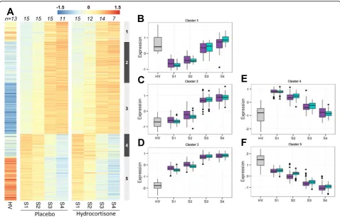
Host response signature to burn shock over time
To study the host response to burn shock over time, in-clusion samples (S1) were compared to 13 HV samples. Then, to study the modulation over the first week, we analyzed only samples from the placebo group (the complete analysis plan is provided in Additional file 1: Figure S1B).
Table S2). Genes from clusters 1 and 4 were initially down-modulated and up-modulated, respectively, and returned to HV levels within the first week. Functional annotation of these transiently modulated genes highlighted interesting functions (Additional file 3: Table S1). Genes involved in the response to hypoxia were down-modulated, whereas several processes related to inflammation and blood vessels were up-modulated.
Modulation of genes from cluster 3 (up-modulated), and 5 (down-modulated) increased over time and was maximal at the last time point. Genes from cluster 2 were up-modulated only at later time points (days 5 and 7 after inclusion). Functional annotation of cluster 5 (down-modulated) highlighted functions related to T cell differentiation and activation, negative regulation of apoptotic process, regulation of the innate immunity and antigen presentation. Interestingly, annotation of the persistently up-modulated genes (clusters 2 and 3) highlighted several genes involved in response to stress, mitochondrial respiratory chain and oxidative
phosphorylation (Additional file 3: Table S1). Altogether, we observed a profound and persistent modulation of gene expression after severe burn shock.
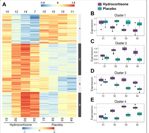
Modulation of gene expression by hydrocortisone after burn shock
[image:4.595.60.545.98.479.2]At inclusion, burn patients exhibited moderate to high levels of plasma cortisol, without a difference be-tween hydrocortisone and placebo groups (respect-ively, 15 μg/dL (8.8–22) and 14 μg/dL (8.4–19); p value = 0.77). The expression of the glucocorticoid re-ceptor (GR) was similar in patients and healthy vol-unteers (Fig. 2a; NR3C1 (alias GR): FC = 0.88, p value = 0.42). Most patients exhibited relative adrenal insuf-ficiency, with small increases in plasma cortisol levels after adrenocorticotropic hormone (ACTH) stimula-tion (Table 1). This was consistent with global down-modulation of the GR pathway genes at inclusion (Fig. 2a and b).
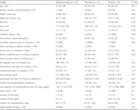
Table 1Clinical description of the study cohort
Variable Hydrocortisone (n = 15) Placebo (n = 15) Total (n = 30) Pvalue
Age, years 47 (42–50) 48 (36–59) 48 (39–55) 0.72
Gender, number of female patients (%) 2 (13%) 6 (40%) 8 (27%) 0.22
Weight (usual), kg 73 (68–86) 80 (65–93) 78 (65–86) 0.84
Weight at inclusion, kg 94 (77–98) 100 (77–112) 94 (77–104) 0.25
TBSA (%) 75 (54–87) 70 (44–76) 70 (48–84) 0.18
Baux score 117 (103–129) 109 (102–119) 110 (102–125) 0.29
ABSI score 12 (11–13) 11 (10–12) 11 (10–12) 0.06
Inhalation injury,n(%) 9 (60%) 3 (20%) 12 (40%) 0.06
Interval (burn injury-inclusion), h 57 (52–66) 46 (41–58) 54 (42–62) 0.11
Etomidate injection prior to inclusion,n(%) 5 (36%) 12 (80%) 17 (58%) 0.03
Blood transfusions before inclusion,n(%) 4 (27%) 3 (20%) 7 (23%) 1
Diuresis prior to inclusion (L/day) 3.2 (2.0–4.2) 3.5 (2.8–4.3) 3.4 (2.2–4.3) 0.47
Plasma creatinine prior to inclusion,μmol/L 86 (78–128) 88 (59–100) 87 (72–105) 0.28
Plasma protein prior to inclusion, g/L 42 (38–45) 41 (39–45) 42 (38–46) 1
Hemoglobin prior to inclusion, g/L 106 (100–117) 113 (90–132) 110 (94–123) 0.72
White blood cells prior to inclusion, 109
/L 5.0 (3.2–7.4) 6.6 (3.5–11.5) 5.3 (3.1–9.6) 0.25
Lymphocytes prior to inclusion, 109/L 0.9 (0.6–1.1) 1.1 (0.8–1.4) 0.9 (0.7–1.3) 0.14
Basal cortisol, µg/dL 15.2 (8.8–21.8) 13.8 (8.4–19.1) 14.5 (8.3–19.9) 0.77
Norepinephrine prior to inclusion,μg/kg/min 0.59 (0.53–0.66) 0.60 (0.51–1.04) 0.60 (0.51–0.78) 0.55
Duration of norepinephrine protocol, h 60 (42–117) 120 (84–141) 102 (56–131) 0.05
Total quantity of norepinephrine over ICU stay,μg/kg 1457 (1132–3705) 1971 (1535–3893) 1771 (1196–4068) 0.27
Septic shock,n(%) 5 (33%) 7 (47%) 12 (40%) 0.71
Number of infections 1 (1–3) 2 (2–3) 2 (1–3) 0.1
Number of skin grafts 4 (1–7) 5 (4–10) 5.00 (1.3–8.5) 0.13
Duration of hospitalization, days 65 (11–77) 67 (51–104) 66 (22–89) 0.11
Deaths before 28 days,n(%) 6 (40%) 2 (13%) 8 (27%) 0.22
Interestingly, hydrocortisone induced only a transient modulation of gene expression over the seven days of treatment. Indeed, only 246 probesets (175 genes) were modulated after hydrocortisone administration (Fig. 3b-e), and 27 probesets (22 genes) were still differentially expressed between the two groups at the last time point (Additional file 2: Table S2B). Hydrocortisone up-modulated most of the genes of the GR pathway (clus-ters 2 to 4, Fig. 3c-e), including the GR itself, and several of its targets (Fig. 2c).
Hydrocortisone modulation of vascular tone at the transcriptional level
As hydrocortisone has been shown to reduce the dur-ation of septic shock [10, 31] and severe burn shock [20], we explored selected mechanisms involved in vas-cular tone control: the modulation of adrenergic and NO pathways at the transcriptional level. As shown in Fig. 2a and b, ADRB2(β2-adrenoreceptor), a direct tar-get of the GR, was down-modulated in patients in com-parison to HV. However, its expression was further down-modulated after hydrocortisone treatment (Fig. 2c).
We also explored alpha-adrenergic receptors, and found no difference between hydrocortisone-treated and placebo-treated groups. Although NO synthase genes were not modulated (NOS1, NOS2andNOS3), we found that the “NO mediated signal transduction pathway” [GO:0007263] was significantly down-modulated by hydrocortisone (S2, S3, and S4,pvalue < 10-6; Fig. 2d).
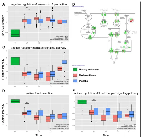
Modulation of the host response toward immunosuppression
[image:5.595.58.537.87.392.2]down-modulation of genes involved in both positive T cell selection (GO:0043368, Fig. 4d; pvalue < 10-6), and regulation of T cell receptor pathway (GO:0050862, Fig. 4e;pvalue < 10-6).
Discussion
Here, we took advantage of a prospective, randomized, double-blind study to assess whole blood transcriptional modulation in severe burn shock and according to hydrocortisone administration. We identified wide and persistent modulation of gene expression over the first week after shock, whereas hydrocortisone-associated
transcriptional modulation was moderate and transient. We also characterized the impact of both shock and hydrocortisone on the immune response, and showed that hydrocortisone transcriptionally enhanced the im-munosuppressive mechanisms that occur after severe burn injury.
[image:6.595.58.543.89.474.2]mesenchymal cells, respectively. Regarding the blood transcriptome of burn patients, a first dataset (GSE19743, buffy-coat samples) from 57 patients and 63 healthy volunteers (analyzed in [37–39]) showed that genes modulated within 10 days after injury were mainly related to immunity. Genes modulated at a later stage (11–49 days) were also involved in metabolism and apoptosis [39]. Our results were consistent regarding the identified functional pathways. We showed that modula-tion was triggered early after burns as most genes were
already modulated within 72 h of injury. A second data-set described the pooled-leukocytes transcriptome of 112 burn patients sampled within 7 days [40]. This study assessed prognostic factors and found a 39-gene signa-ture associated with a burn size >40%. These genes were related to platelet activation, TNF production, cellular adhesion, migration, and degranulation. Interestingly, our dataset shared 8 out of the 10 most modulated genes
(LCN2, LTF, THBS1, ITGA2B, CD24, TCN1, BPI and
SLC51A). Finally, we observed that most of the
[image:7.595.57.546.89.525.2]differentially expressed genes exhibited sustained modu-lation over time after burn injury. This is similar to a previous observation by Seok et al., whereby burn pa-tients exhibited the longest period of transcriptome “ re-covery time”[41].
The beneficial effect of hydrocortisone on the duration of septic shock is now widely accepted. We recently
[image:8.595.58.540.85.550.2]receptor expression in hydrocortisone-treated vs. placebo-treated patients. Several limitations might ex-plain this negative result. First, the effects of hydrocorti-sone might be tissue-specific and not seen in the whole blood transcriptome. Second, the GR is known to have both genomic and non-genomic effects [43], the latter being involved in the density of adrenergic receptors in vessels [44] and the modulation of agonist-induced con-tractions in vascular smooth muscle at multiple sites along signal transduction pathways.
Glucocorticoid receptor may modulate other pathways involved in vascular tone such as NO. Indeed, we ob-served that the NO-mediated signal transduction path-way was up-modulated early after burn, but returned to control values quicker in hydrocortisone-treated than in placebo-treated patients. This result is consistent with former literature, as mice lacking endothelial GR were found to have higher levels of NO, and increased hemodynamic instability [15]. Moreover, hydrocortisone administration to patients with septic shock was associ-ated with reduction in plasma nitrite/nitrate (indicative of lower NO formation) and of vasopressor support [17]. Taken together, these results are in favor of a hydrocortisone-induced modulation of NO balance that may explain positive hemodynamic effects in shock patients.
Glucocorticoids also have many side effects such as hyperglycemia, critical illness polyneuromyopathy [45], delayed wound healing, and immunosuppression. These side effects and the absence of convincing results for the effect on mortality may explain the wide heterogeneity of practice in hydrocortisone administration in septic shock. Wong et al. have recently shown in pediatric pa-tients with septic shock that corticosteroid administra-tion was associated with greater repression of adaptive immunity-related genes [19]. Despite well-matched groups in terms of severity, this study was retrospective, with no control over sampling time. Here, we confirmed the impact of hydrocortisone administration on host im-mune response in a different, but close model of
inflam-matory shock. Moreover, our prospective and
randomized design allowed us to follow the
hydrocortisone-related modulation of gene expression over a week.
Interestingly, along with greater repression of adaptive immunity, we also observed an impact of hydrocortisone on innate immunity. Indeed, the down-modulation of the antigen receptor-mediated pathway (Fig. 4b-c) was significantly greater at day 7 in the hydrocortisone group. This result was reminiscent of repressed mono-cyte expression of HLA-DR seen in various acute in-flammatory responses, including burns, where it was associated with the occurrence of secondary septic shock [46]. These results underline that hydrocortisone
administration may deepen the immunosuppression as-sociated with severe injury.
Interestingly, other groups have reported beneficial ef-fects of hydrocortisone administration in injury-related models. In severe trauma, the incidence of hospital-acquired pneumonia was lower in the hydrocortisone group [47]. The author’s hypothesis was that early hydrocortisone administration could blunt the hyper-inflammatory response associated with trauma, and pre-vent the subsequent associated immunosuppression. However, these results were not confirmed in a second multicenter trial published recently [48]. In combination with our current results, this raises the question of: (1) the timing of hydrocortisone administration after injury, and (2) the duration of hydrocortisone administration. This also underlines the lack of tools to identify/stratify patients who may benefit from hydrocortisone.
Our study has several limitations. Despite an adequate design, the small sample size precluded us from asses-sing associations between hydrocortisone, host-response and outcomes such as mortality or secondary infections. As we selected only patients with severe shock (with >0.5 μg/kg/min norepinephrine), most of them had ex-tensive burns (median TBSA = 70% (48–84), Table 1) and we found no transcriptional modulation according to TBSA. Therefore, we cannot extrapolate to the host response modulation in every patient with burns. How-ever, this provided us with a very homogeneous cohort of patients, allowing us to more precisely decipher the pathways modulated after severe burn injury, and to identify similarities with inflammatory situations such as trauma and septic shock [49]. As described in Table 1, several confounding factors might have impacted the transcriptome modulation over time (ABSI, etomidate administration, etc.). We observed no significant differ-ence in the results when adjusting or not adjusting with these variables but the small sample size precludes a de-finitive conclusion. As all patients received blood trans-fusion during graft surgery, the impact of transtrans-fusion on transcriptome modulation could not be assessed. This deserves more specific evaluation in the future. More-over, as we did not collect whole blood cell counts ex-cept at admission, we were not able to verify if changes in the pattern of blood leukocytes may have impacted longitudinal gene expression. Surprisingly, hydrocorti-sone treatment was only associated with a few modu-lated genes. Our small sample size and stringent thresholds for probe set filtering might explain such re-sults. An additional explanation could be related to the profound basal modulation induced by burn injury,
which might limit our ability to detect all
inflammatory situation, for the first time. Finally, our data were limited to mRNA expression. We were not able to test correlation with either translational modula-tion, or functionality of the immune system. Demonstra-tion of altered immune funcDemonstra-tionality in burn patients is thus still pending.
Conclusions
In conclusion, we assessed the early transcriptional modulation of the host response to burn shock and to hydrocortisone administration. The initial response to burn shock encompasses wide and persistent genomic modulation, with a profound alteration of pathways as-sociated with metabolism and immunity. We identified down-modulation of both innate and adaptive immune responses during the first week after severe burn injury. We believe that these results support the need for more precise evaluation of the benefit/risk ratio of hydrocorti-sone administration in critical illness, where injury-induced immunosuppression may occur.
Additional files
Additional file 1: Figure S1.Flowchart of the study.aSchematic representation of the timing of sampling during the course of administration of hydrocortisone or placebo.dday,Ssample.b
Flowchart of the study describing the number of samples analyzed for each time point, and the pre-processing steps of the bioinformatics analysis. (PDF 61 kb)
Additional file 2: Table S2.Details of modulated probe sets and genes at each time point for analysis 1, comparing modulation of gene expression according to burn injury (A). and analysis 2, comparing the modulation of gene expression according to hydrocortisone treatment (B). Each cell provides the number of modulated probe sets (genes). (DOC 40 kb)
Additional file 3: Table S1.Functional annotation of genes modulated by burn shock, classified according to their modulation pattern. (DOCX 23 kb)
Abbreviations
ACTH:Adrenocorticotropic hormone; ADRB2:β2-Adrenoreceptor gene symbol; cDNA: Complementary Desoxyribo Nucleic Acid; GEO: Gene Expression Omnibus; GO: Gene Ontology; GR: Glucocorticoid receptor; HV: Healthy volunteers; IL: Interleukin; NO: Nitric oxide; NOS: Nitric oxide synthase; RNA: Ribonucleic acid; TNF: Tumor necrosis factor
Acknowledgements
We thank Dr Marc Bertin-Maghit, Dr Christophe Magnin, and Dr Laure Fayolle-Pivot, for their help in recruiting the patients in the prospective study. This project is part of Advanced Diagnostic for New Therapeutic Approaches (ADNA), a program dedicated to personalized medicine, coordinated by Insti-tut Mérieux and supported by the French public agency BPI France.
Funding
Funding was provided by author’s institutions (Hospices Civils de Lyon and bioMérieux) and was part of ADNA program.
Availability of data and materials
MIAME-compliant microarray data are available at the GEO website [GEO:GSE77791].
Authors’contributions
All authors made substantial contributions to the conception or design of the work (FV, GM, ST, JT), or the acquisition (ST, DL, MAC), analysis (MAC, JP, JT), or interpretation of data for the work (FV, JP, GM, AP, JT). They all contributed to drafting the work or revising it critically for important intellectual content. All authors gave final approval of the version to be published and agree to be accountable for all aspects of the work in ensuring that questions related to the accuracy or integrity of any part of the work are appropriately investigated and resolved.
Competing interests
JT, MAC, and AP are employee of bioMérieux. JP is a former employee of bioMérieux, and currently employed by Enterome Bioscience.
Consent for publication
Not applicable.
Ethics approval and consent to participate
The protocol was accepted by ethical committee on 15/02/2005 and was registered at clinicaltrial.gov (NCT00149123). All healthy volunteers and patients (or next-of-kin) gave a written informed consent before inclusion in the study.
Publisher’s Note
Springer Nature remains neutral with regard to jurisdictional claims in published maps and institutional affiliations.
Author details
1
EA7426, Université Claude Bernard Lyon 1, Hospices Civils de Lyon, bioMérieux ;“Pathophysiology of injury induced immunosuppression (PI3)”, hôpital E. Herriot, 5 place d’Arsonval, 69437 Lyon, France.2Hospices Civils de Lyon, Immunology laboratory, hôpital E. Herriot, 5 place d’Arsonval, 69437 Lyon, France.3Hospices Civils de Lyon, Burn ICU, Anesthesia and Critical Care Medicine department, hôpital E. Herriot, 5 place d’Arsonval, 69437 Lyon, France.
Received: 6 September 2016 Accepted: 26 May 2017
References
1. Franchimont D. Overview of the actions of glucocorticoids on the immune response: a good model to characterize new pathways of
immunosuppression for new treatment strategies. Ann NY Acad Sci. 2004; 1024:124–37.
2. Galon J, Franchimont D, Hiroi N, Frey G, Boettner A, Ehrhart-Bornstein M, et al. Gene profiling reveals unknown enhancing and suppressive actions of glucocorticoids on immune cells. FASEB J. 2002;16:61–71.
3. van de Beek D, de Gans J, McIntyre P, Prasad K. Corticosteroids for acute bacterial meningitis. Cochrane Database Syst Rev. 2007;12:CD004405. 4. Vanderheyde N, Verhasselt V, Goldman M, Willems F. Inhibition of human
dendritic cell functions by methylprednisolone. Transplantation. 1999;67:1342–7. 5. DeKruyff RH, Fang Y, Umetsu DT. Corticosteroids enhance the capacity of
macrophages to induce Th2 cytokine synthesis in CD4+ lymphocytes by inhibiting IL-12 production. J Immunol. 1998;160:2231–7.
6. Barrat FJ, Cua DJ, Boonstra A, Richards DF, Crain C, Savelkoul HF, et al. In vitro generation of interleukin 10-producing regulatory CD4(+) T cells is induced by immunosuppressive drugs and inhibited by T helper type 1 (Th1)- and Th2-inducing cytokines. J Exp Med. 2002;195:603–16.
7. Kalil AC, Sun J. Low-dose steroids for septic shock and severe sepsis: the use of Bayesian statistics to resolve clinical trial controversies. Intensive Care Med. 2011;37:420–9.
8. Bone RC, Fisher CJ, Clemmer TP, Slotman GJ, Metz CA, Balk RA. A controlled clinical trial of high-dose methylprednisolone in the treatment of severe sepsis and septic shock. N Engl J Med. 1987;317:653–8.
9. Slotman GJ, Fisher CJ, Bone RC, Clemmer TP, Metz CA. Detrimental effects of high-dose methylprednisolone sodium succinate on serum
10. Annane D, Sébille V, Charpentier C, Bollaert P-E, François B, Korach J-M, et al. Effect of treatment with low doses of hydrocortisone and fludrocortisone on mortality in patients with septic shock. JAMA. 2002;288:862–71. 11. Marik PE. Glucocorticoids in sepsis: dissecting facts from fiction. Crit Care.
2011;15:158.
12. Sherwin RL, Garcia AJ, Bilkovski R. Do low-dose corticosteroids improve mortality or shock reversal in patients with septic shock? A systematic review and position statement prepared for the American Academy of Emergency Medicine. J Emerg Med. 2012;43:7–12.
13. Dellinger RP, Levy MM, Rhodes A, Annane D, Gerlach H, Opal SM, et al. Surviving Sepsis Campaign: international guidelines for management of severe sepsis and septic shock, 2012. Intensive Care Med. 2013;39:165–228. 14. Annane D, Bellissant E, Bollaert PE, Briegel J, Keh D, Kupfer Y. Corticosteroids
for treating sepsis. Cochrane Database Syst Rev. 2015;12:CD002243. 15. Goodwin JE, Feng Y, Velazquez H, Sessa WC. Endothelial glucocorticoid
receptor is required for protection against sepsis. Proc Natl Acad Sci U S A. 2013;110:306–11.
16. Duma D, Silva-Santos JE, Assreuy J. Inhibition of glucocorticoid receptor binding by nitric oxide in endotoxemic rats. Crit Care Med. 2004;32:2304–10. 17. Keh D, Boehnke T, Weber-Cartens S, Schulz C, Ahlers O, Bercker S, et al.
Immunologic and hemodynamic effects of“low-dose”hydrocortisone in septic shock. Am J Respir Crit Care Med. 2003;167:512–20.
18. Hotchkiss RS, Monneret G, Payen D. Immunosuppression in sepsis: a novel understanding of the disorder and a new therapeutic approach. Lancet Infect Dis. 2013;13:260–8.
19. Wong HR, Cvijanovich NZ, Allen GL, Thomas NJ, Freishtat RJ, Anas N, et al. Corticosteroids are associated with repression of adaptive immunity gene programs in pediatric septic shock. Am J Respir Crit Care Med. 2014;189:940–6. 20. Venet F, Plassais J, Textoris J, Cazalis M-A, Pachot A, Bertin-Maghit M, et al.
Low-dose hydrocortisone reduces norepinephrine duration in severe burn patients: a randomized clinical trial. Crit Care. 2015;19:21.
21. Gentleman RC, Carey VJ, Bates DM, Bolstad B, Dettling M, Dudoit S, et al. Bioconductor: open software development for computational biology and bioinformatics. Genome Biol. 2004;5:R80.
22. R Development Core Team. A language and environment for statistical computing. Vienna: R foundation for statistical Computing; 2008. 23. Wilson CL, Miller CJ. Simpleaffy: a BioConductor package for Affymetrix
Quality Control and data analysis. Bioinformatics. 2005;21:3683–5. 24. Kauffmann A, Gentleman R, Huber W. arrayQualityMetrics–a bioconductor
package for quality assessment of microarray data. Bioinformatics. 2009;25:415–6. 25. Culhane AC, Thioulouse J, Perrière G, Higgins DG. MADE4: an R package for
multivariate analysis of gene expression data. Bioinformatics. 2005;21:2789–90. 26. Wu Z, Irizarry RA. Stochastic models inspired by hybridization theory for
short oligonucleotide arrays. J Comput Biol. 2005;12:882–93.
27. Chen M, Shi L, Kelly R, Perkins R, Fang H, Tong W. Selecting a single model or combining multiple models for microarray-based classifier development? A comparative analysis based on large and diverse datasets generated from the MAQC-II project. BMC Bioinformatics. 2011;12 Suppl 10:S3.
28. Smyth GK. Linear models and empirical bayes methods for assessing differential expression in microarray experiments. Stat Appl Genet Mol Biol. 2004;3:Article3.
29. Benjamini Y, Hochberg Y. Controlling the false discovery rate: a practical and powerful approach to multiple testing. J R Stat Soc B. 1995;57:289–300. 30. Gene Ontology Consortium. Gene Ontology Consortium: going forward.
Nucleic Acids Res. 2015;43:D1049–1056.
31. Moreno R, Sprung CL, Annane D, Chevret S, Briegel J, Keh D, et al. Time course of organ failure in patients with septic shock treated with hydrocortisone: results of the Corticus study. Intensive Care Med. 2011;37: 1765–72.
32. Tzika AA, Mintzopoulos D, Mindrinos M, Zhang J, Rahme LG, Tompkins RG. Microarray analysis suggests that burn injury results in mitochondrial dysfunction in human skeletal muscle. Int J Mol Med. 2009;24:387–92. 33. Dasu MRK, Barrow RE, Herndon DN. Gene expression changes with time in
skeletal muscle of severely burned children. Ann Surg. 2005;241:647–53. 34. Greco III JA, Pollins AC, Boone BE, Levy SE, Nanney LB. A microarray analysis
of temporal gene expression profiles in thermally injured human skin. Burns. 2010;36:192–204.
35. Ou S, Liu G-D, Tan Y, Zhou L-S, Bai S-R, Xue G, et al. A time course study about gene expression of post-thermal injury with DNA microarray. Int J Dermatol. 2015;54:757–64.
36. Peterson JR, De La Rosa S, Eboda O, Cilwa KE, Agarwal S, Buchman SR, et al. Treatment of heterotopic ossification through remote ATP hydrolysis. Sci Transl Med. 2014;6:255ra132.
37. Zhou B, Xu W, Herndon D, Tompkins R, Davis R, Xiao W, et al. Analysis of factorial time-course microarrays with application to a clinical study of burn injury. Proc Natl Acad Sci U S A. 2010;107:9923–8.
38. Zhang Y, Tibshirani R, Davis R. Classification of patients from time-course gene expression. Biostatistics. 2013;14:87–98.
39. Li Z, Wang Q, Yu H, Zou K, Xi Y, Mi W, et al. Screening of key genes in severe burn injury at different stages via analyzing gene expression data. J Burn Care Res. 2014;37:e254–62.
40. Sood RF, Gibran NS, Arnoldo BD, Gamelli RL, Herndon DN, Tompkins RG, et al. Early leukocyte gene expression associated with age, burn size, and inhalation injury in severely burned adults. J Trauma Acute Care Surg. 2015; 80:250–7.
41. Seok J, Warren HS, Cuenca AG, Mindrinos MN, Baker HV, Xu W, et al. Genomic responses in mouse models poorly mimic human inflammatory diseases. Proc Natl Acad Sci U S A. 2013;110:3507–12.
42. Whitworth JA, Schyvens CG, Zhang Y, Mangos GJ, Kelly JJ. Glucocorticoid-induced hypertension: from mouse to man. Clin Exp Pharmacol Physiol. 2001;28:993–6.
43. Prigent H, Maxime V, Annane D. Science review: mechanisms of impaired adrenal function in sepsis and molecular actions of glucocorticoids. Crit Care. 2004;8:243–52.
44. Haigh RM, Jones CT. Effect of glucocorticoids on alpha 1-adrenergic receptor binding in rat vascular smooth muscle. J Mol Endocrinol. 1990;5:41–8. 45. Kress JP, Hall JB. ICU-acquired weakness and recovery from critical illness. N
Engl J Med. 2014;370:1626–35.
46. Venet F, Tissot S, Debard A-L, Faudot C, Cramp C, Pachot A, et al. Decreased monocyte human leukocyte antigen-DR expression after severe burn injury: Correlation with severity and secondary septic shock. Crit Care Med. 2007; 35:1910–7.
47. Roquilly A, Mahe P, Seguin P, et al. Hydrocortisone therapy for patients with multiple trauma: the randomized controlled hypolyte study. JAMA. 2011; 305:1201–9.
48. Asehnoune K, Seguin P, Allary J, Feuillet F, Lasocki S, Cook F, et al. Hydrocortisone and fludrocortisone for prevention of hospital-acquired pneumonia in patients with severe traumatic brain injury (Corti-TC): a double-blind, multicentre phase 3, randomised placebo-controlled trial. Lancet Respir Med. 2014;2:706–16.
49. Xiao W, Mindrinos MN, Seok J, Cuschieri J, Cuenca AG, Gao H, et al. A genomic storm in critically injured humans. J Exp Med. 2011;208:2581–90.
• We accept pre-submission inquiries
• Our selector tool helps you to find the most relevant journal
• We provide round the clock customer support
• Convenient online submission
• Thorough peer review
• Inclusion in PubMed and all major indexing services
• Maximum visibility for your research
Submit your manuscript at www.biomedcentral.com/submit
![Fig. 2 Modulation of the glucocorticoid receptor and nitric oxide signaling pathways.patients (pathway [GO:0007263] in HV (differences was assessed using repeated measures analysis of variance taking into account treatment, time and gene (probes) effects](https://thumb-us.123doks.com/thumbv2/123dok_us/8349723.309028/6.595.58.543.89.474/modulation-glucocorticoid-receptor-signaling-differences-assessed-repeated-treatment.webp)