0022-538X/10/$12.00 doi:10.1128/JVI.02412-09
Copyright © 2010, American Society for Microbiology. All Rights Reserved.
p62/Sequestosome-1 Associates with and Sustains the Expression of
Retroviral Restriction Factor TRIM5
␣
䌤
†
Christopher O’Connor,
4Thomas Pertel,
1Seth Gray,
4Seth L. Robia,
2Joanna C. Bakowska,
3Jeremy Luban,
1and Edward M. Campbell
4*
Department of Microbiology and Molecular Medicine, University of Geneva, Geneva, Switzerland1; Department of Physiology,
Stritch School of Medicine, Loyola University Chicago, Maywood, Illinois2; Department of Pharmacology, Stritch School of
Medicine, Loyola University Chicago, Maywood, Illinois3; and Department of Microbiology and
Immunology, Stritch School of Medicine, Loyola University Chicago, Maywood, Illinois4
Received 14 November 2009/Accepted 19 March 2010
TRIM5 proteins mediate a potent block to the cross-species transmission of retroviruses, the most well known being the TRIM5␣protein from rhesus macaques, which potently inhibits human immunodeficiency virus type 1 (HIV-1) infection. This restriction occurs at an early stage in the replication cycle and is mediated by the binding of TRIM5 proteins to determinants present in the retroviral capsid. TRIM5␣, as well as other TRIM family proteins, has been shown to be regulated by interferons (IFN). Here we show that TRIM5␣ associates with another IFN-induced gene, sequestosome-1/p62 (p62). p62 plays a role in several signal transduction cascades that are important for maintaining the antiviral state of cells. Here we demonstrate that p62 localizes to both human and rhesus macaque TRIM5␣cytoplasmic bodies, and fluorescence resonance energy transfer (FRET) analysis demonstrates that these proteins closely associate in these structures. When p62 expression was knocked down via small interfering RNA (siRNA), the number of TRIM5␣cytoplasmic bodies and the level of TRIM5␣protein expression were reduced in cell lines stably expressing epitope-tagged versions of TRIM5␣. In accordance with these data, p62 knockdown resulted in reduced TRIM5␣-mediated retroviral restriction in cells expressing epitope-tagged TRIM5␣or expressing endogenously expressed human TRIM5␣. p62 may therefore operate to enhance TRIM5␣-mediated retroviral restriction, contributing to the antiviral state of cells following IFN treatment.
The TRIM family of proteins, which are defined by the presence of atripartitemotif, consisting of RING, B-box 2, and coiled-coil domains (31), are emerging as components of an innate antiviral response that operate in several ways to inhibit viral replication (reviewed in reference 28). TRIM5␣is a mem-ber of the TRIM family of proteins that has evolved to bind determinants present on retroviral capsids, an interaction that potently inhibits retroviral replication at an early postentry stage of infection (reviewed in references 19 and 35). Since the discovery of TRIM5␣as a retroviral restriction factor, numer-ous other TRIM family proteins have been shown to possess antiviral activities that act at several stages of the viral life cycle (4, 14, 16, 42, 43).
TRIM5␣proteins localize to discrete accumulations in the cytoplasm referred to as cytoplasmic bodies (6, 39, 40). Many other TRIM family proteins form similar accumulations in the cytoplasm or nucleus (31). We have previously utilized live-and fixed-cell microscopy to demonstrate an interaction occur-ring between fluorescently labeled human immunodeficiency virus type 1 (HIV-1) virions and rhesus macaque TRIM5␣ (rhTRIM5␣) cytoplasmic bodies (8). These studies also found that proteasome inhibitors caused the stable accumulation of HIV-1 virions in enlarged cytoplasmic bodies that stained
pos-itively for ubiquitin. These data are in accordance with our biochemical studies that have shown that proteasome inhibi-tion relieves the TRIM5␣-mediated block of viral reverse tran-scription (RT) but does not relieve the ability of TRIM5␣to prevent infection (1, 46). It has also been reported that TRIM5␣ is rapidly turned over, in a proteasome-sensitive manner, following the addition of restriction-sensitive virus to cells (33). Collectively, these findings suggest that restriction occurs via a two-step mechanism. The first step, which includes binding of TRIM5␣to the viral capsid, is sufficient to prevent infection. The second step, which is sensitive to proteasome inhibition, results in the abortive disassembly of the viral capsid in a manner that prevents the formation of viral reverse tran-scription products.
It has also recently been demonstrated that TRIM5␣(3, 10, 34) and numerous other members of the TRIM family (9) are transcriptionally upregulated following interferon (IFN) treat-ment. Therefore, the TRIM family of proteins may collectively contribute to the cellular antiviral state induced by interferon treatment.
Sequestosome-1/p62 (p62) is an interferon-inducible protein (24) that has been implicated in various cellular processes, including regulation of cellular degradative pathways and mod-ulation of signaling pathways (reviewed in references 27 and 37). Notably, p62 contains a ubiquitin binding domain (UBA) through which it binds ubiquitinated cargoes and facilitates their proteasomal degradation via an interaction of its N ter-minus with proteasomal subunits (17, 27, 36, 44). p62 also functions as a scaffolding/adaptor protein in signaling cascades involved in regulating cytokine production or signaling. p62
* Corresponding author. Mailing address: 2160 S. First Ave., May-wood, IL 60153. Phone: (708) 216-3345. Fax: (708) 216-3913. E-mail: ecampbell@lumc.edu.
† Supplemental material for this article may be found at http://jvi .asm.org/.
䌤Published ahead of print on 31 March 2010.
5997
on November 8, 2019 by guest
http://jvi.asm.org/
regulates the activity of interferon regulatory factor 8 (IRF8) (24) and p38 mitogen-activated protein kinase (MAPK) fol-lowing cytokine stimulation (20, 41). p62 also interacts with tumor necrosis factor receptor-associated protein 6 (TRAF6) and enhances its activity (24, 45). TRAF6 is an E3 ubiquitin ligase that undergoes enhanced autoubiquitylation in the presence of p62, which activates the NF-B signaling pathway (11, 24, 45). This p62-mediated clearance of ubiquiti-nated proteins by the proteasome, as well as the ability to modulate signaling pathways associated with the innate im-mune response, prompted us to examine what role p62 may play in the ability of TRIM5␣, itself an E3 ligase capable of autoubiquitination (47), to restrict retroviral infection.
In this study, we demonstrate that p62 associates with both rhesus macaque and human forms of TRIM5␣. p62 localizes to TRIM5␣cytoplasmic bodies in cells. We further demonstrate an interaction between these two proteins by using immunoprecipitation and fluorescence resonance energy transfer (FRET). As p62 has been reported to bind polyubiq-uitinated proteins and facilitate their proteasomal degradation (36) and we have previously demonstrated a proteasome-sen-sitive step in the restriction process (1, 8, 46), we originally hypothesized that p62 may play a role in the process of restric-tion. However, small interfering RNA (siRNA) knockdown of p62 decreased TRIM5␣expression in the absence of restric-tion-sensitive virus. This reduced TRIM5␣-mediated restric-tion in cells exogenously expressing epitope-tagged versions of TRIM5␣ as well as in cells expressing endogenous levels of TRIM5␣. Collectively, these results suggest that TRIM5␣ is stabilized by its interaction with p62. As both of these proteins are IFN inducible, this interaction may act to enhance the expression of TRIM5␣ following IFN treatment, thereby en-hancing the antiviral potential of TRIM5␣.
MATERIALS AND METHODS
Cells and pharmaceuticals.293T and HeLa cell lines were obtained from the American Type Culture Collection. Hemagglutinin-tagged rhesus macaque TRIM5␣(HA-rhTRIM5␣)- and human TRIM5␣(HA-huTRIM5␣)-expressing HeLa cells were gifts from J. Sodroski (Harvard Medical School, Boston, MA). Cells expressing yellow fluorescent protein-tagged rhTRIM5␣(YFP-rhTRIM5␣) have been described previously (6). 293T cells stably expressing HA-rhTRIM5␣ were a gift from C. Aiken (33). Cells were cultured in Dulbecco’s modified Eagle’s medium (DMEM) supplemented with 10% fetal bovine serum (FBS) (HyClone), 100 U/ml penicillin, 100g/ml streptomycin, and 10g/ml cipro-floxacin. Cells were maintained in the presence of 5% CO2at 37°C. HeLa cells were transfected with Lipofectamine 2000 (Invitrogen) according to the manu-facturer’s protocol.
cDNA constructs.Green fluorescent protein-tagged p62 (GFP-p62) was a generous gift from Marie Wooten (Auburn University, Auburn, AL). GFP was replaced with YFP and mCherry in this construct, utilizing the flanking AgeI and BsrGI sites. rhTRIM5␣and huTRIM5␣plasmids have been described previously (6).
Immunofluorescence microscopy.Cells were allowed to adhere to fibronectin-treated glass coverslips and fixed with 3.7% formaldehyde (Polysciences) in 0.1 M PIPES [piperazine-N,N⬘-bis(2-ethanesulfonic acid)], pH 6.8. We used the following primary antibodies: mouse anti-p62 antibody (BD Life Sciences), rab-bit anti-p62 antibody (Santa Cruz Biotechnology), and rabrab-bit and mouse anti-HA antibodies (Sigma). Primary antibodies were secondarily labeled with fluoro-phore-conjugated donkey anti-mouse or anti-rabbit antibody (Jackson Immu-noResearch). Images were collected with a DeltaVision microscope (Applied Precision) equipped with a digital camera (CoolSNAP HQ; Photometrics), using a 1.4-numerical aperture (NA) 100⫻objective lens, and were deconvolved with SoftWoRx deconvolution software (Applied Precision).
Transfections.Plasmid transfections were performed using Lipofectamine 2000 (Invitrogen) according to the manufacturer’s protocol. For FRET
ex-periments, pcDNA 3.1, cyan fluorescent protein-tagged rhTRIM5␣ (CFP-rhTRIM5␣), and YFP-p62 were transfected in a 1:1:1 ratio, and FRET analysis was performed within 18 h of transfection. Control and p62-specific siRNAs (Santa Cruz Biotechnology) were transfected using Lipofectamine 2000 twice within a 24-h period.
Immunoprecipitation assay.ZsGreen1 (Clontech), p62 isoform 1 (GenBank accession no. NM_003900), and p62 isoform 2 (GenBank accession no. NM_ 001142298) cDNAs were cloned into pcDNA3.1 (XbaI-NotI) containing a 3⫻ FLAG motif in order to create N-terminal epitope-tagged proteins. Human TRIM5␣(GenBank accession no. AY625000) cDNA was cloned into pcDNA3.1 (XbaI-XhoI) containing a flexible SGGGG linker and a Myc-6⫻His tag in order to generate a C-terminal Myc-His-tagged fusion protein. Subconfluent 293T cells were transfected with 16g total plasmid DNA by use ofTransIT-LT1 (Mirus) according to the manufacturer’s instructions. Thirty-six hours after transfection, cells were lysed in lysis buffer containing 50 mM Tris, pH 7.4, 150 mM NaCl, and 1% CHAPSO (Pierce), with a cocktail of protease (Roche) and phosphatase (Sigma) inhibitors. Cells were lysed on ice for 20 min, and lysates were clarified by centrifugation at 14,000⫻gfor 15 min at 4°C. Two micrograms of mouse anti-FLAG (clone M2; Sigma) was conjugated to 50l Dynabeads protein G magnetic beads (Invitrogen) and added to 1 ml of clarified lysate. After 2 h of incubation at 4°C, the beads were washed five times with lysis buffer. The beads were resuspended in 100l of 1⫻Laemmli sample buffer and incubated at 100°C for 5 min. After sodium dodecyl sulfate-polyacrylamide gel electrophoresis (SDS-PAGE), proteins were transferred overnight onto a polyvinylidene diflu-oride (PVDF) membrane (Bio-Rad), which was probed with polyclonal rabbit anti-c-Myc and rabbit anti-FLAG antibodies (Sigma).
Fluorescence resonance energy transfer.FRET was quantified by acceptor photobleaching, performed as previously described (22). Briefly, images were obtained by automated fluorescence microscopy using motorized filter wheels and a cooled charge-coupled-device (CCD) camera. Progressive acceptor pho-tobleaching was performed as follows: 60 images were obtained at 10-s intervals for both the donor (CFP: excitation, 427/10 nm; emission, 473/30 nm; 100-ms exposure) and the acceptor (YFP: excitation, 504/12 nm; emission, 542/27 nm; 40-ms exposure), with a period of acceptor photobleaching (excitation, 504/12 nm) between each acquisition. The CFP/YFP fluorescence intensity of each cell in the field was quantified using Metamorph (Molecular Devices Corp., Down-ingtown, PA), and FRET efficiency (E) was calculated from the CFP initial (Fprebleach) and final (Fpostbleach) fluorescence values, according to the equation
E⫽1⫺(Fprebleach/Fpostbleach).
Fluorescence imaging was performed with an inverted microscope equipped with a 1.49-numerical aperture objective lens and a back-thinned CCD camera (iXon 887; Andor Technology, Belfast, Northern Ireland). The detector was cooled to⫺100°C, using a recirculating liquid coolant system (Koolance, Inc., Auburn, WA). Image acquisition and acceptor photobleaching were automated with custom software macros in Metamorph that controlled motorized excita-tion/emission filter wheels (Sutter Instrument Co., Novato, CA) with filters for CFP/YFP/mCherry (Semrock, Rochester NY). The progressive photobleaching protocol was as follows: 100-ms acquisition of the CFP image and 40-ms acqui-sition of the YFP image, followed by a 10-s exposure to YFP-selective photo-bleaching (504/12-nm excitation).
Virus production and infections.To produce virus, a 10-cm plate of 293T cells was transfected with 12g of R7⌬EnvGFP and 8g of vesicular stomatitis virus glycoprotein (VSV-g) to make HIV-1 reporter virus. Cells were transfected with 7g pCigB or pCigN packaging plasmid, 7g GFP or YFP reporter vector, and 7g VSV-g to generate murine leukemia virus (MLV) reporter virus. Transfec-tions were performed using polyethylenimine (PEI) (molecular weight, 25,000; Polysciences) as previously described (8), utilizing a PEI-DNA ratio of 2:1 for each transfection. To assess virus infectivity, equivalent numbers of cells in a 24-well plate were infected for 14 h; then, virus was removed, normal medium was added, and GFP expression was determined 48 to 72 h after infection by using a fluorescence-activated cell sorter (FACS) Canto II flow cytometer (Bec-ton Dickinson). MLV was titrated on CRFK cells to normalize virus input, as previously described (1).
Quantitative PCR.Quantitation of viral RT products was performed as pre-viously described (1, 7). Briefly, cells were plated in equivalent numbers in a 12-well plate and infected for 18 h overnight. Following incubation with RNase A, genomic DNA was harvested using a DNeasy tissue kit (Qiagen) according to the manufacturer’s instruction. Genomic DNA was digested with DpnI to re-move residual plasmid DNA. Proviral DNA was quantified using primers specific for HIV-1 late RT products (7) or the GFP cassette present in MLV vectors (1) with SYBR green PCR reagent (Applied Biosystems) on an Eppendorf Real-plex2
ep gradient Mastercycler and normalized to the number of genomic-actin
5998 O’CONNOR ET AL. J. VIROL.
on November 8, 2019 by guest
http://jvi.asm.org/
copies present in an identical sample to calculate the amount of proviral DNA per-actin copy.
Western blot analysis.Whole-cell lysates were prepared by treating 1⫻105 cells with lysis buffer (100 mM Tris, pH 8.0, 1% NP-40, 150 mM NaCl) containing protease inhibitor cocktail (Roche) for 15 min on ice. The protein concentration was determined using a Coomassie Plus Bradford assay (Thermo Scientific). After lysis of cells, 2⫻SDS sample buffer was added and samples were boiled for 5 min. Equal amounts of protein were loaded into a 10% polyacrylamide gel for SDS-PAGE. After separation of proteins via SDS-PAGE, proteins were trans-ferred to PVDF membranes and detected by incubation with the following antibodies: anti-p62 (BD Life Sciences), anti-HA (clone 3F10) conjugated to horseradish peroxidase (HRP) (Roche), and anti-GFP (Invitrogen). Secondary antibodies conjugated to HRP (Thermo Scientific) were used where necessary, and antibody complexes were detected using SuperSignal West Femto chemilu-minescent substrate (Thermo Scientific). Chemiluminescence was detected using a UVP EC3 imaging system (UVP LLC).
Image analysis.Deconvolved images were analyzed for p62 mean fluorescence intensity (MFI) in cytoplasmic bodies by use of the Surface Finder function of the Imaris software package (Bitplane). Surfaces for cytoplasmic bodies in all samples analyzed were defined by using a fluorescence threshold (600 relative fluorescence units) for YFP-TRIM5␣, and all YFP-rhTRIM5␣bodies over a volume of 0.011m3were used in the analysis. For each YFP-rhTRIM5␣ cytoplasmic body, the p62 MFI was determined and the data were plotted in Prism (Graphpad Software Inc.) for statistical analysis. An unpairedttest with Welch’s correction was used to determine the statistical significance between samples (Pⱕ0.05). To determine the number of cytoplasmic bodies per cell, cytoplasmic-body surfaces were selected as mentioned above. The number of cytoplasmic bodies was determined for each treatment studied and plotted in Prism for statistical analysis.
RESULTS
p62 localizes in rhTRIM5␣cytoplasmic bodies. p62/seques-tosome-1 (p62) is an adapter protein that has been reported to bind ubiquitinated proteins and facilitate their proteasomal degradation (17, 36). Using an antibody to endogenously ex-pressed p62, we investigated whether p62 localized to cytoplas-mic bodies in HeLa cells stably expressing epitope-tagged rhTRIM5␣proteins (6, 40).
As shown in Fig. 1, p62 localized prominently to YFP-tagged rhTRIM5␣cytoplasmic bodies. Similarly, p62 also localized to the cytoplasmic bodies present in a HA-rhTRIM5␣HeLa cell line (see Fig. S1A in the supplemental material). p62 also localized to punctate accumulations in HeLa cells not express-ing exogenous TRIM5␣ protein (see Fig. S1C in the supple-mental material).
We also transduced the monocytic leukemia cell line THP-1 with YFP-rhTRIM5␣ and activated these cells with phorbol myristate acetate (PMA) to differentiate them into a macro-phage-like cell line. YFP-rhTRIM5␣also colocalized with p62 in activated THP-1 cells (see Fig. S1B in the supplemental material). These results demonstrate that p62 colocalization was not limited to HeLa-derived cell lines.
[image:3.585.45.542.71.368.2]We utilized immunofluorescence to observe the localization of p62 in a HeLa cell line stably expressing a HA-tagged version
FIG. 1. p62 localizes to rhTRIM5␣cytoplasmic bodies. HeLa cells stably expressing YFP-tagged rhTRIM5␣(6) were fixed and immunostained with mouse anti-p62 (␣p62) monoclonal antibody (MAb).Z-stack images were collected and deconvolved using DeltaVision deconvolution software. Images shown are from one individualZsection. Individual channel images shown in the left and center panels have been superimposed in the merged panels on the right. The lower three panels represent enlarged magnifications of the areas in the white boxes in the upper panels. The data shown here are representative of three independent experiments.
on November 8, 2019 by guest
http://jvi.asm.org/
of human TRIM5␣(HA-huTRIM5␣). As we have reported pre-viously (6), when GFP-tagged versions of huTRIM5␣were uti-lized, HA-huTRIM5␣localized to both filamentous and spher-ical cytoplasmic accumulations in HeLa cells (see Fig. S2 in the supplemental material). When these cells were stained for p62 expression, p62 localized to both spherical (see Fig. S2A) and filamentous (see Fig. S2B) accumulations of huTRIM5␣ pro-tein. Noticeably, p62 was observed to localize to the tips of the filamentous accumulations of huTRIM5␣.
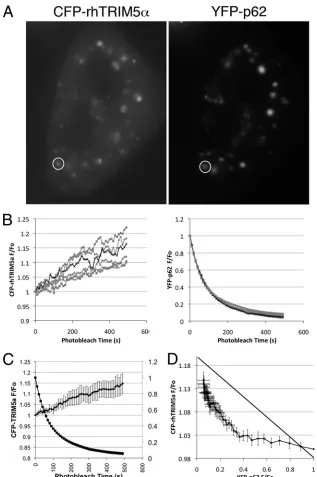
Direct interaction of p62 and TRIM5␣ in HeLa cells. To determine whether p62 and rhTRIM5␣physically interact, we transiently transfected 293T cells with huTRIM5␣containing a C-terminal Myc epitope tag (huTRIM5␣-Myc) and a FLAG-tagged p62 or control ZsGreen1 vector. Cell lysates were then immunoprecipitated using an anti-FLAG antibody to immunoprecipitate FLAG-tagged p62 proteins and analyzed by Western blotting for the presence of huTRIM5␣-Myc in these complexes. As shown in Fig. 2 (top), TRIM5␣ coimmu-noprecipitated with p62 isoform 1, whereas TRIM5␣failed to coimmunoprecipitate with ZsGreen1 or p62 isoform 2, which lacks the first 84 amino acids of the N-terminal region of isoform 1. Therefore, the PB1 domain of p62 isoform 1 (amino acids 3 to 102 [SMART; http://smart.embl-heidelberg.de/]), which is involved in p62 oligomerization and protein-protein interactions (26), appears to be required for binding TRIM5␣. We also utilized FRET to examine the interaction occurring between rhTRIM5␣and p62 in the smaller cytoplasmic bodies in which both of these proteins localize. FRET is the nonra-diative energy transfer from an excited fluorescent donor to an acceptor fluorophore in very close proximity. The 5-nm Fo¨rster transfer distance (R0) for the CFP-YFP pair (32) is on the molecular scale; thus, CFP-YFP FRET suggests that the target
proteins are bound to one another or to the same macromo-lecular complex. FRET is commonly measured by acceptor photobleaching, wherein FRET is quantified as an increase in the fluorescence of the donor fluorophore following the pho-tobleaching of the acceptor fluorophore (23). We therefore measured FRET to examine the interaction occurring between rhTRIM5␣ and p62 in 11 independent cytoplasmic bodies in which both of these proteins could be observed to localize. We utilized CFP-rhTRIM5␣and YFP-p62 in acceptor photo-bleaching FRET experiments. In these experiments, YFP-p62 (acceptor) was iteratively photobleached, and the fluores-cence intensities of CFP-rhTRIM5␣ (donor) and YFP-p62 were measured in regions of the cell in which both CFP-rhTRIM5␣and YFP-p62 could be observed to localize to an individual, immobile cytoplasmic body (one of the 11 cytoplasmic bodies is represented in Fig. 3A; also see Movie S1 in the sup-plemental material). We observed an exponential decrease in YFP-p62 fluorescence, with a concomitant increase in CFP-rhTRIM5␣ fluorescence, in all 11 cytoplasmic bodies analyzed (Fig. 3B). We also examined the stoichiometry of this complex by studying the relationship between CFP and YFP fluorescence in our experiments. Notably, the increase in CFP-rhTRIM5␣ fluo-rescence lagged significantly behind the decrease in YFP-p62 fluorescence (Fig. 3C), and consequently the relationship be-tween CFP-rhTRIM5␣and YFP-p62 was highly nonlinear (Fig. 3D). Such a deviation from linearity is observed for multimeric complexes containing multiple FRET acceptors (5, 22). The de-gree of curvature is related to the number of probes in the com-plex and the probe separation distance (25). When the average ratios of the measured fluorescence relative to the original fluo-rescence (F/F0) for CFP-rhTRIM5␣and YFP-p62 (Fig. 3C) were replotted to examine the donor fluorescence versus the acceptor fluorescence, this plot deviated from linearity (Fig. 3D). Such a deviation is expected if the ratio of acceptor subunits (YFP-p62) to donor subunits (CFP-rhTRIM5␣) is ⬎2. This suggests that more than one p62 protein is associated with each rhTRIM5␣ protein in these cytoplasmic-body complexes. Collectively, these data suggest that TRIM5␣and p62 are closely associated in the cytoplasmic bodies in which they both localize.
p62 knockdown reduces rhTRIM5␣cytoplasmic-body local-ization and protein expression.To ensure that the protein iden-tified as localizing to TRIM5␣ cytoplasmic bodies was indeed endogenously expressed p62, we performed siRNA knockdown of p62 in HeLa cells stably expressing YFP-rhTRIM5␣. Follow-ing 2 transfections with control or p62-specific siRNA, the immunoreactive protein identified by p62 antibodies was re-duced in cells treated with p62-specific siRNA but not in cells treated with control siRNA (see Fig. S3 in the supplemental material). We utilized quantitative image analysis methods to measure the amount of p62 fluorescence associated with YFP-rhTRIM5␣ cytoplasmic bodies. Three-dimensional surfaces de-fining YFP-rhTRIM5␣cytoplasmic bodies were identified in an automated fashion, using fixed fluorescence intensity and size criteria. The p62 fluorescence associated with these sur-faces was then quantified (Fig. 4A). In untreated cells or cells treated with control siRNA, the vast majority of cytoplasmic bodies exhibited p62 fluorescence greater than that observed in cells stained with secondary antibody only. Eighty-nine percent of the bodies in untreated cells and 86% of the bodies trans-fected with control siRNA exhibited p62-associated
fluores-FIG. 2. huTRIM5␣coimmunoprecipitates with p62. huTRIM5␣ -Myc was transfected along with the indicated FLAG-tagged constructs in 293T cells. Detergent extracts were incubated with mouse anti-FLAG antibody conjugated to protein G magnetic beads. Immuno-complexes were isolated and analyzed by SDS-PAGE/Western blotting (WB). The top panel represents coimmunoprecipitated TRIM5␣-Myc probed with rabbit anti-c-Myc antibody, the middle panel represents the TRIM5␣-Myc input (total lysate) probed with rabbit anti-c-Myc antibody, and the bottom panel represents immunoprecipitated (IP) FLAG-tagged proteins probed with rabbit anti-FLAG antibody. The data shown here are representative of three independent experiments. Iso, isoform.
6000 O’CONNOR ET AL. J. VIROL.
on November 8, 2019 by guest
http://jvi.asm.org/
[image:4.585.92.237.68.241.2]cent signals greater than background, which was defined as the maximum mean fluorescence measured in the 765 cytoplasmic bodies analyzed in cells treated with only secondary antibody. In contrast, 0.6% of the cytoplasmic bodies analyzed in cells transfected with p62-specific siRNA contained p62 staining that was greater than background. Moreover, the amount of p62 protein present in these bodies, measured as the mean
fluorescence intensity of the bodies present in cells trans-fected with p62-specific siRNA, was significantly reduced (P ⬍ 0.0001). We can therefore conclude that the protein identified by our p62-specific antibodies is indeed endog-enously expressed p62.
[image:5.585.133.450.65.542.2]In these experiments, we also observed that cells that had been transfected with p62-specific siRNA appeared to have
FIG. 3. FRET signal is induced by the interaction between rhTRIM5␣ and p62. HeLa cells were transiently transfected with equivalent amounts of YFP-p62 and CFP-rhTRIM5␣. (A) Individual cytoplasmic bodies containing both CFP-rhTRIM5␣and YFP-p62 were examined. The levels of CFP and YFP fluorescence associated with an individual cytoplasmic body (circled) were monitored every 10 s as a YFP photobleaching protocol was performed. (B) The relative fluorescence levels, plotted as the ratio of the measured fluorescence relative to the original fluorescence (F/F0) of individual cytoplasmic bodies, examined in 11 separate experiments, are shown for CFP-rhTRIM5␣ and YFP-p62. The black line
represents the data from the cytoplasmic body circled in panel A. (C) Progressive acceptor photobleaching resulted in an exponential decrease in YFP-p62 (squares) and an exponential increase in CFP-rhTRIM5␣(circles). (D) The relationship between CFP-rhTRIM5␣and YFP-p62 was highly nonlinear.
on November 8, 2019 by guest
http://jvi.asm.org/
fewer YFP-rhTRIM5␣ cytoplasmic bodies compared to cells transfected with control siRNA. This phenomenon was also visible using automated image analysis. The numbers of cyto-plasmic bodies identified per cell were similar for untreated cells, control siRNA-transfected cells, and cells that had not been stained with p62 antibody (Fig. 4B). However, the aver-age number of cytoplasmic bodies per cell was significantly
reduced in cells transfected with p62-specific siRNA (P ⫽
0.0013).
[image:6.585.111.473.68.483.2]These results suggest that p62 may play a role in regulating rhTRIM5␣-mediated restriction in these cells by affecting TRIM5␣protein localization and/or expression. To better un-derstand these results, we examined rhTRIM5␣ expression levels in cells following transfection with p62-specific or control
FIG. 4. p62 knockdown reduces rhTRIM5␣expression. (A) HeLa cells stably expressing YFP-rhTRIM5␣were untreated or transfected with control or p62 siRNA. Cells were immunostained with anti-p62 and Cy5-labeled secondary antibody (the “Secondary Only” sample was immunostained with Cy5 secondary antibody alone). FifteenZ-stack sections containing one or more cells were obtained for each condition. Immunofluorescence images were deconvolved, and cytoplasmic-body three-dimensional surfaces were analyzed using the Imaris image analysis software package. The p62 mean fluorescence intensity was measured for each cytoplasmic body and plotted using Prism statistical analysis software. The bar represents the mean for each sample (n⫽ 765). (B) The number of cytoplasmic bodies per cell for each condition was determined from the surface analysis. Error bars represent standard errors of the means (n⫽15 samples). (C) HeLa and 293T cells stably expressing YFP-rhTRIM5␣or HA-rhTRIM5␣were transfected with control or p62-specific siRNA, and TRIM5␣expression was detected by Western blotting 24 to 48 h posttransfection. (D) HeLa cells expressing HA-rhTRIM5␣were transfected with control or p62-specific siRNA, and HA-rhTRIM5␣expression was measured at 1-h intervals after cycloheximide treatment. The Western blot (inset) was analyzed using ImageJ software for the relative protein band intensity at each time point. Data were normalized to the protein band intensity at the first time point and are presented as percentages of the initial relative TRIM5␣protein band intensity versus time. The data shown here are representative of three independent experiments.
6002 O’CONNOR ET AL. J. VIROL.
on November 8, 2019 by guest
http://jvi.asm.org/
siRNA. As can be observed in Fig. 4C, p62 knockout reduced the level of rhTRIM5␣in HeLa cells stably expressing HA- or YFP-tagged versions of rhTRIM5␣. We also see reduced ex-pression of HA-rhTRIM5␣in 293T cells (33), demonstrating that the p62-dependent loss of rhTRIM5␣ expression is not cell type specific. Therefore, these data collectively suggest that p62 acts to stabilize rhTRIM5␣expression in cells stably ex-pressing epitope-tagged TRIM5␣proteins.
The decreased expression of rhTRIM5␣in the absence of p62 suggested that the turnover rate of rhTRIM5␣ was en-hanced. To test this hypothesis, cells transfected with p62 or control siRNA were treated with cycloheximide to stop protein synthesis, and rhTRIM5␣expression was measured over 2 to 3 h at 1-h intervals. We measured the relative intensities of the rhTRIM5␣ bands on the Western blot (Fig. 4D, inset) and normalized the data as percentages of initial protein present. As shown in Fig. 4D, we see enhanced turnover of rhTRIM5␣ in cells lacking p62. When p62 is present, the half-life for rhTRIM5␣ is around 2.5 to 3 h, whereas without p62, the half-life of rhTRIM5␣is 1 to 1.5 h. This enhanced turnover rate suggests that p62 association with rhTRIM5␣either sta-bilizes or protects rhTRIM5␣from cellular degradative path-ways.
p62 knockdown partially relieves rhTRIM5␣ restriction of HIV-1.To examine the role of p62 in rhTRIM5␣-mediated restriction of HIV-1, HeLa cells stably expressing HA-rhTRIM5␣ (40) were transfected with siRNA directed against p62 mRNA or a control scrambled siRNA. As assessed by Western blot-ting, p62 siRNA but not control siRNA significantly reduced the expression of p62 in these cells (Fig. 5A.)
We then utilized cells transfected with p62-specific or con-trol siRNA to determine the susceptibilities of these cells to HIV-1 infection. Cells were infected with HIV-1 GFP (HIV-GFP) reporter virus. p62 knockdown was able to moderately relieve the rhTRIM5␣-mediated block of HIV-1 reverse tran-scription in these cells (Fig. 5B). We also could observe this relief of rhTRIM5␣-mediated restriction at the level of infec-tion by measuring GFP expression. When infecinfec-tion was quan-tified by flow cytometry, an increase in infection was observed in cells transfected with p62-specific siRNA relative to the level for control siRNA-transfected cells (Fig. 5C).
[image:7.585.319.518.76.533.2]p62 knockdown reduces retroviral restriction mediated by endogenously expressed huTRIM5␣.We also examined whether p62 knockdown affected the restriction activity of endogenously expressed huTRIM5␣ in HeLa cells. huTRIM5␣ has been shown to restrict N-tropic murine leukemia virus (N-MLV) but not B-tropic MLV (B-MLV) (18, 21, 30). We therefore trans-fected HeLa cells with control siRNA or p62 siRNA and in-fected these cells with N-MLV and B-MLV vectors expressing GFP following infection (1). Similarly to the results obtained in the experiments using rhTRIM5␣-expressing HeLa cells, p62 knockdown reduced but did not eliminate the restriction of N-MLV reverse transcription in these cells (Fig. 6A). The amount of B-MLV reverse transcription was relatively unaf-fected (Fig. 6A), demonstrating that p62 knockout specifically modulates the TRIM5␣-mediated activity in these cells. The effect of p62 knockdown on huTRIM5␣restriction of N-MLV could also be observed by measuring the level of GFP expres-sion in these cells 48 h following transduction. p62 knockdown increased the transduction of these cells by N-MLV, while
FIG. 5. p62 knockdown results in reduced rhTRIM5␣-mediated restriction. (A) Cells expressing YFP-rhTRIM5␣were transfected with control or p62-specific siRNA. Cells were harvested at numerous times following the second siRNA transfection, and p62 expression was an-alyzed by Western blotting. siRNA-transfected cells were plated in equivalent numbers and infected with VSV-g pseudotyped HIV-1 re-porter virus overnight. (B) Eighteen hours following infection, HeLa and YFP-rhTRIM5␣-expressing HeLa cells were harvested and viral DNA products were analyzed by quantitative PCR. Values were nor-malized to the amounts of-actin DNA in identical samples. Error bars represent the standard deviations from triplicate samples. Values given above the columns represent the fold enhancement relative to the level for the untreated sample of that cell type. (C) Thirty-six to 48 h following infection with the HIV-1 GFP reporter virus, untreated (squares), control siRNA-transfected (circles), and p62 siRNA-trans-fected (triangles) cells were harvested and analyzed for GFP expres-sion by flow cytometry. Ten thousand events/sample were analyzed. The percentage of GFP-positive cells is indicated for each sample. The dilution factor of virus used is given on thexaxis. The data shown here are representative of three independent experiments.
on November 8, 2019 by guest
http://jvi.asm.org/
having little effect on B-MLV transduction (Fig. 6B). We con-sistently observed a slight increase in B-MLV infectivity and reverse transcription activity in cells lacking p62 compared to the levels for our control wild-type cells. It has also been observed that knockdown of huTRIM5␣has a similar effect on B-MLV infectivity (J. Luban, unpublished results). These ob-servations lend more credence to the idea that knockdown of p62 affects endogenous TRIM5␣expression in these cells. It has been difficult to examine the cell biology of endogenously expressed TRIM5␣proteins, because antibodies that reliably detect endogenous TRIM5␣have been difficult to generate. These results indirectly suggest that the loss of p62 also reduces the amount of endogenously expressed huTRIM5␣ protein. The loss of p62 leads to lower expression of TRIM5␣ and subsequent relief of restriction.
DISCUSSION
In this study, we have identified an interaction between the restriction factor TRIM5␣ and p62/sequestosome-1. We can visualize endogenously expressed p62 localizing to TRIM5␣ cytoplasmic bodies by indirect immunofluorescence in cell lines stably expressing epitope-tagged versions of TRIM5␣(Fig. 1; also see Fig. S1 in the supplemental material). These two proteins appear to interact directly in these cells, as indicated by FRET analysis (Fig. 3). The consequence of this interaction appears to be a stabilization of TRIM5␣protein expression (Fig. 4), as p62 knockdown resulted in a reduction of TRIM5␣protein in stable cell lines (Fig. 5). We also demonstrate that p62 knock-down results in reduced restriction mediated by endogenously expressed huTRIM5␣(Fig. 6).
We have recently demonstrated that proteasome inhibition prevents the TRIM5␣-mediated inhibition of viral reverse transcription in target cells but does not affect the ability of TRIM5␣to inhibit viral infection (1, 46). We have also found that proteasome inhibition leads to the accumulation of HIV-1 virions in enlarged, rhTRIM5␣ cytoplasmic bodies that also contain ubiquitin (8). p62 has previously been described as a scaffolding protein that binds ubiquitinated proteins and facil-itates their proteasomal degradation by interacting with pro-teasomal subunits (17, 29, 36, 44). We therefore hypothesized that p62 may play some role in facilitating the proteasomal degradation of rhTRIM5␣or other proteins present in cyto-plasmic bodies during restriction. Such a scenario predicts that p62 knockout should affect restriction in a manner similar to that of proteasome inhibitors, allowing reverse transcription to proceed but not alleviating the TRIM5␣-mediated block to infection. While we speculated that p62 knockdown would relieve the TRIM5␣-mediated block to reverse transcription but not relieve the ability of TRIM5␣to restrict infection, we did not observe this effect. Instead, we observed that p62 knockdown partially relieved TRIM5␣-mediated restriction at the level of reverse transcription and infection (Fig. 5 and 6). Western blot analysis of our cell lines indicates that TRIM5␣ protein levels are reduced following p62 knockdown (Fig. 4), thus explaining why we observed a reduced level of restriction in these cells.
[image:8.585.60.266.82.549.2]The ability of p62 to stabilize TRIM5␣protein expression is likely relevant to maintaining the antiviral state induced in cells following IFN treatment. The TRIM5␣half-life has previously
FIG. 6. p62 knockdown results in reduced huTRIM5␣-mediated restriction in HeLa cells. (A) HeLa cells were transfected with control or p62-specific siRNA, and the extent of knockdown was analyzed by Western blotting. (B) siRNA-transfected cells were plated in equiva-lent numbers and infected with B-tropic or N-tropic MLV overnight. Eighteen hours following infection, cells were harvested and viral DNA products were analyzed by quantitative PCR. Values were nor-malized to the amounts of-actin DNA in identical samples. Error bars represent the standard deviations from triplicate samples. Values given above the columns represent the fold enhancement relative to the level for the untreated sample of that cell type. (C) Thirty-six to 48 h following infection, untreated (squares), control siRNA-trans-fected (circles), and p62 siRNA-transsiRNA-trans-fected (triangles) cells were har-vested and analyzed for GFP expression by flow cytometry. Ten thou-sand events/sample were analyzed. The percentage of GFP-positive cells is indicated for each sample. The dilution factor of virus used is given on thexaxis. The data shown here are representative of three independent experiments.
6004 O’CONNOR ET AL. J. VIROL.
on November 8, 2019 by guest
http://jvi.asm.org/
been estimated to be approximately 60 to 90 min by testing cells treated with cycloheximide (12, 13) or using radioactive pulse-chase analysis (46). TRIM5␣transcription is increased in response to IFN treatment (3, 10, 34). Similarly, p62 is upregu-lated following IFN-␥treatment in macrophages (24). p62 may therefore extend the duration of TRIM5␣upregulation follow-ing IFN signalfollow-ing. In this regard, p62 has been shown to bind TRAF6, which is also a RING finger E3 ubiquitin ligase, and thereby stabilizes its expression and enhances its ubiquitination activity (11, 24, 45).
p62 modulates diverse signaling cascades associated with the innate immune system. The association between p62 and TRAF6 enhances TRAF6 ubiquitination of IB, resulting in stimulation of NF-B activity (2, 24, 45). p62 also modulates the activity of p38 MAPK following cytokine stimulation (20, 41). Finally, p62 interacts with Ro52/TRIM21, a TRIM family protein, in a manner that leads to ubiquitination and deg-radation of interferon regulatory factor 8 (IRF8) (24). It is reasonable to hypothesize that the association between p62 and TRIM5␣is relevant to the modulation of other, yet to be reported signaling pathways. In addition to TRIM21, other TRIM family proteins, notably TRIM30␣and TRIM25 (15), are known to modulate cellular signaling pathways activated in response to pathogenic determinants. TRIM30␣ has been shown to negatively regulate NF-B activation induced follow-ing Toll-like receptor signalfollow-ing (38). TRIM25 is required for the activity of RIG-I, a cytosolic sensor of viral RNA that stimulates the type I IFN response (15). It is feasible that TRIM5␣may participate in a signaling pathway and that the interaction between p62 and TRIM5␣may facilitate the reg-ulation of that pathway.
This work identifies a mechanism by which the expression of TRIM5␣may be fine-tuned in the context of an innate immune response. As p62 has recently been identified as associating with and affecting the activity of TRIM21, p62 may posttrans-lationally regulate the effects on other TRIM family members as well. Future studies might reveal that other TRIM proteins relevant to the innate immune response are similarly affected by p62. Understanding the way in which p62 regulates TRIM proteins and the consequence of this regulation will increase our understanding of the role TRIM family proteins play in the innate immune response.
ACKNOWLEDGMENTS
We thank Marie Wooten for providing plasmids used in this study. This work was supported by National Institutes of Health grant K22 AI078757 to E. M. Campbell and NIH grant RO1AI59159 and Swiss National Science Foundation grant 3100A0-128655 to J. Luban.
REFERENCES
1.Anderson, J. L., E. M. Campbell, X. Wu, N. Vandegraaff, A. Engelman, and T. J. Hope.2006. Proteasome inhibition reveals that a functional preintegra-tion complex intermediate can be generated during restricpreintegra-tion by diverse TRIM5 proteins. J. Virol.80:9754–9760.
2.Ang, E., N. J. Pavlos, S. L. Rea, M. Qi, T. Chai, J. P. Walsh, T. Ratajczak, M. H. Zheng, and J. Xu.2009. Proteasome inhibitors impair RANKL-induced NF-kappaB activity in osteoclast-like cells via disruption of p62, TRAF6, CYLD, and IkappaBalpha signaling cascades. J. Cell. Physiol.220:
450–459.
3.Asaoka, K., K. Ikeda, T. Hishinuma, K. Horie-Inoue, S. Takeda, and S. Inoue.2005. A retrovirus restriction factor TRIM5alpha is transcriptionally regulated by interferons. Biochem. Biophys. Res. Commun.338:1950–1956. 4.Barr, S. D., J. R. Smiley, and F. D. Bushman.2008. The interferon response inhibits HIV particle production by induction of TRIM22. PLoS Pathog.
4:e1000007.
5.Bossuyt, J., S. Despa, F. Han, Z. Hou, S. L. Robia, J. B. Lingrel, and D. M. Bers.2009. Isoform specificity of the Na/K-ATPase association and regula-tion by phospholemman. J. Biol. Chem.284:26749–26757.
6.Campbell, E. M., M. P. Dodding, M. W. Yap, X. Wu, S. Gallois-Montbrun, M. H. Malim, J. P. Stoye, and T. J. Hope.2007. TRIM5 alpha cytoplasmic bodies are highly dynamic structures. Mol. Biol. Cell18:2102–2111. 7.Campbell, E. M., R. Nunez, and T. J. Hope.2004. Disruption of the actin
cytoskeleton can complement the ability of Nef to enhance human immu-nodeficiency virus type 1 infectivity. J. Virol.78:5745–5755.
8.Campbell, E. M., O. Perez, J. L. Anderson, and T. J. Hope.2008. Visualiza-tion of a proteasome-independent intermediate during restricVisualiza-tion of HIV-1 by rhesus TRIM5alpha. J. Cell Biol.180:549–561.
9.Carthagena, L., A. Bergamaschi, J. M. Luna, A. David, P. D. Uchil, F. Margottin-Goguet, W. Mothes, U. Hazan, C. Transy, G. Pancino, and S. Nisole.2009. Human TRIM gene expression in response to interferons. PLoS One4:e4894.
10.Carthagena, L., M. C. Parise, M. Ringeard, M. K. Chelbi-Alix, U. Hazan, and S. Nisole.2008. Implication of TRIM alpha and TRIMCyp in interferon-induced anti-retroviral restriction activities. Retrovirology5:59.
11.Deng, L., C. Wang, E. Spencer, L. Yang, A. Braun, J. You, C. Slaughter, C. Pickart, and Z. J. Chen.2000. Activation of the IkappaB kinase complex by TRAF6 requires a dimeric ubiquitin-conjugating enzyme complex and a unique polyubiquitin chain. Cell103:351–361.
12.Diaz-Griffero, F., A. Kar, M. Perron, S.-H. Xiang, H. Javanbakht, X. Li, and J. Sodroski.2007. Modulation of retroviral restriction and proteasome in-hibitor-resistant turnover by changes in the TRIM5␣B-box 2 domain. J. Vi-rol.81:10362–10378.
13.Diaz-Griffero, F., X. Li, H. Javanbakht, B. Song, S. Welikala, M. Stremlau, and J. Sodroski.2006. Rapid turnover and polyubiquitylation of the retro-viral restriction factor TRIM5. Virology349:300–315.
14.Eldin, P., L. Papon, A. Oteiza, E. Brocchi, T. G. Lawson, and N. Mechti.
2009. TRIM22 E3 ubiquitin ligase activity is required to mediate antiviral activity against encephalomyocarditis virus. J. Gen. Virol.90:536–545. 15.Gack, M. U., Y. C. Shin, C. H. Joo, T. Urano, C. Liang, L. Sun, O. Takeuchi,
S. Akira, Z. Chen, S. Inoue, and J. U. Jung.2007. TRIM25 RING-finger E3 ubiquitin ligase is essential for RIG-I-mediated antiviral activity. Nature
446:916–920.
16.Gao, B., Z. Duan, W. Xu, and S. Xiong.2009. Tripartite motif-containing 22 inhibits the activity of hepatitis B virus core promoter, which is dependent on nuclear-located RING domain. Hepatology50:424–433.
17.Geetha, T., M. L. Seibenhener, L. Chen, K. Madura, and M. W. Wooten.
2008. p62 serves as a shuttling factor for TrkA interaction with the protea-some. Biochem. Biophys. Res. Commun.374:33–37.
18.Hatziioannou, T., D. Perez-Caballero, A. Yang, S. Cowan, and P. D. Bien-iasz.2004. Retrovirus resistance factors Ref1 and Lv1 are species-specific variants of TRIM5alpha. Proc. Natl. Acad. Sci. U. S. A.101:10774–10779. 19.Huthoff, H., and G. J. Towers.2008. Restriction of retroviral replication by
APOBEC3G/F and TRIM5alpha. Trends Microbiol.16:612–619. 20.Kawai, K., A. Saito, T. Sudo, and H. Osada.2008. Specific regulation of
cytokine-dependent p38 MAP kinase activation by p62/SQSTM1. J. Bio-chem.143:765–772.
21.Keckesova, Z., L. M. Ylinen, and G. J. Towers.2004. The human and African green monkey TRIM5alpha genes encode Ref1 and Lv1 retroviral restriction factor activities. Proc. Natl. Acad. Sci. U. S. A.101:10780–10785. 22.Kelly, E. M., Z. Hou, J. Bossuyt, D. M. Bers, and S. L. Robia.2008.
Phos-pholamban oligomerization, quaternary structure, and sarco(endo)plasmic reticulum calcium ATPase binding measured by fluorescence resonance en-ergy transfer in living cells. J. Biol. Chem.283:12202–12211.
23.Kenworthy, A. K.2001. Imaging protein-protein interactions using fluores-cence resonance energy transfer microscopy. Methods24:289–296. 24.Kim, J. Y., and K. Ozato.2009. The sequestosome 1/p62 attenuates cytokine
gene expression in activated macrophages by inhibiting IFN regulatory factor 8 and TNF receptor-associated factor 6/NF-kappaB activity. J. Immunol.
182:2131–2140.
25.Li, M., L. G. Reddy, R. Bennett, N. D. Silva, Jr., L. R. Jones, and D. D. Thomas.1999. A fluorescence energy transfer method for analyzing protein oligomeric structure: application to phospholamban. Biophys. J.76:2587– 2599.
26.Moscat, J., M. T. Diaz-Meco, A. Albert, and S. Campuzano. 2006. Cell signaling and function organized by PB1 domain interactions. Mol. Cell
23:631–640.
27.Moscat, J., M. T. Diaz-Meco, and M. W. Wooten.2007. Signal integration and diversification through the p62 scaffold protein. Trends Biochem. Sci.
32:95–100.
28.Ozato, K., D. M. Shin, T. H. Chang, and H. C. Morse III.2008. TRIM family proteins and their emerging roles in innate immunity. Nat. Rev. Immunol.
8:849–860.
29.Pankiv, S., T. H. Clausen, T. Lamark, A. Brech, J. A. Bruun, H. Outzen, A. Overvatn, G. Bjorkoy, and T. Johansen.2007. p62/SQSTM1 binds directly to Atg8/LC3 to facilitate degradation of ubiquitinated protein aggregates by autophagy. J. Biol. Chem.282:24131–24145.
30.Perron, M. J., M. Stremlau, B. Song, W. Ulm, R. C. Mulligan, and J.
on November 8, 2019 by guest
http://jvi.asm.org/
Sodroski.2004. TRIM5alpha mediates the postentry block to N-tropic mu-rine leukemia viruses in human cells. Proc. Natl. Acad. Sci. U. S. A.101:
11827–11832.
31.Reymond, A., G. Meroni, A. Fantozzi, G. Merla, S. Cairo, L. Luzi, D. Riganelli, E. Zanaria, S. Messali, S. Cainarca, A. Guffanti, S. Minucci, P. G. Pelicci, and A. Ballabio. 2001. The tripartite motif family identifies cell compartments. EMBO J.20:2140–2151.
32.Rizzo, M. A., G. Springer, K. Segawa, W. R. Zipfel, and D. W. Piston.2006. Optimization of pairings and detection conditions for measurement of FRET between cyan and yellow fluorescent proteins. Microsc. Microanal.
12:238–254.
33.Rold, C. J., and C. Aiken.2008. Proteasomal degradation of TRIM5alpha during retrovirus restriction. PLoS Pathog.4:e1000074.
34.Sakuma, R., A. A. Mael, and Y. Ikeda.2007. Alpha interferon enhances TRIM5alpha-mediated antiviral activities in human and rhesus monkey cells. J. Virol.81:10201–10206.
35.Sebastian, S., and J. Luban. 2007. The retroviral restriction factor TRIM5alpha. Curr. Infect. Dis. Rep.9:167–173.
36.Seibenhener, M. L., J. R. Babu, T. Geetha, H. C. Wong, N. R. Krishna, and M. W. Wooten.2004. Sequestosome 1/p62 is a polyubiquitin chain binding protein involved in ubiquitin proteasome degradation. Mol. Cell. Biol.24:
8055–8068.
37.Seibenhener, M. L., T. Geetha, and M. W. Wooten.2007. Sequestosome 1/p62—more than just a scaffold. FEBS Lett.581:175–179.
38.Shi, M., W. Deng, E. Bi, K. Mao, Y. Ji, G. Lin, X. Wu, Z. Tao, Z. Li, X. Cai, S. Sun, C. Xiang, and B. Sun.2008. TRIM30 alpha negatively regulates TLR-mediated NF-kappa B activation by targeting TAB2 and TAB3 for degradation. Nat. Immunol.9:369–377.
39.Song, B., H. Javanbakht, M. Perron, D. H. Park, M. Stremlau, and J. Sodroski.2005. Retrovirus restriction by TRIM5alpha variants from Old World and New World primates. J. Virol.79:3930–3937.
40.Stremlau, M., C. M. Owens, M. J. Perron, M. Kiessling, P. Autissier, and J. Sodroski.2004. The cytoplasmic body component TRIM5alpha restricts HIV-1 infection in Old World monkeys. Nature427:848–853.
41.Sudo, T., M. Maruyama, and H. Osada.2000. p62 functions as a p38 MAP kinase regulator. Biochem. Biophys. Res. Commun.269:521–525. 42.Uchil, P. D., B. D. Quinlan, W. T. Chan, J. M. Luna, and W. Mothes.2008.
TRIM E3 ligases interfere with early and late stages of the retroviral life cycle. PLoS Pathog.4:e16.
43.Wolf, D., and S. P. Goff.2007. TRIM28 mediates primer binding site-tar-geted silencing of murine leukemia virus in embryonic cells. Cell131:46–57. 44.Wooten, M. W., T. Geetha, J. R. Babu, M. L. Seibenhener, J. Peng, N. Cox, M. T. Diaz-Meco, and J. Moscat.2008. Essential role of sequestosome 1/p62 in regulating accumulation of Lys63-ubiquitinated proteins. J. Biol. Chem.
283:6783–6789.
45.Wooten, M. W., T. Geetha, M. L. Seibenhener, J. R. Babu, M. T. Diaz-Meco, and J. Moscat.2005. The p62 scaffold regulates nerve growth factor-induced NF-kappaB activation by influencing TRAF6 polyubiquitination. J. Biol. Chem.280:35625–35629.
46.Wu, X., J. L. Anderson, E. M. Campbell, A. M. Joseph, and T. J. Hope.2006. Proteasome inhibitors uncouple rhesus TRIM5alpha restriction of HIV-1 reverse transcription and infection. Proc. Natl. Acad. Sci. U. S. A.103:7465– 7470.
47.Yamauchi, K., K. Wada, K. Tanji, M. Tanaka, and T. Kamitani.2008. Ubiquitination of E3 ubiquitin ligase TRIM5 alpha and its potential role. FEBS J.275:1540–1555.
6006 O’CONNOR ET AL. J. VIROL.