of Gag: a Second-Site Reversion in Matrix Can Restore Both Processes
in Dimerization-Impaired Mutant Viruses
Anne L’Hernault, Eva U. Weiss, Jane S. Greatorex,* and Andrew M. Lever
Department of Medicine, University of Cambridge, Cambridge, United Kingdom
A unique feature of retroviruses is the packaging of two copies of their genome, noncovalently linked at their 5
=
ends.
In vitro
,
dimerization of human immunodeficiency virus type 2 (HIV-2) RNA occurs by interaction of a self-complementary sequence
exposed in the loop of stem-loop 1 (SL-1), also termed the dimer initiation site (DIS). However, in virions, HIV-2 genome
dimerization does not depend on the DIS. Instead, a palindrome located within the packaging signal (Psi) is the essential motif
for genome dimerization. We reported previously that a mutation within Psi decreasing genome dimerization and packaging
also resulted in a reduced proportion of mature particles (A. L’Hernault, J. S. Greatorex, R. A. Crowther, and A. M. Lever,
Retro-virology 4:90, 2007). In this study, we investigated further the relationship between HIV-2 genome dimerization, particle
matu-ration, and infectivity by using a series of targeted mutations in SL-1. Our results show that disruption of a purine-rich
(
392-GGAG-
395) motif within Psi causes a severe reduction in genome dimerization and a replication defect. Maintaining the
extended SL-1 structure in combination with the
392-GGAG-
395motif enhanced packaging. Unlike that of HIV-1, which can
rep-licate despite mutation of the DIS, HIV-2 replication depends critically on genome dimerization rather than just packaging
effi-ciency. Gag processing was altered in the HIV-2 dimerization mutants, resulting in the accumulation of the MA-CA-p2
process-ing intermediate and suggestprocess-ing a link between genome dimerization and particle assembly. Analysis of revertant SL-1 mutant
viruses revealed that a compensatory mutation in matrix (70TI) could rescue viral replication and partially restore genome
dimerization and Gag processing. Our results are consistent with interdependence between HIV-2 RNA dimerization and the
correct proteolytic cleavage of the Gag polyprotein.
A
ll known exogenous retroviruses except for
spumaretrovi-ruses encapsidate two copies of their RNA genome, linked at
their 5
=
ends through the dimer linkage site (DLS) (8). Genome
dimerization plays an important role during several steps of the
virus life cycle, including reverse transcription and packaging
(re-viewed in references 25 and 51), and allows for greater genetic
diversity by facilitating recombination during reverse
transcrip-tion (30). In human immunodeficiency virus type 1 (HIV-1), the
cis
-acting elements required for genome dimerization and
encap-sidation overlap and have been mapped to four stem-loops (SL-1
to SL-4) located in the 5
=
leader of the genome, suggesting that the
two processes may be linked (6, 7, 12, 13, 28, 39, 50, 54). The
commonly accepted model for the initiation of HIV-1 genome
dimerization is through a loop-loop interaction at the dimer
ini-tiation site (DIS)—a palindromic sequence located in the apical
loop of SL-1 (14, 38, 47, 52, 57)—though there is some suggestion
that the DIS may not be required for HIV-1 dimer formation (7,
29). Although
in vitro
studies suggested that a similarly sited
pal-indrome in the loop of SL-1 might initiate HIV-2 RNA
dimeriza-tion (20, 35), previous work in our laboratory demonstrated that
the DIS is dispensable for dimerization of the HIV-2 genome
in
vivo
(41). Instead, a second palindrome, termed
pal
(
392-GGAGU
GCUCC-
401), located within the encapsidation signal Psi and part
of SL-1, can promote RNA dimerization both
in vitro
and
in vivo
(34, 41).
In both HIV-1 and HIV-2, mutations within the packaging
signal have been shown to impair genomic RNA dimerization and
packaging and to adversely affect viral replication (26, 34, 36, 41).
Long-term culture and phenotypic reversions of
pal
-mutated
HIV-2 have led to the suggestion that an extended SL-1, formed
through the base-pairing of stem B, is essential for efficient HIV-2
replication and RNA packaging (36). The 3
=
end of
pal
(GCUCC-3
=
) is required for the formation of stem B and the extended SL-1.
However, infectivity and viral replication are significantly
im-paired by the sole mutation of the 5
=
end of
pal
(5
=
-GGAGU),
which is not part of stem B (5, 41). Therefore, the precise sequence
and structural elements of SL-1 required for HIV-2 genome
dimerization, packaging, and viral replication remain
incom-pletely defined.
Rous sarcoma virus and HIV-1 Gag have been shown to
assem-ble
in vitro
in the absence of nucleic acid (10); however, it has been
suggested that the retroviral RNA genome plays a role in particle
assembly (9, 24), and there is increasing evidence that the Gag
protein needs to bind both the RNA genome and the plasma
membrane for the correct folding and assembly of the polyprotein
to occur (17, 48). RNA dimerization, in particular the maturation
step, depends on the viral protease and the proteolytic processing
of Gag (22). Specifically, the primary cleavage of Gag between p2
(also known as SP1) and the nucleocapsid (NC) is required for the
Received18 January 2012 Accepted16 February 2012
Published ahead of print14 March 2012
Address correspondence to Andrew M. Lever, [email protected].
* Present address: Department of Clinical Microbiology, Health Protection Agency, Cambridge, United Kingdom.
Copyright © 2012, American Society for Microbiology. All Rights Reserved.
doi:10.1128/JVI.00124-12
The authors have paid a fee to allow immediate free access to this article.
on November 7, 2019 by guest
http://jvi.asm.org/
maturation of the HIV-1 RNA dimer (55). HIV-1 dimerization
mutants have been shown to have a delay in the cleavage of capsid
(CA)-p2 (42), and simian immunodeficiency virus macaque
(SIV
mac239) SL-1 mutants with reduced genome dimerization and
encapsidation showed increases in levels of the intermediate Gag
cleavage products matrix (MA)-CA and CA-NC, as well as
aber-rant morphology (59). It was recently proposed that HIV-1 p2
undergoes a coil-to-helix structural refolding required for particle
assembly (18), although the elements or trigger necessary for this
structural change remain unknown. However, the suggestion that
interactions of Gag with itself, nucleic acid, and the plasma
mem-brane are all required for full extension of the protein during
as-sembly (17), together with evidence that the introduction of a
leucine zipper could supplant the NC-RNA interaction and lead to
particle assembly (15, 31, 60), has raised the possibility that the
dimeric genome serves as a scaffold during assembly, helping to
bring the Gag and Gag-Pol molecules into close proximity and the
correct orientation to allow the required conformational change
to occur.
In this study, we set out to investigate further the exact
se-quences and/or structures involved in HIV-2 genome
dimeriza-tion and viral replicadimeriza-tion and to analyze the role of the dimeric
genome during HIV-2 particle assembly and maturation. Using a
series of substitution mutations in SL-1, we analyzed the
con-formation of the genome and the packaging efficiencies of
mu-tant viruses and assessed their replication in T cells. Our results
show that a purine-rich motif within the Psi region (
392-GGAG-
395) is
required for HIV-2 replication, as suggested previously by an
in
vivo
selection approach (5), and that this motif is essential for
genome dimerization and encapsidation. Independently of
dimerization, stem B enhances encapsidation. However, we
dem-onstrate that it is genome dimerization rather than packaging that
is critical for HIV-2 replication. A moderate packaging defect with
a dimeric genome is compatible with efficient replication.
Analysis of Gag processing in the SL-1 mutant viruses revealed
alteration of the rate of Gag processing in the dimerization
mu-tants, resulting in the accumulation of the intermediate product
MA-CA-p2. Revertant viruses harboring compensatory
muta-tions in Gag were identified, including a matrix
threonine-to-iso-leucine mutation at position 70 (MA 70TI), which had been
re-ported previously in SIV
mac239in the context of an SL-1
dimerization mutant (58) and which in HIV-1 rescues a
mem-brane-binding and assembly defect (49). Introduction of the MA
70TI mutation into the HIV-2 dimerization mutants rescued viral
replication, significantly restored the ability of the virus to form a
dimeric genome, and corrected the processing defect of Gag.
Taken together, the data presented here highlight the dependence
of HIV-2 replication on the process of RNA dimerization and
support a model in which the dimeric conformation of the
pack-aged genome is required for the correct assembly of HIV-2 virions,
possibly by acting as a scaffold to allow the correct folding and
processing of the Gag polyprotein.
MATERIALS AND METHODS
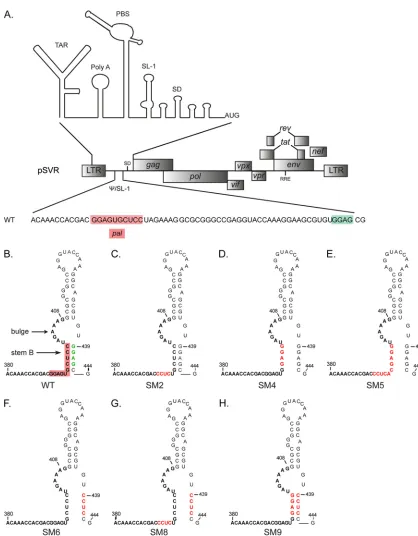
Plasmid constructs.pSVR is an infectious proviral clone of HIV-2 ROD containing a simian virus 40 origin of replication (44). pSVR⌬NB con-tains a 550-nucleotide (nt) deletion in theenvopen reading frame (ORF) between positions 6369 and 6919 (26). Restriction sites and nucleotide numbering, where given, are relative to the first nucleotide of the viral RNA. Mutations in the 5=leader were introduced into a subclone of
HIV-2, pGRAXS (26), by site-directed mutagenesis using the QuikChange II kit (Stratagene). The sequences of the mutagenic primers are available on request. Sequences from the resulting subclones were introduced into the provirus by exchanging an AatII-XhoI fragment, gen-erating the corresponding proviral constructs. All proviral constructs were verified by sequencing. pSVRNCm contains alanine substitutions for all cysteine and histidine residues in the two zinc fingers of the NC domain of Gag and has been described previously (32). Plasmid KS2ES, used to generate the antisense riboprobe to detect HIV-2 genomic RNA (positions 4915 to 5284) by Northern blotting, has been described previ-ously (33).
Cell culture.293T (European Collection of Cell Cultures [ECACC]) and C-33A (American Type Culture Collection [ATCC]) cells were maintained in Dulbecco’s modified Eagle medium (DMEM) (PAA Laboratories) supplemented with 10% fetal calf serum (FCS; PAA), penicillin, and streptomycin (PAA) (complete DMEM). PM1 T cells (Centre for AIDS Reagents [CFAR], National Institute for Biological Standards and Control [NIBSC], United Kingdom) were maintained in RPMI 1640 (PAA) supplemented with 10% FCS, penicillin, and streptomycin (complete RPMI).
Virus preparation.293T or C-33A cells were transfected with proviral constructs by using TransIT-LT1 (Mirus Bio) according to the manu-facturer’s instructions. Cells and supernatants were harvested 48 h later, and virus production was assessed with a reverse transcriptase (RT) assay (53). Virus-containing supernatants were purified through an 8.4% OptiPrep (Sigma-Aldrich) cushion by ultracentrifugation at 98,000⫻gfor 1 h at 4°C. Virus pellets were resuspended either in 500
l of complete RPMI for infection or in 200l of proteinase K buffer for RNA extraction.
Virus infection and evolution study.Wild-type (WT) and mutant viruses were prepared as described above by transfection of 293T cells with the appropriate proviral plasmid. An equivalent of 500 RT units of virus was used to infect 1⫻106PM1 cells in a total volume of 5 ml. Viral
replication was followed by measurement of the RT activity in the culture supernatant every 2 to 4 days, and cells were passaged 1/5 every 4 days. During the evolution study, upon replication of previously replication incompetent virus, 1 ml of cells was harvested, and 0.5 ml of the cell-free supernatant was used to infect 1⫻106fresh PM1 cells
as described above, while the cell pellet and remaining supernatant were stored at⫺20°C until reversion was confirmed by an improved replication kinetic. The cell pellet was then resuspended in 0.5 ml phosphate-buffered saline (PBS) and was lysed by the addition of 100
l of lysis buffer (10 mM Tris-HCl [pH 8], 1 mM EDTA, 0.5% Tween 20) and 6l of 20-mg/ml proteinase K. The mixture was mixed gently and was incubated at 56°C for 1 h. After inactivation of the proteinase K for 10 min at 95°C, 1l of the genomic DNA preparation was used in PCR with different sets of HIV-2-specific primers (sequences avail-able on request) and high-fidelity Accuzyme DNA polymerase (Bio-line). PCR products were purified by using the PCR purification kit (Qiagen) and were sent for sequencing analysis.
RNA preparation.293T cells were transfected with a WT or mutant proviral plasmid as described above, and cytoplasmic RNA was harvested 48 h later by using the RNeasy minikit (Qiagen) according to the manu-facturer’s instructions. Viral RNA was extracted from purified virions by lysis of virus particles in 200l of proteinase K buffer (50 mM Tris-HCl [pH 7.5], 100 mM NaCl, 10 mM EDTA, 1% sodium dodecyl sulfate [SDS], 100g/ml of proteinase K, 100g/ml of tRNA) for 30 min at 37°C. After extraction with acid-buffered phenol-chloroform, followed by one extraction with chloroform-isoamyl alcohol (IAA), the RNA was precip-itated with ethanol at⫺80°C. Cytoplasmic and virion RNAs used for quantitative RT- PCR (qRT-PCR) were subsequently treated with 30 U of Turbo DNase (Life Technologies) for 2 h at 37°C, extracted once with phenol-chloroform and chloroform-IAA, and resuspended in 50 l RNase-free H2O. Virion RNAs used in Northern blotting were
on November 7, 2019 by guest
http://jvi.asm.org/
pended in 20l of a buffer containing 140 mM NaCl, 10 mM Tris-HCl [pH 7.5], and 5 mM MgCl2.
Northern blotting. Northern blotting was performed using the NorthernMax-Gly kit (Life Technologies) as recommended by the man-ufacturer. Virion-extracted RNAs (20l) were separated on a 0.8% aga-rose gel for 4 h at 4°C in 1⫻Gel Prep/Running Buffer, alongside the RiboRuler high-range RNA ladder (Fermentas). RNAs were transferred to a BrightStar-Plus positively charged nylon membrane (Life Technolo-gies), cross-linked by baking for 20 min at 80°C, and detected using the BrightStar Biodetect kit according to the manufacturer’s instructions. The biotinylated riboprobe used for the detection of HIV-2 genomic RNA was transcribedin vitrofrom the linearized (BamHI) KS2ES plasmid using T3 RNA polymerase (Promega) and a biotin RNA labeling mix (Roche). Following DNase treatment (with 10 U of Turbo DNase for 30 min at 37°C), the riboprobe was purified through a G-50 Sephadex column, ali-quoted, and stored at⫺20°C.
Quantification of genomic RNA by qRT-PCR.Cytoplasmic and vi-rion RNAs were normalized to total-RNA concentration and RT activity, respectively, prior to quantification by one-step qRT-PCR using the Su-perScript III One-Step RT-PCR system (Life Technologies) in a Rotor-Gene Q instrument (Qiagen). HIV-2 genomic RNA was detected using a
gag-specific fluorescent probe, 6-carboxyfluorescein (FAM)-CCCAAGTCC CGCAG-minor groove binder (MGB) (Life Technologies), and primers 5=-GCAGGGCTGCTGGAAGTGTGGTAA-3=and 5=-ACTGGGGGTGC TGTTGGTGTCA-3=(Sigma-Aldrich). Human PGK1 (Phosphoglycerate Kinase 1) Endogenous Control (VIC/MGB probe, primer limited; Life Technologies) was used as an endogenous control in the cytoplasmic RNA samples. Cycling conditions were as follows: an initial reverse transcrip-tion step of 30 min at 50°C, followed by 2 min at 95°C and 45 cycles of 95°C for 15 s and 60°C for 1 min. The specific amplification of a 225-nt fragment (positions 1772 to 1997 of the HIV-2 genome) was validated by gel electrophoresis.
Western blotting.Forty-eight hours after the transfection of 293T cells, virions were purified as described above, resuspended in 50l PBS, and lysed by the addition of 2⫻Laemmli buffer (Sigma-Aldrich). Samples were boiled and proteins resolved on 12% SDS-polyacrylamide gel elec-trophoresis (PAGE) gels. Gag was detected using a mouse anti-SIVp57/ p27 antibody (ARP3005; CFAR, NIBSC, United Kingdom) followed by a horseradish peroxidase (HRP)-conjugated anti-mouse antibody (Santa Cruz Biotechnology). A mouse anti--actin antibody (Abcam) was used to verify equal loading of the cytoplasmic fractions.
Pulse-chase metabolic labeling of Gag.C-33A cells were transfected with env-deleted proviral constructs in 6-well plates as described above. After 24 h, cells were washed twice in PBS and were starved for 60 min in 0.5 ml of DMEM lacking methionine and cysteine (Met-Cys-free DMEM; Sigma-Aldrich) supplemented with 4 mM glutamine (Gln; PAA), 10% FCS, penicillin, and streptomycin. Cells were then pulse-labeled for 30 min with 0.5 ml of Met-Cys-free DMEM supple-mented with 110Ci of EasyTag Express35S protein labeling mix (11
mCi/ml; Perkin-Elmer) and 4 mM Gln, 10% FCS, penicillin, and strep-tomycin. The radioactive medium was removed (time point zero), and cells were chased in 0.5 ml of complete DMEM supplemented with 2 mM Met and 2 mM Cys for 30, 60, 90, and 120 min. At each time point, the virus-containing supernatants were harvested and their concentra-tions adjusted to 1% Triton X-100, 1% sodium deoxycholate, and 0.1% SDS. The capsid (CA) and Gag proteins were immunoprecipi-tated using an anti-SIVp57/p27 antibody (ARP3061; CFAR, NIBSC United Kingdom) diluted at 1/150 overnight at 4°C. One hundred microliters of a mixture of protein A- and protein G-Sepharose beads (Sigma-Aldrich) prepared in radioimmunoprecipitation assay (RIPA) buffer (50 mM Tris-HCl [pH 7.5], 100 mM NaCl, 1% sodium deoxy-cholate, 0.1% SDS, 1% Triton X-100, protease inhibitor cocktail [Roche]) was added, and samples were rotated for a further 1 h 30 min at 4°C. Samples were spun at 6,000⫻gfor 2 min, washed three times in RIPA buffer, and resuspended in 50l 2⫻Laemmli buffer. Samples
were boiled for 5 min, and 25l was loaded onto a 13% acrylamide gel alongside a Precision Plus protein marker (Bio-Rad). Gels were fixed in 40% methanol–10% acetic acid for 30 min, incubated in Amplify (GE Healthcare) for 30 min, and dried prior to autoradiography. Quantification (densitometry analysis) was performed using ImageJ software.
RESULTS
Mutation of the HIV-2 leader RNA.
To investigate the exact
re-quirement for the palindrome
pal
and the SL-1 structure and in
particular to establish the precise role of stem B in HIV-2 viral
replication, we introduced a series of mutations into the SL-1
region of the leader RNA of an infectious HIV-2 molecular clone
(pSVR) (Fig. 1A), disrupting either the
pal
sequence or the
struc-ture of SL-1, or both (Fig. 1B to H). A previously characterized
replication- and dimerization-deficient mutant, SM2 (
392-CCUC-
395)
(41), was included for comparison (Fig. 1C). The SM4 and SM5
mutations both lead to disruption of stem B, with SM4 also
dis-rupting
pal
, while SM5 retains a palindromic sequence (Fig. 1D
and E). In SM6, a GGAG motif at the 3
=
end of SL-1 was mutated
to disrupt stem B while leaving
pal
unaffected (Fig. 1F). To assess
the relative importance of the stem B structure compared to its
constituent sequences, the mutations of SM4 and SM6 were
com-bined (SM9) (Fig. 1H). Finally, since several GGAG motifs are
present in SL-1 and similar purine tetrads have been implicated in
RNA packaging and Gag binding in both HIV-1 (1, 3, 11, 16, 19,
40, 56) and HIV-2 (16), the mutations of SM2 and SM6 were
combined (SM8) (Fig. 1G).
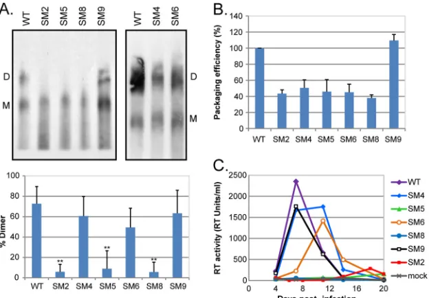
Defects in HIV-2 genome dimerization, but not RNA
encap-sidation, correlate with impaired viral replication.
We wanted to
clarify and reconcile previous
in vitro
and
in vivo
data on HIV-2
dimerization (37, 41) and the association with viral infectivity
(41). Thus, we analyzed the ability of the mutant viruses to form
genomic RNA dimers
in vivo
. WT and mutant virions were
puri-fied from transfected cells, and the viral genomic RNA was
ana-lyzed by native Northern blotting (Fig. 2A). Disruption of stem B
(mutants SM4 and SM6) had no significant effect on the
propor-tion of dimeric genomes present in the virions, indicating that the
structure of SL-1 does not play a role in HIV-2 RNA dimerization.
Replacement of the original
pal
sequence by an alternative
palin-drome (SM5) did affect RNA dimerization, suggesting that the
native sequence is important. However, the self-complementarity
of
pal
is disrupted in SM9, which dimerized as efficiently as the
WT. To clarify this discrepancy, we compared the dimerization of
two
pal
mutants, the previously described SM2 mutant (41), in
which the 5
=
end of
pal
is mutated, and SM4, in which the 3
=
end of
pal
is mutated. The dimerization efficiencies of these mutants
in-dicated that only the 5
=
end of
pal
(
392-GGAG-
395) was required
for HIV-2 genome dimerization. The level of dimers detected for
the SM2 mutant differed slightly between the current study and
our previously published study (41). This is most likely due to
differences in the RNA extraction protocol. In this study, a more
stringent extraction method was used, which would potentially
increase the dissociation of loose dimers but would have little or
no effect on the integrity of tight dimers. The levels of wild-type
dimer are similar in the two studies, consistent with this
interpre-tation. Mutations of both GGAG motifs present in Psi/SL-1 (SM8
virus) drastically reduced genome dimerization. Comparison of
the SM2 and SM6 viruses (Fig. 2A) (41) strongly suggests that
392
-GGAG-
395is the critical element for HIV-2 genome
on November 7, 2019 by guest
http://jvi.asm.org/
tion (Table 1). While mutants lacking the
392-GGAG-
395motif
have an unequivocal dimerization defect, it should be noted that
the method used does not allow us to determine whether the RNA
extracted from virions is truly monomeric or represents unstable
dimer that is disrupted by the extraction procedure. Increasing
evidence that the HIV-1 genome is packaged as a dimer (45, 46),
together with the disparities in the levels of dimer obtained for the
SM2 mutant by different extraction methods, would suggest that
FIG 1Structure of stem-loop 1 in wild-type and mutant HIV-2. (A) Schematic of the molecular clone pSVR of the HIV-2 ROD genome, with the structure of the 5=leader RNA (20) shown above and the sequence of the packaging signal (Psi)/stem-loop 1 (SL-1) region detailed below. The palindromepal(residues 392 to 401) and the GGAG motif at position 439 are highlighted in red and green, respectively. TAR,trans-activator response; PBS, primer binding site; SD, major splice donor. (B to H) Structures of SL-1 in the WT (B) and substitution mutants (C to H) based on the previously described structure of SL-1 (4). The palindromepalis highlighted in red on the WT structure. Stem B and the distal bulge (residues 402 to 409) are indicated. SM2 (C) (residues 392 to 395) has been described and characterized previously (41). Red letters represent mutated residues.
on November 7, 2019 by guest
http://jvi.asm.org/
[image:4.585.78.496.58.594.2]loose dimers are initially formed but are not stable enough to
withstand the more stringent extraction procedure.
It was proposed previously that stem B is required for efficient
packaging of the HIV-2 genome and viral replication (36). Since
stem B does not appear to play a role in genome dimerization, we
decided to measure the packaging efficiencies of our Psi/SL-1
mu-tants relative to that of the WT (Fig. 2B). Our data confirmed the
requirement for stem B in HIV-2 RNA encapsidation, but only in
combination with the
392-GGAG-
395motif (Table 1). Indeed, the
formation of stem B alone is not sufficient to promote efficient
RNA packaging, as illustrated by the poor packaging efficiency of
the SM2 mutant (5, 41; this study).
[image:5.585.137.449.66.283.2]We next assessed the abilities of the SL-1 mutant viruses to
replicate in T cells (Fig. 2C). We observed a striking direct
corre-lation between the efficiency of genome dimerization and viral
replication, although a packaging efficiency of 100% is not
abso-lutely necessary for HIV-2 propagation. A 2-fold reduction in
RNA packaging had no significant effect on the viral replication of
the SM4 and SM6 mutants, whereas a major reduction in the level
of dimeric genome abrogated replication of the SM2, SM5, and
SM8 viruses. These results are in agreement with previous data
showing that a GGRG motif at position 392 is required for HIV-2
replication, but they do not support an essential role for stem B in
viral replication (5). The data presented here do strongly suggest
FIG 2HIV-2 replication is dependent on efficient genome dimerization and the presence of the392-GGAG-395motif in the RNA leader. (A) (Top) Native Northern blot analysis of HIV-2 genomic RNA; (bottom) quantification (mean⫹standard deviation) (n⫽3) of the percentage of dimeric RNA in the virions. 293T cells were transfected with WT and mutant proviral constructs, and viral RNA was extracted at 48 h posttransfection and was analyzed by native Northern blotting using an antisense riboprobe specific forpol. The positions of the dimeric (D) and monomeric (M) RNA species are indicated. Asterisks indicate a significant difference from the WT (**,P⬍0.002) by an unpaired Studentttest. (B) Packaging efficiencies of the mutant viruses relative to that of the WT, measured by qRT-PCR using the standard-curve method of quantification. The packaging efficiency of the WT was set to 100%. (C) WT and mutant HIV-2 virions were produced by transfection of 293T cells with the corresponding proviral plasmid, purified through 8.4% OptiPrep, and used to infect 1⫻106PM1 T cells. Virus input was normalized on the reverse transcriptase (RT) activity of the virus preparation, and an equivalent of 500 RT units was used. Viral replication was followed by measuring the RT activity in the culture supernatant every 4 days. Mock, mock-infected cells.TABLE 1Correlation between viral replication, RNA dimerization, and packaging and the sequences and structural elements located within HIV-2 SL-1
Virus
Viral replicationa
Genome dimerization (%)b
Packaging efficiencyc
Presence or absence of:
pal Stem B 392-GGAG-395 439-GGAG-442
WT ⫹⫹ 72.7 1.00 ⫹ ⫹ ⫹ ⫹
SM4 ⫹⫹ 60.6 0.51 ⫺ ⫺ ⫹ ⫹
SM6 ⫹ 49.4 0.45 ⫹ ⫺ ⫹ ⫺
SM9 ⫹⫹ 63.2 1.10 ⫺ ⫹ ⫹ ⫺
SM2 ⫺ 5.8 0.44 ⫺ ⫹ ⫺ ⫹
SM5 ⫺ 8.9 0.46 ⫹d ⫺ ⫺ ⫹
SM8 ⫺ 5.6 0.38 ⫺ ⫺ ⫺ ⫺
aReplication in PM1 T cells was followed by measuring the RT activity in the culture supernatant. Symbols indicate the presence (⫹⫹) or absence (⫺) of detectable RT activity at 4
to 15 days postinfection (n⫽3).
bPercentage of dimeric genome in the virion as measured by native Northern blotting (n⫽3). c
Relative to that of the WT (set to 1) as measured by qRT-PCR (n⫽3).
dAlternative palindromic sequence in place of WTpal.
on November 7, 2019 by guest
http://jvi.asm.org/
[image:5.585.41.545.577.677.2]that the conformation of the genomic RNA encapsidated in the
virion, rather than the packaging efficiency, is critical for HIV-2
replication.
A compensatory mutation in matrix can rescue the
replica-tion of dimerizareplica-tion mutants.
To seek reversion mutations with
restored replication kinetics, T cells infected with the dimerization
mutants were cultured for more than 3 weeks; eventually,
repli-cating virus was detected in all three cultures (data not shown).
Cell-free supernatant was used to infect fresh cells, and a faster
replication kinetic than that with the original infection was
ob-served, suggesting that a reversion event had occurred. The
in-fected cells were harvested at the peak of infection; total DNA was
isolated; and the 5
=
untranslated region (5
=
UTR) and
gag
gene of
the integrated proviruses were sequenced. Every virus sequenced
had retained the original mutation in the Psi/SL-1 region.
How-ever, several second-site mutations were identified in the
gag
gene,
most notably a C-to-T mutation at position 754, resulting in a
Thr-to-Ile mutation at position 70 of matrix (MA 70TI), which
was identified in both SM5 and SM8 and had been observed
pre-viously in SIV
mac239in the context of an SL-1
dimerization-defi-cient mutant also harboring an 184MV mutation in RT (58).
The MA 70TI mutation was therefore introduced into the
SM2, SM5, and SM8 proviral constructs, and the revertant viruses
were phenotypically characterized (Fig. 3). The replication of WT
HIV-2, the three dimerization mutants, and their corresponding
revertant viruses was followed over a period of 4 weeks (Fig. 3A).
The three MA 70TI revertant viruses were able to rescue the viral
replication of SM2, SM5, and SM8, bringing it close to the WT
level. Genome dimerization was then assessed by native Northern
blotting (Fig. 3B). The MA mutation was able to rescue RNA
ge-nome dimerization by the SL-1 mutants (
P
,
⬍
0.05 by an unpaired
Student
t
test). Introduction of the MA 70TI mutation into WT
proviral DNA had no significant effect on viral replication or
ge-nome dimerization (Fig. 3A and B).
Overall, these data show that a second-site mutation at
posi-tion 70 in matrix can rescue the replicaposi-tion of dimerizaposi-tion- and
packaging-deficient mutant viruses and can partially restore
ge-nome dimerization.
During the course of our evolution study, other second-site
mutations were identified, including MA 68VE, CA 26VI, CA
98TA, p6 35EK, p6 42EK, and p6 53EK. The p6 EK mutations
alone are not sufficient to rescue infectivity—introduction of
the 42EK mutation into the SM5 background did not rescue
viral replication—indicating that other second-site mutations
may have arisen elsewhere in the genome at the same time and
may have been necessary for the improved replication fitness
observed (data not shown). This corroborates a previous study
with SIV
mac239in which a p6 49EK compensatory mutation was
able to rescue the replication of an SL-1 dimerization mutant
only in combination with two second-site mutations, nt 423AG
in the DIS loop and CA 197KR (27). However, none of our
evolution studies yielded reversion of the RNA 5
=
UTR or any
similar mutation in CA.
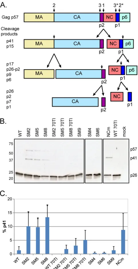
Dimerization-deficient mutants show a processing defect
that is partially rescued by the MA 70TI reversion mutation.
Proteolytic processing of Gag is known to be associated with the
RNA dimerization process, promoting the maturation of the
dimer, as shown by analysis of protease-deficient HIV-1 and
mu-rine leukemia virus (MLV) virions (22, 23). Conversely,
muta-tions reducing HIV-1 genome dimerization have been shown to
impair Gag processing by delaying the CA-p2 cleavage (42). With
this in mind, we investigated the effects of our SL-1 mutations on
Gag synthesis and processing.
We first analyzed the levels of Gag and its cleavage products
(Fig. 4A) in the particles released at 48 h posttransfection (Fig. 4B).
While all the Gag protein has been cleaved into CA in the WT
virus, the dimerization mutants showed a delay in Gag processing,
with a small yet significant accumulation of the p41 (MA-CA-p2)
intermediate (Fig. 4C). The replication-competent SL-1 mutants
and the MA 70TI revertant viruses all showed normal processing
of Gag (Fig. 4B and C). To examine further the contribution of
dimer formation and Gag-RNA interaction to Gag processing, we
analyzed an HIV-2 Gag mutant harboring cysteine-to-alanine and
histidine-to-alanine substitutions in the two zinc finger domains
of NC (NCm), which abrogate Gag-RNA binding (32). This
tant displayed the same phenotype as the SL-1 dimerization
mu-tants (Fig. 4B and C), suggesting that binding of Gag to the
genomic RNA is important for the correct processing of the
pro-tein.
The precise step at which Gag processing is disrupted is
diffi-cult to ascertain with the “snapshot” approach described above, so
we investigated this question further by use of pulse-chase
meta-bolic labeling (Fig. 5). Transfected cells were pulse-labeled for 30
min and were chased for as long as 120 min. Gag and its
CA-containing cleavage products were immunoprecipitated and
ana-lyzed by autoradiography (Fig. 5A). Confirming the results
ob-FIG 3A compensatory mutation in MA (70TI) can rescue the replication ofdimerization mutants and partially restore genome dimerization. (A) PM1 T cells were infected with an equivalent of 500 RT units of WT, dimerization mutant, and MA 70TI mutant viruses. Viral replication was followed by mea-suring the RT activity in the culture supernatant every 2 to 4 days. 70TI, mu-tation in matrix at position 70; mock, mock-infected cells. (B) (Left) Native Northern blot analysis of MA 70TI mutant viruses; (right) quantification (means⫹standard deviations) (n⫽2) of the percentages of dimeric RNA in
the virions. The WT data are reported from Fig. 2A. D, dimer; M, monomer.
on November 7, 2019 by guest
http://jvi.asm.org/
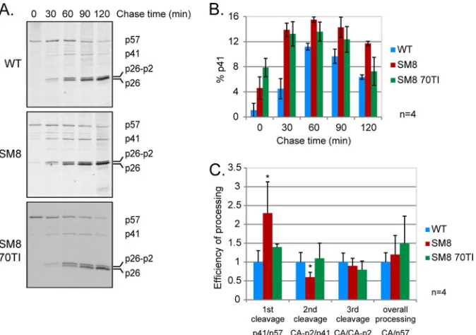
[image:6.585.61.268.64.324.2]tained by Western blotting (Fig. 4B), the dimerization-deficient
mutant SM8 showed a significant accumulation of the p41
inter-mediate compared to that in the WT virus (Fig. 5A and B). The
MA revertant of SM8, SM8 70TI, partially rescued this phenotype
after 2 h. The efficiency of each individual cleavage, taken as the
ratio of cleaved to uncleaved product, was calculated for each virus
at 2 h postlabeling and is shown in Fig. 5C. The dimerization
mutant SM8 was twice as efficient as the WT virus at cleaving Gag
into p41 but was significantly slower at the second cleavage site,
which explains the observed accumulation of the p41
intermedi-ate. In accordance with the results shown in Fig. 4, the MA 70TI
mutation was able to restore the correct rate of Gag processing in
the SM8 mutant. The rates of the third cleavage of CA-p2 into CA
and p2 were similar for all viruses, as were the overall processing
efficiencies (CA/Gag ratios), suggesting that the improper rate of
processing and the subsequent accumulation of p41—rather than
decreases in the proportions of cleaved MA and CA—are
respon-sible for the reduced infectivity.
DISCUSSION
In this study, we evaluated the requirement for sequence and
structural elements within the SL-1 region of the HIV-2 RNA
leader for viral replication, genome dimerization, and
encapsida-tion. Using substitution mutants that interfered with the
palin-drome
pal
or stem B, we have shown that neither of these elements
is indispensable for HIV-2 replication (Table 1). However, the
392
-GGAG-
395motif is critical for viral replication (as previously
proposed [5]), and we show that this correlates precisely with
genome dimerization. In addition, we have observed an absolute
dependence on RNA dimerization, but not packaging, for HIV-2
replication. These findings posit a model in which
in vivo
dimerization occurs prior to RNA capture and encapsidation, as
suggested for HIV-1 (46) and MLV (21). Surprisingly, the
pres-ence of stem B does not appear to influpres-ence viral replication, in
contrast with the conclusions of a previous report in which the
formation of an extended SL-1 was proposed to be important for
viral replication and encapsidation (36). One explanation for this
discrepancy lies in the fact that the role of stem B was derived from
evolution studies of a randomized sequence, whereas our results
were obtained from a mutagenesis approach whereby specific
res-idues were replaced in order to alter the structure of SL-1. Our
results do, however, confirm that stem B and the extended
struc-ture of SL-1 can enhance HIV-2 genome encapsidation, albeit
exclusively in combination with the
392-GGAG-
395motif.
Mainte-nance of the stem-loop structure immediately downstream of the
nt 392-to-395 purine-rich motif may serve to ensure that the motif
is optimally presented for Gag binding. The recent report that the
HIV-1 leader RNA can adopt multiple conformations which are
involved in the regulation of key processes in the virus life cycle
(43) provides another explanation for the apparent differences
between this and a previous study (36), as well as for those
be-tween the WT and SL-1 mutant viruses. Indeed, one could
envis-age a similar situation in HIV-2 where a structural switch in the
genomic RNA regulates the transition between translation and
genome dimerization and packaging.
Analysis of the HIV-2 dimerization mutants by Western
blot-ting revealed an accumulation of the Gag intermediate cleavage
product p41 (MA-CA-p2), a result confirmed by metabolic
label-ing and Gag immunoprecipitation. The rate of Gag processlabel-ing was
altered in the dimerization mutant from that in the WT,
explain-FIG 4Mutations affecting RNA dimerization and Gag-RNA interactionresult in suboptimal processing and the accumulation of the p41 interme-diate. (A) Schematic of the Gag polyprotein (p57) and its proteolytic pro-cessing. The sites and order of cleavage are indicated above the p57 precur-sor and each intermediate product. p17, matrix (MA); p26, capsid (CA); p2, spacer peptide p2, or SP1; p7, nucleocapsid (NC); p1, spacer peptide p1, or SP2; p41, MA-CA-p2; p15, NC-p1-p6; p9, NC-p1. (B) Western blot analysis of virion proteins, extracted at 48 h posttransfection, by use of an anti-SIVp57/27 antibody. The positions of Gag (p57), MA-CA-p2 (p41), and CA (p26) are indicated on the right. WT, wild type; 70TI, mutation in matrix at position 70; NCm, mutation of all Cys and His residues to Ala in the two NC zinc fingers. (C) Quantification of the percentage of the p41 cleavage intermediate in the virion (means⫹standard deviations) (n⫽3). Asterisks indicate significant differences from the wild type (*,P⬍0.05) by the unpaired Studentttest.
on November 7, 2019 by guest
http://jvi.asm.org/
[image:7.585.43.284.65.535.2]ing the increase in the p41 level. Indeed, initial cleavage of the Gag
polyprotein was faster, while cleavage of p41 was slower, in the
mutant than in the WT. The overall processing efficiency
re-mained the same, as did the level of CA, suggesting that the
incor-rect timing of processing leading to the accumulation of p41 is
responsible for the poor infectivity of the mutant virus.
Evolution study of the dimerization mutants resulted in the
emergence of several revertant viruses harboring compensatory
mutations in the
gag
gene. Interestingly, a threonine-to-isoleucine
mutation at position 70 of matrix, analogous to those previously
described for HIV-1 (49) and SIV
mac239(58), was obtained in two
out of three dimerization mutants. In HIV-1, a MA 69TI mutation
was able to compensate for the viral release defect of the MA 6VR
mutant, possibly by affecting the global conformation of the
pro-tein and correcting an alteration in the structure introduced by the
original mutation. More relevant to the present study is the
res-cued replication of the SIV
mac239SL-1 mutant by the MA 70TI
reversion. SIV
mac239is closely related to HIV-2, and it is
interest-ing that, as with our findinterest-ings for HIV-2, the SIV SL-1 mutant
showed a lower level of dimeric genome and lower infectivity than
the WT virus. However, the authors did not report on the effect of
the compensatory mutation on genome dimerization.
When the MA 70TI compensatory mutation was introduced
into the genomes of WT and mutant viruses, we observed rescue
of the viral replication of the dimerization mutants but no
signif-icant effect on replication in the WT virus. This rescue was
asso-ciated with a largely restored ability to form dimeric genomes and
restoration of the correct rate of Gag processing, with levels of p41
similar to that of the WT at 2 h postlabeling. While residue 70 of
matrix does not appear to be particularly involved in the structure
of the protein, in the HIV-1 structure it is located near the MA-CA
interface, and this spatial positioning may be responsible for the
effect on Gag processing that was observed. In addition, it was
shown recently that upon binding of HIV-1 matrix to the genomic
RNA, certain residues (including residues 68 and 70) become
more reactive (2), indicating a potential change of the
conforma-tion of this region upon MA-RNA interacconforma-tion. The effects of the
MA mutation on the replication, RNA dimerization, and Gag
pro-cessing of HIV-2 mutants—together with the recently proposed
model whereby HIV-1 NC and MA both bind to the genomic
RNA and, following binding to the plasma membrane, the Gag
protein extends and viral particles assemble (17)—provide
addi-tional insight into the role of matrix in HIV particle assembly.
Taking our findings together, this study provides further
evi-dence of the role of the genomic RNA in retrovirus assembly and
highlights the importance of genome dimerization for HIV-2
rep-lication. Targeting this process through the
cis
-acting RNA
ele-ments located in the highly conserved (and thus less prone to
escape mutations) 5
=
UTR may open the way to new therapeutic
approaches.
ACKNOWLEDGMENTS
We thank John Sinclair for critical reading of the manuscript.
The anti-SIVp57/p27 antibody (ARP3004) and the PM1 cells
FIG 5The rate of Gag processing is altered in a replication-incompetent SL-1 mutant with impaired RNA dimerization. Pulse-chase metabolic labeling and immunoprecipitation of Gag were performed 24 h after transfection of C33-A cells with the WT, SM8, and SM8 MA 70TI proviral plasmids. Cells were starved of methionine (Met) and cysteine (Cys) for 60 min, pulse-labeled with [35S]Met-Cys for 30 min, and chased in cold medium for 30 to 120 min. Capsid-containing Gag proteins were immunoprecipitated from the culture supernatant at each time point with anti-SIVp57/p27, resolved by SDS-PAGE, and visualized by autoradiography, prior to quantification by densitometry. (A) Representative results of pulse-chase metabolic labeling, immunoprecipitation, and gel electro-phoresis are shown for each virus. The chase times are given above the gel, and the positions of Gag (p57) and its cleavage products MA-CA-p2 (p41), CA-p2 (p26-p2), and CA (p26) are indicated on the right. (B) Quantification of the percentage of the p41 cleavage intermediate for each virus (means⫾standard deviations) (n⫽4). (C) Efficiency of processing at 120 min postlabeling for each cleavage of Gag, as diagramed in Fig. 4A, corresponding to the ratio of the cleaved to the uncleaved product. Means⫹standard deviations are shown (n⫽4). Asterisks indicate significant differences from the WT (*,P⬍0.05) by an unpaired
Studentttest.
on November 7, 2019 by guest
http://jvi.asm.org/
[image:8.585.125.462.66.303.2](ARP057) were obtained from the Programme EVA Centre for AIDS Re-agents, NIBSC, United Kingdom, supported by the EC FP6/7 Europrise Network of Excellence, the AVIP and NGIN consortia, and the Bill and Melinda Gates GHRC-CAVD Project, and were donated by S. Osmanov and M. Reitz, respectively. This work was supported by the United King-dom Medical Research Council (award G0800142) and the Biomedical Research Centre (grant RG52162).
REFERENCES
1.Aldovini A, Young RA.1990. Mutations of RNA and protein sequences involved in human immunodeficiency virus type 1 packaging result in production of noninfectious virus. J. Virol.64:1920 –1926.
2.Alfadhli A, et al.2011. HIV-1 matrix protein binding to RNA. J. Mol. Biol.410:653– 666.
3.Amarasinghe GK, et al.2000. NMR structure of the HIV-1 nucleocapsid protein bound to stem-loop SL2 of the psi-RNA packaging signal. Impli-cations for genome recognition. J. Mol. Biol.301:491–511.
4.Baig TT, Lanchy JM, Lodmell JS.2007. HIV-2 RNA dimerization is regulated by intramolecular interactions in vitro. RNA13:1341–1354. 5.Baig TT, Lanchy JM, Lodmell JS.2009. Randomization and in vivo
selection reveal a GGRG motif essential for packaging human immuno-deficiency virus type 2 RNA. J. Virol.83:802– 810.
6.Baudin F, et al.1993. Functional sites in the 5=region of human immu-nodeficiency virus type 1 RNA form defined structural domains. J. Mol. Biol.229:382–397.
7.Berkhout B, van Wamel JL.1996. Role of the DIS hairpin in replication of human immunodeficiency virus type 1. J. Virol.70:6723– 6732. 8.Berkowitz R, Fisher J, Goff SP.1996. RNA packaging. Curr. Top.
Mi-crobiol. Immunol.214:177–218.
9.Campbell S, Rein A.1999. In vitro assembly properties of human immu-nodeficiency virus type 1 Gag protein lacking the p6 domain. J. Virol.
73:2270 –2279.
10. Campbell S, Vogt VM.1995. Self-assembly in vitro of purified CA-NC proteins from Rous sarcoma virus and human immunodeficiency virus type 1. J. Virol.69:6487– 6497.
11. Clavel F, Orenstein JM.1990. A mutant of human immunodeficiency virus with reduced RNA packaging and abnormal particle morphology. J. Virol.64:5230 –5234.
12. Clever J, Sassetti C, Parslow TG.1995. RNA secondary structure and binding sites forgaggene products in the 5=packaging signal of human immunodeficiency virus type 1. J. Virol.69:2101–2109.
13. Clever JL, Parslow TG.1997. Mutant human immunodeficiency virus type 1 genomes with defects in RNA dimerization or encapsidation. J. Virol.71:3407–3414.
14. Clever JL, Wong ML, Parslow TG.1996. Requirements for kissing-loop-mediated dimerization of human immunodeficiency virus RNA. J. Virol.
70:5902–5908.
15. Crist RM, et al.2009. Assembly properties of human immunodeficiency virus type 1 Gag-leucine zipper chimeras: implications for retrovirus as-sembly. J. Virol.83:2216 –2225.
16. Damgaard CK, Dyhr-Mikkelsen H, Kjems J.1998. Mapping the RNA binding sites for human immunodeficiency virus type-1 Gag and NC pro-teins within the complete HIV-1 and -2 untranslated leader regions. Nu-cleic Acids Res.26:3667–3676.
17. Datta SA, et al.2011. HIV-1 Gag extension: conformational changes require simultaneous interaction with membrane and nucleic acid. J. Mol. Biol.406:205–214.
18. Datta SA, et al.2011. On the role of the SP1 domain in HIV-1 particle assembly: a molecular switch? J. Virol.85:4111– 4121.
19. De Guzman RN, et al.1998. Structure of the HIV-1 nucleocapsid protein bound to the SL3 psi-RNA recognition element. Science279:384 –388. 20. Dirac AM, Huthoff H, Kjems J, Berkhout B.2001. The dimer initiation
site hairpin mediates dimerization of the human immunodeficiency virus, type 2 RNA genome. J. Biol. Chem.276:32345–32352.
21. D’Souza V, Summers MF.2004. Structural basis for packaging the di-meric genome of Moloney murine leukaemia virus. Nature431:586 –590. 22. Fu W, Gorelick RJ, Rein A.1994. Characterization of human immuno-deficiency virus type 1 dimeric RNA from wild-type and protease-defective virions. J. Virol.68:5013–5018.
23. Fu W, Rein A.1993. Maturation of dimeric viral RNA of Moloney murine leukemia virus. J. Virol.67:5443–5449.
24. Ganser BK, Li S, Klishko VY, Finch JT, Sundquist WI.1999. Assembly and analysis of conical models for the HIV-1 core. Science283:80 – 83. 25. Greatorex J.2004. The retroviral RNA dimer linkage: different structures
may reflect different roles. Retrovirology1:22.
26. Griffin SD, Allen JF, Lever AM.2001. The major human immunodefi-ciency virus type 2 (HIV-2) packaging signal is present on all HIV-2 RNA species: cotranslational RNA encapsidation and limitation of Gag protein confer specificity. J. Virol.75:12058 –12069.
27. Guan Y, et al.2001. Partial restoration of replication of simian immuno-deficiency virus by point mutations in either the dimerization initiation site (DIS) or Gag region after deletion mutagenesis within the DIS. J. Virol.
75:11920 –11923.
28. Harrison GP, Lever AM.1992. The human immunodeficiency virus type 1 packaging signal and major splice donor region have a conserved stable secondary structure. J. Virol.66:4144 – 4153.
29. Hill MK, et al.2003. The dimer initiation sequence stem-loop of human immunodeficiency virus type 1 is dispensable for viral replication in pe-ripheral blood mononuclear cells. J. Virol.77:8329 – 8335.
30. Hu WS, Temin HM.1990. Genetic consequences of packaging two RNA genomes in one retroviral particle: pseudodiploidy and high rate of genetic recombination. Proc. Natl. Acad. Sci. U. S. A.87:1556 –1560.
31. Johnson MC, Scobie HM, Ma YM, Vogt VM. 2002. Nucleic acid-independent retrovirus assembly can be driven by dimerization. J. Virol.
76:11177–11185.
32. Kaye JF, Lever AM.1999. Human immunodeficiency virus types 1 and 2 differ in the predominant mechanism used for selection of genomic RNA for encapsidation. J. Virol.73:3023–3031.
33. Kaye JF, Lever AM.1998. Nonreciprocal packaging of human immuno-deficiency virus type 1 and type 2 RNA: a possible role for the p2 domain of Gag in RNA encapsidation. J. Virol.72:5877–5885.
34. Lanchy JM, Ivanovitch JD, Lodmell JS.2003. A structural linkage be-tween the dimerization and encapsidation signals in HIV-2 leader RNA. RNA9:1007–1018.
35. Lanchy JM, Lodmell JS.2002. Alternate usage of two dimerization initi-ation sites in HIV-2 viral RNA in vitro. J. Mol. Biol.319:637– 648. 36. Lanchy JM, Lodmell JS.2007. An extended stem-loop 1 is necessary for
human immunodeficiency virus type 2 replication and affects genomic RNA encapsidation. J. Virol.81:3285–3292.
37. Lanchy JM, Rentz CA, Ivanovitch JD, Lodmell JS. 2003. Elements located upstream and downstream of the major splice donor site influence the ability of HIV-2 leader RNA to dimerize in vitro. Biochemistry42: 2634 –2642.
38. Laughrea M, Jette L.1994. A 19-nucleotide sequence upstream of the 5= major splice donor is part of the dimerization domain of human immu-nodeficiency virus 1 genomic RNA. Biochemistry33:13464 –13474. 39. Laughrea M, et al.1997. Mutations in the kissing-loop hairpin of human
immunodeficiency virus type 1 reduce viral infectivity as well as genomic RNA packaging and dimerization. J. Virol.71:3397–3406.
40. Lever A, Gottlinger H, Haseltine W, Sodroski J.1989. Identification of a sequence required for efficient packaging of human immunodeficiency virus type 1 RNA into virions. J. Virol.63:4085– 4087.
41. L’Hernault A, Greatorex JS, Crowther RA, Lever AM.2007. Dimerisa-tion of HIV-2 genomic RNA is linked to efficient RNA packaging, normal particle maturation and viral infectivity. Retrovirology4:90.
42. Liang C, et al.1999. Deletion mutagenesis within the dimerization initi-ation site of human immunodeficiency virus type 1 results in delayed processing of the p2 peptide from precursor proteins. J. Virol.73:6147– 6151.
43. Lu K, et al.2011. NMR detection of structures in the HIV-1 5=-leader RNA that regulate genome packaging. Science334:242–245.
44. McCann EM, Lever AM.1997. Location ofcis-acting signals important for RNA encapsidation in the leader sequence of human immunodefi-ciency virus type 2. J. Virol.71:4133– 4137.
45. Moore MD, et al.2007. Dimer initiation signal of human immunodefi-ciency virus type 1: its role in partner selection during RNA copackaging and its effects on recombination. J. Virol.81:4002– 4011.
46. Moore MD, et al.2009. Probing the HIV-1 genomic RNA trafficking pathway and dimerization by genetic recombination and single virion analyses. PLoS Pathog.5:e1000627.
47. Muriaux D, Girard PM, Bonnet-Mathoniere B, Paoletti J. 1995. Dimerization of HIV-1Lai RNA at low ionic strength. An autocomple-mentary sequence in the 5=leader region is evidenced by an antisense oligonucleotide. J. Biol. Chem.270:8209 – 8216.
on November 7, 2019 by guest
http://jvi.asm.org/
48. Muriaux D, Mirro J, Harvin D, Rein A.2001. RNA is a structural element in retrovirus particles. Proc. Natl. Acad. Sci. U. S. A.98:5246 –5251. 49. Ono A, Huang M, Freed EO.1997. Characterization of human
immu-nodeficiency virus type 1 matrix revertants: effects on virus assembly, Gag processing, and Env incorporation into virions. J. Virol.71:4409 – 4418. 50. Paillart JC, et al.1996. A dual role of the putative RNA dimerization
initiation site of human immunodeficiency virus type 1 in genomic RNA packaging and proviral DNA synthesis. J. Virol.70:8348 – 8354. 51. Paillart JC, Shehu-Xhilaga M, Marquet R, Mak J.2004. Dimerization of
retroviral RNA genomes: an inseparable pair. Nat. Rev. Microbiol.2:461– 472.
52. Paillart JC, Skripkin E, Ehresmann B, Ehresmann C, Marquet R.1996. A loop-loop “kissing” complex is the essential part of the dimer linkage of genomic HIV-1 RNA. Proc. Natl. Acad. Sci. U. S. A.93:5572–5577. 53. Potts B.1990. Mini RT assay, p 103–106.InAldovini A, Walker B (ed),
Techniques in HIV research. Stockton Press, New York, NY.
54.Sakuragi J, Ueda S, Iwamoto A, Shioda T. 2003. Possible role of dimerization in human immunodeficiency virus type 1 genome RNA packaging. J. Virol.77:4060 – 4069.
55. Shehu-Xhilaga M, et al. 2001. Proteolytic processing of the p2/ nucleocapsid cleavage site is critical for human immunodeficiency virus type 1 RNA dimer maturation. J. Virol.75:9156 –9164.
56. Shubsda MF, Paoletti AC, Hudson BS, Borer PN.2002. Affinities of packaging domain loops in HIV-1 RNA for the nucleocapsid protein. Biochemistry41:5276 –5282.
57. Skripkin E, Paillart JC, Marquet R, Ehresmann B, Ehresmann C.1994. Identification of the primary site of the human immunodeficiency virus type 1 RNA dimerization in vitro. Proc. Natl. Acad. Sci. U. S. A.91:4945– 4949.
58. Whitney JB, Oliveira M, Detorio M, Guan Y, Wainberg MA.2002. The M184V mutation in reverse transcriptase can delay reversion of attenu-ated variants of simian immunodeficiency virus. J. Virol.76:8958 – 8962. 59. Whitney JB, Wainberg MA. 2006. Impaired RNA incorporation and dimerization in live attenuated leader-variants of SIVmac239. Retrovirology
3:96.
60. Zhang Y, Qian H, Love Z, Barklis E.1998. Analysis of the assembly function of the human immunodeficiency virus type 1 Gag protein nu-cleocapsid domain. J. Virol.72:1782–1789.
on November 7, 2019 by guest
http://jvi.asm.org/