0022-538X/11/$12.00 doi:10.1128/JVI.01391-10
Copyright © 2011, American Society for Microbiology. All Rights Reserved.
Recombinant Infectious Bursal Disease Virus Carrying
Hepatitis C Virus Epitopes
䌤
Chitra Upadhyay,
1Arun Ammayappan,
1Deendayal Patel,
3Imre Kovesdi,
2and Vikram N. Vakharia
1*
University of Maryland Biotechnology Institute, Center of Marine Biotechnology, 701 East Pratt Street,
Suite 236, Baltimore, Maryland 212021; VectorLogics, Inc., Birmingham, Alabama 352942;
and Hauptman-Woodward Medical Research Institute,
700 Ellicott Street, Buffalo, New York 142033
Received 2 July 2010/Accepted 10 November 2010
The delivery of foreign epitopes by a replicating nonpathogenic avian infectious bursal disease virus (IBDV) was explored. The aim of the study was to identify regions in the IBDV genome that are amenable to the introduction of a sequence encoding a foreign peptide. By using a cDNA-based reverse genetics system, insertions or substitutions of sequences encoding epitope tags (FLAG, c-Myc, or hepatitis C virus epitopes) were engineered in the open reading frames of a nonstructural protein (VP5) and the capsid protein (VP2). Attempts were also made to generate recombinant IBDV that displayed foreign epitopes in the exposed loops (PBC and PHI) of the VP2 trimer. We successfully recovered recombinant IBDVs expressing c-Myc and two different virus-neutralizing epitopes of human hepatitis C virus (HCV) envelope glycoprotein E in the VP5 region. Western blot analyses with anti-c-Myc and anti-HCV antibodies provided positive identification of both the c-Myc and HCV epitopes that were fused to the N terminus of VP5. Genetic analysis showed that the recombinants carrying the c-Myc/HCV epitopes maintained the foreign gene sequences and were stable after several passages in Vero and 293T cells. This is the first report describing efficient expression of foreign peptides from a replication-competent IBDV and demonstrates the potential of this virus as a vector.
Infectious bursal disease virus (IBDV), a pathogen causing an immunosuppressive disease in chickens (26), has been used as a therapeutic agent without any toxicity in clinical trials with patients suffering from acute and chronic hepatitis C virus (HCV) infections (2, 8). IBDV belongs to the genus
Avibirna-virusof theBirnaviridaefamily, and its genome consists of two
segments of double-stranded RNA (11). The smaller segment, B, encodes VP1, a 97-kDa multifunctional protein with poly-merase and capping enzyme activities (21, 27). The larger segment, A, encodes a 110-kDa precursor polyprotein in a single large open reading frame (ORF) which is cotranslation-ally processed by the viral protease VP4 (4, 24) into the VP2 precursor (pVP2), VP3, and VP4. pVP2 is further processed by several proteolytic cleavages at its C terminus for conversion into mature VP2 (10). Segment A also encodes VP5, a 17-kDa nonstructural (NS) protein, in a small ORF partly preceding and overlapping the polyprotein ORF. VP5 is dispensable for viral replicationin vitroandin vivo(29), which makes it a prime candidate for the construction of marked vaccines carrying deletions. These marked vectors could be easily distinguished from the wild-type virus and could also trigger a specific cel-lular immune response in the host species. The available struc-tural data for VP2 (7, 14, 17) reveal that the protein is folded into three different domains (base B, shell S, and projection P). Expression of VP2 by itself leads to dodecahedral subviral particles (SVPs) containing 20 VP2 trimers (6) and exposing the four loops of the P domain (named PBC, PDE, PFG, and
PHI). Here, we explore the possibility of displaying foreign epitopes stably in recombinant IBDV by either inserting or replacing sequences in the PBCand PHIloops of VP2.
In this study, we used a 10-amino-acid linear c-Myc epitope (EQKLISEEDL) derived from the C terminus of human c-Myc protein and an 8-amino-acid linear FLAG epitope (DY KDDDDK). c-Myc and FLAG epitope tags were selected as they are well characterized and are recognized by specific monoclonal antibody (MAb) Myc1-9E10 (12) and MAb M2 (5), respectively. These epitope tags allow for systematic de-termination of sites potentially amenable to insertions or sub-stitutions that are tolerated by the virus during assembly. The specific sites for insertion/substitution of c-Myc or FLAG se-quences within segment A were chosen to investigate the fol-lowing: (i) insertion/substitution of epitopes at sites that are exposed on the surface of the virus (the loops); (ii) substitu-tion, which does not dramatically alter the size or length of segment A (N terminus of the VP5 or VP2 protein), rather than insertion; or (iii) insertion of the tag at the N terminus of VP5 or VP2, which would increase the length of segment A by 30 nucleotides (nt), assuming that it does not interfere in viral packaging. We further explored the vector potential of IBDV by inserting or substituting HCV envelope glycoprotein E2 epitopes, amino acid residues 523 to 535 [HCV(523–535)] and 412 to 419 [HCV(412–419)], which induce broadly neutralizing anti-HCV antibodies, in VP5 and the external loops of VP2. Consequently, we investigated a series of modifications made in segment A of the IBDV to determine the feasibility of expressing exogenous epitopes.
Generation of recombinant IBDV carrying foreign epitopes.
Construction of the full-length cDNA clones of IBDV seg-ments A and B of strain D78 has been described previously
* Corresponding author. Mailing address: University of Maryland Biotechnology Institute, Center of Marine Biotechnology, 701 East Pratt St., Baltimore, MD 21202-3101. Phone: (410) 234-8880. Fax: (410) 234-8896. E-mail: vakharia@umbi.umd.edu.
䌤Published ahead of print on 24 November 2010.
1408
on November 7, 2019 by guest
http://jvi.asm.org/
(19), and these clones were used as templates to generate pIBDVA and pIBDVB plasmids. The genome fragments were amplified using the respective primers, as shown in Table 1, and the segments were fused to the cytomegalovirus (CMV) promoter transcription start site of the pCI vector (Promega) at their 5⬘ ends and a hepatitis delta virus (HDV) ribozyme sequence at their 3⬘ ends, as described previously (3). The integrity of the plasmid DNA was verified by sequencing, and the proper clones were used for further manipulations. Ten different constructs of IBDV segment A were created by in-serting or substituting various foreign sequences in the VP5 or VP2 region, as shown in Fig. 1. First, we replaced the nucleo-tides encoding the N termini of VP5 (nt 100 to 129) and VP2 (nt 134 to 163) with 30 nucleotides of a c-Myc sequence, thus generating plasmids pIBDV-1 and pIBDV-2, respectively. Sec-ond, we inserted the c-Myc sequence between nt 99 and 100 (for pIBDV-3) and nt 133 and 134 (for pIBDV-4) of segment A. We also inserted HCV(523–535)and HCV(412–419)sequences between nt 99 and 100 to make the plasmids pIBDV-5 and pIBDV-6, respectively. Third, we modified the PBC loop of VP2 by mutating the amino acids at positions 222 and 223 to Ala and Ser and inserting the FLAG epitope (for pIBDV-7) and the HCV(523–535)epitope (for pIBDV-8). We also modi-fied the PHIloop of VP2 by replacing PHIloop amino acids 316 to 323 with the FLAG epitope (for pIBDV-9) and the
HCV(412–419)epitope (for pIBDV-10). All the manipulations
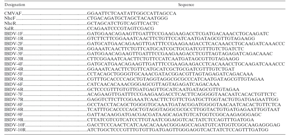
were done by performing overlapping PCR using the respec-tive primers (Table 1). Briefly, two primer pairs (CMVAF/ IBDV-1R and IBDV-1F/NheR) were used to construct pIBDV-1. These primer pairs yielded fragments of 800 and 550 bp. These fragments were combined and subsequently ampli-fied using the flanking primers CMVAF and NheR. The am-plified fragment was digested by EcoRI and NheI restriction enzymes and inserted into the EcoRI- and NheI-digested frag-ment of pIBDVA to yield plasmid pIBDV-1. Similarly, plasmid
pIBDV-2 was constructed using the CMVAF/IBDV-2R and IBDV-2F/NheR primer pairs. The construction of plasmids pIBDV-3, pIBDV-4, pIBDV-5, and pIBDV-6 was performed similarly using the respective primers (Table 1). Plasmids pIBDV-7 to pIBDV-10 were generated as described above using the flanking primers NheF and SalR along with the respective primers (Table 1). All the plasmids were sequenced to confirm the desired sequence changes in segment A. Using a cDNA-based reverse genetics system developed for IBDV, we generated recombinant viruses by transfecting Vero cells with 1g each of a plasmid carrying segment A and a plasmid carrying segment B (pIBDVA and pIBDVB). Successful virus recovery was achieved when cells were cotransfected with the plasmid carrying segment B and with the constructs pIBDVA, pIBDV-3, pIBDV-5, and pIBDV-6 (Table 2), yielding IBDV, IBDV-3, IBDV-5, and IBDV-6, respectively. Generated vi-ruses were passaged further in Vero cells to generate the stock viruses.
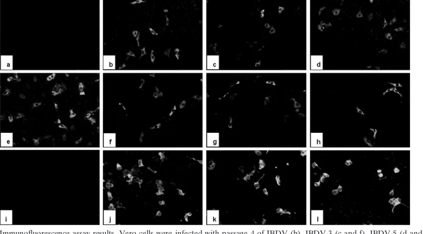
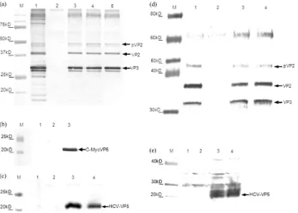
Analysis of the recovered viruses. The recovered viruses were characterized by both immunofluorescence and Western blotting analyses. Vero cells infected with IBDV and IBDV-3 were incubated with rabbit anti-IBDV serum and mouse c-Myc MAb (Sigma), respectively, and cells infected with IBDV-5 and IBDV-6 were incubated with HCV polyclonal antibody (My-BioSource, CA); cells were then stained with fluorescein-con-jugated secondary antibody. The IBDV polyclonal antibody recognized IBDV antigen in the IBDV-, IBDV-3-, IBDV-5-, and IBDV-6-infected cells. In contrast, the anti-c-Myc MAb readily detected viral antigens in IBDV-3-infected cells but not in mock-infected or IBDV-infected controls (Fig. 2). Similarly, HCV polyclonal antibody reacted with viral antigens in IBDV-5- and IBDV-6-infected cells but not in mock-infected or IBDV-infected controls. Western blot analyses of infected Vero cell lysates with anti-IBDV polyclonal antibody, c-Myc MAb, and HCV polyclonal antibody also confirmed the
ex-TABLE 1. Oligonucleotides used for construction of various plasmids
Designation Sequence
CMVAF...GGAATTCTCAATATTGGCCATTAGCCA NheF ...CTGACAGATGCTAGCTACAATGGG NheR...GCTAGCATCTGTCAGTTCACTC SalR...CCAGAATCCCGTAGTCGACG
IBDV-1F...GATGGAACAGAAGTTGATTTCCGAAGAAGACCTCGATGACAAACCTGCAAGATC IBDV-1R ...GTCTTCTTCGGAAATCAACTTCTGTTCCATCAATGATAGCGTTGTAGAAGG
IBDV-2F...GATGCATGAACAGAAGTTGATTTCCGAAGAAGACCTCACAAACCTGCAAGATCAAACCC IBDV-2R ...GGAAATCAACTTCTGTTCATGCATCGCTGCGATCGTTTGTCTGATCTC
IBDV-3F...GATGGAACAGAAGTTGATTTCCGAAGAAGACCTCGTTAGTAGAGATCAGACAAAC IBDV-3R ...CTTCGGAAATCAACTTCTGTTCCATCAATGATAGCGTTGTAGAAGG
IBDV-4F...GATGCATGAACAGAAGTTGATTTCCGAAGAAGACCTCACAAACCTGCAAGATCAAACCC IBDV-4R ...GGAAATCAACTTCTGTTCATGCATCGCTGCGATCGTTTGTCTGAT
IBDV-5F...CCTACAGCTGGGGTGCAAACGATACGGACGTTAGTAGAGATCAGACAAA IBDV-5R ...CGTTTGCACCCCAGCTGTAGGTAGGCGCGCCCATCAATGATAGCGTTGTAGAA IBDV-6F...CATCAACACAAACGGGAGCGTTAGTAGAGATCAGACAAA
IBDV-6R ...GCTCCCGTTTGTGTTGATGAGTTGCATCAATGATAGCGTTGTAGAA
IBDV-7F...ACAGAAGTTGATTTCCGAAGAAGACCTCACTTCAGGGGTAACAATCACACTGTTCTC IBDV-7R ...GAGGTCTTCTTCGGAAATCAACTTCTGTTCTGATGCTTGGTACTGTGATGAGAATTGG IBDV-8F...GCCTACCTACAGCTGGGGTGCAAATGATACGGATGGGGTAACAATCACACTGTTCTCA IBDV-8R ...TCATTTGCACCCCAGCTGTAGGTAGGCGCGCCTTGGTACTGTGATGAGAATTGGTAAT IBDV-9F...GATTACAAGGATGACGACGATAAGCAGATGTCATGGTCGGCAAGAGGGAGC IBDV-9R ...CTTATCGTCGTCATCCTTGTAATCGGAGGTCACTATCTCCAGTTTGATGGA
IBDV-10F...GACCTCCCAACTCATCAACACAAACGGGAGCCAGATGTCATGGTCGGCAAGAGGGAG IBDV-10R ...ATCTGGCTCCCGTTTGTGTTGATGAGTTGGGAGGTCACTATCTCCAGTTTGATGG
VOL. 85, 2011 NOTES 1409
on November 7, 2019 by guest
http://jvi.asm.org/
[image:2.585.35.541.76.319.2]pression of c-Myc (fused to VP5) and HCV epitopes by the recovered viruses (Fig. 3). The recovery of the viruses was further confirmed by reverse transcription-PCR (RT-PCR) analysis of viral RNA template. Total cellular RNA from in-fected and mock-inin-fected cells was extracted and analyzed by RT-PCR. Sequence analysis of the RT-PCR products con-firmed the presence of desired alterations in segment A of the generated IBDV-3, IBDV-5, and IBDV-6 viruses. Taken to-gether, these results suggest that IBDV can stably sustain the insertion of small foreign epitopes in VP5 protein without interference in assembly or virus attachment/entry.
Growth kinetics and genetic stability of the recovered vi-ruses. To compare the replication kinetics of the recovered viruses, Vero cells were infected with IBDV, IBDV-3, IBDV-5, and IBDV-6 at a multiplicity of infection (MOI) of 1.0.
In-fected cell cultures were harvested at different time points, and the titer of infectious virus present in the culture was determined by a focus-forming assay. Briefly, Vero cells were infected with different dilutions of recovered viruses and incubated for 1 h at 37°C. After incubation, the cells were rinsed, overlaid with Dulbecco’s modified Eagle medi-um–5% fetal calf serum, and incubated further at 37°C. After 24 h, the cells were fixed with methanol acetone (1:1), incubated with anti-IBDV polyclonal rabbit antiserum for 1 h, and stained with fluorescein-labeled secondary antibody and foci were counted by using fluorescence microscopy with a Zeiss Axioplan microscope. The results showed that the kinetics and magnitude of replication for IBDV-3 were very similar to those for the wild-type IBDV and that the final virus yields in Vero cells were comparable. However,
FIG. 1. Schematic representation of IBDV segment A and B plasmids. The cDNAs of both segments were placed such that transcription from the CMV promoter starts at the first 5⬘-end nucleotide, whereas the HDV ribozyme sequence was introduced downstream of both segments to ensure the generation of authentic 3⬘ends. Segment A was further modified to construct the plasmids with a c-Myc/FLAG/HCV epitope, which is shown in bold letters. Plasmid characteristics are as follows: pIBDV-1, replacement of the 5⬘terminus of the VP5 coding sequence with the c-Myc sequence; pIBDV-2, replacement of the 5⬘terminus of the VP2 coding sequence with the c-Myc sequence; pIBDV-3, insertion of the c-Myc sequence at the 5⬘terminus of the VP5 coding sequence; pIBDV-4, insertion of the c-Myc sequence at the 5⬘terminus of the VP2 coding sequence; pIBDV-5, insertion of the HCV(523–535)epitope at the 5⬘terminus of the VP5 coding sequence; pIBDV-6, insertion of the HCV(412–419)epitope
at the 5⬘terminus of the VP5 coding sequence; pIBDV-7, mutation and insertion of a FLAG epitope in the PBCloop of VP2; pIBDV-8, mutation
and insertion of the HCV(523–535)epitope in the PBCloop of VP2; pIBDV-9, replacement of the PHIloop of VP2 by the FLAG epitope; and
pIBDV-10, replacement of the PHIloop of VP2 by the HCV(412–419)epitope. Vero cells were transfected with various IBDV segment A constructs,
plus segment B. The virus infectivity was monitored by immunofluorescence and Western blotting analyses.
on November 7, 2019 by guest
http://jvi.asm.org/
IBDV-5 and IBDV-6 showed a slight delay in growth and had titers 1 log lower than IBDV (Fig. 4). To determine the genetic stability of the transfectant virusesin vitro, the vi-ruses were propagated in Vero cells (up to 5 passages), total RNA was isolated, and the region corresponding to the modified portion of VP5 was amplified by RT-PCR. Se-quence analysis of the RT-PCR product confirmed the ex-pected modifications in the VP5 genes of the recovered viruses. The stability of the recombinant viruses expressing foreign epitopes was further confirmed by immunofluores-cence and Western blot analyses of the cell lysates.
The ability of IBDV to propagate in primate cells (16) sug-gests that IBDV might be capable of replication in humans. Additionally, replication of IBDV in human cells will establish its potential to be used as vectors for prophylactic purposes.
With this objective, we infected HEK 293 cells, a specific cell line originally derived from human embryonic kidney cells, with IBDV, IBDV-5, and IBDV-6, and virus replication was confirmed by an immunofluorescence assay (Fig. 2) and West-ern blot analysis (Fig. 3).
[image:4.585.42.544.79.202.2]The use of recombinant viral vaccines is a relatively novel and promising approach to combating infectious diseases in humans as well as in veterinary medicine. An efficient viral vector is expected to provide preferentially stable and long-term transgene expression. Several viral systems, including hepatitis B virus, poliovirus, Newcastle disease virus, adenovi-rus, and influenza viadenovi-rus, have been used as vectors to express foreign epitopes and induce protective immunity against unre-lated pathogens (15, 18, 20, 25, 28). Previous studies have shown that vaccination of humans with short synthetic HCV
TABLE 2. Recovery of infectious viruses using different IBDV segment A constructsa
Construct name Description or change in segment A Recovery resultb
pIBDVA Strain D78 segment A ⫹
pIBDV-1 Replacement of 5⬘terminus of VP5 coding sequence (nt 100–129) with c-Myc sequence ⫺ pIBDV-2 Replacement of 5⬘terminus of VP2 coding sequence (nt 134–163) with c-Myc sequence ⫺ pIBDV-3 Insertion of c-Myc sequence at 5⬘terminus of VP5 coding sequence (between nt 99 and 100) ⫹ pIBDV-4 Insertion of c-Myc sequence at 5⬘terminus of VP2 coding sequence (between nt 133 and 134) ⫺ pIBDV-5 Insertion of HCV(523–535)epitope at 5⬘terminus of VP5 coding sequence (between nt 99 and 100) ⫹
pIBDV-6 Insertion of HCV(412–419)epitope at 5⬘terminus of VP5 coding sequence (between nt 99 and 100) ⫹
pIBDV-7 Mutation and insertion of FLAG epitope in PBCloop of VP2 (corresponding to nt 794–800) ⫺
pIBDV-8 Mutation and insertion of HCV(523–535)epitope in PBCloop of VP2 (corresponding to nt 794–800) ⫺
pIBDV-9 Substitution of FLAG epitope in PHIloop of VP2 (corresponding to nt 1076–1100) ⫺
pIBDV-10 Substitution of HCV(412–419)epitope in PHIloop of VP2 (corresponding to nt 1076–1100) ⫺
a
Amino acid sequences of epitopes were as follows: c-Myc epitope, EQKLISEEDL; FLAG epitope, DYKDDDDK; HCV(523–535)epitope, GAPTYSWGANDTDV;
and HCV(412–419)epitope, QLINTNGS. Vero cells were transfected with various IBDV segment A constructs, plus segment B.
b
Results for recovery of virus are denoted as⫹(virus was recovered) and⫺(no virus was recovered).
FIG. 2. Immunofluorescence assay results. Vero cells were infected with passage 4 of IBDV (b), IBDV-3 (c and f), IBDV-5 (d and g), and IBDV-6 (e and h) at an MOI of 1.0. Uninfected Vero cells were used as a negative control (a). At 24 h postinfection, the cells were fixed and analyzed by immunofluorescence staining with rabbit anti-IBDV (a, b, c, d, and e), c-Myc monoclonal antibody (f), and HCV polyclonal antibody (g and h). HEK293 cells were infected with IBDV (j), IBDV-5 (k), and IBDV-6 (l), and uninfected HEK293 cells were used as a negative control (i). At 24 h postinfection, the cells were fixed and analyzed by immunofluorescence staining with rabbit anti-IBDV antibody (i, j, k, and l).
VOL. 85, 2011 NOTES 1411
on November 7, 2019 by guest
http://jvi.asm.org/
[image:4.585.81.507.440.676.2]peptides has generated both humoral and cellular immunity (13, 30, 31). In the present study, we explored the potential utility of IBDV to carry foreign epitopes with the aim of de-veloping a safe and efficient viral vector platform. Using a plasmid-based reverse genetics system, we generated a recom-binant IBDV (IBDV-3) expressing a c-Myc epitope by intro-ducing the epitope sequence into the VP5 coding region of IBDV, thereby increasing the length of genome segment A by 30 nucleotides. After the recovery of IBDV-3, we further explored the VP5 region by inserting the hepatitis C vi-rus epitopes [HCV(523–535), GAPTYSWGANDTDV, and
HCV(412–419), QLINTNGS] and successfully recovered the
[image:5.585.81.499.67.366.2]vi-ruses harboring the HCV epitopes, thus increasing the length of segment A by 42 and 24 nucleotides, respectively. We fur-ther explored the VP5 region by inserting the M2e gene (en-coding the sequence SLLTEVETPIRNEWGCRCNDSS) from influenza type A but were unsuccessful in recovering this virus (data not shown). We speculate that the reason for the failure
FIG. 3. Immunoblot analysis of IBDV proteins synthesized in virus-infected Vero/HEK293T cells. (a to c) Vero cells were cotransfected with pIBDVB and pIBDVA, pIBDV-3, pIBDV-5, or pIBDV-6. Five days posttransfection, virus was harvested after three cycles of freeze-thawing. Proteins in the cell lysates were separated by SDS–12.5% PAGE; blotted onto nitrocellulose; reacted with polyclonal anti-IBDV serum (a), c-Myc MAb (b), and hepatitis C virus glycoprotein E polyclonal antibody (c); and detected with alkaline-phosphatase and naphthol phosphate fast red color development reagents. (a) Lanes: 1, sample from IBDV-infected cells; 2, sample from mock-infected cells; 3, sample from IBDV-3-infected cells; 4, sample from IBDV-5-infected cells; and 5, sample from IBDV-6-infected cells. (b) Lanes: 1, sample from IBDV-infected cells; 2, sample from mock-infected cells; and 3, sample from IBDV-3-infected cells. (c) Lanes: 1, sample from IBDV-infected cells; 2, sample from mock-infected cells; 3, sample from IBDV-5-infected cells, and 4, sample from IBDV-6-infected cells. (d and e) HEK293T cells were infected with IBDV, IBDV-5, or IBDV-6. Five days postinfection, virus was harvested after three cycles of freeze-thawing. Proteins in the cell lysates were separated by SDS–4 to 20% PAGE, blotted onto nitrocellulose, reacted with polyclonal anti-IBDV serum (d) and hepatitis C virus polyclonal antibody (e), and detected with horseradish peroxidase color development reagents. Lanes: 1, sample from IBDV-infected cells; 2, sample from mock-infected cells; 3, sample from IBDV-5-infected cells; and 4, sample from IBDV-6-infected cells. The positions of pVP2, VP2, VP3, c-Myc–VP5, HCV epitope-VP5, and marker proteins (M) are indicated in kilodaltons.
FIG. 4. Replication kinetics of the recovered viruses. Monolayers of Vero cells were infected with the indicated viruses at an MOI of 1.0 and harvested at the indicated time points. Infectious titers were de-termined by a focus-forming-unit (FFU) assay.
on November 7, 2019 by guest
http://jvi.asm.org/
[image:5.585.42.284.550.687.2]to recover the virus with the flu sequence inserted into VP5 may be the size of the epitope. The coding sequence is 66 nucleotides, longer than the c-Myc or HCV epitope sequence, and the longer foreign sequence may have hindered the effi-cient packaging of the IBD virus. This indicates the limitation of IBDV in its intolerance of longer foreign sequences, as well as the importance of the amino acid sequences selected for insertion, which may interfere with protein folding, assembly, or replication of the IBDV genome. Based on the crystal struc-ture of VP2, we also explored insertion into the PBCand PHI loops but failed to recover any recombinant virus. Recently, it was shown that the foot-and-mouth disease virus (FMDV) immunodominant epitope could be effectively inserted into the PBCloop of IBDV to produce IBDV SVPs with chimeric VP2 in insect cells (23). The inability to recover any of the recom-binant viruses with modification in the loops may suggest that though it is possible to generate chimeric SVPs, any insertion of sequences into these loops may limit or interfere with effi-cient packaging or the function of VP2 in virus attachment/ entry. The same may be true for the lack of recovery of virus from cells transfected with pIBDV-2 and pIBDV-4 constructs, suggesting the importance of protein sequences in viral func-tion. Although the tolerable size and sites for the insertion were not fully explored in this study, IBDV certainly could be exploited for the expression of small therapeutic epitopes. One of the fundamental requirements for currently available viral vectors is their safety. IBDV is an avian virus and, despite its worldwide distribution in domestic fowl, is not known to be a hazard for transmission to other species. While remaining cau-tious, we believe that this may not be a risk to humans. The use of IBDV as a therapeutic agent in patients suffering from chronic hepatitis infections has already been documented (2, 8). The results of a phase II clinical trial of IBDV coinfection therapy in 42 acute hepatitis patients (infected with hepatitis B virus and HCV) showed the safety and efficacy of IBDV ther-apy and provided encouraging evidence that the progression to chronic infection was marginally reduced in IBDV-treated pa-tients compared to the controls (9). It has also been reported that IBDV therapy improved the condition of an HCV patient that had become resistant to conventional treatment, devel-oped decompensated chronic viral hepatitis, and received dis-ability status (2). Importantly, during the treatment of patients, it emerged that to ensure an “artificial viremia” with IBDV, the viral preparation needs to be given in large doses and continuously over a long period (1). These findings further support the therapeutic application of IBDV in humans with-out detrimental effects. IBDV is not known to multiply in human cells but is able to propagate in primate cells. To es-tablish the potential of an IBDV vector for prophylactic pur-poses in HCV infections, we infected HEK293T cells, a specific cell line originally derived from human embryonic kidney cells, and compared the growth of IBDV in Vero cells to that in HEK293T cells. The virus replicated to yield similar titers in the two cell lines, and the replication was confirmed by immu-nofluorescence and Western blot analyses. The ability of IBDV-HCV vectors to replicate in human cells ascertains the potential of IBDV to be useful as an expression vector in humans. A recent study described mice as a potential carrier for IBDV (22). The study also confirmed that IBDV main-tained its viability and pathogenicity in mice and that virus
excreted from mice induced clinical disease in chickens (22), suggesting that IBDV might be capable of replication in hu-mans. Additionally, most human and animal populations are not exposed to IBDV, in contrast to vaccinia virus or adeno-virus and polioadeno-virus, on which some viral vectors and vaccines are based. Therefore, in vivo expression of a heterologous immunogen from an IBDV-based vaccine vector would not be limited by prior immunity. Furthermore, because of its natural heat stability, ease of production, and widespread use, IBDV lends itself as a low-cost recombinant veterinary vaccine, es-pecially for poultry. In this study, for the first time we demon-strated the insertion of foreign epitopes in segment A of IBDV, thus generating a replication-competent recombinant virus. Further exploration of the insertion sites used in the present study and other sites with different antigenic epitopes will shed more light on the development of novel IBDV vec-tors.
The project was supported in part by the Maryland Industrial Part-nerships (MIPS) and VectorLogics, Inc., Rockville, MD, through UMBI award no. MIPS 4103.
REFERENCES
1.Bakacs, T., and J. N. Mehrishi.2004. Examination of the value of treatment of decompensated viral hepatitis patients by intentionally coinfecting them with an apathogenic IBDV and using the lessons learnt to seriously consider treating patients infected with HIV using the apathogenic hepatitis G virus. Vaccine23:3–13.
2.Bakacs, T., and J. N. Mehrishi.2002. Intentional coinfection of patients with HCV infection using avian infection bursal disease virus. Hepatology36:255. 3.Ben Abdeljelil, N., B. Delmas, and H. Mardassi. 2008. Replication and packaging of an infectious bursal disease virus segment A-derived minig-enome. Virus Res.136:146–151.
4.Birghan, C., E. Mundt, and A. E. Gorbalenya.2000. A non-canonical Lon proteinase lacking the ATPase domain employs the Ser-Lys catalytic dyad to exercise broad control over the life cycle of a double-stranded RNA virus. EMBO J.19:114–123.
5.Brizzard, B. L., R. G. Chubet, and D. L. Vizard. 1994. Immunoaffinity purification of FLAG epitope-tagged bacterial alkaline phosphatase using a novel monoclonal antibody and peptide elution. Biotechniques16:730–735. 6.Caston, J. R., et al.2001. C terminus of infectious bursal disease virus major capsid protein VP2 is involved in definition of the T number for capsid assembly. J. Virol.75:10815–10828.
7.Coulibaly, F., et al.2005. The birnavirus crystal structure reveals structural relationships among icosahedral viruses. Cell120:761–772.
8.Csatary, L. K., R. Schnabel, and T. Bakacs.1999. Successful treatment of decompensated chronic viral hepatitis by bursal disease virus vaccine. Anti-cancer Res.19:629–633.
9.Csatary, L. K., L. Telegdy, P. Gergely, B. Bodey, and T. Bakacs.1998. Preliminary report of a controlled trial of MTH-68/B virus vaccine treatment in acute B and C hepatitis: a phase II study. Anticancer Res.18:1279–1282. 10.Da Costa, B., et al. 2002. The capsid of infectious bursal disease virus contains several small peptides arising from the maturation process of pVP2. J. Virol.76:2393–2402.
11.Delmas, B., et al.2004. Birnaviridae, p. 561–569.InC. M. Facquet, M. A. Mayo, J. Maniloff, U. Desselberger, and A. L. Ball (ed.), Virus taxonomy. Academic Press, London, United Kingdom.
12.Evan, G. I., G. K. Lewis, G. Ramsay, and J. M. Bishop.1985. Isolation of monoclonal antibodies specific for human c-mycproto-oncogene product. Mol. Cell. Biol.5:3610–3616.
13.Firbas, C., et al.2006. Immunogenicity and safety of a novel therapeutic hepatitis C virus (HCV) peptide vaccine: a randomized, placebo controlled trial for dose optimization in 128 healthy subjects. Vaccine24:4343–4353. 14.Garriga, D., et al.2006. The 2.6-angstrom structure of infectious bursal
disease virus-derived T⫽1 particles reveals new stabilizing elements of the virus capsid. J. Virol.80:6895–6905.
15.Girard, M., A. Martin, and S. van der Werf.1993. Potential use of poliovirus as a vector. Biologicals21:371–377.
16.Jackwood, D. H., Y. M. Saif, and J. H. Hughes.1987. Replication of infec-tious bursal disease virus in continuous cell lines. Avian Dis.31:370–375. 17.Lee, C. C., et al.2006. Crystal structure of infectious bursal disease virus VP2
subviral particle at 2.6A resolution: implications in virion assembly and immunogenicity. J. Struct. Biol.155:74–86.
18.Mebatsion, T., et al.2002. Newcastle disease virus (NDV) marker vaccine:
VOL. 85, 2011 NOTES 1413
on November 7, 2019 by guest
http://jvi.asm.org/
an immunodominant epitope on the nucleoprotein gene of NDV can be deleted or replaced by a foreign epitope. J. Virol.76:10138–10146. 19.Mundt, E., and V. N. Vakharia. 1996. Synthetic transcripts of
double-stranded Birnavirus genome are infectious. Proc. Natl. Acad. Sci. U. S. A. 93:11131–11136.
20.Palese, P., F. Zavala, T. Muster, R. S. Nussenzweig, and A. Garcia-Sastre. 1997. Development of novel influenza virus vaccines and vectors. J. Infect. Dis.176(Suppl. 1):S45–S49.
21.Pan, J., V. N. Vakharia, and Y. J. Tao.2007. The structure of a birnavirus polymerase reveals a distinct active site topology. Proc. Natl. Acad. Sci. U. S. A.104:7385–7390.
22.Park, M. J., J. H. Park, and H. M. Kwon.2010. Mice as potential carriers of infectious bursal disease virus in chickens. Vet. J.183:352–354.
23.Re´mond, M., et al.2009. Infectious bursal disease subviral particles dis-playing the foot-and-mouth disease virus major antigenic site. Vaccine 27:93–98.
24.Sanchez, A. B., and J. F. Rodriguez.1999. Proteolytic processing in infectious bursal disease virus: identification of the polyprotein cleavage sites by site-directed mutagenesis. Virology262:190–199.
25.Scho¨del, F. P. D., J. Hughes, R. Wirtz, and D. Milich.1996. Hybrid hepatitis B virus core antigen as a vaccine carrier moiety. I. Presentation of foreign epitopes. J. Biotechnol.44:91–96.
26.van den Berg, T. P., N. Eterradossi, D. Toquin, and G. Meulemans.2000. Infectious bursal disease (Gumboro disease). Rev. Sci. Tech.19:509–543. 27.von Einem, U. I., et al.2004. VP1 of infectious bursal disease virus is an
RNA-dependent RNA polymerase. J. Gen. Virol.85:2221–2229. 28.Worgall, S., et al.2005. Protection against P. aeruginosa with an adenovirus
vector containing an OprF epitope in the capsid. J. Clin. Invest.115:1281– 1289.
29.Yao, K., M. A. Goodwin, and V. N. Vakharia.1998. Generation of a mutant infectious bursal disease virus that does not cause bursal lesions. J. Virol. 72:2647–2654.
30.Yutani, S., et al.2009. Phase I clinical study of a peptide vaccination for hepatitis C virus-infected patients with different human leukocyte antigen-class I-A alleles. Cancer Sci.100:1935–1942.
31.Yutani, S., et al.2007. Phase I clinical study of a personalized peptide vaccination for patients infected with hepatitis C virus (HCV) 1b who failed to respond to interferon-based therapy. Vaccine25:7429–7435.


