1
Dissertation on
“A STUDY TO CORRELATE ELEVATED CREATININE PHOSPHOKINASE WITH SNAKE BITE AND ACUTE KIDNEY INJURY”
Submitted in partial fulfillment of requirements of
MD DEGREE GENERAL MEDICINE
BRANCH – I
GOVERNMENT RAJAJI HOSPITAL, MADURAI MEDICAL COLLEGE
MADURAI- 20
THE TAMILNADU
Dr. M.G.R. MEDICAL UNIVERSITY
CHENNAI
2
CERTIFICATE FROM THE DEAN
This is to certify that this dissertation entitled “A STUDY TO CORRELATE ELEVATED CREATININE PHOSPHO KINASE WITH SNAKE BITE AND
ACUTE KIDNEY INJURY” is the bonafide work of Dr. K. KIRTHIKA in partial fulfillment of the university regulations of the Tamil Nadu Dr. M.G.R. Medical University, Chennai, for M.D General Medicine Branch I examination to be held in April 2018.
Dr. D. MARUTHUPANDIAN MS
THE DEAN,
3
CERTIFICATE FROM THE HOD
This is to certify that this dissertation entitled “A STUDY TO CORRELATE ELEVATED CREATININE PHOSPHO KINASE WITH SNAKE BITE AND
ACUTE KIDNEY INJURY’’ is the bonafide work of Dr. K. KIRTHIKA in partial fulfillment of the universityregulations of the Tamil Nadu Dr. M.G.R. Medical University, Chennai, for M.D General Medicine Branch I examination to be held in April 2018.
DR.V.T.PREM KUMAR MD
Professor and HOD,
4
CERTIFICATE FROM THE GUIDE
This is to certify that this dissertation entitled “A STUDY TO CORRELATE ELEVATED CREATININE PHOSPHO KINASE WITH SNAKE BITE AND
ACUTE KIDNEY INJURY’’ is the bonafide work of Dr. K. KIRTHIKA in partial fulfillment of the universityregulations of the Tamil Nadu Dr. M.G.R. Medical University, Chennai, for M.D General Medicine Branch I examination to be held in April 2018.
DR.V.T.PREM KUMAR MD Professor and HOD,
Department Of General Medicine, Government Rajaji Hospital, Madurai Medical College, Madurai.
5
DECLARATION
I, Dr. K. KIRTHIKA declare that, I carried out this work on” A STUDY TO CORRELATE ELEVATED CREATININE PHOSPHO KINASE WITH
SNAKE BITE AND ACUTE KIDNEY INJURY’’ at the Department of Medicine, Govt. Rajaji Hospital duringthe period JANUARY 2016 TO MAY 2016. I also declare that this bonafide work or a part of this work was not submitted by me or any others for any award, degree or diploma to any other University, Board either in India or abroad.
This dissertation is submitted to The Tamil Nadu Dr. M.G.R. Medical University, Chennai in partial fulfillment of the rules and regulations for the award of M.D Degree General MedicineBranch- I; examination to be held in April 2018.
6
ACKNOWLEDGEMENTS
At the outset, I wish to thank our Dean Dr. D. MARUTHUPANDIAN MS, for permitting me to use the facilities of Madurai Medical College and Government Rajaji Hospital to conduct this study.
I shall remain eternally grateful to my beloved teacher, my guide, my unit chief and Head of the department of general medicine, Prof. Dr. V. T. PREMKUMAR MD who has given me his moral support and encouragement throughout the
conduct of the study. His valuable words of advice has guided me during my study and through my internship in college.
.
I also sincerely thank our beloved professors . Dr.M.Natarajan MD,
Dr.R.Balajinathan.MD.,Dr.G.Bagialakshmi.M.D., Dr.J.Sangumani. MD,
Dr.C.Dharmaraj. MD.,and Dr.R.Prabhakaran. MD for their par excellence clinicalteaching and constant support.
I offer my thanks to my unit Assistant Professors for their constant
encouragement, timely help and critical suggestions throughout the study and also for making my stay in the unit informative.
I am extremely thankful to Prof Dr. Mohan Kumar MD, Head of the department of biochemistry & Prof Dr. Arul MD DM, Head of the department of
7
I express my thanks to Dr. Sruthi R.S., Dr. Vinoj, Dr. Gokul, Dr. Arun, for their help and support in my dissertation work.
My patients, who form the most integral part of the work, were always kind and cooperative. I pray to God give them courage and strength to endure their illness, hope all of them go into complete remission.
I thank my dad, mom and my brother who have stood by me during my times of need. Their help and support have always been invaluable to me.
8
1
INTRODUCTION
2
AIMS AND OBJECTIVES
3
REVIEW OF LITERATURE
4
MATERIALS AND METHODS
5
OBSERVATION AND RESULTS
6
DISCUSSION
7
SUMMARY
8
CONCLUSION
9 ANNEXURES
Bibliography
Abbreviations
Proforma
Master Chart
Ethical Clearance letter
9
INTRODUCTION
“ India , similar to global incidence in estimation to have the highest snakebite mortality. WHO estimated the number of bites to be 83,000 with 11,000 deaths per annum. Much of the fatalities are due to the victims not reaching the primary care in time where treatment can be administered”.
“ Moreover people are not much informed about the occupational risks and the measures which will prevent snake bites. They continue to adopt harmful practices such as tourniquets ,or cutting and sucking , etc. Observations reveals that the primary care centres do not treat snakebite patients primarily due to lack of expertise and confidence”.
“But At the secondary and tertiary care level, more than 2 region specific protocols are followed for anti-snake venom (ASV) treatment”
“Snake is the most commonly used term for the “serpentes” family of reptiles .Out of which only about 20% of snakes are venomous and hence medically important”.
10
family of Elapidae, Viperidae, Colubridae, and Atractaspididae. But most of the burden is caused by bites of vipers and elapids. Much commonly, vipers
significantly causes coagulopathy, renal failure when compared with elapids which causes neurotoxicity”.
RUSSELL’S VIPER
11
“It might be clear that in most parts of the South East Asia, they form an important emergency and reason for hospital admissions. This can result in either death or disability of most of the younger and working population, most commonly those involved in the farming or plantation works. The truer scale of mortality and even morbidity from snake-bites remain not to be certain because of not adequate reporting in most of the country”.
“To remedy this situation, strong recommendations should be made as snake bite to be a notifiable disease in all countries of South East Asian regions”.
“Much of Snake-bite is are seen in people who involve in occupations of farmers, plantation workers, herdsmen, fisherman, daily labourer and other producer. Therefore it is a medical problem that has an important implication for mainly nutrition and economy of the country where it occurs most commonly”.
12
“ Inspite of its importance, there have been very few clinical studies of snake-bite when compared to that of any other tropical diseases. Snakebite probably cause more deaths in the landscape region than do amoebiasis. But only about a small fraction of the research contribution in amoebiasis has been given to the snake-bite”.
“Clinical Diagnosis of species of snakes which is responsible for bite will be important for ideal clinical treatment”.
“If the bite has neurotoxic features with bulbar and respiratory involvement, anti snake venom only cannot be beleived upon to prevent rapid death due to asphyxiation. Neostigmine and ventilatiory support is necessary in such cases”.
“in cases of AKI, Conservative management or rarely, hemodialysis is effective along with supportive treatment in victims of Russell’s or , humped-nosed viper or sea snake-bites”.
13
is present or a high intra compartmental pressure should be confirmed by invasive/ direct measurement”.
15
“ Anti venom, only effective antidote for venom. The essential part of treating systemic envenoming, but may be insufficient on own to save patient.
16
EPIDEMEOLOGY
“Mostly the incidence of snake-bites depends critically on the frequency of contact between snakes and humans. Except during rains, snakes are elusive and reclusive and so contact with humans is likely only when people move into the snakes’ favoured environment”.
“Example :
Rice fields in the case of Russell’s vipers and cobras;
rubber and coffee plantations in the case of Malayan pit vipers)”
“Nocturnally active snakes are trodden upon by people walking along dark paths. Seasonal peaks of snake-bite incidence are usually associated with increases in agricultural activity or monsoon rains”.
“Different species of snakes vary in their willingness to strike when disturbed. Typically7 “irritable” species include Russell’s vipers (Daboia russelii
and D. siamensis) and saw-scaled vipers (Echis)”.
17
“cobras (Naja) which may live in roof spaces or under the floor and by kraits (Bungarus) which enter human dwellings at night in search of their prey like chicken but may bite people who move in their sleep”.
“The risk of envenoming after bites by venomous snakes varies with the species but is on an average it is about 50%. Bites in which the fangs pierce the skin but no envenoming results in a condition known as “dry bite”
“Epidemics of snake-bite may result from heavy flooding, as has been reported from India and Myanmar, and when the snakes’ habitat is invaded by a large workforce involved in road building or logging and as a result of irrigation plans that alter the climate making it attractive both for snakes and farmers”.
“There was no immediate increase in snake-bites in Myanmar after Cyclone Nargis but an increase was recorded in the aftermath 9-12 months later”.
“Males are more often bitten than females, but where the work force predominantly is females (e.g. tea and coffee picking). The peak age for bites is children and young adults”.
18
“There is some evidence that peak case fatality is in young children and the elderly. In pregnancy, snake-bite carries definite risks to mother and fetus,in the form of bleeding and abortion”.
“ Most snake-bites are inflicted on the feet and ankles of agricultural workers”.
CIRCUMSTANCES :
“Mostly snake-bites happen when the snake is pulled along, either in the dark or in undergrowth, by someone who is bare-footed or wearing sandals.”
“The snake may be picked up either unintentionally in a handful of foliage or intentionally by someone who is trying to show off.”
“Some bites occurs when the snake (usually a krait) comes in to the home at night in search of its prey (other snakes, lizards, frogs, mice) and someone
sleeping on the floor rolls over onto the snake in their sleep. Not all the snake-bites happen in rural areas”.
19
“who sleep in small huts (jhuggies) are frequently bitten by kraits during sleep and wake with paralysis.”
“The risk of snake-bite is very strongly associated with
Occupations like farming (rice), plantation work (rubber, coffee), herding, hunting, fishing and fish farming, catching and handling snakes for food (in snake restaurants), displaying and performing with snakes (snake charmers), manufacturing leather (especially sea snakes)”
“OCCUPATION AFFECTED
Farmers (rice)
Plantation workers (rubber, coffee) Herdsmen
Hunters
Snake-handlers
Fishermen and fish farmers
Sea-snake catchers (for sea snake skins, leather)
20
“The numbers of snake-bite fatalities in India has long been controversial”.
“Estimates as low as 61 507 bites and 1,124 deaths in 2006 and 76,948 bites and 1,359 deaths in 2007 and as high as 50 000 deaths each year have
been published”.
“The Registrar-General of India’s “Million Death Study” 2001-2003, is expected to provide reliable evidence of substantial mortality
(exceeding 50 000 per year) because it is based on Representative, Re-sampled, Routine Household Interview of Mortality with Medical Evaluation (“RHIME”)”
“ It covers all age groups across the entire country with geographical, seasonal and also occupational data”.
“Previous studies included a field survey in randomly selected villages in Barddhaman (Burdwan) district, West Bengal that suggested that among the total population of nearly five million people, around 8 000 with bites and 800 killed by snakes each year, an average incidence of 16.4 deaths/100 000/year . In
21
“Category 1:
Elapidae: Bungarus caeruleus; Naja kaouthia (northeast),
CATEGORY 1 Naja naja (throughout)”
“Viperidae: Daboia russelii, Echis carinatus; Hypnale
hypnale (south-west)” Category 2:
“Elapidae: Bungarus fasciatus, Bungarus niger,
CATEGORY 2 Bungarus sindanus, Bungarus walli; Naja oxiana” “Viperidae: Cryptelytrops albolabris, Cryptelytrops
purpureomaculatus (east), Trimeresurus
(south-west), Trimeresurus gramineus (south India)” “Death from snake-bite
Contributing factors”
22
“complicating infections, and failure to observe patients closely after they were admitted to hospital”.
“Time between snake-bite and death
Although very rapid death after snake-bite has rarely been reported and it is clear that large series of snake-bite deaths that many hours usually elapse between bite and death in the case of elapid envenoming, and several days in the case of viper envenoming”
“SIGNS AND SYMPTOMS
Bites in which the fangs pierce the skin but no envenoming results are known as “dry bites”.
“The explanation for dry bites is either mechanical inefficiency of the venom apparatus striking at an unnatural angle (or through clothing) or perhaps voluntary retention of venom by the snake”
23 “(2) Systemic envenoming
(3) Effects of anxiety prompted by the frightening experience of being bitten.
(4) Effects of first-aid and other pre-hospital treatments that may cause misleading results”.
“Local symptoms and signs in the bitten part:
fang marks local pain local bleeding bruising
lymphangitis (raised red lines tracking up the bitten limb) lymph node enlargement
inflammation (swelling, redness, heat) blistering
local infection, abscess formation necrosis”
“Generalized (systemic) symptoms and signs
24
“Nausea, vomiting, malaise, abdominal pain, weakness, drowsiness, Prostration”.
“Cardiovascular (Viperidae)
Visual disturbances, dizziness, faintness, collapse, shock, hypotension,
cardiac arrhythmias, pulmonary oedema, conjunctival oedema (chemosis)”
“Bleeding and clotting disorders (Viperidae)
Traumatic bleeding from recent wounds (including prolonged bleeding from
the fang marks ( venipunctures) and from old partly-healed wounds”
“Spontaneous systemic bleeding - from gums, epistaxis, bleeding into the
tears, intracranial haemorrhage (meningism from subarachnoid
haemorrhage, lateralizing signs and/or coma from cerebral haemorrhage, haemoptysis, haematemesis), rectal bleeding or melaena, haematuria, vaginal bleeding, ante-partum haemorrhage in pregnant women, bleeding into the mucosae (e.g. conjunctivae), skin (petechiae, purpura, discoid haemorrhages – and ecchymoses) and retina”.
25
“Cerebral arterial thrombosis (western Russell’s viper Daboia
russelii)”
“Thrombotic strokes, confirmed by neuro imaging, angiography, are reported rarely after envenoming”.
“Neurotoxic (Elapidae, Russell’s viper)
Drowsiness, paraesthesiae, abnormalities of taste and smell, “heavy” eyelids, ptosis , external ophthalmoplegia(manifested as double vision), paralysis of facial
muscles and other muscles innervated by lower cranial nerves, nasal voice or aphonia, regurgitation of feeds, difficulty in swallowing secretions, respiratory weakness and generalised flaccid quardriparesis”.
Skeletal muscle breakdown (sea snakes, some krait species –
Bungarus niger and B. candidus, western Russell’s viper Daboia russelii)
Generalized pain, stiffness and tenderness of muscles, trismus, myoglobinuria, hyperkalaemia, cardiac arrest, acute renal failure.
26
“Loin (lower back) pain, haematuria, haemoglobinuria, myoglobinuria,
oliguria/anuria, symptoms and signs of uraemia like acidotic breathing, hiccups, nausea, pleuritic chest pain etc”.,
“Endocrine features
Acute pituitary/adrenal insufficiency from infarction of the anterior pituitary (Russell’s viper in Myanmar and South India) due to pituitary apoplexy.”
“Acute phase: Shock, hypoglycaemia, seizures, altered sensorium
Chronic phase (months to years after the bite): Weakness, loss of secondary sexual hair, loss of libido, amenorrhoea, testicular atrophy, hypothyroidism, generalized lethargy, depressed mood etc”.
“Long-term complications (sequelae) of snake-bite
At the bite site, loss of tissue may result from sloughing or surgical débridement of necrotic areas. Amputation can occur. Chronic ulceration, infection, osteomyelitis, contractures, arthrodesis or arthritis may persist and causes severe physical disability”.
27
“Symptoms and signs of sea snake bites
Envenoming by sea snakes (Hydrophiinae) and sea kraits (Laticaudinae): unprovoked and bite is usually not painful and may not be noticed by the
swimmer. Fangs and teeth may be left in the wound. There is minimal or no local swelling and there is no localized lymphadenopathy like in hemotoxic bites.
Generalized rhabdomyolysis is the dominant effect of envenoming by these snakes although patients without this feature have been described”.
“ Earliest symptoms include headache and thirst.
Generalized aching, stiffness and tenderness of the muscles becomes noticeable between 30 minutes and 3½ hours after the bite.”
“Trismus can also occur. Even Passive stretching of the muscles is painful . Later, there is progressive flaccid paralysis starting with ptosis, as in other neurotoxic bites. The patient remains conscious until the respiratory muscles are sufficiently affected to cause respiratory failure”.
28
“Myoglobin and potassium released from damaged skeletal muscles may cause” “renal failure, while hyperkalaemia developing within 6–12 hours of the bite may precipitate cardiac arrest”
“Venom composition
Around 90% of snake venom (dry weight) are proteins. Each venom contains a little more than a hundred different proteins: enzymes (constituting 80-90% of viperid and 25-60% of elapid venoms), enzymatic polypeptide toxins, non-toxic proteins such as nerve growth factor.”
Venom enzymes
These include hydrolases, hyaluronidase, and activators or inactivators of
physiological processes, such as kininogenase. Most venoms contain l-amino acid oxidase, phosphor diesterases, 5’-nucleotidase, DNAase, NAD-nucleosidase, phospholipase A2 and peptidases.
Zinc metalloproteinase haemorrhagins: Damage vascular endothelium, causes bleeding.
Procoagulant enzymes:
Venoms of Viperidae and some Elapidae contains serine proteases and
procoagulant enzymes that are thrombin-like or activate factor X, prothrombin and other factors.
29
“This process results in incoagulable blood since most of the fibrin clot is broken down by the body’s own plasmin fibrinolytic system.
Within 30 minutes of the bite, the levels of clotting factors are so depleted
resulting in consumption coagulopathy.
Some venoms contain multiple anti-haemostatic factors.
For example, Russell’s viper venom contains toxins that activate factors V, X, IX and XIII, protein C, affects platelet aggregation and haemorrhage”.
“Phospholipase A2 (lecithinase): The most extensively studied of all venom enzymes. It damages mitochondria, erythrocytes, leucocytes, platelets, peripheral nerve endings, skeletal muscle,endothelium, and other membranes, produces presynaptic neurotoxic activity, opioid like sedative effects, leads to the autopharmacological release of histamine and anti-coagulants.
Acetylcholinesterase: Although found in most elapid venoms, it doesn’t contribute to the neurotoxicity”.
“Hyaluronidase: Promotes the spread of venom through tissues.Proteolytic enzymes like metalloproteinases, endopeptidases or hydrolases and polypetide cytotoxins”
30
“Venom polypeptide toxins (“neurotoxins”)
Postsynaptic (α) neurotoxins such as α-bungarotoxin and cobrotoxin, consists of 66-74 amino acids.
They bind to acetylcholine receptors at the motor endplate.
Presynaptic (β) neurotoxins such as β-bungarotoxin, crotoxin, and taipoxin, contain 120 amino acids and a phospholipase A subunit.
They release acetylcholine at neuromuscular junctions and then damage the endings, preventing further release of transmitter.”
“Quantity of venom injected at a bite, “dry bites”
“Depends on the species and size of the snake, the mechanical efficiency of
31
“About 50% of bites by Malayan pit vipers and Russell’s vipers, 30% of
bites by cobras and 5%-10% of bites by saw-scaled vipers does not result in any symptoms or signs of envenoming”.
“Snake doesn’t exhaust the store of venom, even after several strikes, and
they are still venomous after eating their prey”.
Although large snakes tend to inject more venom than smaller specimens of
the same species, the venom of young vipers may be richer in some dangerous components, causing more hemotoxicity.
32
35
“Venom-Induced Consumption Coagulopathy
VICC is a simple term used to describe any coagulopathy resulting in the consumption of clotting factors due to a procoagulant toxin in venom. It can be caused by the bite of range of snakes including Viperid snakes, Elapidae snakes, and some”
“Colubrid snakes.
Before, the coagulopathy associated with snake envenoming was often referred to as a disseminated intravascular coagulation. However, the word disseminated intravascular coagulation is not appropriate for coagulopathy due to snakebite. DIC results from widespread immune, endothelial, cellular, and clotting cascade activation and has a very high mortality, whereas VICC has a completely different pathogenesis (specific enzyme activation),and is also treated differently, with a much lower mortality.”
“Venom-induced consumption coagulopathy is characterized by the activation of the clotting pathway due to procoagulant toxins in the snake venom. They are referred to as procoagulant toxins because in vitro, they result in rapid clot
formation, but in vivo, they lead to severe factor consumption and lead to bleeding risk.”
36
“pathway by targeting different clotting factors. Despite the differences, all procoagulant toxins cause a similar clinical picture of both clotting factor consumption and coagulopathy.”
“The clinical severity of VLCC dependent on which clotting factors are consumed. For example, a milder form appears to occur when there is only fibrinogen consumption,”
“whereas a more severe form is usually associated with multiple
factor consumption, such as is seen with Australian elapid envenoming which results in fibrinogen, FV, FVIII consumption
But in Russell's viper envenoming with FV, FVIII, FX, and fibrinogen consumption”.
“The toxins in snake venom that produce VICC are classically categorized by where they act on the clotting pathway, with the most important ones being prothrombin activators, factor V and X activators, and thrombin-like enzymes (TLEs), the latter referred to as fibrinogenases”.
Prothrombin Activators
“Two large groups of snakes contain prothrombin activators:
37
“[Hoplocephalus spp], and taipans [Oxyuranus spp]) and the Echis genus, which includes carpet vipers and the sawscaled viper.
Prothrombin activators is classified into 4 groups (A-D) depending on their structure, function, and cofactor requirements.”
“Australian snakes contains the group C and D prothrombin activators which are serine proteases. Group C prothrombin activators are found in the brown snake and taipan and resemble the human prothrombinase complex or factor Xa Va complex which converts prothrombin to thrombin as in extrinsic pathway”.
“Group D toxins are found in tiger snake, rough-scale snake, and
broadheaded snakes and contain a toxin similar to human factor FXa,without the FVa cofactor.”
“Group A and B toxins are found in the Echis species of snakes and
directly activated prothrombin but convert it to meizothrombin rather than the fully active thrombin.”
“Activation of the clotting cascade by prothrombin activators results in multiple factor deficiencies due to the positive and negative feedback loops activated when prothrombin (II) is converted to thrombin (IIa)”
38
“Thrombin also activates fibrinogen to fibrin consuming all of the fibrinogen complex.
Measurements of factor levels after envenoming have shown severe deficiencies of factor V and factor VIII, with an associated not recordable prothrombin time
(PT)/international normalized ratio (INR) with an undetectable fibrinogen levels, consistent with fullb lown”
VICC.
“A partial VICC has been recognized in patients with low, but recordable
fibrinogen levels where the pathway has not been activated completely and not all of the fibrinogen has been converted to fibrin”.
“Factor X and V Activators Russell's viper (Daboia genus) venom contains factor V and X activators. Most important toxin is the factor X activator, which converts factor X to Xa resulting in the formation of the prothrombinase complex and subsequent conversion of fibrinogen to fibrin”.
“Because the factor X activation acts earlier in the coagulation pathway (before common pathway), it results in positive and negative feedback loops leading to indirect consumption of factors V and VIII.”
39
Thrombin-Like Enzymes
“Thrombin-like enzymes are different to the prothrombin activators and factor Xa/Va activator toxins because they simply consume fibrinogen rather than activating the clotting pathway. For this reason,
TLEs only produce an isolated deficiency of fibrinogen and uncommonly affects other clotting factors.
They usually cause a milder form of VICC, but they will result in non recordable PT/INR and bleeding complications if there is undetectable fibrinogen.
There are more than 67 different TLE toxins that have been identified, mostly being zinc
metalloproteinase that cleaves the α chain of fibrinogen resulting in consumption of fibrinogen without the production of (or conversion to) fibrin.
Other TLEs cleave the β chain and a few cleave both chains, but they do not result in intact fibrin.”
40
Clinical Manifestations of VICC
“The clinical outcome for patients who develops VICC is not only dependent on the severity of the coagulopathy, but other factors such as trauma, platelet count and function, and vascular injury are important.
Arguably the most important issues in patients with VICC is whether physical trauma occurs and anything that potentially causes vessel damage and the release of tissue factor may result in hemorrhage.
This may result in significant morbidity or death in case of intracranial or major organ hemorrhage. Bleeding can range from minor, such as oozing at the bite site or cannula insertion site, to major life-threatening hemorrhage requiring blood transfusion.”
“Intracranial hemorrhage can be seen after an associated head injury, but spontaneous bleeds also occur, often in the setting of hypertension. Thoracic cavity bleeding or bleeding into other body compartments can occur after trauma”.
“The metalloproteinase prothrombin activator toxin ecarin found in Echis species not only activates the clotting pathway but also acts as a hemorrhagin. These
41 “the risk of bleeding associated with VICC.
This explains why spontaneous and more severe bleeding is reported with Echis envenoming compared with Australian elapid envenoming, despite the
prothrombin”
activators being more potent in the latter.
“The duration of VICC depends on the type of toxin and the administration of antivenom.”
“Venom-induced consumption coagulopathy resulting from Australian elapid envenoming appears to resolve after 24 to 48 hours independent of antivenom treatment, and the time to recovery appears to depend only on the production of new clotting factors”.
“ On the other hand, Echis-induced VICC has been shown to continue for days if not treated with antivenom. However, after Echis envenoming is treated with antivenom, the time of recovery is also 24 to 48 hours demonstrating that once the toxin is neutralized, there is normal recovery of clotting factors.”
An understanding of expected natural course of VICC is determined by the type of snake and procoagulant toxins which may influence the need for antivenom,
duration of
42 Anticoagulant Coagulopathy
A less common condition related to snake envenoming is an anticoagulant-type coagulopathy. The Australian black snake (Pseudechis) genus venom has been shown to inhibit clotting in vitro and cause a mild to moderate elevation of the aPTT. In a series of
17 mulga snake envenomings in Australia, 10 patients developed an anticoagulant coagulopathy evidenced by an aPTT median rise of 82 seconds (interquartile range, 55-91 seconds). Four of these patients had a small INR rise (b3.0), with the rest having an INR within the normal limits. Fibrinogen and D-dimer levels were normal.
No patients developed any significant clinical bleeding, supporting the idea that this is
not a significant coagulopathy. A similar anticoagulant coagulopathy was also found in red-bellied black snake (Pseudechis porphyriacus) bites.
43
The early administration of antivenom has been shown to prevent myotoxicity. The occurrence of anticoagulant coagulopathies in other snakes remains unclear, mainly because the 20-minute WBCT is unlikely to be abnormal, except in the most severe cases. Anticoagulant toxins have been identified in a number of snakes, including cobras (Naja spp) and the southern copperhead (Agkistrodon genus). The venom of the southern copperhead contains a protein C activator, but does not appear to have any clinical effects in humans.
Thrombotic Microangiopathy
A proportion of patients who develop VICC can also go on to develop an
envenoming-related thrombotic microangiopathy, of which the pathogenesis is not completely understood. This is characterized by thrombocytopenia,
microangiopathic hemolytic anemia, and an acute kidney injury which continues beyond the resolution of VICC
and has been noted in a number of different snake species, including Australasian elapids and many viper species. From the information available in cases, it is difficult to define where snakebite-induced thrombotic microangiopathy fits on the spectrum
44
uremic syndrome—and it is likely to be a distinct syndrome. The combination of thrombocytopenia with mainly renal involvement is unusual.
In the full blown syndrome which occurs in less than 5% of cases, patients develop severe thrombocytopenia and acute anuric renal failure requiring renal replacement therapy for 2 to 6 weeks. However, most cases are less severe and snakebite-induced thrombotic microangiopathy appears to be a spectrum of
disorders, with some patients only having a brief period of thrombocytopenia and a moderate rise in creatinine
without oliguria.
Treatment is supportive in all cases, with renal replacement therapy being the most important intervention in severe cases. Plasmapheresis has been used in a number of more severe cases but does not appear to change the natural course of the
thrombocytopenia, anemia, or renal injury. The lack of benefit of plasmapheresis is probably due to the underlying cause being different and not related to ADAMTS-13
function or antibodies like in thrombotic thrombocytopenic purpura.
An important consideration is that plasmapheresis will remove the antivenom. It is unclear whether antivenom alters the course of thrombotic microangiopathy, but is potentially beneficial for any toxin-related injury resulting in the
45
ACUTE KIDNEY INJURY PATHOGENESIS :
It is mainly observed in snakes which belong to the viperidae group and it is seen less with sea snake bites and bites of colubridae group. Most of the Indians are victims of Russell’s viper or echiscarinatus bites, which causes AKI.
It is an important complication of snake bite and a proper supportive management after the antivenom administration is of utmost importance for a good patient outcome.
Tubular necrosis and cortical necrosis are the important causes of renal failure. The renal failure after bite is usually reversible, but if acute cortical necrosis occurs, it may lead to an incomplete recovery.
The main cause of this “unacceptable incidence” of snake bite fatalities is becausepeople try out bizarre remedies initially instead of going to a primary health care.
There are very less number of studies on the development and outcome of AKI following snake bite in India.
The exact pathogenesis for ARF is not well established. However a number of factors contribute like
46
Disseminated intravascular coagulation Intravascular hemolysis
Nephrotoxicity of the venom Nephrotoxic drugs in treatment
Cellulitis causing myoglobinuria and tubular necrosis Acute interstitial nephritis is also described
Albuminuria can be present in most of the cases. This shows that the toxin induced breakdown of the renal filtration barrier. However, this finding is more useful in followup of these patients as a persistent albuminuria can serve as a marker of residual renal dysfunction after recovery from acute kidney injury. Other common findings would be thrombocytopenia, metabolic acidosis and coagulopathy is itself an independent marker since it is an indirect marker for damaged renal vasculature.
RHABDOMYOLYSIS
The entity rhabdomyolysis associated with acute renal failure is to underlie three basic mechanisms.
Renal vasoconstriction
Direct heme protein induced cytotoxicity
47
Renal vasoconstriction : it is a characterstic feature of myoglobinuric AKI and it can be explained by several mechanisms. First, when there is extensive
myonecrosis there is fluid accumulation in third space and resultant intravascular volume depletion (IVF)
and aggressive vasoconstriction. Secondly, there is generation of endotoxins like endothelin-1, thromboxane A2, TNF-alfa and they activate endotoxin cytotoxin cascade resulting in vasoconstriction. Thirdly, nitric oxide, a potent endogenous vasodilator is scavenged by the heme protein myoglobin. In the setting of
myoglobinuria, renal hypoperfusion is exacerbated because myoglobin in the setting of myoglobinemia, can cause increase in peripheral vascular resistance. Thus hypovolemia cold not be detected clinically and ischemic tissue injury is aggravated.
Moreover, renal vasoconstriction can facilitate heme toxicity by decreasing GFR and prolonging their half life, promoting proximal tubular uptake and increasing the propensity for cast formation. IV volume depletion stimulates fluid
48
Myoglobin mediated proximal tubular cytotoxicity : heme proteins and myoglobin have a direct cytotoxic effect on proximal tubules. Heme proteins can exacerbate ischemic renal injury by intesyfying renal vasoconstricton in the setting of volume depletion.
They decrease ATP availability via non hemodynamic iron mediated mechanism. Heme protein endocytosis by the proximal tubular cells directly sensitizes the plasma membrane to phospholipase A2 mediated injury in ischemia-reperfusion. Myoglobin contains iron as a ferrous oxide (Fe2+) necessary for blinding with oxygen.However oxidation of ferrous to ferric oxide generates hydroxyl radical that can injure tubular epithelium.
This is further strengthened by the experiments where iron chelators
(desferoxamine) and antioxidants like glutathione have shown protective effect in myoglobinuria induced tubular damage.
Myoglobin itself exhibit peroxidase like enzyme activity and leads to uncontrolled oxidation of biomolecules, lipid peroxidation and generation of isoprostanes.
49
determine formation of tubular casts. Second most important factor is acidic urine. In acidic pH solubility of myoglobin is decreased and it forms aggregates.
Since, tamm horsfall proteins are primarily synthesized in distal tubules and stasis of myoglobin occurs more in distal tubules, they become primary location for cast formation and resultant and tubular obstruction.
Additional mechanisms of myoglobinuric acute renal injury are :
1. Hyperphosphatemia potentiating ischemic and nephrotoxic damage 2. Hyperuricemia contributes to cast formation and distal tubule obstruction 3. Severe crush injury and rhabdomyolysis triggers disseminated IV
coagulation and results in intrarenal micro thrombus formation and aggravation of ischemic damage
Rhabdomyslysis and its link with elevated CPK :
The sarcolemma contains numerous pumps that regulate cellular electrochemical gradient.
This gradient maintains electronegativity within cell, for which ATP is used as an energy source.
50
CREATININE KINASE, potassium, phosphate, and myoglobin
Muscle damage is further amplified by infiltration of neutrophils
Ferrihemate produced from myoglobin at acidic pH produces free radicals and direct nephrotoxicity.
These proteins may enhance vasoconstriction through interactions with nitric oxide and endothelin.
Thereby causing AKI
Renal pathology
Tubulointerstitial lesions, predominantly acute tubular necrosis,are observed in 70% - 80% of patients with acute renal failure.
Biopsies performed during the first week after envenoming reveal dilated tubules lined by flattened epithelium and, in severe cases, tubulorrhexis with cell necrosis and desquamation of the necrotic cells from the basement
membrane. The tubular lumina sometimes contain hyaline or hemoglobin pigment casts. Varying degrees of interstitial edema, inflammatory cell infiltration, and scattered areas
51
Myoglobin casts are seen in patients bitten by the sea snake or the Australian small-eyed black snake Some studies been have carried out detailed ultrastructural analyses of renal tissue from patients with ARF from snake bites.
Electron microscopy reveals dense intracytoplasmic bodies representing
degenerating organelles in the proximal tubules, with areas of denudation of the basement membrane.
Distal tubular cells show a dilated endoplasmic reticulum and many degenerating organelles. Some tubules are completely acellular, consisting of only a collapsed basement membrane.
The interstitium is infiltrated with eosinophils, mast cells, plasma cells,
lymphocytes, and some hyperplastic fibroblasts. Mast cells and eosinophils show both granulated and partially degranulated forms. Thus the lesions arising from snakebite- induced ARF differ from those observed in acute tubular necrosis due to other causes. Features unique to snake-bite induced ARF are the severe tubular and vascular lesions,
52
Acute interstitial nephritis alone has been observed by us in some patients
following Echis carinatus bite and in some patients following Russell's viper bite. A recent report from India described renal papillary necrosis, but the species of snake causing the lesion was not identified.
Two instances of necrotizing arteritis of the interlobular arteries have occurred in association with acute tubular necrosis secondary to Russell's viper bite.
Thrombophlebitis of the arcuate vein and its tributaries also was present in these patients.
Dense deposits of C3 were seen in the walls of afferent and efferent arterioles in these cases, but the significance of this 12% of all cases of cortical necrosis in some studies.
The renal lesion that carries the most sinister prognosis is documented in patients bitten by Russell's viper, Echis canacute cortical necrosis.
Snake bite the second most natus, Agkistrodon hypnale, and bothrops species common cause of acute cortical necrosis in India.
Cortical necrosis can be patchy or diffuse. Autopsy specimens showing diffuse cortical necrosis disclosed a narrow subcapsular zone containing a few tubules and and occasional glomerulus that appeared normal.
53
Fibrinoid necrosis and occlusive thrombosis were seen in lobar and sublobar arteries in about 20% of cases.
Fibrin thrombi were observed in the glomerular capillaries in 10% to 25%. The ischemic zone was separated from the medulla by a zone of tissue that appeared hyperemic and contained a dense leukocytic infiltrate.
The deeper cortical and medullary regions showed changes of extensive tubular necrosis. The histologic changes varied with the duration of illness.
Evidence of healing was seen in the form of fibroblastic proliferation, organization of arterial and arteriolar thrombi, and fibrosis of the arterial wall.
Calcification on either side of the necrotic zone (subcapsular as well as corticomedullary) could be seen both in early and late stages of the infarct. In biopsy specimens showing patchy cortical necrosis, varying numbers of glomeruli were found to be necrotic.
Renal ultrastructure in cortical necrosis following Russell's viper bite has been reported in few patients.
54
necrosis of arterioles and fibrin thrombi are seen mostly of endothelial origin, were present in some capillary loops.
No platelets were visible.
Tubules showed an intact basement membrane surrounding degenerating epithelium, erythrocytes, and leukocytes.
Endothelial swelling of small arterioles and necrosis of peritubular capillaries also were
evident in this patient.
In the second patient, the biopsy was performed 31 days after the bite.
The urinary space contained unidentified cells with large cytoplasmic vacuoles but no erythrocytes.
The cortical tubules were lined by flattened epithelium with large nuclei and a dilated endoplasmic reticulum.
The tubular basement membrane was thickened, and fibroblastic proliferation was seen in the interstitium.
55
In green pit viper envenoming, fibrin thrombi and degenerating platelets have been observed in the glomerular capillaries.
Immunofluorescence data are scanty; Sitprija and colleagues demonstrated granular deposits of C3 without any immunoglobulins in the glomerular mesangium and arterial walls in 2 patients with acute renal failure following Russell's viper bite.
Investigations/laboratory tests
20-minute whole blood clotting test (20minCT)
This useful and informative bedside test requires very little skill and one piece of apparatus – clean, dry, glass vessel (tube or bottle).
Method
Place 2 ml of fresh sampled venous blood in a small, new or heat cleaned, dry, glass tube.
56
If the blood is still liquid (unclotted) and moves out, the patient has hypofibrinogenaemia (“not clotted blood”) as a result of venom-induced consumption coagulopathy.
In the South-East Asia region, not clotted blood is diagnostic of a viper bite and rules out elapid bite.
If the vessel used is not made of an ordinary glass, or if it has been cleaned
with a detergent, it may not stimulate clotting of the blood sample (surface
activation of factor XI – Hageman factor) and test will not be valid
If there is any doubt, repeat the test again, including a “control” like blood from a healthy person such as a relative
Sometimes - in West Papua and the Maluku Islands, envenoming by elapids can cause not clotted blood sample.
Other tests
Haemoglobin concentration/haematocrit:
57
decrease reflecting blood loss or, in the case of Indian, Thai or Sri Lankan Russell’s viper bite, intravascular haemolysis.
Platelet count:
Counts may be decreased in victims of envenoming by vipers and Australasian elapids.
White blood cell count:
There is early neutrophil leucocytosis is evidence of systemic envenoming from any species.
Blood film:
Peripheral smear should be examined for fragmented red cells (“helmet cell”, schistocytes) when there is microangiopathic haemolysis.
Plasma/serum:
It can be pinkish or brownish if there is massive haemoglobinaemia or myoglobinaemia.
58
Aminotransferases and muscle enzymes (creatine kinase, aldolase etc) will be elevated if there are severe local muscle damage or, specifically, if there is generalized muscle damage (sea snake, some krait, Australasian elapid and Sri Lankan and South Indian Russell’s viper bites).
Mild hepatic dysfunction is reflected in slight increase in other serum enzymes. Bilirubin is increased following massive extravasation of blood.
Serum Potassium, creatinine, urea or blood urea nitrogen levels are raised in acute kidney injury of Russell’s viper, hump-nosed viper bites and other sea snakebites. Early potassium change may be seen following extensive rhabdomyolysis in sea snake-bites.
Bicarbonate will be low when there is metabolic acidosis (e.g. renal failure). Hyponatraemia (transient) is reported in victims of krait bites in northern Vietnam (Bungarus candidus and B. multicinctus) due to SIADH or hypopituitarism
Arterial blood gases and pH
It may show evidence of respiratory failure (neurotoxic envenoming) and acidaemia (respiratory or metabolic acidosis).
Contra indication: Arterial puncture is contraindicated in patients with
59 Saturation:
Oxygen saturation can be assessed non-invasively in patients with respiratory failure or shock using pulse oximeter.
Urine examination:
The colour of the urine (pink, red, brown, black) should be examined and it should be tested by dipsticks for blood or haemoglobin or myoglobin.
Standard dipsticks doesn’t distinguish blood from haemoglobin and myoglobin. Haemoglobin and myoglobin can be separated by immunoassays but it is not a easy or reliable test.
Microscopic examinaation will confirm whether there are erythrocytes in the urine. Red cell casts indicate glomerular involvement.
Massive proteinuria is an early sign of the increase in capillary permeability in Russell’s viper envenoming and an early sign of acute kidney injury.
Investigations for VICC
The diagnosis of VICC is made based on a history of and evidence of
60
(often not recordable) INR, or PT, lower undetectable fibrinogen and an elevated D-dimer (at least 10 times the upper
limit)
This definition captures all forms of VICC, from the milder TLE-induced fibrinogen consumption to the more severe factor consumption, seen with prothrombin activator venoms.
Fibrinogen is the most persistently consumed factor in all types of bites causing VICC because it is a common point of both the pathways.
The D-dimer is marker of fibrin degradation from formed cross-linked fibrin clot. Therefore, D-dimer levels are expected to be markedly increased in VICC resulting from procoagulant toxins that activate clotting pathway high up.
In this case, normal fibrin is formed from fibrinogen cross-linked by factor XIII and degraded by plasminogen. In the case of Australian elapids, the values are 100 to 1000 times the upper limit of normal.
For Russell's viper bite, the D-dimer is elevated by 10 to 100 times the upper limit of normal. For the Thrombin Like Enzymes which consume fibrinogen without the production of fibrin, and so there is no formation of cross-linked fibrin there is only a modest elevation of the D-dimer.
non–cross-61
linked fibrinogen degradation products (FDPs) or to some associated endogenous activation of the clotting pathway.
A less specific marker of fibrinogen degradation products, FnDP/FgD or FDP assay, will be 100 times normal in VICC due to TLEs. With only a modest increase in the D-dimer values.
However, these FDP assays are no longer available in diagnostic labs, which is unfortunate for regions where snakes with TLEs occur. The Serial measurement of fibrinogen levels has been used in many studies to demonstrate that the recovery of coagulopathy and is a useful endpoint for research into VICC. Clinically, the measurement of fibrinogen in VICC will confirm that there has been consumption but will not usually add much to the additional information to a
PT/INR or D-dimer.
Fibrinogen is the slowest of all factors to recover, and effective clotting appears to occur with only minimal fibrinogen recovery to 0.5 to 1.0 g/L when the PT/INR values has almost been normalized. This means that it is not a good clinical marker of recovery. The PT/INR is a more useful marker of recovery of VICC because it measures the functional recovery of the clotting pathway and clinicians better understand the risk of bleeding associated with increased INRs.
62
of VICC. The whole blood clotting test (WBCT) is the most commonly used bedside clotting test countrywide. This test works on the principle that blood taken from a patient with VICC is not clotted and so will not clot in a glass tube within a defined time frame, usually
20 minutes at ambient temperature.
Although widely used, there is no standardized procedure for the WBCT, and so, the test is a mislead with errors, misinterpretation, and high false-negativity rates in a busy clinical environment, which is prone to interruption and distraction. Various studies have used end points of 10, 15, 20, and 30 minutes, and there is no universally agreed tube for collection or defined volume of blood to be
collected.
A recent study of the 20-minuteWBCT in patients with Russell's viper bite showed a sensitivity of only 42%, which contributes to delays in antivenom administration. However, it has been suggested that when performed under strict guidelines, the WBCT may be more than accurate and further studies are required to confirm this findings.
63
In a small study of 15 patients, the POC INR was normal in 3 of 7 patients with VICC where a laboratory INR was not recordable.
The reason for this, lies in the difference between the assays from formal laboratories and POC machines.
A standard laboratory PT is measured by adding thromboplastin (tissue factor) to plasma and calculating the time to clot formation by usually identifying a clot by either spectrophotometry/fibrometery (mechanical).
In contrast, POC INR machines does not assesses or measure clot formation, but use thrombin cleavage as a marker of clotting function.
This is achieved by adding tissue factor to a blood sample (similar to laboratory method) to trigger clot formation and then adding an electrochemical substrate which is activated when it is cleaved by the thrombin.
In patients with VICC where factor deficiencies prevent clot formation, there still remains active thrombin, due to activation by the prothrombin activator. Therefore the substrate in POC machines is still able to be cleaved and activated, incorrectly detecting clot formation.
64
FIRST AID TREATMENT
ANTIVENOM
Antivenom treatment for snake-bite was first introduced by Albert Calmette at the Institute.
65
‘Specific’ antivenom, implies that it has been raised against the venom of the snake which has bitten the patient and that it can therefore be expected to contain specific antibodies that will neutralize that particular venom and also the venoms of closely related species (paraspecific effect).
Monovalent antivenom neutralizes the venom of one species of snake. Polyvalent (polyspecific) antivenom neutralizes the venom of different species of snakes, usually the important species, in a particular geographical area.
For example, the Indian manufacturers’ ‘polyvalent anti venom’ is raised in horses using the venoms of the four important venomous snakes in India ( Cobra, Naja naja; Indian krait, Bungaruscaeruleus; Russell’s viper, Daboia russelii; saw-scaled viper, Echis carinatus),
Although the validity of concept of “the big four” is increasingly challenged by discovery of other species that are also important in certain regions
Example. H. hypnale in South-West India Trimeresurus malabaricus in southern India; Echis carinatus sochureki in Rajasthan.
Antivenom reactions:
66
The risk of reactions is dose-related, except in rare cases in which there has been sensitization (IgE Type I hypersensitivity) by previous exposure to serum, example, to equine or tetanus-immunoglobulin or rabies immunoglobulin.
(1)Early anaphylactic reactions: Usually within 10 minutes to 3 hours of
starting antivenom, the patient begins to itch (over the scalp) and develops urticaria, dry cough, fever, nausea, vomiting, abdominal colic, diarrhoea and tachycardia.
A minority of these patients can develop severe life-threatening reactions hypotension, bronchospasm and angio-oedema.
Fatal reactions are probably been under-reported as death after snake-bite is usually attributed to the venom and patients may not be monitored carefully after treatment.
Mostly, these reactions may not be truly “allergic”, because, these are not IgE-mediated type I hypersensitivity reactions to horse proteins as there are no
67
(2)Pyrogenic (endotoxin) reactions: Usually these develop 1-2 hours after
antivenom treatment. Symptoms include shaking chills (rigors), fever, and a fall in blood pressure(due to vasodilatation)
Febrile convulsions may be precipitated in children.
These reactions are caused by pyrogen contamination during the manufacturing process. They are commonly reported.
(3)Late (serum sickness type) reactions: Develop 1-12 (mean 7) days after
treatment. Clinical features include low grade fever, nausea, vomiting, loose stools, itching, recurrent urticaria, arthralgia, myalgia, lymphadenopathy, periarticular swelling, mononeuritis multiplex, proteinuria with immune complex nephritis.
Patients who suffer early reactions and are treated with antihistaminics and corticosteroid are less likely to develop late reactions.
Prediction of antivenom reactions
Since, Skin and conjunctival hypersensitivity tests will reveal IgE mediated Type I hypersensitivity to horse or sheep proteins. However, since majority of early
(anaphylactic) or late (serum sickness type) reactions result from direct
68
Neurotoxic envenoming with respiratory paralysis:
Assisted ventilation with room air or oxygen has been effective, and has been followed by complete recovery, even after periods of more than one month. Manual ventilation (ambu bag) by doctors, medical students, relatives and nurses has also been effective where no mechanical ventilator was available.
Anticholinesterases should always be tried.
Haemostatic abnormalities: Strict bed rest to avoid even minor trauma; transfusion of factors and platelets. Ideally fresh frozen plasma (FFP) and cryoprecipitate or platelet concentrates or, if not available, fresh whole blood transfusions.
Intramuscular injections should be avoided.
Shock, myocardial damage: Hypovolaemia should be treated with colloids, with the observation of the central venous pressure. Ancillary pressor drugs (dopamine or epinephrine-adrenaline) may be needed. Patients with hypotension associated with bradycardia can be treated with atropine.
69
Dark brown urine (myoglobinuria or haemoglobinuria):
70
MATERIALS AND METHODS
STUDY POPULATION :
Patients with history of snake bite who fulfill the inclusion and exclusion criteria getting admitted in General Medicine wards of GOVT RAJAJI HOSPITAL, MADURAI, during the period of JANUARY 2016 TO JUNE 2016.
INCLUSION CRITERIA :
All patients with history of snake bite with signs of envenomation aged 15 to 60 years of both sexes admitted in general medicine wards of Government Rajaji Hospital, Madurai.
EXCLUSION CRITERIA :
Patients with pre existing renal diseases and ischemic heart diseases with present history of snake bite.
Patients with the risk of developing renal diseases due to underlying diseases like hypertension, diabetes, connective tissue diseases and chronic infection. Patients with history of medications (steroids, nephrotoxic drugs) within last 10 days, before the snake bite.Neurotoxic snake bites were not included
71 DATA COLLECTION :
Data will be collected using a pretested proforma meeting the objectives of the study. Detailed history, physical examination, and necessary investigations will be
undertaken.
The purpose of the study will be explained to the patient and informed consent obtained.
Using noninvasive methods acute kidney injury in snake bite patients who fulfill the inclusion criteria is assessed.
The analysis of the data will be done using appropriate statistical methods.
LABORATORY INVESTIGATIONS :
Complete haemogram, Whole blood clotting time, Bleeding time, Blood urea and serum creatinine, CREATINE PHOSPHO KINASE (CPK), USG abdomen and pelvis, Prothrombin time.
DESIGN OF STUDY : Prospective cross sectional study
72 PARTICIPANTS :
Patients admitted with history of snake bite with signs of envenomation in General Medicine wards of Government Rajaji Hospital, Madurai from January 2016 to May 2016.
METHOD :
Around 250 patients admitted to medicine department with history of snake bite with features of hemotoxicity was tested for serum CPK levels, routine blood
investigations and USG abdomen and pelvis was also done to rule out chronic kidney disease.
First and third day creatinine values were measured and the patients were divided into cases and controls for intervention with sodium bicarbonate (1 ampoule in 500 ml NS over 1 hour) with serial monitoring for hypokalemia
73 OBSERVATION AND RESULTS
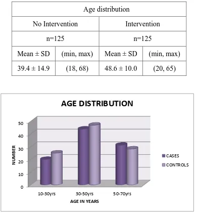
[image:73.612.110.500.174.597.2]AGE DISTRIBUTION OF SNAKE BITE CASES AND CONTROL
TABLE 1 :
This chart shows the maximum age distribution of cases and control is between 30-50 years of age. This carries significance since the working and productive population falls within this group.
Age distribution
No Intervention Intervention
n=125 n=125
Mean ± SD (min, max) Mean ± SD (min, max) 39.4 ± 14.9 (18, 68) 48.6 ± 10.0 (20, 65)
0 10 20 30 40 50
10-30yrs 30-50yrs 50-70yrs
N
UM
B
ER
AGE IN YEARS
AGE DISTRIBUTION
CASES
74
GENDER DISTRIBUTION
Gender
Group
CONTROL CASES
No % No %
Male 96 76.8 106 84.8
Female 29 23.2 19 15.2
Total 125 100.0 125 100.0
85% 15%
Sex distribution in Cases
males: 85%
75
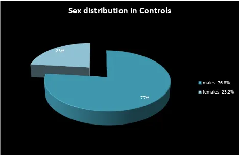
TABLE 2:
This pie diagram shows that the gender affected mostly are males. This is because they remain outdoors than females and so are affected.
77% 23%
Sex distribution in Controls
males: 76.8%
76 TABLE 3
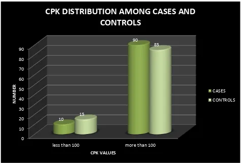
This diagram shows that cases with CPK elevation were mainly included under study. No Intervention Intervention
n=125 n=125
Mean ± SD (min, max) Mean ± SD (min, max) CPK 344.5 ± 240.0 (38, 955) 405.8 ± 147.5 (167, 784)
p-value 0.016 (Significant)
0 10 20 30 40 50 60 70 80 90
less than 100 more than 100 10 90 15 85 N UM B ER CPK VALUES
CPK DISTRIBUTION AMONG CASES AND
CONTROLS
CASES
77
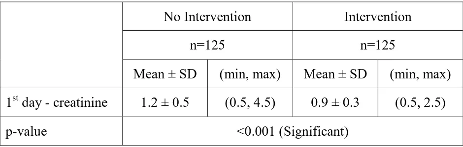
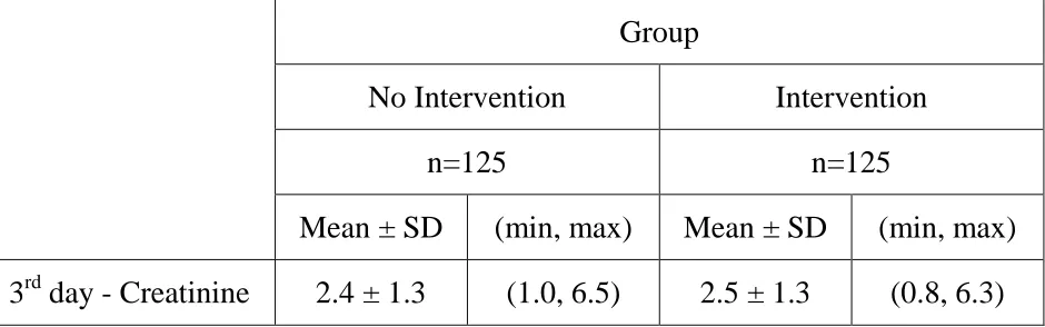
CREATININE LEVELS IN CASES AND CONTROLS
No Intervention Intervention
n=125 n=125
Mean ± SD (min, max) Mean ± SD (min, max) 1st day - creatinine 1.2 ± 0.5 (0.5, 4.5) 0.9 ± 0.3 (0.5, 2.5)
p-value <0.001 (Significant)
[image:77.612.71.541.123.273.2]
TABLE 4 :
This chart shows that there was a significant difference in creatinine values in the controls and cases group which was taken after soda bicarbonate infusion
0 0.5 1 1.5 2 2.5 3 3.5 4 4.5 5
CASES CONTROL
78 TABLE 5:
This chart shows that there was less increase in third day creatinine values in the cases group post soda bicarbonate infusion than in the control group.
0 1 2 3 4 5 6 7 8
CASES CONTROL
3.5 6 CR EA TININE VA LU ES Group
No Intervention Intervention
n=125 n=125
79
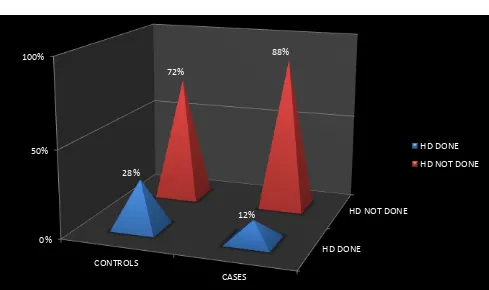
HEMODIALYSIS REQUIREMENT
HD Done
Group
No Intervention Intervention
No % No %
Yes 35 28.0 15 12.0
No 90 72.0 110 88.0
Total 125 100.0 125 100.0
[image:79.612.74.563.295.585.2]p-value p=0.002 (Significant)
TABLE 6:
This chart shows that the requirement of hemodialysis in AKI group with significant creatinine values was less in cases than in controls with statistical significance.
HD DONE
HD NOT DONE
0% 50% 100% CONTROLS CASES 28% 12% 72% 88% HD DONE
80
DISCUSSION
In our study around 250 patients were analysed after applying inclusion and exclusion criteria, clinical examination were done for all of them. Later first and third day urea and creatinine values were done with CPK values and other routine investigations were also done.
The observations made in the study were males formed most of the study population around 75-80% and most of them were within the age group of 35-45 years with around 15% are less than or equal to 25 years, 27% are between 25-35 years of age, 31% between 35-45 years and 27 % are 45 and above years.
And it was also found out that the higher the value of CPK in these patients the more the patients go into to develop AKI. So intervention group was tried with sodium bicarbonate because one of the mechanisms attributed is
81
It was found that the intervention group had much lesser incidence of dialysis requiring creatinine levels with statistical significance. Around 85% of cases recovered following soda bicarbonate infusion without dialysis whereas those without infusion only 67% recovered.
82
LIMITATIONS OF THE STUDY
In this study the total CPK was tested and not the iso enzyme form. So there is a possibility of non specific elevation of CPK due to other factors like MI/ angina. Other parameters of acute kidney injury like NGAL were not measured. The cases were not followed up thereafter to record for any chronic lesions
83
SUMMARY
Around 250 patients 85% were males and 15% were females with most of the males within 35-45 working population.
Around 15% are less than or equal to 25 years, 27% are between 25-35 years of age, 31% between 35-45 years and 27 % are 45 and above years.
More than 90% patients with cellulitis or with history of hemotoxicity developed CPK elevations of more than 150 which was significant
First day creatinine values were higher in the non intervention group than in the intervention groups by around 45%
Third day creatinine values were also greater in the non intervention than post soda bicarbonate infusion group.
84
CONCLUSION