Roles of Cell Signaling Pathways in Cell-to-Cell Contact-Mediated
Epstein-Barr Virus Transmission
Asuka Nanbo,aHaruna Terada,a* Kunihiro Kachi,aKenzo Takada,band Tadashi Matsudaa
Graduate School of Pharmaceutical Sciences, Hokkaido University, Sapporo, Japan,aand Institute for Genetic Medicine, Hokkaido University, Sapporo, Japanb
Epstein-Barr virus (EBV), a human gamma herpesvirus, establishes a life-long latent infection in B lymphocytes and epithelial
cells following primary infection. Several lines of evidence indicate that the efficiency of EBV infection in epithelial cells is
accel-erated up to 10
4-fold by coculturing with EBV-infected Burkitt’s lymphoma (BL) cells compared to infection with cell-free
viri-ons, indicating that EBV infection into epithelial cells is mainly mediated via cell-to-cell contact. However, the molecular
mecha-nisms involved in this pathway are poorly understood. Here, we establish a novel assay to assess cell-to-cell contact-mediated
EBV transmission by coculturing an EBV-infected BL cell line with an EBV-negative epithelial cell line under stimulation for
lytic cycle induction. By using this assay, we confirmed that EBV was transmitted from BL cells to epithelial cells via cell-to-cell
contact but not via cell-to-cell fusion. The inhibitor treatments of extracellular signal-regulated kinase (ERK) and nuclear factor
(NF)-
B pathways blocked EBV transmission in addition to lytic induction. The blockage of the phosphoinositide 3-kinase
(PI3K) pathway impaired EBV transmission coupled with the inhibition of lytic induction. Knockdown of the RelA/p65 subunit
of NF-
B reduced viral transmission. Moreover, these signaling pathways were activated in cocultured BL cells and in epithelial
cells. Finally, we observed that viral replication was induced in cocultured BL cells. Taken together, our data suggest that
cell-to-cell contact induces multiple cell-to-cell signaling pathways in BL cell-to-cells and epithelial cell-to-cells, contributing to the induction of the viral lytic
cycle in BL cells and the enhancement of viral transmission to epithelial cells.
E
pstein-Barr virus (EBV), a human gamma herpesvirus,
estab-lishes a persistent latent infection in B lymphocytes and
epi-thelial cells following primary infection (
19
). EBV has been
impli-cated as a cause of lymphomas and epithelial malignancies, such as
Burkitt’s lymphoma (BL), Hodgkin’s disease, nasopharyngeal
car-cinoma, and gastric cancer (
19
). EBV binds to B lymphocytes
through a direct interaction of the EBV glycoprotein gp350/220
with the complement receptor CD21 (
13
,
22
). In contrast, the
mechanism by which EBV enters epithelial cells remains
unde-fined. Epithelial cells express very low levels of CD21 or are CD21
negative in culture (
12
,
15
), resulting in the lack of an efficient
infection of cell-free viruses.
Several lines of evidence indicate that EBV infection into
epi-thelial cells is mainly mediated by cell-to-cell contact (
5
,
15
,
34
,
35
,
37
,
43
). The rate of EBV infection in epithelial cells increases up to
10
3-fold by coculturing with EBV-positive B cells compared to
infection with cell-free EBV (
5
,
15
,
43
). Moreover, Shannon-Lowe
et al. demonstrated that most EBV virions are retained on cell
surfaces after binding to primary B cells and are transferred to
epithelial cells, resulting in the 10
3- to 10
4-fold increase in
infec-tion compared to cell-free virus infecinfec-tion (
35
). All of these studies
support a model in which EBV-infected B cells migrating into the
epithelial stroma or intraepithelial space contribute to efficient
EBV transmission into epithelium via cell-to-cell contact (
42
,
43
).
However, the molecular mechanisms of cell-to-cell EBV
transmis-sion remain unclear.
In the present study, we establish a novel assay to assess
cell-to-cell contact-mediated EBV transmission by coculturing an
EBV-infected BL cell line with an EBV-negative epithelial cell line
under stimulation for lytic cycle induction in BL cells. By using
this system, we showed that EBV transmission was mediated via
cell-to-cell contact but not via cell-to-cell fusion. We
demon-strated that the treatment of inhibitors of extracellular
signal-reg-ulated kinase (ERK) and nuclear factor (NF)-B pathways
blocked EBV transmission in addition to lytic induction. The
blockage of the phosphoinositide 3-kinase (PI3K) pathway
im-paired EBV transmission coupled with the inhibition of lytic
in-duction. Knockdown of the RelA/p65 subunit of NF-
B also
re-duced the efficiency of viral transmission. Moreover, these cell
signaling molecules were activated in cocultured BL cells and
ep-ithelial cells. Finally, we observed that the viral lytic cycle was
induced in BL cells by coculturing with epithelial cells. The
possi-ble roles of these signaling molecules in cell-to-cell
contact-medi-ated EBV transmission are discussed.
MATERIALS AND METHODS
Cell culture.The African green monkey kidney epithelial Vero-E6 cell line (9,26), which was provided by Ayato Takada, and human gastric adeno-carcinoma cell lines, AGS (2,49) and NU-GC-3 (1,16,22,31,32,49), were grown in high-glucose Dulbecco’s modified Eagle’s medium (DMEM) containing 10% fetal bovine serum (FBS) and antibiotics. BL-derived Akata⫺EBV-eGFP cells, which are latently infected with a recombinant Akata strain of EBV carrying the enhanced green fluorescent protein (eGFP) gene inserted into the viral BXLF1 open reading frame (ORF) (22), were maintained in RPMI 1640 medium containing 10% FBS, anti-biotics, and 800g/ml G418. EBV-positive BL-derived Akata (Akata⫹) cells (39–41) and EBV-negative BL-derived Daudi (Daudi⫺) cells (20,28) were maintained in RPMI 1640 medium containing 10% FBS and antibi-otics. Cells were maintained at 37°C in 5% CO2.
Received5 April 2012Accepted9 June 2012
Published ahead of print20 June 2012
Address correspondence to Asuka Nanbo, nanboa@pharm.hokudai.ac.jp.
* Present address: RIKEN Wako Institute, Wako, Saitama, Japan.
A.N. and H.T. contributed equally to this work.
Copyright © 2012, American Society for Microbiology. All Rights Reserved.
doi:10.1128/JVI.00712-12
on November 7, 2019 by guest
http://jvi.asm.org/
EBV transmission assay.EBV-negative Vero-E6 cells (5⫻104) were
cocultured with Akata⫺EBV-eGFP cells (5⫻105) for 24 h in 24-well
plates in the presence or absence of 0.2% F(ab=)2fragments of goat
anti-human IgG polyclonal antibody (␣hIgG; Dako, Glostrup, Denmark) to induce the viral lytic cycle in Akata⫺EBV-eGFP cells. To remove Akata⫺ EBV-eGFP, Vero-E6 cells were washed, trypsinized, and cultured in 6-well plates for 6 h in the absence of␣hIgG. For the transmission assay with a physical barrier, Vero-E6 cells (1⫻105) were grown in the basolateral
chamber of a 24-well plate with membrane inserts having pore sizes of 0.4
m (Corning, Toledo, OH). Akata⫺EBV-eGFP cells (1⫻105) were
added to the membrane inserts and incubated for 24 h in the presence or absence of␣hIgG. Vero-E6 cells were grown on the coverslips and fixed in 4% paraformaldehyde (PFA). The transmission of EBV-eGFP into Vero-E6 cells was analyzed by a confocal laser scanning microscope (Flu-oview FV10i; Olympus, Tokyo, Japan). Four fields containing approxi-mately 2,000 cells were randomly collected, and the fractions of eGFP-positive Vero-E6 cells were measured. The percentages of eGFP-eGFP-positive cells were also analyzed by flow cytometry (FACSCalibur, Becton, Dick-inson and Company, Franklin Lakes, NJ). In parallel with fluorescence-activated cell sorting (FACS) analysis, the same sample was analyzed by a confocal laser scanning microscope to confirm that the sample did not contain Akata⫺EBV-eGFP cells.
Immunofluorescent staining.Vero-E6 cells were grown on coverslips and cocultured with Akata⫺EBV-eGFP cells under␣hIgG treatment. For analysis of the expression of caveolin-1, EBNA1, and HLA-DR, Vero-E6 cells were washed to remove Akata⫺EBV-eGFP, cultured in 6-well plates for 6 h, and grown on coverslips. For analysis of the phosphorylation of ERK and PI3K and the nuclear translocation of RelA/p65, Vero-E6 cells grown in 35-mm glass-bottomed culture dishes (MatTek Corporation, Ashland, MA) were cocultured with Akata⫺EBV-eGFP cells in the pres-ence or abspres-ence of 0.2%␣hIgG for various times. The cells were fixed with 4% PFA in phosphate-buffered saline (PBS) for 10 min at room temper-ature, permeabilized with PBS containing 0.05% Triton X-100 for 10 min at room temperature, and blocked in PBS containing 1% bovine serum albumin (BSA) and 0.05% Triton X-100 for 20 min at room temperature. The cells were incubated with individual primary antibodies for 1 h at room temperature. After being washed twice in PBS, the cells were incu-bated with Alexa Fluor-labeled secondary antibodies (Life Technologies, Carlsbad, CA) for 1 h at room temperature. After being washed twice in PBS, the nucleus was counterstained with 4=,6-diamidino-2-phenylindole (DAPI). Images were collected with a 60⫻water objective lens (numeric aperture, 1.3) of a confocal laser scanning microscope (Fluoview FV10i) and acquired by using FV10-ASW software (Olympus). For presentation in the manuscript, all images were digitally processed with Adobe Photo-shop. Rabbit polyclonal antibodies for human caveolin-1 and human RelA/p65 were purchased from Abcam (Cambridge, United Kingdom) and Santa Cruz Biotechnology (Santa Cruz, CA), respectively. Mouse monoclonal antibody for EBV-encoded EBNA1 and a phycoerythrin (PE)-labeled monoclonal antibody for human leukocyte antigen (HLA)-DR were provided by Hironori Yoshiyama (Hokkaido University) and Kenji Oritani (Osaka University), respectively. Rabbit monoclonal antibodies for phosphorylated ERK1/2 (The202/Tyr204) and Akt (Ser473) were purchased from Cell Signaling Technology (Danvers, MA). The percentages of signal-activated cells were analyzed by a confocal laser scanning microscope. Four fields containing approximately 1,000 cells were randomly collected, and the fractions of the activated cells were measured.
Flow cytometry.For analysis of FcR expression, Vero-E6 cells (5⫻ 105) or Akata⫹cells (5⫻105) were incubated with␣hIgG (1:100) for 1 h
on ice. The cells were washed twice in PBS and incubated with fluorescein isothiocyanate (FITC)-labeled rabbit anti-goat IgG (Sigma-Aldrich, St. Louis, MO) for 30 min on ice. After two washes in PBS, the binding of
␣hIgG was analyzed by flow cytometry. For analysis of the expression of EBV-encoded gp350, Vero-E6 cells were cocultured with Akata⫺EBV-eGFP cells in the presence or absence of 0.2%␣hIgG. Akata⫺EBV-eGFP cells were
harvested, fixed, permeabilized, and blocked as described above. The cells were incubated with anti-gp350 monoclonal antibody (C-1) (44) for 1 h at room temperature, washed twice in PBS, and incubated with Alexa Fluor 647-labeled secondary antibodies (Life Technologies). After being washed twice in PBS, the expression of gp350 was analyzed by flow cytometry.
Cell-free EBV infection.Akata⫺EBV-eGFP cells were treated with 0.2%␣hIgG for 48 h for viral production. Vero-E6 or Daudi⫺cells (1⫻ 105) were incubated with culture supernatant of␣hIgG-treated Akata⫺
EBV-eGFP cells for 1 h at 37°C. The culture supernatant was replaced with fresh medium, and the cells were further incubated for 24 h. The cells were fixed with 4% PFA, and the infection efficiencies were analyzed by flow cytometry as described above.
Inhibitor treatment. Vero-E6 cells were cocultured with Akata⫺ EBV-eGFP cells and treated with dimethylsulfoxide (DMSO), U0126 (Sigma-Aldrich), LY294002 hydrochloride (Cell Signaling Technology), wortmannin (Sigma-Aldrich), or BAY 11-7082 (Merck, Darmstadt, Ger-many) for 24 h in the presence or absence of 0.2%␣hIgG. U0126, both LY294002 and wortmannin, and BAY11-7082 inhibit the ERK, PI3K, and NF-B pathways, respectively. Vero-E6 cells were washed and cultured in 6-well plates for 6 h in the absence of␣hIgG. The effect of these inhibitors on the transmission of EBV-eGFP into Vero-E6 cells was analyzed by flow cytometry as described for the EBV transmission assay. The effect of these inhibitors on the expression of gp350 in Akata⫺EBV-eGFP cells was analyzed by flow cytometry as described above.
Knockdown of RelA/p65 by shRNA.Vero-E6 cells (2⫻104) grown in 6-well plates were transfected by Fugene HG (Roche, Basel, Switzerland) with 3g of either pGPU6/GFP/Neo short hairpin RNA (shRNA) vector carrying two identical target sequences against the human RelA/p65 sub-unit of NF-B (shRNA 1, GCCCATGGAATTCCAGTACCT; shRNA 2, GAATCCAGTGTGTGAAGAAGC) (Shanghai GenePharma, Shanghai, China) or pGPU6/GFP/Neo shRNA control plasmid possessing the target sequence against human glyceraldehyde-3-phosphate dehydrogenase (GAPDH) (GTATGACAACAGCCTCAAG) (Shanghai GenePharma). The individual G418-resistant cell clones were isolated, and the down-regulation of the RelA/p65 gene was analyzed by reverse transcription-PCR (RT-transcription-PCR) with sense (ATGGACGAACTGTTC) and antisense (TC CTTGGTGACCAGGG) oligonucleotides.
FISH.Fluorescent in situ hybridization (FISH) analysis was per-formed based on the protocol of Nanbo et al. (27), with some modifica-tions. Briefly, Akata⫺EBV-eGFP cells were treated with 0.075 M KCl for 20 min at 37°C, fixed in methanol-acetic acid (3:1) for 30 min at room temperature, and spread on the slides. Slides were treated with 4⫻SSC (1⫻SSC is 0.15 M NaCl plus 0.015 M sodium citrate) containing 0.5% (vol/vol) Nonidet P-40 for 30 min at 37°C, dehydrated in a cold ethanol series (70, 80, and 90%) for 2 min each, air dried, and denatured in 70% formamide–2⫻SSC for 2 min at 72°C. Slides were dehydrated in a cold ethanol series and air dried. Hybridization probes for detection of EBV plasmids were generated by nick translation using biotin-11-dUTP (Roche). Twentyg of a probe was precipitated by ethanol in the presence of 6g salmon sperm DNA (Eppendorf, Hamburg, Germany) and 4g human Cot-1 DNA (Life Technologies), resuspended in hybridization buffer (2⫻SSC, 50% formamide, 10% dextran sulfate), and incubated for 10 min at 70°C, for 5 min at 4°C, and for 1 h at 37°C. A hybridization mix containing 5 ng probe was placed on each sample and incubated overnight at 37°C in a moist chamber. Slides were washed in 2⫻SSC containing 50% formamide for 30 min at 50°C and in 2⫻SSC for 30 min at 50°C. After blocking in 4⫻SSC containing 1% BSA, the hybridized probe was re-vealed by incubation with streptavidin conjugated to Cy3 (Sigma-Aldrich) for 30 min at 37°C. Slides were washed twice in 4⫻SSC contain-ing 0.05% Triton X-100 for 5 min at room temperature. The nucleus was counterstained with DAPI. The percentages of Akata⫺EBV-eGFP cells that underwent the lytic cycle were analyzed by a confocal laser scanning microscope. Four fields containing approximately 1,000 cells were ran-domly collected, and the fractions of the cells that underwent the lytic cycle were measured.
on November 7, 2019 by guest
http://jvi.asm.org/
RESULTS
EBV is transmitted from cocultured BL cells to epithelial cells.
We established a cocultivation system to evaluate the efficiency of
cell-to-cell EBV transmission. We cocultured EBV-negative
Afri-can green monkey kidney epithelial Vero-E6 cells (
9
,
26
) with
BL-derived Akata
⫺EBV-eGFP cells, which are latently infected
with a recombinant Akata strain of EBV the encoding eGFP gene
inserted into the viral genome (
22
) for 24 h. To amplify viral
transmission, we cross-linked the cell surface IgG of Akata
⫺EBV-eGFP by adding F(ab
=
)
2fragments of goat anti-human IgG
poly-clonal antibody (␣hIgG) to induce the viral lytic cycle (
39
–
41
).
The transmission of EBV-eGFP to Vero-E6 cells was analyzed by a
FIG 1EBV is transmitted from BL cells to epithelial cells by cocultivation. (A) EBV transmission from BL cells to epithelial cells is facilitated by␣hIgG treatment. Vero-E6 cells were cocultured with Akata⫺EBV-eGFP cells in the absence (middle) or presence (right) of␣hIgG for 24 h. The infection of EBV-eGFP in Vero-E6 cells (green) was analyzed by a confocal laser scanning microscope. The left panel shows the result without cocultivation. The nucleus was counterstained with DAPI. Scale bars, 100m. (B) A summary of cell-to-cell contact-mediated EBV transmission. Vero-E6 cells were cocultured with Akata⫺EBV-eGFP cells in the absence or presence of␣hIgG for 24 h. The percentages of EBV-eGFP-positive Vero-E6 cells were analyzed by a confocal laser scanning microscope (black bars) or flow cytometry (gray bars). The experiment was performed five times independently, and the averages and standard deviations are shown for each condition. *,P⬍0.05 versus the respective control (Student’sttest). (C) Infection of cell-free EBV-eGFP into Vero-E6 cells. Vero-E6 cells or Daudi⫺cells were incubated for 1 h at 37°C with culture supernatants derived from␣hIgG-treated (gray bars) or untreated (black bars) Akata⫺EBV-eGFP cells. The culture supernatants were replaced with fresh medium, and the cells were further incubated for 48 h. The percentages of eGFP-positive cells were analyzed by flow cytometry. The experiment was performed three times independently. The averages and standard deviations are shown for each condition. **,P⬍0.01 versus the respective control (Student’sttest). (D) EBV-eGFP is transmitted to human gastric epithelial cells. AGS (white bars), NU-GC-3 (gray bars), or Vero-E6 (black bars) cells were cocultured with Akata⫺EBV-eGFP cells in the absence or presence of␣hIgG for 24 h. The percentages of eGFP-positive epithelial cells were analyzed by flow cytometry. The experiment was performed three times independently, and the averages and standard deviations are shown for each condition. *,P⬍0.05 versus the respective control. **,P⬍0.01 versus the respective control (Student’sttest). (E) EBV-eGFP is transmitted to Vero-E6 cells. Vero-E6 cells were cocultured with Akata⫺EBV-eGFP cells under the treatment of␣hIgG for 24 h. The expression of an epithelium marker, caveolin-1, was analyzed by immunofluorescent staining. Caveolin-1 (red; middle) was expressed in eGFP-positive cells (green; left); BL-derived Akata⫺EBV-eGFP cells were caveolin-1 negative (right). The nucleus was counterstained with DAPI. Scale bars, 20m. (F) eGFP-positive Vero-E6 cells are infected with EBV-eGFP. Vero-E6 cells were cocultured with Akata⫺EBV-eGFP cells under treatment with␣hIgG for 24 h. The expression of EBV-encoded nuclear antigen 1 (EBNA1) was analyzed by immunofluorescent staining. eGFP-positive Vero-E6 cells (green; left) were EBNA1 positive (red; right). The nucleus was counterstained with DAPI. Scale bars, 20m.
Molecular Mechanism of Cell-to-Cell EBV Transmission
on November 7, 2019 by guest
http://jvi.asm.org/
[image:3.585.81.504.63.459.2]confocal laser scanning microscope (
Fig. 1A
and
B
, black bars)
and flow cytometry (
Fig. 1B
, gray bars). EBV-eGFP infected
ap-proximately 5% of the cell population by coculturing with Akata
⫺EBV-eGFP (
Fig. 1A
, left and middle, and
B
).
␣hIgG treatment
increased the transmission rate to 9% (
Fig. 1A
, right, and
B
).
We also examined the infection efficiency of cell-free virus into
Vero-E6 cells. We treated Akata
⫺EBV-eGFP cells with
␣
hIgG for
48 h for the production of eGFP. Vero-E6 cells or
EBV-negative BL-derived Daudi
⫺cells (
20
,
28
) were incubated with the
culture supernatant of
␣hIgG-treated Akata
⫺EBV-eGFP cells,
and the percentages of eGFP-positive cells were analyzed by flow
cytometry. The cell-free virus infected Daudi
⫺cells efficiently
(
Fig. 1C
). In contrast, Vero-E6 cells were highly resistant to
cell-free viral infection (
Fig. 1C
).
We also examined the efficiency of EBV transmission into two
human gastric epithelial cell lines, AGS (
2
,
49
) and NU-GC-3 (
1
,
16
,
22
,
31
,
32
,
49
). EBV-eGFP was also transmitted to AGS (
Fig.
1D
, white bars) and NU-GC-3 cells (
Fig. 1D
, gray bars) in the
absence (1.4 and 1.8%, respectively) and in the presence (2.3 and
2.4%, respectively) of
␣hIgG (
Fig. 1D
). Because EBV-eGFP was
transmitted into Vero-E6 cells more efficiently, we used Vero-E6
cells for further study. We confirmed that eGFP-positive cells
ex-pressed an epithelial marker, caveolin-1 (
7
) (
Fig. 1E
, left and
mid-dle). All eGFP-positive cells expressed EBV-encoded nuclear
an-tigen 1 (EBNA1) (
Fig. 1F
), indicating that the expressed eGFP in
Vero-E6 cells was derived from incoming EBV-eGFP.
Flow-cytometric analysis demonstrated that
␣
hIgG bound to
the surface of Vero-E6 cells with low efficiency, suggesting that
Vero-E6 cells express low levels of Fc receptor (FcR) (
Fig. 2A
, left,
and
B
). It is possible that
␣hIgG-mediated cross-linking of the FcR
of Vero-E6 cells initiates cellular signaling pathways and affects
viral transmission. A previous study demonstrated that certain
anti-IgG antibodies coat particles of measles virus and enhance
viral entry into monocytes and macrophages (
14
), suggesting that
␣
hIgG treatment enhances EBV infection of Vero-E6 cells in a
similar way. To test these possibilities, we treated Akata
⫺EBV-eGFP cells with
␣
hIgG for 2 h, replaced the culture medium with
fresh medium to remove free
␣hIgG, and cocultured Akata
⫺EBV-eGFP cells with Vero-E6 cells for an additional 24 h. EBV was
transmitted similarly in the presence and absence of free
␣hIgG
(
Fig. 2C
), indicating that
␣
hIgG-induced enhancement of viral
transmission was independent of
␣hIgG-coated virions and
cross-linking of FcR of Vero-E6 cells.
EBV is transmitted from BL cells to epithelial cells via
cell-to-cell contact.
We further examined whether viral transmission
re-quires physical contact. Vero-E6 cells were grown on the
basolat-eral chamber of transculture plates with membrane inserts having
pore sizes of 0.4
m. Akata
⫺EBV-eGFP cells were added to the
inserts and incubated for 24 h in the presence or absence of
␣
hIgG.
We analyzed the titer of eGFP-EBV in the culture supernatant of
the basolateral chamber released from the insert containing
␣hIgG-treated Akata
⫺eGFP EBV cells by incubating the culture
supernatant with Daudi
⫺cells. We confirmed that 38% of
re-leased eGFP-EBV was transferred to the basolateral chambers
through the membrane (data not shown). Viral transmission did
not occur if physical cell-to-cell contact was impaired (
Fig. 3A
, left
and middle, and B), indicating that physical contact is
indispens-able for efficient viral transmission to epithelial cells.
The EBV lytic cycle induces the expression of the viral
glyco-proteins, which possess membrane fusion activity (
6
,
24
,
33
). To
test the possibility that these surface glycoproteins mediate the
fusion of plasma membranes between Akata
⫺EBV-eGFP cells and
Vero-E6 cells and thus the transfer of virus and/or eGFP to
Vero-E6 cells, we assessed the expression of HLA-DR in
eGFP-positive Vero-E6 cells by immunofluorescent staining. HLA-DR is
the class II histocompatibility molecule constitutively expressed
on antigen-presenting cells, including B cells, and T lymphocytes
only after activation (
45
). We confirmed that Akata
⫺EBV-eGFP
cells expressed HLA-DR and that Vero-E6 cells are HLA-DR
neg-ative (
Fig. 3C
). All eGFP-expressing Vero-E6 cells were HLA-DR
negative (
Fig. 3C
), indicating that EBV-eGFP transmission was
mediated via cell-to-cell contact but not via cell-to-cell fusion.
The effect of inhibitors of cell signaling pathways on
cell-to-cell contact-mediated EBV transmission.
Because the molecular
mechanism of cell-to-cell EBV transmission remains undefined,
we investigated the roles of cellular signaling pathways in this
pro-cess by using specific inhibitors. We cocultured Vero-E6 cells with
Akata
⫺EBV-eGFP cells in the presence and absence of
␣hIgG
under the treatment of inhibitors such as U0126, LY294002,
wort-mannin, and BAY11-7082. U0126, both LY294002 and
wortman-FIG 2Role of free␣hIgG in viral transmission. (A) The binding of␣hIgG to Vero-E6. Vero-E6 cells (left) or Akata⫹cells (right) were incubated with 0.1%
␣hIgG on ice. The binding of␣hIgG was revealed with FITC-labeled secondary antibody (boldface lines) by flow cytometry. As a control, cells were incubated with the secondary antibody (thin lines). (B) Summary of binding of␣hIgG. The efficiency of binding of␣hIgG to Vero-E6 or Akata⫹cells was quantified by flow cytometry (black bars). As a control, cells were incubated with the secondary antibody (gray bars). The experiment was performed three times independently, and the averages and standard deviations are shown for each condition. **,P⬍0.01 versus the respective control (Student’sttest). (C) The role of free␣hIgG in viral transmission. Akata⫺EBV-eGFP cells were treated with␣hIgG for 2 h, washed to remove free␣hIgG, and cocultured with Vero-E6 cells for 24 h. The percentage of eGFP-positive epithelial cells was analyzed by flow cytometry. As a control, Akata⫺EBV-eGFP cells were cocul-tured with Vero-E6 cells in the presence of free␣hIgG. The experiment was performed three times independently, and the averages and standard devia-tions are shown for each condition. **,P⬍0.01 versus the respective control (Student’sttest).
on November 7, 2019 by guest
http://jvi.asm.org/
[image:4.585.301.540.65.314.2]nin, and BAY11-7082 inhibit the ERK (
11
), PI3K (
46
), and NF-B
pathways (
50
), respectively. We treated the cells with these
inhib-itors in various concentrations that did not exhibit cytotoxicity.
␣
hIgG treatment triggers a variety of signaling pathways
concur-rent with induction of the lytic cycle (
36
). To exclude the
possi-bility that the inhibitors reduced
␣
hIgG-induced viral
produc-tion, we also measured the effect of these inhibitors on the
expression of viral lytic gene gp350 (
22
,
30
) by flow-cytometric
analysis. The expression of gp350 was enhanced under treatment
with
␣
hIgG (
Fig. 4A
, bottom). U0126 and BAY11-7082
signifi-cantly inhibited cell-to-cell contact-mediated EBV transmission
(
Fig. 4A
, top) with moderate impairment of gp350 expression
(
Fig. 4A
, bottom), suggesting that the ERK and the NF-B
path-ways contribute to EBV transmission in addition to lytic
induc-tion. Both PI3K inhibitors, LY294002 and wortmannin, blocked
EBV transmission and gp350 expression similarly (
Fig. 4A
),
sug-gesting that the blockage of the PI3K pathway impaired EBV
transmission coupled with inhibition of the lytic induction.
We further examined the role of the NF-B pathway in
cell-to-cell contact-mediated EBV transmission by using shRNA
target-ing the RelA/p65 subunit of NF-B in Vero-E6 cells (
Fig. 4B
). The
knockdown of RelA/p65 significantly reduced viral transmission
(
Fig. 4C
). Taken together, the NF-
B pathway contributes to EBV
transmission and moderately to the viral lytic cycle. The inhibitor
treatments and knockdown of RelA/p65 also reduced viral
trans-mission and lytic induction in the absence of
␣hIgG (
Fig. 4A
and
C
).
Cell signaling molecules activate immediately in the
coculti-vated cells.
Because specific inhibitors of the ERK, PI3K, and
NF-B pathways significantly blocked cell-to-cell
contact-medi-ated EBV transmission, we examined whether these signaling
molecules are activated in cocultured BL cells and epithelial cells.
We assessed the status of the ERK, PI3K, and NF-
B signaling
pathways by immunofluorescent staining in Akata
⫺EBV-eGFP
cells and Vero-E6 cells, respectively. By using a laser scanning
confocal microscope, we confirmed that the individual marker
proteins in Akata
⫺EBV-eGFP cells (HLA-DR) and Vero-E6 cells
(caveolin-1) were detected without leakage of the signal derived
from cocultured cells (
Fig. 5A
).
Under stimulation of the ERK pathway, the activation loop
residues Thr202/Tyr204 and Thr185/Tyr187 of ERK are
phos-phorylated by a sequential protein kinase cascade (
3
). We
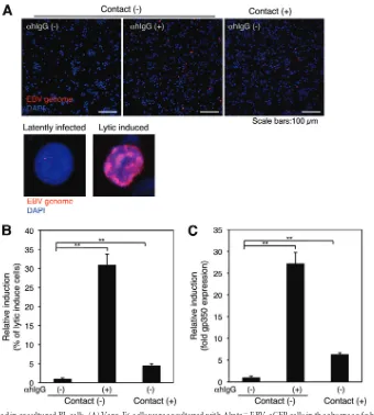
ana-FIG 3EBV is transmitted from BL cells to epithelial cells via cell-to-cell contact. (A) Physical cell-to-cell contact is required for EBV transmission. Vero-E6 cells were grown in the basolateral chamber of transculture inserts with pore sizes of 0.4m. Akata⫺EBV-eGFP cells were added to the inserts and incubated in the presence (left) or absence (middle) of␣hIgG for 24 h. As a control, Vero-E6 cells were cocultured with Akata⫺EBV-eGFP in the presence of␣hIgG (right). The infection of EBV-eGFP into Vero-E6 (green) was analyzed by a confocal laser scanning microscope. The nucleus was counterstained with DAPI. Scale bars, 100
m. (B) A summary of viral transmission with a physical barrier. Vero-E6 cells were grown in the basolateral chamber of transculture plates. Akata⫺EBV-eGFP cells were added to the inserts and incubated in the presence (gray bars) or absence (black bars) of␣hIgG for 24 h. As a control, Vero-E6 cells were cocultured with Akata⫺EBV-eGFP in the presence (gray bars) or absence (black bars) of␣hIgG. The infection of EBV-eGFP in Vero-E6 cells was analyzed by flow cytometry. The experiment was performed three times independently. The averages and standard deviations are shown for each condition. **,P⬍0.01 versus the respective control (Student’sttest). (C) EBV transmits from BL cells to epithelial cells via cell-to-cell contact. Akata⫺EBV-eGFP cells were cocultured with Vero-E6 cells in the presence of␣hIgG for 24 h. The expression of HLA-DR (red) in Akata⫺EBV-eGFP or Vero-E6 cells was analyzed by immunofluorescent staining (left panels). HLA-DR (red) was expressed in eGFP-positive Vero-E6 cells (green) (right panels). The nucleus was counterstained with DAPI. Scale bars, 20m.
Molecular Mechanism of Cell-to-Cell EBV Transmission
on November 7, 2019 by guest
http://jvi.asm.org/
[image:5.585.79.504.63.363.2]lyzed the phosphorylation status of ERK by immunofluorescent
staining. Phosphorylated ERK was observed diffusely in the
cyto-plasm in both Akata
⫺EBV-eGFP cells (
Fig. 5B
and
D
) and
Vero-E6 cells (
Fig. 5C
and
D
) 30 min after cocultivation. The
phosphorylation was transient and disappeared 3 h and 30 min
after cocultivation in Alata-EBV-eGFP and Vero-E6 cells,
respec-tively (
Fig. 5B
to
D
).
To analyze the activation of the PI3K pathway, we assessed the
level of phospho-Ser473 of Akt, which is a key component of
the pathway (
21
). The phosphorylated Akt was distributed in the
plasma membrane of Akata
⫺EBV-eGFP cells 1 h after
cocultiva-tion, and the activation continued stably (
Fig. 6A
and
C
). Akt was
transiently phosphorylated in the cytoplasm of Vero-E6 cells 1 h
after cocultivation (
Fig. 6B
and
C
).
Following stimulation of the NF-
B pathway, the inhibitor of
kB (IKB) inhibitory subunit of the NF-B complex is
phosphor-ylated, ubiquitinated, and degraded, and the transcription factor
subunits of the NF-B complex subunit (RelA/p65 and RelA/p50)
translocate into the nucleus, resulting in the modulation of gene
transcription (
23
). Because its nuclear import is a key event in
NF-
B activation, we assessed the distribution of the RelA/p65
subunit in cocultured cells by immunofluorescent staining. RelA/
p65 transiently translocated to the nucleus in cocultured Vero-E6
cells (
Fig. 7B
and
C
). In contrast, RelA/p65 translocated to the
FIG 4Effect of the inhibitors of cell signaling pathways on cell-to-cell contact-mediated EBV transmission and replication. (A) The effect of the inhibitors on cell-to-cell contact-mediated EBV transmission (top) and EBV replication (bottom). Vero-E6 cells were cocultured with Akata⫺EBV-eGFP cells in the presence (gray bars) or absence (black bars) of␣hIgG under treatment with DMSO, U0126, LY294002, wortmannin, or BAY11-7082 for 24 h. The percentages of eGFP-positive cells were analyzed by flow cytometry. The data were normalized to untreated and DMSO-treated cells (top). Cocultured Akata⫺EBV-eGFP cells were harvested, and the expression of gp350 was analyzed by flow cytometry. The data were normalized to␣hIgG-untreated and DMSO-treated cells (bottom). The experiment was performed three times independently. The averages and standard deviations are shown for each condition. (B) RelA/p65 knockdown by shRNA in Vero-E6 cells. Total RNA was isolated from two Vero-E6 clones stably expressing shRNA against human NF-B RelA/p65 (RelA/p65-shRNAs 1 and 2) and a control expressing shRNA against human glyceraldehyde-3-phosphate dehydrogenase (GAPDH-shRNA). Knockdown of RelA/p65 mRNA was ana-lyzed by RT-PCR (top). As a control, the expression of-actin mRNA is shown (bottom). (C) The effect of RelA/p65 knockdown on cell-to-cell contact-mediated EBV transmission. RelA/p65-shRNA or GAPDH-shRNA was cocultured with Akata⫺EBV-eGFP cells in the presence (gray bars) or absence (black bars) of
␣hIgG for 24 h. Viral transmission was assessed by flow cytometry. The experiment was performed three times independently. The averages and standard deviations are shown for each condition. **,P⬍0.01 versus the respective control (Student’sttest).
on November 7, 2019 by guest
http://jvi.asm.org/
[image:6.585.113.473.61.458.2]FIG 5Activation of the ERK pathway in cocultured cells. (A) Immunofluorescent staining analysis in cocultured cells. Vero-E6 cells grown in 35-mm glass-bottomed culture dishes were cocultured with Akata⫺EBV-eGFP cells for 4 h. The cells were harvested and subjected to immunofluorescent staining to analyze the expression of HLA-DR in Akata⫺EBV-eGFP cells (top left) or caveolin-1 in Vero-E6 cells (bottom middle) under cocultured conditions. The bright-field images are shown (right). Scale bars, 100m. ERK is phosphorylated in cocultured Akata⫺EBV-eGFP (B) or Vero-E6 cells (C). Vero-E6 cells were cocultured with Akata⫺EBV-eGFP cells in the presence (b to e) or absence (f) of␣hIgG for various times. The phosphorylation of ERK in Akata⫺EBV-eGFP (B) or Vero-E6 (C) cells was examined by immunoflu-orescent staining (top). The effect of U0126 treatment (0.2M) on the phosphorylation of ERK is shown in image e. As a control, the status of ERK phosphorylation in untreated Akata⫺EBV-eGFP or Vero-E6 cells is shown in image a. Bright-field images are shown at the bottom. Scale bars, 100m. (D) The percentages of the cells positive for the phosphorylation of ERK were analyzed by a confocal laser scanning microscope. The experiment was performed three times independently. The averages and standard deviations are shown for each condition. **,P⬍0.01 versus the respective control (Student’sttest).
on November 7, 2019 by guest
http://jvi.asm.org/
[image:7.585.80.503.38.622.2]nucleus in Akata
⫺EBV-eGFP cells 24 h after cocultivation (
Fig.
7A
and
C
). Treatment with U0126, LY294002, wortmannin, or
BAY11-7082 specifically blocked the activation of individual
tar-get molecules (
Fig. 5
to
7
). Interestingly, these signaling molecules
were also activated in the cocultured cells in the absence of
␣
hIgG
(
Fig. 5
to
7
).
Cell-to-cell contact induces viral replication.
We observed
that EBV is transmitted to epithelial cells in the absence of
␣hIgG
(
Fig. 1A
to
C
). The inhibitor treatments and the knockdown of
RelA/p65 also reduced viral transmission and lytic induction in
the absence of
␣
hIgG (
Fig. 4A
and
C
). The signaling molecules
were activated in the cocultured cells in the absence of
␣hIgG (
Fig.
FIG 6Activation of the PI3K pathway in cocultured cells. Akt is phosphorylated in cocultured Akata⫺EBV-eGFP (A) or Vero-E6 (B) cells. Vero-E6 cells were cocultured with Akata⫺EBV-eGFP cells in the presence (b to f) or absence (g) of␣hIgG for various times. The phosphorylation of Akt in Akata⫺EBV-eGFP (A) or Vero-E6 (B) cells was examined by immunofluorescent staining (top). The effect of LY294002 treatment (0.2M) (e) or wortmannin treatment (0.1M) (f) on the phosphorylation of Akt is shown. As a control, the status of Akt phosphorylation in Akata⫺EBV-eGFP or Vero-E6 cells not treated with␣hIgG is shown in image a. Bright-field images are shown (bottom). Scale bars, 100m. (C) The percentages of the cells exhibiting phosphorylation of Akt were analyzed by a confocal laser scanning microscope. The experiment was performed three times independently. The averages and standard deviations are shown for each condition. **,P⬍0.01 versus the respective control (Student’sttest).
on November 7, 2019 by guest
http://jvi.asm.org/
[image:8.585.61.520.83.578.2]5A
and
6A
). Therefore, we tested whether cell-to-cell contact
in-duces the viral lytic cycle in the latently infected Akata
⫺EBV-eGFP cells and leads to viral transmission. We cocultured Vero-E6
cells with Akata
⫺EBV-eGFP cells and analyzed the efficiencies of
lytic induction in Akata
⫺EBV-eGFP cells by FISH analyses using
Cy3-labeled probe, which specifically hybridized to the EBV
ge-nome. The EBV genomes were significantly amplified (10
2- to
10
3-fold) in the nucleus under the treatment of
␣hIgG (
Fig. 8A
,
bottom right). FISH analysis showed that less than 1% of the cell
population underwent lytic induction without
␣hIgG treatment
FIG 7Activation of the NF-B pathway in cocultured cells. The nuclear translocation of RelA/p65 in cocultured Akata⫺EBV-eGFP (A) or Vero-E6 (B) cells. Vero-E6 cells were cocultured with Akata⫺EBV-eGFP cells in the presence (b to e) or absence (f) of␣hIgG for various times. The translocation of RelA/p65 in Akata⫺EBV-eGFP (A) or Vero-E6 (B) cells was examined by immunofluorescent staining (top). The effect of BAY11-7082 treatment (2M) on the translo-cation of RelA/p65 is shown in image e. As a control, the status of RelA/p65 translotranslo-cation in Akata⫺EBV-eGFP or Vero-E6 cells not treated with␣hIgG is shown in image a. The nucleus is counterstained with DAPI (bottom). Insets show enlargements of the boxed areas. Scale bars, 100m. (C) The percentages of the cells that contain the translocation of RelA/p65 were analyzed by a confocal laser scanning microscope. The experiment was performed three times independently. The averages and standard deviations are shown for each condition. **,P⬍0.01 versus the respective control (Student’sttest).
Molecular Mechanism of Cell-to-Cell EBV Transmission
on November 7, 2019 by guest
http://jvi.asm.org/
[image:9.585.96.486.70.575.2](
Fig. 8A
, top left, and
B
). By
␣hIgG treatment, 12% of Akata
⫺EBV-eGFP cells underwent the lytic cycle (
Fig. 8A
, top middle,
and
B
). The rate of lytic induction in Akata
⫺EBV-eGFP cells
increased by up to 2% by cocultivation with Vero-E6 cells (
Fig. 8A
,
top right, and
B
), indicating that cell-to-cell contact between BL
cells latently infected with EBV and epithelial cells initiated lytic
induction. Immunofluorescent staining analysis also
demon-strated that gp350 expression was enhanced in Akata
⫺EBV-eGFP
cells by cocultivation with Vero-E6 cells (
Fig. 8C
).
DISCUSSION
In the present study, we demonstrated the roles of the cell
signal-ing pathways in cell-to-cell contact-mediated EBV transmission
by using an
in vitro
cocultivation system. Our observations
indi-cate that (i) cell-to-cell contact without cell-to-cell fusion
contrib-utes to viral transmission (
Fig. 1
to
3
); (ii) the treatment of
inhib-itors of the ERK and NF-B pathways downregulates EBV
transmission in addition to lytic induction, blockage of the PI3K
pathway impairs EBV transmission coupled with the inhibition of
the lytic induction, and the knockdown of RelA/p65 significantly
downregulates cell-to-cell contact-mediated viral transmission
(
Fig. 4
); (iii) the ERK, PI3K, and NF-B pathways are activated in
cocultured BL and epithelial cells (
Fig. 5
to
7
); and (iv) cell-to-cell
contact initiates the induction of the lytic cycle in BL cells (
Fig. 8
).
␣
hIgG treatment amplified EBV transmission into epithelial
cells (
Fig. 1A
,
B
, and
D
), indicating that the synthesis of progeny
virus in Akata
⫺EBV-eGFP cells is indispensable for cell-to-cell
viral transmission. It has been reported that a small fraction of
Akata cells (1%) spontaneously enters into the lytic cycle (
41
) (
Fig.
8A
, left). By cocultivation with Vero-E6 cells, the viral lytic cycle
was induced in Akata
⫺EBV-eGFP cells (
Fig. 8A
, middle, and
B
) in
the absence of
␣hIgG, suggesting that the cell-to-cell
contact-me-diated lytic induction contributes to viral transmission into the
epithelial cells under physiological conditions. It has been shown
FIG 8Lytic cycle is induced in cocultured BL cells. (A) Vero-E6 cells were cocultured with Akata⫺EBV-eGFP cells in the absence of␣hIgG for 24 h (top, right). As a control, Akata⫺EBV-eGFP cells were treated in the presence (top middle) or absence of␣hIgG (top left). Akata⫺EBV-eGFP cells were harvested, and the induction of the lytic cycle was analyzed by FISH using Cy3-labeled probe for the EBV genome (top). Enlarged images of the cell in latent (bottom left) or lytic (bottom right) phase are shown. The nucleus was counterstained with DAPI. Scale bars, 100m. (B) The percentages of Akata⫺EBV-eGFP cells undergoing the lytic cycle were analyzed by a confocal laser scanning microscope. The experiment was performed three times independently. The averages and standard deviations are shown for each condition. **,P⬍0.01 versus the respective control (Student’sttest). (C) The expression of gp350 in cocultured BL cells. The expression of gp350 was analyzed by flow cytometry. The experiment was performed three times independently. The averages and standard deviations are shown for each condition. **,P⬍0.01 versus the respective control (Student’sttest).
on November 7, 2019 by guest
http://jvi.asm.org/
[image:10.585.120.460.63.441.2]that a variety of signal transductions, including the ERK and PI3K
pathways, are synergistically activated during the EBV lytic cycle
(
18
). We showed that the specific inhibitors of the ERK and PI3K
pathways blocked EBV replication (
Fig. 4A
, bottom) and that ERK
and Akt were phosphorylated in cocultured Akata
⫺EBV-eGFP
cells in the absence of
␣
hIgG treatment (
Fig. 5
and
6
). These data
suggest that the activation of the ERK and PI3K pathways
contrib-utes to cell-to-cell contact-mediated viral lytic induction in
Akata
⫺EBV-eGFP cells. It is known that the EBV-encoded
imme-diate-early protein ZTA, which is important for the switch to viral
lytic replication, is inhibited by the RelA/p65 subunit (
4
,
25
).
However, no enhancement of EBV replication was observed
un-der BAY11-7082 treatment in our study (
Fig. 4
, bottom). The
discrepancy may result from a difference in the experimental
sys-tems (e.g., single cultured B cells versus cocultured B cells). The
NF-
B pathway was activated in Akata
⫺EBV-eGFP cells in the
late stage of cell-to-cell contact-mediated EBV transmission (
Fig.
7A
and
C
). The role of the NF-B pathway in B cells in cell-to-cell
contact-mediated EBV transmission remains unclear, and its
fur-ther investigation is required.
Shannon-Lowe and colleagues have demonstrated that EBV
virions loaded on the surface of primary B cells facilitate the
for-mation of a virological synapse (VS)-like intercellular conjugation
between B cells and cocultured epithelial cells (
34
,
35
). The VS is a
tight adhesive junction across which virus can be efficiently
trans-ferred from virus-infected cells to noninfected target cells without
cell-cell fusion. The VS has been intensively studied in the field of
retroviruses (
17
). The ERK pathway appears to enhance the
po-larization of the mitotic organizing center (MTOC) and
contrib-utes to microtubule-dependent trafficking of viral Gag proteins
(
29
). We demonstrated that an ERK inhibitor, U0126,
signifi-cantly blocked EBV transmission with a partial impairment of
induction of the lytic cycle (
Fig. 4A
), and that ERK was
phosphor-ylated in cocultured Akata
⫺EBV-eGFP cells (
Fig. 5A
). These
ob-servations suggest that the ERK pathway plays a role in the
effi-cient trafficking of EBV virions to the intercellular space mediated
by MTOC polarization.
We also demonstrated that the PI3K and NF-
B pathways
con-tribute to viral transmission (
Fig. 4
). Previous observations have
demonstrated an important role of an EBV glycoprotein,
BMRF-2, in the cell-to-cell spread of EBV in polarized oral
epi-thelial cells (
47
,
48
). BMRF-2 interacts with

1 and
␣
v integrins
and initiates PI3K signaling, events that are crucial for the entry of
EBV into B cells (
10
). NF-
B signaling is activated by the
conju-gation of viral gp350/200 with CD21 in B cells (
8
,
38
). It is possible
that the binding of BMRF-2 to the integrins and that of gp350/200
to undefined viral receptors in epithelial cells trigger the PI3K and
NF-
B pathways and promote entry of intercellular EBV virions
into adjacent epithelial cells. We demonstrated that the
phosphor-ylation of Akt and the nuclear translocation of RelA/p65 were
induced in Vero-E6 cells 1 h after cocultivation (
Fig. 6B
and
C
).
The expression of viral glycoproteins and the generation of
prog-eny virions are unlikely to be induced by 1 h after cell-to-cell
contact-mediated lytic induction. The roles of PI3K and NF-B in
the processes of EBV entry into epithelial cells mediated by
cell-to-cell contact currently remain unknown and warrant further
investigation.
In our study, we used a monkey and two human epithelial cell
lines as recipient cells. It would be interesting and informative to
test our findings in EBV-negative primary human epithelial cells
in the future.
Taken together, this study provides a tractable system to
exam-ine cell-to-cell transmission of EBV to epithelial cells. Our
obser-vations indicate that multiple intracellular signaling molecules
play roles in cell-to-cell contact-mediated EBV transmission and
serve as a basis to understand better the mechanism of cell-to-cell
viral transmission.
ACKNOWLEDGMENTS
We thank Ayato Takada (Hokkaido University), Kenji Oritani (Osaka University), and Hironori Yoshiyama (Hokkaido University) for provid-ing us with Vero-E6 cells, PE-labeled anti-human HLA-DR antibody, and anti-EBNA1 polyclonal antibody, respectively. We also thank Bill Sugden (University of Wisconsin, Madison) and Yusuke Ohba (Hokkaido Uni-versity) for critically reviewing the manuscript.
This work was supported by grants in aid from the Japanese Ministry of Education, Science, Sports, Culture, and Technology, Japan.
REFERENCES
1.Akiyama S, et al.1988. Characteristics of three human gastric cancer cell lines, NU-GC-2, NU-GC-3 and NU-GC-4. Jpn. J. Surg.18:438 – 446. 2.Barranco SC, et al.1983. Establishment and characterization of an in
vitro model system for human adenocarcinoma of the stomach. Cancer Res.43:1703–1709.
3.Boulton TG, et al. 1991. ERKs: a family of protein-serine/threonine kinases that are activated and tyrosine phosphorylated in response to in-sulin and NGF. Cell65:663– 675.
4.Brown HJ, et al.2003. NF-kappaB inhibits gammaherpesvirus lytic rep-lication. J. Virol.77:8532– 8540.
5.Chang Y, et al.1999. Requirement for cell-to-cell contact in Epstein-Barr virus infection of nasopharyngeal carcinoma cells and keratinocytes. J. Virol.73:8857– 8866.
6.Connolly SA, Jackson JO, Jardetzky TS, Longnecker R.2011. Fusing structure and function: a structural view of the herpesvirus entry machin-ery. Nat. Rev. Microbiol.9:369 –381.
7.Couet J, Belanger MM, Roussel E, Drolet MC.2001. Cell biology of caveolae and caveolin. Adv. Drug Deliv. Rev.49:223–235.
8.DE Oliveira DE, Ballon G, Cesarman E. 2010. NF-kappaB signaling modulation by EBV and KSHV. Trends Microbiol.18:248 –257. 9.Desmyter J, Melnick JL, Rawls WE.1968. Defectiveness of interferon
production and of rubella virus interference in a line of African green monkey kidney cells (Vero). J. Virol.2:955–961.
10. Dorner M, et al.2010. Beta1 integrin expression increases susceptibility of memory B cells to Epstein-Barr virus infection. J. Virol.84:6667– 6677. 11. Favata MF, et al.1998. Identification of a novel inhibitor of
mitogen-activated protein kinase kinase. J. Biol. Chem.273:18623–18632. 12. Fingeroth JD, Diamond ME, Sage DR, Hayman J, Yates JL. 1999.
CD21-dependent infection of an epithelial cell line, 293, by Epstein-Barr virus. J. Virol.73:2115–2125.
13. Hutt-Fletcher LM.2007. Epstein-Barr virus entry. J. Virol.81:7825–7832. 14. Iankov ID, et al.2006. Immunoglobulin G antibody-mediated enhance-ment of measles virus infection can bypass the protective antiviral im-mune response. J. Virol.80:8530 – 8540.
15. Imai S, Nishikawa J, Takada K.1998. Cell-to-cell contact as an efficient mode of Epstein-Barr virus infection of diverse human epithelial cells. J. Virol.72:4371– 4378.
16. Iwakiri D, Eizuru Y, Tokunaga M, Takada K.2003. Autocrine growth of Epstein-Barr virus-positive gastric carcinoma cells mediated by an Ep-stein-Barr virus-encoded small RNA. Cancer Res.63:7062–7067. 17. Jolly C, Sattentau QJ.2004. Retroviral spread by induction of virological
synapses. Traffic5:643– 650.
18. Kenney SC.2007. Reactivation and lytic replication of EBV.InArvin A, et al (ed), Human herpesviruses: biology, therapy, and immunoprophylaxis, chapter 25. Cambridge University Press, Cambridge, United Kingdom. 19. Kieff E, Rickinson AB.2001. Epstein-Barr virus and its replication, p
2511–2573.InKnipe DM, et al (ed), Fields virology, 4th ed. Lippincott Williams & Wilkins, Philadelphia, PA.
20. Klein E, et al.1968. Surface IgM-kappa specificity on a Burkitt lymphoma cell in vivo and in derived culture lines. Cancer Res.28:1300 –1310.
Molecular Mechanism of Cell-to-Cell EBV Transmission
on November 7, 2019 by guest
http://jvi.asm.org/
21. Liu P, Cheng H, Roberts TM, Zhao JJ.2009. Targeting the phosphoino-sitide 3-kinase pathway in cancer. Nat. Rev. Drug Discov.8:627– 644. 22. Maruo S, Yang L, Takada K.2001. Roles of Epstein-Barr virus
glycopro-teins gp350 and gp25 in the infection of human epithelial cells. J. Gen. Virol.82:2373–2383.
23. Mattioli I, et al.2004. Transient and selective NF-kappa B p65 serine 536 phosphorylation induced by T cell costimulation is mediated by I kappa B kinase beta and controls the kinetics of p65 nuclear import. J. Immunol.
172:6336 – 6344.
24. McShane MP, Longnecker R.2004. Cell-surface expression of a mutated Epstein-Barr virus glycoprotein B allows fusion independent of other viral proteins. Proc. Natl. Acad. Sci. U. S. A.101:17474 –17479.
25. Mothes W, Sherer NM, Jin J, Zhong P.2010. Virus cell-to-cell trans-mission. J. Virol.84:8360 – 8368.
26. Nanbo A, et al.2010. Ebolavirus is internalized into host cells via mac-ropinocytosis in a viral glycoprotein-dependent manner. PLoS Pathog.
6:e1001121. doi:10.1371/journal.ppat.1001121.
27. Nanbo A, Sugden A, Sugden B.2007. The coupling of synthesis and partitioning of EBV’s plasmid replicon is revealed in live cells. EMBO J.
26:4252– 4262.
28. Nanbo A, Yoshiyama H, Takada K.2005. Epstein-Barr virus-encoded poly(A)-RNA confers resistance to apoptosis mediated through Fas by blocking the PKR pathway in human epithelial intestine 407 cells. J. Virol.
79:12280 –12285.
29. Nejmeddine M, et al.2009. HTLV-1-Tax and ICAM-1 act on T-cell signal pathways to polarize the microtubule-organizing center at the virological synapse. Blood114:1016 –1025.
30. Nemerow GR, Mold C, Schwend VK, Tollefson V, Cooper NR.1987. Identification of gp350 as the viral glycoprotein mediating attachment of Epstein-Barr virus (EBV) to the EBV/C3d receptor of B cells: sequence homology of gp350 and C3 complement fragment C3d. J. Virol.61:1416 – 1420.
31. Nishikawa J, et al. 1999. Epstein-Barr virus promotes epithelial cell growth in the absence of EBNA2 and LMP1 expression. J. Virol.73:1286 – 1292.
32. Oda T, Imai S, Chiba S, Takada K.2000. Epstein-Barr virus lacking glycoprotein gp85 cannot infect B cells and epithelial cells. Virology276: 52–58.
33. Omerovic J, Lev L, Longnecker R.2005. The amino terminus of Epstein-Barr virus glycoprotein gH is important for fusion with epithelial and B cells. J. Virol.79:12408 –12415.
34. Shannon-Lowe C, Rowe M.2011. Epstein-Barr virus infection of polar-ized epithelial cells via the basolateral surface by memory B cell-mediated transfer infection. PLoS Pathog.7:e1001338. doi:10.1371/journal.ppat. 1001338.
35. Shannon-Lowe CD, Neuhierl B, Baldwin G, Rickinson AB, Delecluse HJ.2006. Resting B cells as a transfer vehicle for Epstein-Barr virus infec-tion of epithelial cells. Proc. Natl. Acad. Sci. U. S. A.103:7065–7070. 36. Sinclair AJ, Farrell PJ.1995. Host cell requirements for efficient infection
of quiescent primary B lymphocytes by Epstein-Barr virus. J. Virol.69: 5461–5468.
37. Speck P, Longnecker R.2000. Infection of breast epithelial cells with Epstein-Barr virus via cell-to-cell contact. J. Natl. Cancer Inst.92:1849 – 1851.
38. Sugano N, Chen W, Roberts ML, Cooper NR.1997. Epstein-Barr virus binding to CD21 activates the initial viral promoter via NF-kappaB induc-tion. J. Exp. Med.186:731–737.
39. Takada K.1984. Cross-linking of cell surface immunoglobulins induces Epstein-Barr virus in Burkitt lymphoma lines. Int. J. Cancer33:27–32. 40. Takada K, et al.1991. An Epstein-Barr virus-producer line Akata:
estab-lishment of the cell line and analysis of viral DNA. Virus Genes5:147–156. 41. Takada K, Ono Y.1989. Synchronous and sequential activation of
la-tently infected Epstein-Barr virus genomes. J. Virol.63:445– 449. 42. Tao Q, et al.1995. Evidence for lytic infection by Epstein-Barr virus in
mucosal lymphocytes instead of nasopharyngeal epithelial cells in normal individuals. J. Med. Virol.45:71–77.
43. Tao Q, Srivastava G, Chan AC, Ho FC. 1995. Epstein-Barr-virus-infected nasopharyngeal intraepithelial lymphocytes. Lancet345:1309 – 1310.
44. Thorley-Lawson DA, Geilinger K.1980. Monoclonal antibodies against the major glycoprotein (gp350/220) of EpsteBarr virus neutralize in-fectivity. Proc. Natl. Acad. Sci. U. S. A.77:5307–5311.
45. Trowsdale J, Ragoussis J, Campbell RD.1991. Map of the human MHC. Immunol. Today12:443– 446.
46. Walker EH, et al.2000. Structural determinants of phosphoinositide 3-kinase inhibition by wortmannin, LY294002, quercetin, myricetin, and staurosporine. Mol. Cell6:909 –919.
47. Xiao J, Palefsky JM, Herrera R, Berline J, Tugizov SM. 2009. EBV BMRF-2 facilitates cell-to-cell spread of virus within polarized oral epi-thelial cells. Virology388:335–343.
48. Xiao J, Palefsky JM, Herrera R, Berline J, Tugizov SM. 2008. The Epstein-Barr virus BMRF-2 protein facilitates virus attachment to oral epithelial cells. Virology370:430 – 442.
49. Yoshiyama H, Imai S, Shimizu N, Takada K.1997. Epstein-Barr virus infection of human gastric carcinoma cells: implication of the existence of a new virus receptor different from CD21. J. Virol.71:5688 –5691. 50. Zanotto-Filho A, et al.2010. The pharmacological NFkappaB inhibitors
BAY117082 and MG132 induce cell arrest and apoptosis in leukemia cells through ROS-mitochondria pathway activation. Cancer Lett.288:192– 203.
on November 7, 2019 by guest
http://jvi.asm.org/