Exploration of the patterns of microbial colonization of
intravascular devices in severely ill patients
By
David Boon Chai Koh
Bachelor of Nursing (Honours)
Graduate Certificate in Nursing
Submitted in fulfilment of the requirements for the degree of
Doctor of Philosophy
Candidate Declaration
I certify that the thesis entitled: “Exploration of the patterns of microbial colonization of intravascular devices in severely–ill patients” submitted for the fulfilment of the degree of Doctorate in Philosophy is the result of my own research, and contains no material which has been accepted for a degree or diploma by this university or any other institution. To the best of my knowledge and belief, this thesis contains no material previously published or written by another person except where due acknowledgement is made in the text of the thesis.
Statement of authority of access
The publishers of the papers comprising Chapters 3, 4 and 5 hold the copyright for that content, and access to the material should be sought from respective journal websites. The remaining non published content of the thesis may be made available for loan and limited copying and communication in accordance with the Copyright Act 1968.
Statement of ethical conduct
The research associated with this thesis abides by the international and Australian codes on human and animal experimentation, the guidelines by the Human Research Ethics Committee (Tasmania) Network.
Acknowledgements
I wish to extend my sincere gratitude and appreciation to everyone who has been involved in the underlying work of this thesis, with special thanks to:
Dr Iain Robertson, my research supervisor and facilitator, as well as biostatistician consultant for this research, for encouraging me to persevere throughout the journey and imparting valuable knowledge and assistance in the sometimes confounding world of statistics.
Dr Marianne Watts, co-supervisor, who instructed and supervised my medical microbiological laboratory work, and taught me the art of scientific writing.
Prof Rosalind Bull, co-supervisor, who facilitated the research for my last study, and provided advice and encouragement to fulfil my goal.
Assoc Prof Dominic Geraghty, Graduate Research Coordinator, who often had to deal with the complications of supervision, and my research-induced panic attacks.
Dr Kiran Ahuja, for kindly assisting me to format and put the entire thesis together.
Dr Andrew Davies, co-supervisor, for guidance and direction in the early half of this research.
Dr John Gowardman, co-supervisor, and the then Director, Intensive Care Unit, Launceston General Hospital, for the inception of the preliminary study of this research.
Assoc Prof Claire Rickard, co-supervisor, who organised my enrolment as a PhD candidate and facilitated my application for a scholarship with The Clifford Craig Medical Research Trust, Tasmania.
The Clifford Craig Medical Research Trust, Tasmania, for providing my scholarship to study.
Nursing and medical colleagues who supported my work in the Launceston General Hospital.
The participants, without whom I would not have the raw data needed to proceed with my studies.
My beloved wife, Sally, and my dearest children, Shevonne and Donovan, for their faithful, continual love and understanding, and for supporting me during my lowest moments.
Members of Punchbowl Christian Centre, for their continual prayer and encouragement.
I dedicate this thesis to my parents, Lawrence Kong Choo Koh and Polly Poh Lee Wong.
Table of Contents
Candidate Declaration ... i
Statement of authority of access ... i
Acknowledgements ... ii
Table of Contents ... iv
List of Tables ... vii
List of Figures . ... viii
List of publications and presentations... ix
List of abbreviations ... x
Abstract ... xi
CHAPTER 1 ... 1
General introduction ... 1
1.1 Research purpose ... 2
1.2 Research problem ... 3
1.3 Research questions.. ... 3
1.4 Research aim ... 3
1.5 Research objectives ... 3
CHAPTER 2 ... 6
Literature Review on Intravascular Devices, Microbial Colonization, and Intravascular Devices-Related Bloodstream Infection ... 6
2.1 Introduction ... 6
2.2 Intravascular devices ... 6
2.3 Microbial colonization . ... 9
2.4 IVD-Related Bloodstream Infection ... 13
CHAPTER 3 ... 25
Preliminary study: Prospective study of peripheral arterial catheter infection and comparison with concurrently-sited central venous catheters ... 25
CHAPTER 4 ... 43
Study 1: A retrospective study to determine whether accessing frequency affects the incidence of microbial colonisation in peripheral arterial catheters ... 43
CHAPTER 5 ... 56
Study 2: Where does colonization occur in intravascular devices? A prospective study to determine the degree of microbial colonization on the external and internal surfaces of concurrently-sited intravascular devices. ... 56
CHAPTER 6 ... 79
CHAPTER 7 ... 104
Discussion ... 104
7.1 Introduction ... 104
7.2 Future Directions ... 111
CHAPTER 8 ... 116
Conclusion ... 116
REFERENCES ... 119
APPENDIX 1: Information sheet of Study 3 ... 132
APPENDIX 2: Consent form of Study 3 ... 133
List of Tables
Table 1: IVDs used for venous and arterial access ... 8
Table 2: CDC definitions for catheter-related infection ... 14
Table 3: A summary of the diagnostic techniques used to detect CRBSI in IVDs ... 20
Table 4: Categories of preventive strategies for IVD-related BSI ... 23
Table 5: Colonization and CRBSI associated with arterial catheters (ACs) and central venous catheters (CVCs)... 34
Table 6: Arterial catheter (AC) colonization when catheters were removed at different time periods ... 35
Table 7: Absolute and relative rates of arterial catheter (AC) colonization in patients with different reasons for admission ... 37
Table 8: Comparison of potentially confounding variables between uncolonized and colonized groups of patients ... 50
Table 9: Types of IVD used ... 62
Table 10: Comparison of principal admission diagnoses between uncolonized & colonized patients ... 65
Table 11: Comparison of colonization rates between surfaces and segments of intravascular devices... 67
Table 12: Comparison of questionnaire's Categorical Questions with Centers for Disease Control Guidelines ... 84
Table 13: Questionnaire response rates from respective clinical areas ... 87
List of Figures
Figure 1: The various routes where micro-organisms enter the bloodstream via an
intravascular device and cause bloodstream infection. ... 12
Figure 2: Proportion of arterial (AC) and central venous catheters (CVC) remaining uncolonized on removal...………..……..34
Figure 3: Proportion of arterial catheters (ACs) remaining uncolonized on removal in relation to insertion in intensive care unit (ICU), operating theatre (OT), or department of emergency medicine (DEM) ... 36
Figure 4: Proportion of arterial catheters (ACs) remaining uncolonized on removal in relation to anatomical insertion site. ... 36
Figure 5: Comparison of Low, Medium, and High Rates of Accessing the AC ... 51
Figure 6: Comparison of colonization rates in ACs, CVCs, and PICCs... 68
Figure 7: Comparison of colonization rates between different types of CVCs ... 69
Figure 8: Comparison of colonization rates between different types of PICCs ... 70
List of publications and presentations
Publications arising out of this work
Koh, D.B.C., Gowardman, J.R., Rickard, C.M., Robertson, I.K. and Brown, A. Prospective study of peripheral arterial catheter infection and comparison with concurrently-sited central venous catheters. Critical Care Medicine, 2008. 36(2): p. 397-402.
Koh, D.B.C., Robertson, I.K., Watts, M. and Davies, A.N. A retrospective study to determine whether accessing frequency affects the incidence of microbial colonisation in peripheral arterial catheters. Anaesthesia and Intensive Care, 2010. 38(4): p. 678-84.
Koh, D.B.C., Robertson, I.K., Watts, M., Davies, A.N.. “Where does colonization occur in intravascular devices? A prospective study to determine the density of microbial colonization on the external and internal surfaces of concurrently-sited intravascular devices.” American Journal of Critical Care, 2011. (in press).
Presentation to Learned Societies arising from this work
List of abbreviations
AC(s) arterial catheter(s)
AICA Australian Infection Control Association
APACHE Acute Physiology And Chronic Health Evaluation
cm centimetres
CDC Centers for Disease Control CFU(s) colony forming unit(s) CNE(s) clinical nurse educator(s)
CRBSI catheter-related bloodstream infection CVC(s) central venous catheter(s)
DEM Department of Emergency Medicine
HR hazards ratio
IAD(s) intravascular access device(s) ICU(s) intensive care unit(s)
IQR interquartile range IVD(s) intravascular device(s)
mm millimetres
OR odds ratio
OT operating theatre
PICC(s) peripherally-inserted central catheter(s) RN(s) registered nurse(s)
SAPS Simplified Acute Physiology Score SD standard deviation
USA United States of America
US United States
$ dollars
Abstract
Innovations in healthcare have led to survival of a higher proportion of critically-ill, elderly and immuno-compromised patients. Intravascular devices (IVDs) are indispensable in providing safe, reliable vascular access and continuous haemodynamic monitoring of these patients in the intensive care unit. Unfortunately, many healthcare-acquired or nosocomial infections in severely ill patients can be caused by the very medical devices that are implanted to provide life-sustaining care.
IVDs comprising peripheral arterial catheters (ACs), non-tunnelled short-term central venous catheters (CVCs) and peripherally-inserted central catheters (PICCs) breach the skin and provide a potential avenue for external micro-organisms to invade the tissue or bloodstream. All IVDs are associated with a risk of both local and systemic catheter-related bloodstream infection (CRBSI).
Few studies have been conducted on colonization rates of ACs and their potential to cause CRBSI. Therefore, in a preliminary study, we compared the colonization rates of ACs with CVCs which were concurrently managed in a defined cohort of patients. This study revealed that both AC colonization and CRBSI rates were comparable to those in concurrently-sited and identically managed CVCs. Therefore, ACs should be accorded the same degree of importance as CVCs as a potential source of sepsis. This observation led to the development of 3 studies to critically examine a number of aspects of this problem.
Study 1: To determine the predominant mechanism of ACs colonization by comparing ACs accessing frequency to colonization rate
Study 3: To determine the degrees of concordance of nursing care and management of IVDs with Centers for Disease Control and Prevention (CDC) guidelines and institutional protocols, and how the deficit in adherence to these protocols may impact on IVD colonization.
There are currently three explanations for the process of microbial colonization in IVDs. The first suggests colonization by micro-organisms occurs on the outside of the catheter, either via downward colonization of micro-organisms from the patient’s skin surface on the outside surface of the catheter, or via upward colonization where the micro-organisms are inoculated on the tip of the IVD at the time of insertion. The second suggests micro-organisms are introduced via the inside surface of the IVD, either via a contaminated infusate, or via contamination of the port or hub connected to the IVD. The third suggests that micro-organisms are disseminated from some other part of the patient’s body, and carried via the bloodstream to both the inside and outside surfaces of the catheter.
My next study focused on determining the degree of microbial colonization on the external and internal surfaces of concurrently-sited IVDs, and to establish if a relative difference in microbial growth existed on a particular segment of the IVD at the time of removal. This involved determining the colony count at six different sites on each individual IVD, allowing repeated-measures comparison of each IVD with itself. Degree of colonization was greatest at the proximal, external surface of the intravascular segment of all IVD types compared to the middle or distal segments. Overall degree of colonization on the IVDs’ internal surfaces was also less than on the external surfaces. This suggests that the wound site created by IVD insertion may be a significant source of colonization and CRBSI. This finding raised the question if IVD wound-site care practices might contribute to the likelihood of colonization.
In summary, the major findings of this work are:
1) Establishing that AC colonization rates and CRBSI rates were similar to CVCs, reiterating the need to accord the same degree of importance to ACs as CVCs as a potential source of sepsis.
2) Dispelling the notion that the more frequently an IVD is accessed, the greater the likelihood of contamination and colonization.
3) IVD colonization via the intra-luminal route was less common when compared to the mechanism of microbial colonization on the external surface of the IVD.
4) Microbial colonization is heaviest on the external surface of the proximal segment of all IVD types compared to the middle or distal segments, and that overall degree of colonization on the IVDs’ internal surfaces was also less than on the external surfaces.
5) Discrepancies in concordance between the CDC guidelines and current nursing practice exist
CHAPTER 1
General introduction
Innovative advances in healthcare have resulted in a higher proportion of critically-ill, elderly and immuno-compromised patients being treated. The advent of modern medicine in both developed and developing countries brings with it increasing numbers of invasive technological procedures and therapies [1, 2]. Safe and reliable vascular access is an essential requirement for the management of critically and chronically ill patients in modern-day medicine [3, 4].
IVDs are indispensable tools that provide safe, reliable, vascular access and continuous haemodynamic monitoring of patients in the intensive care unit (ICU) [5]. Their usage has substantially increased not only in the hospital setting, but also in the outpatient setting [6]. Unfortunately, many nosocomial or healthcare-acquired infections in severely ill patients are attributed to the very medical devices that are implanted to provide life-sustaining care [7-9]. All IVDs breach the patient’s skin integrity and potentially provide an avenue for external micro-organisms to invade the tissue or bloodstream, increasing the risk of both local infection and CRBSI [7, 8, 10, 11].
CRBSI, which form part of healthcare-acquired infections, incur human, economic and social costs [12]. Costs include treating the infection (e.g. antibiotics, surgery), increased laboratory investigations, extended lengths of stay in intensive care units, additional care costs in the ward, and also in the community after hospital discharge [13-15]. Social costs include the loss of work productivity, dependence on social welfare and support mechanisms, and the experience of suffering and disability [16].
While there are several preventive measures implemented to contain the incidence of CRBSI in the ICU, IVD colonization, a precursor of CRBSI, is common. There is a wealth of literature about the pathophysiological processes of infection, how the human body interacts with invading microbes, and how foreign objects inserted into the human body, such as IVDs provide a link between the two pathological developments [5, 10, 17]. However, it appears that the understanding and knowledge of CRBSI and IVD colonization may have not been used in a very systematic manner to drive forward the investigation of IVD colonization as cause of disease, to produce an effective methodology to control it. This thesis attempts to identify the gaps in what is understood about IVD colonization, and accord some clarity to the issue of CRBSI.
The multi-factorial elements contributing to the risk of IVD colonization and CRBSI has given rise to many strategies and recommendations to guide the care and management of IVDs in the clinical area [18]. Many of these are incorporated into guidelines or protocols, which provide registered nurses (RNs) with an evidence base to support clinical decision-making in caring for and managing patients with IVDs. It is however, unknown how concordant RNs are with these guidelines when managing and caring for patients with IVDs in the clinical area.
This thesis will provide an inquiry into current nursing care and management of IVDs in the clinical area, and attempt to identify any deficits which may affect the delivery of quality healthcare in the clinical area. The details of the research will be outlined next.
1.1 Research purpose
1.2 Research problem
The research problem identified is:
There are currently three theories explaining the likely routes of IVD colonization, but there is a lack of knowledge of how microbial colonization develops on the entire length of the IVD, and it is unclear which of one of these three routes is more predominant than the others.
1.3 Research questions
The questions that arise from the research problem are: Does microbial colonization occur on all IVDs? How do the colonization rates compare between ACs, CVCs and PICCs? Is there a more commonly-occurring route of microbial colonization in IVDs? Is there a relative difference in microbial growth existing on a particular segment of IVD at the time of its removal, and what are the implications of this finding? How concordant to guidelines and protocols is the routine care and management of IVDs in the clinical areas, and how do these findings impact on microbial colonization?
1.4 Research aim
The aim of the research is to explore the mechanisms of occurrence of IVD microbial colonization in order to achieve insights that might be useful in its prevention. Specifically, the research examines the process that occurs inside the body of the patient when colonization occurs; and what aspects of nursing practice, occurring outside the patient’s body, might influence microbial colonization.
1.5 Research objectives The objectives of the research are:
- To determine the predominant mechanism of AC colonization by comparing AC accessing frequency to colonization rate
- To determine the degree of microbial colonization on the external and internal surfaces of concurrently-sited IVDs, and to establish if microbial growth is greater on a particular segment of the IVDs at the time of removal
- To determine the degrees of concordance of nursing care and management of IVDs with current guidelines and institutional protocols, and how if any deficit in adherence to these protocols may impact on IVD colonization
1.6 Development of thesis
This thesis is comprised of work published in peer reviewed journals between 2008 and 2010 by David Koh and duly acknowledged co-authors, and recently completed work which has been submitted or about to be submitted for publication. The impetus of my research arose from my involvement in a service surveillance audit conducted by Dr John Gowardman, the then Director of Intensive Care in the Launceston General Hospital; which focused on monitoring colonization and CRBSI rates in CVCs. Findings from this preliminary study led to the development of three other studies which comprise the main body of work being undertaken for this thesis at the School of Human Life Sciences, University of Tasmania.
Chapter 2 is a review of the literature on IVDs, microbial colonization, and CRBSI. It explores the indispensability and risk factors associated with IVDs in critical care. The chapter also examines the phenomenon of microbial colonization in IVDs, and how this process can sometimes cause CRBSI. An overview of CRBSI is presented and the gaps where there is a lack of further inquiry and knowledge are highlighted.
entirety, which covers the aims, methodology, results, discussion and conclusion of each study. Chapter 5 is presented in the format of a submitted manuscript which is in press awaiting publication by the American Journal of Critical Care. Chapter 6 is a prepared manuscript in the format for submission to the Journal of Advanced Nursing. Some repetition between the chapters is the result of these chapters being written as scientific papers for publication in various peer-reviewed journals (details on page ix).
Chapter 7 brings together the key findings of the four studies and integrates a general
CHAPTER 2
Literature Review on Intravascular Devices, Microbial Colonization, and Intravascular Devices-Related Bloodstream Infection
2.1 Introduction
This chapter provides an overview and critique of the literature surrounding the use of IVDs and CRBSI. The usage, physical differences, and complications of ACs, CVCs and PICCs will be discussed. The process of biofilm formation leading to microbial formation on IVDs will be examined. CRBSI definitions, incidence, outcome, pathogenesis and risk factors will be reviewed while preventive measures pertaining to catheter selection, insertion techniques, pre- and post-insertion care and treatment options will also be discussed.
2.2 Intravascular devices
2.2.1 IVD usage
IVDs are used regularly in critical care, most frequently in ICUs [19] with recent trends of increased usage in the community as well [8]. The precise number of IVDs used internationally is unknown, however estimates of IVDs usage in the ICUs of the United States of America (USA) were at least 150 million inserted annually [20], with 15 million IVD days of treatment per year in the USA alone [21]. IVD usage figures in Australia were harder to obtain, but can be estimated at 345,000 IVDs used annually based on data extrapolated from these studies [22, 23].
2.2.2 Risks associated with IVD usage
hospitalization stay of between 2-3 weeks [16, 27, 28]. Treatment of CRBSI incurs significantly higher institutional costs estimated to range between 3,000 United States (US) dollars ($) to US$ 56,167 per case in the USA [21, 29, 30], contributing to an annual financial burden of US$ 6.5 billion [26]. In Australia, the estimated cost of healthcare-acquired infections related to IVD-related bloodstream infections is estimated to cost the Australian government an extra $32 to $54 million per year in healthcare expenditure [23].
2.2.3 Types of IVDs
Many types of IVDs are used to obtain vascular access in the health care system. Terminology used in the identification of IVDs is complicated, because naming tends to be based on the different features of the IVD, its insertion site, or aspects relevant to communication between healthcare professionals for the management of IVDs [31]. For example, an IVD can be designated by the type of vessel it resides in (e.g. peripheral venous, central venous, or arterial); its intended lifespan (e.g. temporary or short-term versus permanent or long-term); its site of insertion (e.g. subclavian, femoral, internal jugular, peripherally inserted central, peripheral); its pathway from skin to vessel (e.g. tunnelled versus non-tunnelled); its physical length (e.g. long versus short); its physical characteristics (single lumen versus multiple lumens); or by some special characteristic the IVD possess (e.g. cuff versus non-cuffed, impregnated with heparin, antibiotics or antiseptics). Each is designed slightly differently to suit its purpose (e.g. high or low volume infusions; long or short-term use) and intended insertion site (e.g. central or peripheral; venous or arterial) [31]. This thesis will focus primarily on peripheral ACs, CVCs (non-cuffed and non-tunnelled) and PICCs.
2.2.4 ACs, CVCs and PICCs
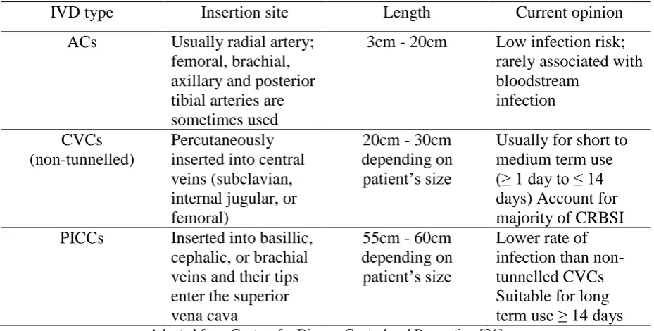
20cm) and their selection and usage is determined by the site of insertion and operators’ preferences (Table 1).
Table 1: IVDs used for venous and arterial access
IVD type Insertion site Length Current opinion
ACs Usually radial artery; femoral, brachial, axillary and posterior tibial arteries are sometimes used
3cm - 20cm Low infection risk; rarely associated with bloodstream
infection CVCs
(non-tunnelled)
Percutaneously inserted into central veins (subclavian, internal jugular, or femoral)
20cm - 30cm depending on patient’s size
Usually for short to medium term use (≥ 1 day to ≤ 14 days) Account for majority of CRBSI PICCs Inserted into basillic,
cephalic, or brachial veins and their tips enter the superior vena cava
55cm - 60cm depending on patient’s size
Lower rate of infection than non-tunnelled CVCs Suitable for long term use ≥ 14 days
Adapted from Centers for Disease Control and Prevention [31]
CVCs are inserted percutaneously with their tips usually ending within a major vein. They measure 20cm - 30cm, have between three to five lumens, and are used for the administration of intravenous fluids and electrolytes, blood transfusion, total parenteral nutrition, chemotherapy, and antibiotic therapy; as well as indirect monitoring of cardiac function and hydration status via central venous pressure [25]. CVCs, particularly anti-microbial CVCs are more expensive than peripheral catheters [37] (Table 1).
There is a real risk for an IVD to become colonized and hence increase infection risk the longer the duration it is left in situ [35, 39, 40]. These IVDs were found to have coatings of fibrin and thrombus formations along their surfaces, increasing the susceptibility of the IVD to microbial colonization.
2.3 Microbial colonization
2.3.1 Biofilm formation
A biofilm is not a static, flimsy, slime layer but rather is a coordinated living community of micro-organisms which may be composed of multiple species of bacteria and their secreted polysaccharide matrix and components deposited from bodily fluids [17, 41]. A conditioning film, dependent on the type of fluid the device interacts with, is formed on the surface of the IVD [10, 42, 43]. For example, IVDs rapidly acquire a sleeve of fibrin and fibronectin, components commonly found in blood, whereas urinary catheters tend to become encrusted with protein, electrolytes, and other organic molecules from the patient’s urine [17, 44]. Once this conditioning film is formed, the IVD surface becomes susceptible to microbial attachment and adhesion of micro-organisms to the IVD surface will depend on long-range bacteria-biomaterial surface interactions such as Van Der Waal’s forces, electrostatic interactions and hydrophobic interactions [10, 45]. These forces also influence protein adhesion on the surface of the IVD [10].
strongly fixing the micro-organism to the surface of the catheter [46-48]. Other micro-organism cell structures such as pili and flagella also facilitate attachment.
Microbial colonization occurs when attached or sessile micro-organisms divide to form micro-colonies of bacteria which in turn secrete large amounts of extracellular polysaccharide matrix (or slime) forming the architectural structure of the biofilm [43]. Slime is an extensive and diffuse polyanionic matrix that surrounds the micro-organisms’ cells [17, 45]. The cells in their three-dimensional matrix form thick pillars separated by fluid-filled spaces [43]. The pillars can be compared to as apartment buildings, and the fluid-filled spaces as streets between buildings through which the organisms receive nutrients, diffuse away wastes, and send chemical signals to each other [43]. This slimy substance protects the bacteria against host defence systems and antimicrobial agents, and clumping factor produced by some micro-organisms such as Staphylococcus aureus, makes it thrombogenic [10, 17].
Other species of bacteria may move into the biofilm, and interaction by quorum sensing mechanisms between the species can in turn produce different micro-environments within a given biofilm, facilitating plasmid exchange and enhancing antimicrobial resistance [17, 49-51]. If environmental conditions should become unfavourable; such as the exhaustion of nutrients or overcrowding, sessile micro-organisms can detach themselves and become free-floating, or planktonic in the bloodstream of the patient [17].
2.3.2 Types of micro-organisms
species [61], Enterococcus [57, 58], Coliforms [11, 61], Corynebacterium species [60], and Pseudomonas species [62, 63]. While it is interesting to take into account the different microbial species cultured, it is also worthwhile investigating further the relationship between the micro-organisms co-habitating in the same IVD which may influence the degree of virulence, thereby impacting on CRBSI.
2.3.3 Mechanisms of microbial colonization
Micro-organisms enter the bloodstream at the time of IVD insertion or at any time during therapy, from endogenous sources such as skin flora, or through extrinsic sources such as contaminated health care workers’ hands, inadequate or incorrect skin disinfection prior to IVD insertion or when the IVD is in situ (Figure 1).
There are currently three explanations describing the process of microbial colonization in IVDs. The first suggests colonization by micro-organisms occurs on the outside of the catheter (extra-luminally), either via downward colonization of micro-organisms from the patient’s skin surface on the outside surface of the catheter [17, 20, 45, 57, 64-66] or via upward colonization where the micro-organisms are inoculated on the tip of the IVD at the time of insertion [67] (Figure 1).
The second mechanism suggests micro-organisms are introduced via the inside surface of the IVD (intra-luminally), either via contamination of the port or hub connected to the IVD [59, 61, 64, 68-70], or via a contaminated infusate, [71, 72] although this latter source is now relatively rare (Figure1).
Figure 1: The various routes where micro-organisms enter the bloodstream via an intravascular device and cause bloodstream infection.
Adapted from Crnich and Maki [3] HCW = health care worker
The literature has revealed three mechanisms of IVD colonization, however, it remains unclear if one major route of IVD colonization occurs more frequently than another major route of colonization. While some past studies have suggested that microbial colonization originated from the skin surface surrounding the point of IVD entry, tracking downwards the external surface of the IVD, these studies fall short in examining the IVD in its entirety, failing to determine if there was a pattern of heavier microbial colonization on a particular segment of the IVD.
2.3.4 Incidence of Colonization
healthcare-acquired infection [1, 31]. Colonization is a more frequent event compared to CRBSI, and is therefore regarded as a necessary but not sufficient condition for the diagnosis of CRBSI. Although the focus of this thesis is on the exponential growth of micro-organisms on the surfaces of IVDs to determine if a pattern or trend of microbial growth exists, it is necessary to review the literature on CRBSI and expose how a lack of understanding in the pattern of microbial colonization has led to an array of diagnostic techniques for CRBSI which may not always be effective. Consequently, different diagnostic techniques have resulted in different ways of presenting data and results, adding to more confusion to the complexities that CRBSI already present.
2.4 IVD-Related Bloodstream Infection 2.4.1 Definitions
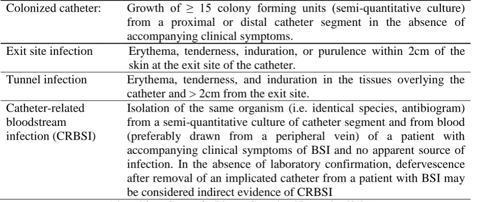
Table 2: CDC definitions for catheter-related infection
Colonized catheter: Growth of ≥ 15 colony forming units (semi-quantitative culture) from a proximal or distal catheter segment in the absence of accompanying clinical symptoms.
Exit site infection Erythema, tenderness, induration, or purulence within 2cm of the skin at the exit site of the catheter.
Tunnel infection Erythema, tenderness, and induration in the tissues overlying the catheter and > 2cm from the exit site.
Catheter-related bloodstream infection (CRBSI)
Isolation of the same organism (i.e. identical species, antibiogram) from a semi-quantitative culture of catheter segment and from blood (preferably drawn from a peripheral vein) of a patient with accompanying clinical symptoms of BSI and no apparent source of infection. In the absence of laboratory confirmation, defervescence after removal of an implicated catheter from a patient with BSI may be considered indirect evidence of CRBSI
Adapted from Centers for Disease Control and Prevention [31]
In keeping with the CDC definition, it has become increasingly well accepted practice to define a positive catheter culture with ≥15 colony forming units (CFUs) as catheter colonization [31]. A catheter is colonized based on the laboratory criteria of a minimum of ≥15 CFUs using a semi-quantitative culture technique [80] or ≥100 CFUs using a qualitative culture technique [31, 81, 82]. Catheter contamination is sometimes clarified by a growth of <15 CFUs from the catheter segment cultured [76]. The CDC definitions for “exit site infection” and “tunnel infection” are clinically based and relatively straightforward, and even if they do occur, such infections are however classified as wound infections in the presence of a foreign body rather than CRBSI because of the diagnostic criteria applied to diagnose CRBSI.
IVD contamination is mostly attributed to skin commensals around the exit site of the IVD. Current CDC recommended protocols of dressing changes using chlorhexidine 0.5% in 70% alcohol appear to contain IVD contamination to a minimum. However, a contaminated IVD
can develop into a colonized IVD (≥15 CFUs) through time, and the offending micro-organism is eradicated with antibiotic therapy to prevent further microbial
infection. Microbial IVD colonization is contained by the prompt removal of IVDs when their usage have ceased, routine IVD replacement (after 10-14 days in situ as practised in some clinical areas) and also by the increased use of antibiotic-impregnated IVDs. While not all colonized IVDs will cause CRBSI, by definition CRBSI can only occur from a colonized IVD, and therefore the surveillance of IVD colonization rates may be a useful preventive measure of reducing the risk of CRBSI by identifying rising trends in IVD colonization whose cause might be identified and eliminated. A patient with a heavily colonized IVD is more likely to have the device removed instead of waiting for signs and symptoms of CRBSI to develop. The diagnostic criteria of CRBSI are clearly defined in Table 2.
A difficulty in reviewing IVD-related bloodstream infections is the large variation in what is considered to be an “infected device or catheter” with much debate existing as to precisely what is a significant quantity of bacterial growth [83]. Counts or CFUs of micro-organisms can range from 5 CFUs to 1000 CFUs depending on the type of diagnostic method used [81, 83, 84].
2.4.2 Prevalence of CRBSI in the clinical area
the usage of multi-lumen IVDs, the use of jugular and femoral insertion sites make them more susceptible to acquire CRBSI [8, 85-87].
Maki and colleagues [8] conducted a systematic review of 200 prospective studies that used appropriate criteria to determine the incidence of CRBSI in different IVDs. The risk estimates of IVD-related BSI from these studies were:
Catheter site Risk per 1000 catheter days
Peripheral intravenous catheters 0.5 (95% CI, 0.2- 0.7) Non medicated, non-tunnelled and non-cuffed
CVCs 2.7 (95% CI, 2.6-2.9)
Non-medicated, tunnelled and non-cuffed CVCs 1.7 (95% CI, 1.2-2.3)
Cuffed and tunnelled CVCs 1.6 (95% CI 1.5-1.7)
Arterial Catheters 1.7 (95% CI, 1.2-2.3)
Pulmonary arterial catheters 3.7 (95% CI, 2.4-5.0) Peripherally inserted central catheters 1.1 (95% CI, 0.9-1.3) Peripherally inserted midline catheters 0.2 (95% CI, 0.0-0.5)
Adapted from Maki et al. [8] CVCs = central venous catheters
CRBSI occurrence was estimated to occur at a rate of 5.0 per 1000 catheter days in the ICU setting [24].
2.4.3 Methods of reporting CRBSI
literature, many studies have favoured clinical diagnosis over microbiological confirmation, and the reverse is also true for surveillance literature.
There is also no standardized method used in reporting results from these studies, with some choosing to present their incidence rates in raw percentages [34, 35, 53, 56, 88-91] and others expressing their incidence rates per 1000 catheter days [54, 92, 93]. Raw percentages or incidence rates per 1,000 catheter days, however, do not take into account varying IVD durations and therefore may produce misleading comparisons. Other differences, such as study designs, heterogeneity of patient populations, catheter indwelling times, terminology, and techniques for microbial analyses, also contributed to this lack of standardization in reporting results and hence complicate comparison of different studies [8, 34, 35, 53, 54, 56, 88-90, 92, 93]. Surveillance studies within a particular ICU and comparisons between
different ICUs also tend to utilise calculation methods for incidence rates which are insensitive to the mix of diseases and their severity and to the duration the IVDs
remain in situ.
The reporting of colonisation and CRBSI rates is usually accomplished as raw percentages (e.g. 12% of all IVDs colonized) or as rates per 1,000 patient days (measured by Poisson or negative binomial regression). Neither of these methods is appropriate, as both assume that catheter colonization is a random event that suddenly occurs at a fixed level of probability. It is much more likely that colonization is governed by a growth pathway where the likelihood of the event increases exponentially.
defined rather inelegantly as a terminating event (as opposed to an incidental observation). This also avoids assuming that there is a linear association between likelihood of colonization and IVD in situ time, when it is more likely that the association will be some sort of
exponential (if in situ time was used as an adjustment factor in a multivariate Poisson regression model).
Ideally, analysis would involve comparison in different patient / catheter groups of the exponential rate of microbial growth by non-linear regression, using multiple measurements of colonization whilst the IVDs are still in situ. However, the technology for such measurements does not currently exist.
2.4.4 How is CRBSI detected?
The current ‘gold standard’ markers for making a definitive diagnosis of CRBSI or IVD bloodstream infection derived from the CDC guidelines of 2002 requires the following three criteria to be present.
1) A positive catheter culture
2) A positive peripheral blood culture taken before IVD removal 3) The same micro-organism isolated from (1) & (2)
peripheral blood load at the time of sampling [83]. Other studies have also revealed that blood culture contamination by skin micro-organisms is common, and that there is a low positive predictive value of positive blood cultures for micro-organisms, such as coagulase negative Staphylococci [97, 98].
The latest publication of clinical practice guidelines for the diagnosis of CRBSI available have made clearer recommendations for the diagnosis of CRBSI based on catheter culture and blood culture techniques, with recommendations to manage and treat CRBSI based on the micro-organism cultured [99]. It would be interesting if the CDC adopts some of these recommendations. As it stands, an array of diagnostic techniques to diagnose CRBSI exist.
2.4.5 Diagnostic Techniques
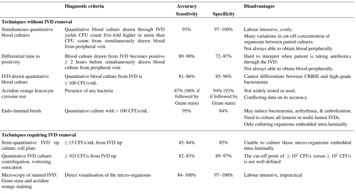
There are several diagnostic methods used to culture microbial content of catheters. However, there is no ‘gold standard’ method to which all techniques utilised for the diagnosis of CRBSI can be compared, and thus it is difficult to compare results from different studies using different diagnostic techniques to determine CRBSI. Diagnostic techniques can be divided into 2 classifications; (i) techniques which do not require removal of the IVD; and (ii) those that require IVD removal. A summary of these techniques, their different diagnostic criteria, accuracy, and disadvantages are presented in Table 3.
Table 3: A summary of the diagnostic techniques used to detect CRBSI in IVDs
Diagnostic criteria Accuracy Disadvantages
Sensitivity Specificity
Techniques without IVD removal
Simultaneous quantitative blood cultures
Quantitative blood culture drawn through IVD yields CFU count five-fold higher or more than CFU count from simultaneously drawn blood from peripheral vein
93% 97–100% Labour intensive, costly.
Many variations in cut-off concentration of organisms between paired cultures.
Not always able to obtain blood peripherally Differential time to
positivity
Blood culture drawn from IVD becomes positive ≥ 2 hours before simultaneously drawn blood culture from peripheral vein
89–90% 72–87% Hard to interpret when patient is taking antibiotics through the IVD.
Not always able to obtain blood peripherally. IVD-drawn quantitative
blood culture
Quantitative blood culture from IVD is ≥ 100 CFUs/mL
81–86% 85–96% Cannot differentiate between CRBSI and high-grade bacteraemia
Acridine orange leucocyte cytosine test
Presence of any bacteria 87% (96% if followed by Gram stain)
94% (92% if followed by
Gram stain)
Not widely tested or used. Conflicting data on its accuracy.
Endo-luminal brush Quantitative culture with > 100 CFUs/mL 95% 84% May induce bacteraemia, arrhythmia, & embolization. Need to culture all lumens in multi-lumen IVDs. Only culturing organisms embedded intra-luminally
Techniques requiring IVD removal
Semi-quantitative IVD tip culture, roll plate
≥ 15 CFUs/mL from IVD tip 45–84% 85% Unable to culture those micro-organisms embedded intra-luminally
Quantitative IVD culture: centrifugation, vortexing, sonication
≥ 103 CFUs from IVD tip 82–83% 89–97% The cut-off point of ≥ 103 CFUs versus ≥ 102 CFUs is not well defined
Microscopy of stained IVD: Gram stain and acridine orange staining
Direct visualisation of the micro-organisms 84–100% 97–100% Labour intensive, impractical
Secondly, different cut-off points for concentrations of micro-organisms between the paired blood cultures to positively predict the incidence of CRBSI exist [100-102]. This range from a 3-fold [102], 4-fold [101], 5-fold [100] difference in concentrations of micro-organisms drawn from the IVD and peripheral site, making comparison and interpretation of results difficult [103]. Besides, in 25-50% of cases of suspected CRBSI, it is often not possible to withdraw blood through these IVDs and the sensitivity of this technique is questioned [83].
Traditionally, the IVD tip has been cultured and depending on which diagnostic technique is utilised, microbial yields in the form of CFUs are used to determine if the IVD is contaminated, colonized, or colonized beyond a pre-determined number of CFUs to indicate a diagnosis of CRBSI. This thesis raises a question mark concerning this current practice of culturing only the tip of the IVD as the proxy for determining if an IVD is contaminated or colonized or potentially likely to cause CRBSI, postulating that if microbial colonization originated and developed from the skin downwards, then it may be possible that the proximal intravascular segment of the IVD would become colonized earlier and have a heavier density of microbial colonization compared to the middle or distal segments of the IVD. The nature in which microbial biofilm develops on the surfaces of the IVD has not been extensively examined, and there is a possibility that microbial colonization on the proximal intravascular segment of any IVD may be releasing free-floating planktonic colonies of micro-organisms into the bloodstream with a potentiality to cause CRBSI, well before the same biofilm colonizes downwards towards the tip through time to effect a finding of microbial colonization at the IVD tip. Future research is required to determine whether such a finding may impact on the current diagnostic technique used to determine IVD colonization or CRBSI which rely heavily on culturing only the IVD tip.
Finally, the length of the catheter segment to be cultured needs to be standardized. In the original description of the Maki roll-plate technique, a range of 5cm-7cm of segments of intravascular catheters were sampled [80] whereas in other studies, only 3-4cm of the IVD tip was cultured [84, 107].
2.4.6 Prevention of CRBSI
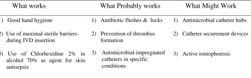
[image:38.595.75.510.271.407.2]Considerable progress has been made in the area of prevention of IVD-related bloodstream infection. Various novel preventive strategies have been developed because the pathogenesis of IVD bloodstream infections is amenable to interventions at many steps [3] These novel strategies can be categorised into three areas based on the level of evidence of (1) what works, (2) what probably works, and (3) what might work (Table 4).
Table 4: Categories of preventive strategies for IVD-related BSI What works What Probably works What Might Work
1) Good hand hygiene 1) Antibiotic flushes & locks 1) Antimicrobial catheter hubs 2) Use of maximal sterile barriers
during IVD insertion
2) Prevention of thrombus formation
2) Catheter securement devices 3) Use of Chlorhexidine 2% in
alcohol 70% as agent for skin antisepsis
3) Antimicrobial-impregnated catheters in specific conditions
3) Active iontophoresis 4) Designated IVD therapy team
Adapted from Cicalini et al., Hammarskjold et al., Sherertz et al., and Eggimann et al.[64, 105, 114, 115 ]
The “least novel” of preventive strategies against IVD-related bloodstream infections, are basic infection control practices such as good hand-washing, use of sterile gown, gloves and drapes during IVD insertion, using appropriate skin disinfectant, removing the IVD when no longer required, and avoiding using the femoral site for IVD are measures which effectively work [3, 7, 87, 105, 116-118].
efficacy of anti-microbial and antibiotic-coated IVD, using several combinations of antibiotics, such as minocycline [119-121], silver zeolite [122], silver sulfadiazine [75, 123-125], vancomycin [3, 126], rifampin [119-121], and chlorhexidine [75, 104, 123, 125] each presenting with supposedly efficacies.
Anti-coagulants such as heparin and enoxaparin have also been used as agents to prevent thrombus formation, which in turn prevents biofilm formation in IVDs. There have also been studies and trials introducing the use of antibiotic locks and hubs for IVDs but the effectiveness of these interventions are still unproven [68, 127].
CHAPTER 6
Study 3: Intravascular Devices: A Survey of Clinical Practices
Evidence-based practice in healthcare is perceived as a process of inducting evidence arising from research into clinical decision making with the main emphasis of “doing the right things right” [154, 155]. It supports healthcare professionals in their decision making to eliminate ineffective, inappropriate, and potentially dangerous practices; improving clinical practice, health service management and patients’ choice [155, 156].
Practice guidelines and institutional infection control protocols provide a reference point for nurses involved with the care and management of IVDs to implement best practice. However, little is known about how closely nurses adhered to the guidelines and protocols when caring and managing IVDs, and if any variations in practice contributes to increased microbial colonization in IVDs. This final study seeks to determine the degree of concordance of current nursing practice to evidence-based practice guidelines, as a proxy for actual adherence to protocols, and how partial or non-adherence to protocols may impact on IVD colonization.
The study’s information sheet, consent form, questionnaire and answer template will be found in the Appendix section of this thesis.
Statement of contribution:
I was responsible for the study design, data collection, statistical analysis, interpretation and writing of this paper with consultations with Assoc Prof Rosalind Bull, Dr Robertson, and Dr Marianne Watts.
ABSTRACT
Objective: This study explores current care and management of intravascular devices (IVDs) by registered nurses (RNs) in a regional hospital to determine the degree of concordance with evidence-based practice guide IVDs are indispensable for the management of critically-ill patients, but carry an associated risk of catheter-related bloodstream infection (CRBSI) increasing morbidity, mortality and health care costs. CDC Guidelines for the Prevention of Intravascular Catheter-Related Infections and institutional infection control practices are available, but there may be variations in clinical protocols and practice preferences resulting in varying degrees of concordance.
Design: A questionnaire was developed using the CDC Guidelines and current institutional practice protocols as benchmarks. Responses were analysed for degrees of concordance with guidelines in different clinical areas.
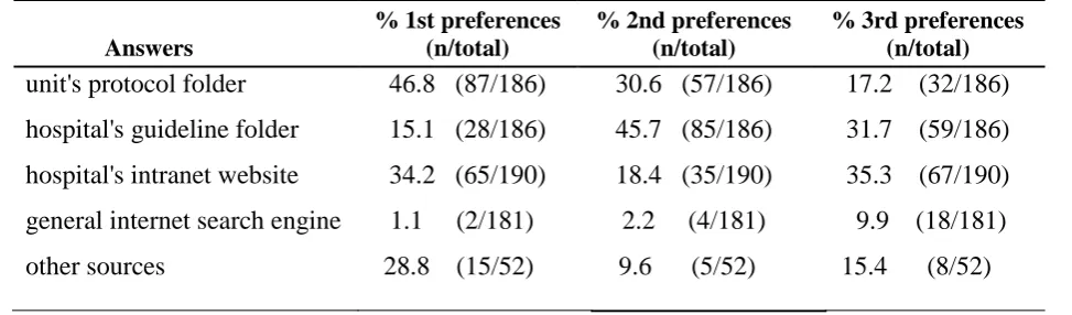
Results: Overall median concordance ranged from 28.6% - 66.7%. There was variation in the care and management of IVDs between clinical areas, and nursing practice reflected only partial concordance with CDC guidelines. Variation was present amongst RNs selection of IVD dressing supplies and equipment, as well as the frequency of dressing changes. Also, the mode of information dissemination appeared to be inimical with the respondents’ preferences, suggesting access to practice protocols and guidelines may need to be addressed.
Introduction
Intravascular devices (IVDs) are indispensable for the management of critically and chronically ill patients. They provide safe, reliable vascular access, continuous haemodynamic monitoring, and are used in both the hospital and outpatient setting [8, 148]. Unfortunately, many healthcare-acquired infections in severely ill patients are caused by these medical devices [38, 60, 157]. Cases of catheter-related bloodstream infections (CRBSI) occur 250,000 - 500,000 times annually in hospitals in the United States [1, 83], and over 3,000 times annually in Australian hospitals [22], resulting in extended lengths of hospitalisation [16, 27], increased healthcare costs [13-15], and higher morbidity and mortality (12% - 35%) [13, 16, 27].
IVDs are inserted by physicians or specially-trained nurses, whilst the post-insertion care and management of IVDs are predominantly nursing responsibilities [158]. As an evidence-based profession, nursing draws on such protocols as those of the Centers for Disease Control and Prevention (CDC). The CDC reviews the evidence for effective preventive measures against IVD-related infection and makes recommendations for best practice [20, 31]. The CDC’s recommendations are drawn on to develop effective nursing practice protocols to promote patient safety and decrease preventable infections. The protocols cover a variety of related topics such as IVD site care, dressing regimes, frequency of changing of administration sets, disinfection of IVD ports, to specific guidelines for care and management of arterial catheters (ACs), central venous catheters (CVCs), and peripherally-inserted central catheters (PICCs)[20, 31]. These guidelines help to inform nursing practice and provide a framework of evidence to enable appropriate clinical decision-making.
revealed variations in IVD colonization rates in the different clinical practice areas. Three major routes of IVD colonization (i.e. extra-luminal, intra-luminal, endogenous spread) are postulated to contribute to IVD colonization. As nursing care and management of IVDs are implicit to the risk of extra-luminal and/or intra-luminal routes of colonization, the present study focused on determining if any variations in practice exist. Little is known about how closely nurses adhere to CDC guidelines and institutional protocols when caring and managing IVDs.
The Study Aim
The aim of the study was to determine the degree of concordance of current nursing practices involving the care and management of IVDs by registered nurses (RNs) with the CDC recommended guidelines.
Design
A prospective, cross-sectional, anonymous survey was conducted in a 350-bed Australian regional, teaching hospital, from January to March 2010.
Sample
All RNs (n=308) involved with the care and management of patients with IVDs were invited to participate, with representation from medical, surgical, rehabilitation wards and the intensive care unit. RNs on leave during the study period were excluded.
Questionnaire
(Appendix 3). Questions were then grouped into the following categories to provide an indication of the RNs concordance with CDC protocols:
1) Selection of supplies when changing IVD dressings;
2) The frequency of IVD dressing changes;
3) The rationales for IVD dressing changes;
4) The management of IVDs (i.e. external components of the IVD);
5) General knowledge base concerning the care & management of IVDs;
6) Infection control awareness;
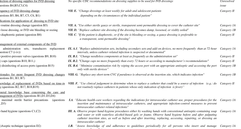
Table 12: Comparison of questionnaire's Categorical Questions with Centers for Disease Control Guidelines
Infection Control Procedure Centers for Disease Control Guidelines [20, 31] Level of Evidence *
Selection of dressing supplies for IVD dressing No specific CDC recommendations on dressing supplies to be used for IVD dressings Unresolved
(questions B4,B5,C3,C4) issue
Frequency of IVD dressing change VII. E. “Change dressings at least weekly for adult and adolescent patients Category II
(questions B5, B6, B7, C5, C6, B1) depending on the circumstances of the individual patient”
Indications for application of dressing to IVD site:
(i) routine dressing change (question B5) VII. A. "Use either sterile gauze or sterile, transparent semi-permeable dressing to cover the catheter site" Category 1A
(ii) loose dressing, or IVD site bleeding or oozing VII. D. “Replace catheter-site dressing if the dressing becomes damp, loosened, or visibly soiled" Category 1B
(ii) diaphoretic patient (question B8) VII. C. "If the patient is diaphoretic, or if the site is bleeding or oozing, a gauze dressing is preferable to a transparent, semi-permeable dressing"
Category II
Management of external components of the IVD (i) administration sets, transducers replacement (question C7 (i-iv))
IX. A.1. "Replace administration sets, including secondary sets and add-on devices, no more frequently than at 72-hour intervals, unless catheter-related infection is suspected or documented"
Category 1A
(ii) positive-pressure valves (questions B9, B10) IX. B.1. “Change needleless components at least as frequently as the administration set" Category II
(iii) caps (questions B10, B11,) IX. B.2. "Change caps no more frequently than every 72 hours or according to manufacturer’s recommendations" Category II
(iv) disinfecting of access ports (question B15) IX. B.4. “Minimize contamination risk by wiping the access port with an appropriate antiseptic and accessing the port only with sterile devices”
Category 1B
Rationales for more frequent IVD dressing changes (questions B1, B3, B7)
VIII. G. “Replace any short-term CVC if purulence is observed at the insertion site, which indicates infection" Category 1B
Knowledge of replacement of IVDs based on time-in situ (questions B2, B17, B18,B19)
VIII. F. “Use clinical judgement to determine when to replace a catheter that could be a source of infection (e.g.: do not routinely replace catheters in patients whose only indication of infection is fever)”
Category II
General knowledge base concerning the care and management of IVDs (questions B1,D1.D3,D4) (i) maximal sterile barrier precautions (questions D1,D3)
I.A. “Educate health-care workers regarding the indications for intravascular catheter use, proper procedures for the insertion and maintenance of intravascular catheters, and appropriate infection-control measures to prevent intravascular catheter-related infections"
Category 1A
(ii) hand hygiene (questions C1,C2) III. A. Observe proper hand-hygiene procedures either by washing hands with conventional antiseptic-containing soap and water or with waterless alcohol-based gels or foams. Observe hand hygiene before and after palpating catheter insertion sites, as well as before and after inserting, replacing, accessing, repairing, or dressing an intravascular catheter
Category 1A
(iii)Aseptic technique (question D2) 1.B. “Assess knowledge of and adherence to guidelines periodically for all persons who insert and manage intravascular catheters
Category 1A
The internal construct validity of the questionnaire was reviewed initially by an RN-academic with expertise in wound care and infection control policies and a medical biostatistician with intensive care and public health experience to determine that the research questions reflected the phenomena of interest [159]. The questionnaire was pilot tested by clinical nurse educators (CNEs) (n=7) from each clinical area. A revised version of the questionnaire was then distributed to all RNs (n=308) who met the inclusion criteria.
A prescribed template of correct answers to all questions in the questionnaire was used to ascribe concordance scores to all responses received. The prescribed template of correct answers was based on a combination of the CDC recommended guidelines and institutional protocols (in instances whereby the CDC guidelines do not provide any suggestions). The prescribed template of correct answers and concordance scores was created by the researcher and approved by an RN-academic with expertise in wound care and infection control policies. All questionnaire responses were compared to the prescribe template of answers for accuracy.
concordance, high partial concordance, moderate partial concordance, low partial concordance, and non concordance.(Appendix 3)
Data Collection
Information sessions (n= 26) were conducted for RNs in all clinical areas at different times to accommodate all shift-working staff. Distribution of questionnaires was varied in the clinical areas and the final mode of distribution was determined after close consultation with the respective ward’s CNE. Completed questionnaires were returned to the researcher via institutional internal mail. No subject identifier information was collected.
Ethical And Etiquette Considerations
Human Research Ethics Committee approval (H0010857) was obtained and confidentiality of individual participants was protected. Questionnaires were administered without collection of subject identifiers. Permission to conduct the study and administer the questionnaires was gained from the Directors of Nursing of the hospital and nurse unit managers of the different clinical areas.
Data Management & Statistical Analysis Predictor variables
Demographic data of respondents and their clinical areas was obtained. The hierarchical rank of respondent, employment status (part-time or full-time), clinical area, institution or agency staff, numbers of years in nursing, whether trained locally or elsewhere, and if possessed post-registration qualifications were recorded.
Outcome measures
multivariate analysis of variance) expressed as odds ratios (OR) and 95% confidence intervals (95% CI) for nominal categories or odds ratios for trends for continuous values or rank-order categories, corrected where appropriate for repeated measures and for multiple comparisons by the Holm method. Backward stepwise ordinal logistic regression was used to select multivariate models of association of predictor variables (P for removal = 0.2, P for entry = 0.12). Analyses were performed using Stata IC 10.1™ for Windows™ (StataCorp, College Station, Texas, USA).
Results Sample
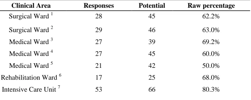
[image:49.595.77.512.439.613.2]202 (65.6%) out of 308 distributed questionnaires were returned. There was a marginally higher response from ICU nurses (OR, 2.48; 95% CI, 1.05 - 5.83; p = 0.23; overall Fisher’s exact test for distribution p = 0.05) Table 13.
Table 13: Questionnaire response rates from respective clinical areas Clinical Area Responses Potential Raw percentage
Surgical Ward 1 28 45 62.2%
Surgical Ward 2 29 46 63.0%
Medical Ward 3 27 39 69.2%
Medical Ward 4 27 45 60.0%
Medical Ward 5 21 42 50.0%
Rehabilitation Ward 6 17 25 68.0%
Intensive Care Unit 7 53 66 80.3%
1
General surgical ward with specialization in colorectal, abdominal, and urological surgery.
2
General surgical ward with specialization in orthopaedics, maxilla-facial, ear, nose, throat (ENT), and gynaecological surgery.
3
General medical ward with specialization in renal medicine and endocrinology.
4
General medical ward with specialization in oncology, haematology and immunology.
5
General medical ward with specialization in cardiology and neurology.
6
Rehabilitation ward containing a mixture of post-surgical and medical patients.
7
Category 1: Selection of supplies when changing of IVD dressings
Figure 10: Box plot showing overall concordance to questions asked in categories 1, 2 & 5
Category 2: Frequency of IVD dressing changes
Median overall concordance was 48.8% (IQR, 36.9% - 59.4%). Rehabilitation ward RNs were most concordant to the recommended protocol of weekly dressing changes for CVCs (OR, 1.51; 95% CI, 1.00 - 2.29; p = 0.05); and PICCs (OR, 1.39; 95% CI, 0.95 - 2.04; p = 0.09). RNs who possessed higher post-registration nursing qualifications also tended to be
more concordant to the recommended protocol of weekly CVC dressing changes (OR, 0.77; 95% CI, 0.57 - 1.03; p = 0.08) when compared to those nurses with basic nursing
(OR, 0.11; 95% CI, 0.04 - 0.32; p < 0.001) and ACs (95.83% (46 out of n=48) preferring to change their AC dressings every 4 days or more frequently).
Respondents were asked to select as many clinical scenarios which they deemed appropriate to necessitate an IVD dressing change out of the recommended protocol of weekly change. The scenarios were: (i) when the IVD site is oozing with blood underneath the dressing; (ii) when the IVD site is red and inflamed; (iii) when the transparent dressing of the IVD site is partially lifting; and (iv) when the patient requests the nurse to do so. General surgical ward RNs were more concordant to protocol concerning weekly frequency of changing IVD dressings (OR, 3.79; 95% CI, 1.42 - 10.11; p = 0.008), when the IVD site was oozing, looked red or inflamed, and had blood underneath the transparent dressing, compared to their colleagues from other clinical areas. RNs who did their pre-registration preparation locally (OR, 0.61; 95% CI, 0.39 - 0.95; p = 0.03) also tended to be more concordant to changing IVD dressings more frequently under these clinical scenarios than those who trained elsewhere. ICU RNs scored the least concordance in this question as they tended to change the IVD dressing more frequently under all of these clinical scenarios (OR, 0.11; 95% CI, 0.008 - 1.51; p = 0.099), resulting in a poor overall concordance score (OR, 0.10; 95% CI, 0.04 - 0.22; p < 0.001) in this category when compared to RNs from all other clinical areas (Figure 10)
Category 3: The rationales or reasons for IVD dressing changes
Median overall concordance was 50% (IQR, 41.7% - 58.3%). There are many reasons why nurses change IVD dressings. Respondents indicated observing for the following characteristics during routine IVD site inspection:
b) Line: Ensuring patency and functionality of the IVD, to inspect if the IVD was dislodged, or if there was leaking around the skin site of IVD insertion.
c) Dressing: Inspecting the site for IVD dressing integrity, condition of dressing, etc. d) Other: Other reasons for IVD site inspection such as, as per protocol or to find out
the date and time of the last dressing change, etc.
ICU RNs (OR, 3.98; 95% CI, 1.94 - 8.14; p < 0.001 and OR, 2.00; 95% CI, 1.08 - 3.73; p = 0.03) provided the most concordant answers to these 2 questions, followed by higher ranked RNs (OR for trend, 1.25; 95% CI, 0.95 - 1.66; p = 0.11 and OR for trend, 1.43; 95% CI, 1.04 - 1.98; p = 0.03) and those who have had a longer service in nursing (OR for trend, 0.76; 95% CI, 0.54 - 1.07; p = 0.11 and OR for trend, 0.78; 95% CI, 0.58 - 1.04; p = 0.09). 96% (n=199) of respondents indicated that an IVD site which is red and inflamed, or a CVC or PICC which has been in situ for more than 7 days (83.4%) may be potential indicators of infection. 34.7% of respondents attributed a spike in body temperature above 38⁰C and an IVD site which had an exudate or was leaking (33.7%) as predictors of infection. Medical and surgical ward RNs (OR, 2.55; 95% CI, 1.30 - 5.00; p = 0.006) and ICU RNs (OR, 2.25; 95% CI, 1.18 - 4.32; p = 0.014) regarded routine IVD dressing changes as a lesser priority of routine nursing care in instances where they became busy with other nursing care, when compared to nursing staff from the medical or rehabilitation wards. ICU RNs (OR, 4.14; 95% CI, 1.91 - 8.96; p < 0.001) had better overall concordance in this category than their colleagues from other clinical areas.
Category 4: The management of IVDs