Proton Transfer, Hydrogen Bonding, and Disorder: Nitrogen
Near-Edge X
‑
ray Absorption Fine Structure and X
‑
ray Photoelectron
Spectroscopy of Bipyridine
−
Acid Salts and Co-crystals
Joanna S. Stevens,
*
,†Lauren K. Newton,
†Cherno Jaye,
∥Christopher A. Muryn,
§Daniel A. Fischer,
∥and Sven L. M. Schroeder
*
,†,‡,§†School of Chemical Engineering and Analytical Science, The University of Manchester, Oxford Road, Manchester M13 9PL, U.K. ‡School of Chemical and Process Engineering, University of Leeds, Leeds LS2 9JT, U.K.
§School of Chemistry, The University of Manchester, Brunswick Street, Manchester M13 9PL, U.K.
∥National Measurement Laboratory, National Institute of Standards and Technology, Gaithersburg, Maryland 20899, United States
*
S Supporting InformationABSTRACT: The sensitivity of near-edge X-ray absorption
fine structure (NEXAFS) spectroscopy to Brønsted donation and the protonation state of nitrogen in the solid state is investigated through a series of multicomponent bipyridine− acid systems alongside X-ray photoelectron spectroscopy (XPS) data. A large shift to high energy occurs for the 1s→ 1π*resonance in the nitrogen K-edge NEXAFS with proton transfer from the acid to the bipyridine base molecule and allows assignment as a salt (CNH+), with the peak ratio
providing the stoichiometry of the types of nitrogen species present. A corresponding binding energy shift for CNH+is
observed in the nitrogen XPS, clearly identifying protonation and formation of a salt. The similar magnitude shifts observed
with both techniques relative to the unprotonated nitrogen of co-crystals (CN) suggest that the chemical state (initial-state) effects dominate. Results from both techniques reveal the sensitivity to identify proton transfer, hydrogen bond disorder, and even the potential to distinguish variations in hydrogen bond length to nitrogen.
■
INTRODUCTIONProton (hydrogen) transfer can be thought of as one of the simplest chemical reactions, ranging from complete transfer from an acidic to a basic moiety (protonation through Brønsted donation) to varying degrees of sharing through hydrogen bonding. Whether Brønsted proton transfer occurs has a profound effect on the location of protons in crystal structures and influences chemical and physical properties. These interactions can be employed to target properties of solid forms (crystal engineering), with particular relevance to the pharmaceutical industry where acid/base guest molecules can be combined with active ingredients to tailor properties such as solubility and bioavailability through formation of salts and co-crystals.1−4 Other relevant fields are organic ferroelectrics,5 energetic materials,6 and the design of materials with targeted optical properties such as color7 and luminescence.8,9 Even among hydrogen bonds, the level of interaction with the donor and acceptor atoms can vary significantly, from relatively weak to strong with quasi-covalent character,10and there is also the possibility of disordered hydrogen bonds.11 While X-ray diffraction (XRD) and solid-state nuclear magnetic resonance spectroscopy (ssNMR) are often techniques of choice for
structural characterization,2,11−14they are not always unambig-uous with regard to proton locations (although further clarity can often be obtained by neutron diffraction).11,13−16 The importance of accurate characterization of salts vs co-crystals based on this relatively small difference in proton location should not be underestimated, particularly with the wider implications for intellectual property and regulatory control in the pharmaceutical industry.4,17
X-ray photoelectron spectroscopy (XPS) has recently been shown to unequivocally identify whether intermolecular proton transfer occurs in a range of two-component systems and distinguish protonation (salts) from H-bonding (co-crys-tals),14,18−21 as well as the presence of zwitterionic species.22 This reflects the influence of the local chemical state on the core level binding energy,19,23−25resulting in a large positive chemical shift due to proton transfer to nitrogen.14,18−21 Near-edge X-ray absorption fine structure (NEXAFS) is likewise highly dependent on the local electronic structure, as excitation
Received: December 16, 2014 Revised: February 17, 2015 Published: February 23, 2015
Article
pubs.acs.org/crystal
environment of the excited (nitrogen) atom and whether proton transfer has occurred (NH+vs N···H−O). Novelty for organic crystal growth research arises from exploiting the sensitivity of NEXAFSπ*resonances to proton transfer. The level of crystallographic information obtainable is explored by looking into the impact of varying nitrogen−hydrogen distance and disorder on NEAXFS and XPS chemical shifts.
■
EXPERIMENTAL METHODSMaterials. 4,4′-Bipyridine (4BPY), 2,2′-bipyridine (2BPY), 5-sulfosalicylic acid (sulfo) dihydrate, 1,2,3,4-butanetetracarboxylic acid (buTA), and malonic acid were obtained with >99% purity, thiodipropionic acid (thio) with >97% purity, benzene-1,2,4,5-tetracarboxylic acid (BTA) with >96% purity, and phthalic acid and adipic acid with >99.5% purity (Sigma-Aldrich, UK). The salts and co-crystals were formed as follows.
2,2′-Bipyridine/Benzene-1,2,4,5-tetracarboxylic Acid Salt (2BPY/ BTA). 2,2′-Bipyridine (156.0 mg, 0.999 mmol) and
benzene-1,2,4,5-′
to 25 mL of water and stirred to give a suspension. The buTA solution was added to the heated 4,4′-bipyridine suspension dropwise over 15 min while stirring. The mixture was left overnight and thenfiltered to yield the 1:1 co-crystal with some excess starting materials. This initial product was then recrystallized as follows: 123.0 mg of the resulting powder was added to a hot aqueous solution (80 mL, 70 °C) and stirred; additional hot water was added until the product dissolved (total 300 mL). The solution was left to cool to RT, with a small amount of the original product added as a seed the following day. Small, clear crystals started to form after 2 weeks and werefiltered after 3 weeks to give the 1:1 co-crystal.
4,4′-Bipyridine/Phthalic Co-crystal (4BPY/phthalic). Stoichiomet-ric amounts of 4,4′-bipyridine (94.4 mg, 0.604 mmol) and phthalic acid (100.3 mg, 0.604 mmol) were milled in a Retsch MM200 mixer mill for 40 min at a rate of 30 Hz, using two 5 mL stainless steel jars, each containing one 7 mm-diameter stainless steel ball, to yield the 1:1 co-crystal.
4,4′-Bipyridine/Adipic Co-crystal (4BPY/adipic). 4,4′-Bipyridine (218.1 mg, 1.396 mmol) was dissolved in 4 mL of methanol, and adipic acid (205.0 mg, 1.403 mmol) was dissolved in 6 mL of methanol. On combination of the two solutions and stirring, the solution went cloudy and wasfiltered under vacuum after 2.5 h to give the 1:1 co-crystal.
4,4′-Bipyridine/Thiodipropionic Co-crystal (4BPY/thio).4,4′ -Bipyr-idine (187.1 mg, 1.198 mmol) was dissolved in 5 mL of methanol, and thiodipropionic acid (213.2 mg, 1.194 mmol) was dissolved in 7 mL of methanol. On combination of the two solutions and stirring, the solution went cloudy and wasfiltered under vacuum after 4 h to give the 1:1 co-crystal.
4,4′-Bipyridine/Malonic Co-crystal (4BPY/malonic). 4,4′ -Bipyri-dine (250.3 mg, 1.603 mmol) was dissolved in 4.5 mL of methanol, and malonic acid (166.6 mg, 1.601 mmol) was dissolved in 5.5 mL of methanol. On combination of the two solutions, the solution went cloudy and wasfiltered under vacuum after 3 h to give the 1:1 co-crystal.
Successful formation of co-crystals and salts was confirmed by XPS and comparison of X-ray powder diffraction patterns (available as Supporting Information) with those simulated from previously reported single crystal structures.35−37 The new 4BPY/sulfo system was revealed as a 1:1 salt and 4BPY/buTA system as a 1:1 co-crystal initially by XPS, and this was confirmed by both NEXAFS and single crystal X-ray diffraction (CCDC 1038154 and 1038155, Supporting Information).
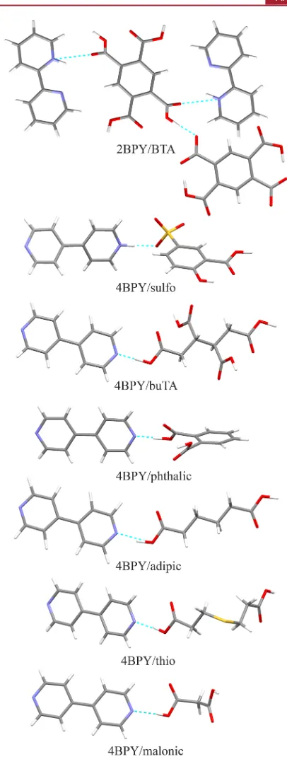
[image:2.625.60.301.216.530.2]Powder X-ray Diffraction (PXRD). Powder X-ray diffraction (PXRD) patterns (available as Supporting Information) were collected using a Rigaku Miniflex instrument utilizing Cu Kα radiation (λ = 1.5406 Å), operating over 5−40°2θat 1.5°min−1with a 0.03°step, 30 kV voltage, and 15 mA current. Typically 5 mg of sample was placed on a small sample attachment and smoothed to achieve a level surface. Single Crystal X-ray Diffraction (XRD). Single crystal X-ray diffraction (XRD) data was collected for 4BPY/sulfo and 4BPY/buTA at 100 K with an Oxford Diffraction X-Calibur 2 diffractometer utilizing Mo Kα radiation (λ = 0.71073 Å) and an Oxford CryoSystems Cryostream Controller 700. Data was recorded at 50 kV and 40 mA with a CCD detector. Data reduction, cell refinement, Figure 1. Chemical structures comprising the two component
and multiscan absorption corrections were carried out using the program CrysAlis RED (Oxford Diffraction Ltd., version 1.171.34.44, 2010). The structure was solved with SHELXS-97 and refined onF2 against all reflections with SHELXL-97. All non-hydrogen atoms were refined by direct methods anisotropically; all hydrogen atoms were located in difference Fourier maps and refined isotropically. Full crystallographic details are provided within the CIF files in the Supporting Information, along with tables summarizing the main crystallographic data. The CIFs are also deposited with the Cambridge Crystallographic Data Centre (CCDC): CCDC 1038154 and 1038155 contain the supplementary crystallographic data for this paper. These data can be obtained free of charge from The Cambridge Crystallographic Data Centre via www.ccdc.cam.ac.uk/data_request/ cif.
X-ray Photoelectron Spectroscopy (XPS). XP spectra of the powder samples (∼1 mg) were recorded with a Kratos Axis Ultra instrument employing a monochromatic Al Kα source (1486.69 eV).21,38High resolution spectra were measured within the spectral range of interest (ca.±20 eV around the core level emission peaks) with 0.1 eV steps and 1000 ms dwell time per data point. Analysis of the data was carried out with Casa XPS software39 using a Shirley background and GL(30) line shape (70% Gaussian, 30% Lorent-zian).39Samples were referenced to the lowestEBcomponent CC at
284.8 eV,40 and the COOH/COO− peak position checked for successive scans; repeatability of the peak positions was within±0.05 eV. Repeats were carried out to check for radiation damage.
[image:3.625.110.513.62.323.2]Near Edge X-ray Absorption Fine Structure (NEXAFS) Spectroscopy.NEXAFS measurements were performed at the U7a beamline of the National Synchrotron Light Source (NSLS) at Brookhaven National Laboratory, NY. Partial electron yield (PEY) spectra for the nitrogen K-edge were collected via a channeltron electron multiplier with the sample (few mg) at the magic angle (54.7°) relative to the incident beam. An entrance grid bias of−150 V was used for PEY collection and a monochromator with a 600 lines/ mm grating, providing energy resolution of∼0.15 eV. After collection, the spectra were normalized by the simultaneously recorded drain current from anin situgold-coated, 90% transmission grid (I0) placed in the incident X-ray beam to eliminate the effect of incident beam intensity fluctuations and beamline optics absorption features. The monochromator energy scale was calibrated using the 400.6 eVfirstπ* resonance of a titanium nitride grid located in the path of the incident X-ray beam. Repeatability of the peak positions was within±0.05 eV. Repeats were carried out to check for radiation damage.
Figure 2.Nitrogen XPS (left) and NEXAFS (right) for bipyridine salts and co-crystals, showing the shift to high energy with proton transfer to nitrogen (salt formation) for both 1s binding energy (XPS) and 1s→1π*energy (NEXAFS). Note that transition to higher orderπ*molecular orbitals in NEXAFS results in an additional low intensity resonance around 401 eV (clearly visible as a separate peak for the co-crystals and as asymmetry of the 1π*CNH+peak for the salts).
Table 1. Nitrogen XPS and NEXAFS Peak Assignments and Positionsa
XPS N 1s binding energy, eV NEXAFS N 1s→1π*energy, eV complex (base/acid) ΔpKa d(N−H), Å CN CNH+ shift CN CNH+ shift
2BPY/BTA salt 2.48 0.916 399.15 401.54 +2.39 397.60 399.72 +2.12 4BPY/5-sulfosalicylic salt 3.87 0.962 399.40 401.33 +1.93 397.73 399.52 +1.79 4BPY/buTA co-crystal −0.13 1.390 399.46 397.96
4BPY/Phthalic co-crystal 0.32 1.449 399.41 397.85 4BPY/Adipic co-crystal −1.17 1.627 399.30 397.78 4BPY/Thiodipropionic co-crystal −0.76 1.673 399.26 397.75 4BPY/Malonic co-crystal 0.44 1.798 399.20 397.71
aNitrogen−hydrogen distances are shown for CNH+of the salts and CN···HOOC of the co-crystals,35−37along with theΔpK
avalues [ΔpKa= pKa(base)−pKa(acid)].1,13,43,44.
[image:3.625.61.571.396.502.2]the co-crystals (CN···HOOC, Figure 3). XPS of 4,4′ -bipyridine/1,2,3,4-butanetetracarboxylic acid (4BPY/buTA) also shows a single peak for the presence of CN atoms and the formation of a co-crystal (Figures 2 and 3). For 2,2′ -bipyridine/benzene-1,2,4,5-tetracarboxylic acid (2BPY/BTA), however, the photoemission signal is split into two peaks of approximately equal area, with a shift of around 2 eV to high energy for the new peak (Figure 2, left). With formation of a salt, nitrogen acquires a positive charge through proton transfer (donation of hydrogen) from the acid component: CN → CNH+. The nitrogen XPS thus reveals that one of the nitrogen atoms of bipyridine has been protonated (CNH+),
shifting its photoemission to higher energy (Table 1), in agreement with the single crystal X-ray diffraction (XRD) structure,35 with 2BPY/BTA forming a NH+, COO− salt [(2BPY+)
2·BTA2−·BTA] (Figure 3). Comparison of the
nitrogen XPS for the new complex of 4,4′ -bipyridine/5-sulfosalicylic acid (4BPY/sulfo) shows two photoemission signals of equivalent area at 399.40 and 401.33 eV (Figure 2, left; Table 1), revealing that a proton has been transferred from 5-sulfosalicylic acid to one of the 4,4′-bipyridine nitrogen atoms, forming a 1:1 salt. This is confirmed by the subsequently obtained single crystal XRD, with 4BPY/sulfo involving NH+,
SO3−[BPY+, sulfo−] (Figure 3 and Supporting Information). NEXAFS.Whereas XPS probes emission of an electron from the core N 1s level (Figure 4), the sharp resonances in the pre-edge region of NEXAFS arise from promotion of the N 1s electron to unoccupied valence orbitals ofπ*character (N 1s
→π*, Figure 4). An edge (step up) in the spectrum occurs when sufficient energy is absorbed to promote the 1s electron beyond the ionization potential (IP) of its atom. Past the edge, some additional fine structure arises from broader σ* shape resonances, which arise from multiple scattering of the electron offneighboring atoms. In the nitrogen K-edge spectra for the
five co-crystals, the pyridine nitrogen CN atoms result in the 1π*resonance around 397.7 eV, arising from N 1s→LUMO (lowest unoccupied molecular orbital) transitions (N 1s → 1π*, Figure 2, right; Table 1), followed by a low intensity, higher orderπ*resonance around 401 eV from transitions to higher-energy unoccupied π*molecular orbitals. For the two salts, the 1π*resonance around 397.7 eV arising from the C N nitrogen environment is reduced in intensity and a second 1π*resonance of half the intensity is visible at higher energy around 399.6 eV (Figure 2, right; Table 1). This shift to high energy reflects the change associated with proton transfer to nitrogen (CNH+, Figure 4), and the 1:1 ratio of the peaks (Figure 2, right) shows that only one of the two CN nitrogen atoms of bipyridine has been protonated, in agreement with what was found by XPS and XRD (note that the higher order
π*resonance∼401 eV also occurs for the salts, but is obscurred
by the 1π* CNH+ peak and thus presents as asymmetry
toward high energy).
■
DISCUSSION [image:4.625.341.544.55.595.2]The ability to identify when protonation has occurred and distinguish between nitrogen-containing co-crystals and salts with nitrogen NEXAFS via π* resonances is clearly demonstrated through comparisons with XPS and XRD (Figures 2 and 3), and for XPS and NEXAFS this can be prior to (or even in the absence of) crystal structure
information. In recent years, pKadifferences have been used as
a tool for predicting whether co-crystals or salts are expected [ΔpKa = pKa(base) − pKa(acid)] and correlations with
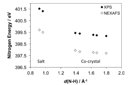
hydrogen bond strengths (including the proposed pKa slide rule).13,41,42 A plot of the XPS N 1s binding energies and NEXAFS N 1s→1π*energies of the nitrogen acceptors of the seven base−acid systems as a function of ΔpKa (Figure 5)
shows the transition from hydrogen-bonding (associated with co-crystal formation) to proton transfer (salt formation) as the
ΔpKa increases through the region between 0 and +3, within which it is difficult to predict whether co-crystal or salt formation takes place.13For the current systems, the salts cover a range ofΔpKafrom 2.48 to 3.87 and the co-crystals−1.17 to
[image:5.625.342.546.66.213.2]0.44 (with 4BPY/phthalic and 4BPY/malonic in the intermediate 0−3 region, Table 1). There is a clear separation between the respective XPS/NEXAFS nitrogen energies for the hydrogen bonded co-crystals and those for the protonated salts
(Figure 5).14,19 Both techniques unambiguously determine whether protonation has occurred, with a nitrogen energy difference of around +2 eV arising from protonation with both techniques (Figure 5).
In addition to identifying salt vs co-crystal, XPS and NEXAFS can provide information on cases associated with disorder. The previously reported XRD crystal structure of 4BPY/buTA45had short NHO hydrogen bonds (2.565(3) Å) and was refined as disordered over two distinct positions, with site occupancy of 0.59 (O−H···N with d(N−H) 1.73 Å) and 0.41 (O···H−N+ with d(N−H) 0.88 Å). The XPS and
NEXAFS in the current study show the presence of only one, sharp peak occurring in the region for CN nitrogen atoms (Figure 2, Table 1). If there was any disorder of the NHO hydrogen bond, so that both CN (O−H···N) and CNH+
(O···H−N+) types of nitrogen environment were present, there would be a second, higher energy signal from CNH+around
401.5 eV for XPS and 399.5 eV for NEXAFS (the ultrafast nature of the electronic transitions (∼10−15−10−16 s) means that every detected event represents a snapshot rather an average over vibrational, conformational, or structural changes, thereby permitting population analysis, even when transient species coexist26,46−48). Moreover, the relative (area) intensity of the two peaks would directly inform the site occupancy of hydrogen over the O−H···N and O···H−N+ states. The
complete absence of this peak here reveals no disorder in the product obtained from the current synthesis and redetermina-tion, but rather a 1:1 co-crystal (confirmed by subsequent single crystal XRD, Figure 3 and Supporting Information) with a short nitrogen acceptor-hydrogen length of 1.39(4) Å for the NHO hydrogen bond (2.559(3) Å). The two structures were collected at the same temperature (100 K), negating the inference of temperature-related disorder. The differences in the products obtained in the previous45and current study are most likely attributable to the difficulty with which this complex is formed: recrystallization over several weeks is required to generate only a small amount of co-crystal product here while the previous crystallization generated disordered material45 (and was not reproducible). It is also notable that the pKa
[image:5.625.97.268.79.477.2]difference for 4BPY/buTA is extremely close to the range (ΔpKa0−3) for which salt/co-crystal is not easily predictable (−0.13, Table 1).
Figure 4.Schematic of photoemission from the nitrogen 1s core level for XPS such that the system is ionized through complete ejection of an electron and promotion from the nitrogen 1s core level to the lowest unoccupied molecular orbital (LUMO) for NEXAFS (N 1s→ 1π*, where the antibonding π* MO is pulled below the ionization potential (IP) by the increased Coulomb potential on creation of a core hole26).
Figure 5. Correlation between the XPS N 1s binding energy and NEXAFS N 1s → 1π* energy and ΔpKa, illustrating that both techniques clearly distinguish between protonated (salt) and unprotonated nitrogen (co-crystal). ΔpKa = pKa (base) − pKa (acid).1,13,43,44
dominate over final-state effects, with only slightly smaller magnitudes in the NEXAFS shifts (Table 1).
Further comparison of the energy shifts between the signals arising from the two types of nitrogen in the salts (CN and CNH+) reveals that the energy shift occurring with protonation is larger for 2BPY/BTA (+2.39 eV) than for 4BPY/sulfo (+1.93 eV) with XPS, and this is also reflected in the NEXAFS shifts (Table 1), visible as a widening separation between the peaks arising from CN and CNH+ nitrogen environments (Figure 2). In the 2BPY/BTA salt, single crystal XRD35 shows the donated hydrogen (from COOH) to be closer to nitrogen than that donated by SO3H in the 4BPY/ sulfo salt (Table 1). This can be viewed as a more complete proton transfer from the acid and leads to decreased electron density (higher effective positive charge) at the protonated nitrogen CNH+ for 2BPY as it interacts more strongly with
[image:6.625.69.297.438.582.2]the hydrogen atom. More energy must therefore be provided to eject or promote a nitrogen core electron, leading to the higher CNH+ energy for 2BPY/BTA compared with 4BPY/sulfo (Figure 6). Interestingly, the energy for the remaining,
non-protonated CN bipyridine nitrogen in the salts is slightly reduced for 2BPY/BTA compared with 4BPY/sulfo in both techniques (perhaps slightly more evident with XPS, Table 1), and this may be related to the nature of the hydrogen bonding (or differing electron density around nitrogen with varying position within the pyridine rings). In the 2,2′-bipyridine system, the non-protonated CN nitrogen is involved in a long, intramolecular bridging CN···H−N+(between the two
2,2′-nitrogen atoms, d(N−H) 2.189 Å),35 as opposed to the shorter intermolecular CN···H−O in 4BPY/sulfo (d(N−H) 1.461 Å, Supporting Information), and variation in hydrogen
hydrogen (or the extent of proton transfer from the acid COOH), this effect appears enough to be reflected in the chemical shifts (Figure 6).
The identification of whether a salt or co-crystal has been formed can be particularly important in terms of regulatory requirements in the pharmaceutical industry,4,17and the nature and nitrogen state stoichiometry of the complex provided by NEXAFS adds it to one of only a few techniques capable of unambiguous assignment.20The level of sensitivity shown for the nitrogen XPS and NEXAFS chemical shifts, through their dependence on the nitrogen−hydrogen bond length and a clear method of detecting the presence of disordered NHO hydrogen bonds, widens the use of these techniques to more crystallographic applications.
■
CONCLUSIONSThe ability of nitrogen NEXAFS to identify Brønsted proton transfer and therefore co-crystals vs salts has been illustrated for bipyridine systems alongside XPS. Additional π* (NEXAFS) and N 1s photoemission (XPS) peaks occur when proton transfer from the acid to nitrogen of the base results in the formation of a salt (CNH+). The intensity ratio between the
peaks associated with the protonated and unprotonated form (CN) provides the stoichiometry between the two. The absence of the CNH+ peak for the 4,4′ -bipyridine/1,2,3,4-butanetetracarboxylic acid system reveals that no disorder across the NHO hydrogen bond is present in the sample synthesized for this study, in contrast to previously obtained disordered material, while the product of crystallization of 4,4-bipyridine with 5-sulfosalicylic acid was identified as a 1:1 salt. The similarity of the chemical shifts for NEXAFS with XPS indicates that the core level (initial-state) tends to dominate in these organic systems. Comparison of the XPS and NEXAFS nitrogen resonances with ΔpKa values clearly shows the
transition from co-crystal to salt, with a shift of around +2 eV for protonation of nitrogen. There is also a monotonic dependence of the chemical shifts on the nitrogen−hydrogen distance (or amount of proton transfer) and the nature of hydrogen bonding at the non-protonated nitrogen. This demonstrates the sensitivity of NEXAFS to proton transfer (Brønsted donation) and provides insight into the local interactions of hydrogen with nitrogen, and more widely, the important influence of the chemical state on theπ*region of NEXAFS.
■
ASSOCIATED CONTENT*
S Supporting InformationX-ray crystallographic informationfiles (CIF) for the new 4′ 4-bipyridine/5-sulfosalicylic acid (4BPY/sulfo) salt and 4,4′ bipyr-idine/1,2,3,4-butanetetracarboxylic acid (4BPY/buTA)
crystal, along with tables summarizing the crystallographic data and powder XRD patterns for the co-crystals and salts. This material is available free of charge via the Internet at http:// pubs.acs.org. Crystallographic information files are also available from the Cambridge Crystallographic Data Centre (CCDC) upon request (http://www.ccdc.cam.ac.uk, CCDC deposition numbers 1038154−1038155).
■
AUTHOR INFORMATIONCorresponding Authors
*Joanna S. Stevens. Tel: +44 (0)161-306-4362. E-mail: joanna. stevens@manchester.ac.uk.
*Sven L. M. Schroeder. Tel: +44 (0)113-343-2401. Fax +44 (0)113-343-2384. E-mail: s.l.m.schroeder@leeds.ac.uk.
Notes
The authors declare no competingfinancial interest.
■
ACKNOWLEDGMENTSWe acknowledge funding from the EPSRC under the previous PhD+ fellowship for J.S.S. and current support for J.S.S. and S.L.M.S. through the Critical Mass Grant EP/1013563/1. NEXAFS beamtime was provided at the U7a beamline of the National Synchrotron Light Source (NSLS), NY. Use of the National Synchrotron Light Source, Brookhaven National Laboratory, was supported by the U.S. Department of Energy, Office of Science, Office of Basic Energy Sciences, under Contract No. DE-AC02-98CH10886. L.K.N. acknowledges
financial support from EPSRC through a DTA Ph.D. studentship and the NowNano Doctoral Training Centre (Grant Number EP/G03737X/1).
■
REFERENCES(1)Handbook of Pharmaceutical Salts: Properties, Selection, and Use; Wiley-VCH: Weinheim, Germany, 2002.
(2) Schultheiss, N.; Newman, A.Cryst. Growth Des.2009,95, 499− 516.
(3) Vishweshwar, P.; McMahon, J. A.; Bis, J. A.; Zaworotko, M. J.J. Pharm. Sci.2006,95, 499−516.
(4) Pharmaceutical Salts and Co-crystals; The Royal Society of Chemistry: Cambridge, U.K., 2012.
(5) Horiuchi, S.; Kumai, R.; Tokura, Y.Chem. Commun.2007, 2321− 2329.
(6) Bolton, O.; Matzger, A. J.Angew. Chem., Int. Ed.2011,50, 8960− 8963.
(7) Jones, C. L.; Wilson, C. C.; Thomas, L. H.CrystEngComm2014, 16, 5849−5858.
(8) Yan, D.; Bučar, D.-K.; Delori, A.; Patel, B.; Lloyd, G. O.; Jones, W.; Duan, X.Chem.Eur. J.2013,19, 8213−8219.
(9) Yan, D.; Fan, G.; Guan, Y.; Meng, Q.; Li, C.; Wang, J.Phys. Chem. Chem. Phys.2013,15, 19845−19852.
(10) Gilli, G.; Gilli, P. The Nature of the Hydrogen Bond; Oxford University Press: Oxford, U.K., 2009.
(11) Wilson, C.Crystallogr. Rev.2007,13, 143−198.
(12) Aakeröy, C. B.; Fasulo, M. E.; Desper, J. Mol. Pharmaceutics 2007,4, 317−322.
(13) Childs, S. L.; Stahly, G. P.; Park, A.Mol. Pharmaceutics2007,4, 323−338.
(14) Stevens, J. S.; Byard, S. J.; Seaton, C. C.; Sadiq, G.; Davey, R. J.; Schroeder, S. L. M.Phys. Chem. Chem. Phys.2014,16, 1150−1160.
(15) Taylor, R.; Kennard, O. Acta Crystallogr., Sect. B: Struct. Sci. 1983,39, 133−138.
(16) Schmidtmann, M.; Coster, P.; Henry, P. F.; Ting, V. P.; Weller, M. T.; Wilson, C. C.CrystEngComm2014,16, 1232−1236.
(17) FDA Guidance for Industry: Regulatory Classification of Pharmaceutical Co-crystals; Food and Drug Administration: Silver
Spring, MD, http://www.fda.gov/downloads/Drugs/Guidances/ UCM281764.pdf, 2013.
(18) Stevens, J. S.; Byard, S. J.; Schroeder, S. L. M.J. Pharm. Sci. 2010,99, 4453−4457.
(19) Stevens, J. S.; Byard, S. J.; Seaton, C. C.; Sadiq, G.; Davey, R. J.; Schroeder, S. L. M.Angew. Chem., Int. Ed.2011,50, 9916−9918.
(20) Stevens, J. S.; Byard, S. J.; Muryn, C. A.; Schroeder, S. L. M.J. Phys. Chem. B2010,114, 13961−13969.
(21) Stevens, J. S.; Byard, S. J.; Schroeder, S. L. M.Cryst. Growth Des. 2010,10, 1435−1442.
(22) Stevens, J. S.; de Luca, A. C.; Pelendritis, M.; Terenghi, G.; Downes, S.; Schroeder, S. L. M.Surf. Interface Anal.2013,45, 1238− 1246.
(23) Bagus, P. S.; Illas, F.; Casanovas, J.Chem. Phys. Lett.1997,272, 168−172.
(24) Tu, G.; Tu, Y.; Vahtras, O.; Agren, H.Chem. Phys. Lett.2009, 468, 294−298.
(25) Garcia-Gil, S.; Arnau, A.; Garcia-Lekue, A.Surf. Sci.2013,613, 102−107.
(26) Stöhr, J.NEXAFS Spectroscopy; Springer-Verlag: Berlin, 1992. (27) Lehmann, J.; Solomon, D.; Brandes, J.; Fleckenstein, H.; Jacobson, C.; Thieme, J., Synchrotron-Based Near-Edge X-Ray Spectroscopy of Natural Organic Matter in Soils and Sediments. In Biophysico-Chemical Processes Involving Natural Nonliving Organic Matter in Environmental Systems; Senesi, N., Xing, B., Huang, P. M., Eds.; John Wiley & Sons, Inc.: Hoboken, NJ, 2009; Chapter 17, pp 741−748.
(28) Solomon, D.; Lehmann, J.; Kinyangi, J.; Liang, B.; Heymann, K.; Dathe, L.; Hanley, K.; Wirick, S.; Jacobsen, C.Soil Sci. Soc. Am. J.2009, 73, 1817−1830.
(29) Leinweber, P.; Kruse, J.; Walley, F. L.; Gillespie, A.; Eckhardt, K.-U.; Blyth, R. I. R.; Regier, T.J. Synchrotron Radiat.2007,14, 500− 511.
(30) Vairavamurthy, A.; Wang, S. Environ. Sci. Technol. 2002,36, 3050−3056.
(31) Cooney, R. R.; Urquhart, S. G. J. Phys. Chem. B2004, 108, 18185−18191.
(32) Urquhart, S. G.; Ade, H.J. Phys. Chem. B 2002,106, 8531− 8538.
(33) Stevens, J. S.; Seabourne, C. R.; Jaye, C.; Fischer, D. A.; Scott, A. J.; Schroeder, S. L. M.J. Phys. Chem. B2014,118, 12121−12128.
(34) Booth, A. M.; Braun, S.; Lonsborough, T.; Purton, J.; Patel, S.; Schroeder, S. L. M.AIP Conf. Proc.2007,882, 325−327.
(35) Mrvos-Sermek, D.; Popovic, Z.; Matkovic-Calogovic, D.Acta Crystallogr., Sect. C: Cryst. Struct. Commun.1996,52, 2538−2541.
(36) Tomura, M.; Yamashita, Y.Chem. Lett.2001,30, 532−533. (37) Pedireddi, V. R.; Chatterjee, S.; Ranganathan, A.; Rao, C. N. R. Tetrahedron1998,54, 9457−9474.
(38) Stevens, J. S.; Schroeder, S. L. M.Surf. Interface Anal.2009,41, 453−462.
(39) Fairley, N.; Carrick, A.The Casa Cookbook - Part 1: Recipes for XPS Data Processing; Acolyte Science: Knutsford, Cheshire, 2005.
(40) The XPS of Polymers Database; Surface Spectra Ltd.: Manchester, 2000.
(41) Gilli, P.; Pretto, L.; Gilli, G.J. Mol. Struct.2007,844−845, 328− 339.
(42) Gilli, P.; Pretto, L.; Bertolasi, V.; Gilli, G.Acc. Chem. Res.2009, 42, 33−44.
(43) Experimental pKavalues were obtained using the ACD/I-Lab Web service:ACD/pKa DB 12.0; Advanced Chemistry Development, Inc. (ACD/Labs): Toronto, 2010.
(44) Predicted pKa values were obtained using: ACD/PhysChem. Suite v12.0; Advanced Chemistry Development, Inc. (ACD/Labs): Toronto, 2010.
(45) Najafpour, M. M.; Holynska, M.; Lis, T.Acta Crystallogr., Sect. E: Struct. Rep. Online2008,64, o985.
(46) Sawatzky, G. A.; Antonides, E.J. Phys., Colloq.1976,37, C4-117−C4-123.