This is a repository copy of
PET iterative reconstruction incorporating an efficient positron
range correction method
.
White Rose Research Online URL for this paper:
http://eprints.whiterose.ac.uk/103074/
Version: Accepted Version
Article:
Bertolli, O, Eleftheriou, A, Cecchetti, M et al. (3 more authors) (2016) PET iterative
reconstruction incorporating an efficient positron range correction method. Physica
Medica, 32 (2). pp. 323-330. ISSN 1120-1797
https://doi.org/10.1016/j.ejmp.2015.11.005
© 2016, Elsevier. Licensed under the Creative Commons
Attribution-NonCommercial-NoDerivatives 4.0 International
http://creativecommons.org/licenses/by-nc-nd/4.0/
Reuse
Unless indicated otherwise, fulltext items are protected by copyright with all rights reserved. The copyright exception in section 29 of the Copyright, Designs and Patents Act 1988 allows the making of a single copy solely for the purpose of non-commercial research or private study within the limits of fair dealing. The publisher or other rights-holder may allow further reproduction and re-use of this version - refer to the White Rose Research Online record for this item. Where records identify the publisher as the copyright holder, users can verify any specific terms of use on the publisher’s website.
Takedown
If you consider content in White Rose Research Online to be in breach of UK law, please notify us by
PET Iterative Reconstruction Incorporating An
Efficient Positron Range Correction Method
Ottavia Bertollia,b,∗, Afroditi Eleftherioua,c, Matteo Cecchettib, Niccol`o
Camarlinghib, Nicola Belcarib, Charalampos Tsoumpasa
aDivision of Biomedical Imaging, University of Leeds, United Kingdom b
University of Pisa and INFN Pisa, Italy
c
Department of Physics, National and Kapodistrian University of Athens, Greece
Abstract
Positron range is one of the main physical effects limiting the spatial resolution
of Positron Emission Tomography (PET) images. If positrons travel inside a
magnetic field, for instance inside a nuclear Magnetic Resonance (MR)
tomo-graph, the mean range will be smaller but still significant. In this investigation
we examined a method to correct for the positron range effect in iterative image
reconstruction by including tissue-specific kernels in the forward projection
op-eration. The correction method was implemented within STIR library (Software
for Tomographic Image Reconstruction).
In order to obtain the positron annihilation distribution of various
radioac-tive isotopes in water and lung tissue, simulations were performed with the
Monte Carlo package GATE [1] simulating different magnetic field intensities
(0T, 3 T, 9.5 T and 11T) along the axial scanner direction. The positron range
kernels were obtained for68Ga in water and lung tissue for 0 T and 3 T magnetic
field voxellizing the annihilation coordinates into a three-dimensional matrix.
The proposed method was evaluated using simulations of material-variant and
material-invariant positron range corrections for the HYPERImage preclinical
PET-MR scanner. The use of the correction resulted in sharper active region
boundary definition, albeit with noise enhancement, and in the recovery of the
∗Corresponding author
true activity mean value of the hot regions. Moreover, in the case where a
mag-netic field is present, the correction accounts for the non-isotropy of the positron
range effect, resulting in the recovery of resolution along the axial plane.
Keywords: Positron range, iterative reconstruction, PET, PET/MR, STIR
1. Introduction
After its emission, the positron travels a finite distance interacting with the
surrounding media. The length of its path depends on the energy of the positron,
that has a characteristic emission spectrum dependent on the radiotracer. The
photon-producing event therefore occurs outside the radioactive nucleus and the
actual position of the radiotracer is different from the annihilation position. In
the reconstruction process it is assumed, with sufficient approximation, that the
radiotracer resides somewhere along the Line Of Response (LOR) defined by the
two crystals detecting the annihilation photons. The spatial blurring caused by
the positron range, however, limits the validity of the LOR modeling, affecting
both image resolution and accuracy.
When inside a magnetic field, a positron moving with velocity~vexperiences
the Lorentz force:
~
F =q~v×B~
which only acts on the component of the velocity perpendicular to the direction
of the magnetic field B~ . The positron experiences a centripetal force F~B =
qvBsinθ that will make it follow a helicoidal trajectory around the direction
ofB~, confining its path in the plane perpendicular to it. Simultaneous hybrid
imaging using nuclear Magnetic Resonance tomography and Positron Emission
Tomography (PET-MR) is expected to substantially improve the PET image
resolution in the plane perpendicular to the static magnetic field of the scanner,
due to the confined positron trajectory and particularly when inside a very
strong magnetic field [2, 3, 4]. However, this is not the case for the resolution
non-Therefore it is decisive to consider approaches to reduce the positron range effect
for both PET and PET-MR systems.
1.1. Background
The blurring due to the positron range effect can be approached in different
ways [5]. One common way is to describe the detected data as the ideal integral
of activity (i.e. the projections) convolved with a function that represents the
positron range, therefore in the case of analytic reconstruction methods the
range effect can be removed by dividing the Fourier transform of the measured
projection data by the Fourier transform of the positron range function [6].
Within the iterative reconstruction framework the correction can be achieved by
incorporating the positron range probability distribution as part of the forward
projection matrix [7] with two different approaches: (A) isotope-specific and
spatially variant point spread function (PSF) in resolution modeling [8, 3]; or
(B) by convolving the object with a positron range based kernel during the
forward projection operation [9, 10, 11].
1.2. Aim
In this work we implement a method to take into account the positron range
effect in iterative image reconstruction following the efficient approach proposed
by Cal-Gonzalez [9] and Kraus [10]. The presented correction method is
suit-able for every kind of scanner, although its practical utility would be more
relevant for pre-clinical systems or organ-specific systems, given their
high-est spatial resolution. The implementation is compatible with and without
the presence of a magnetic field and it is validated for material-variant and
material-invariant positron range corrections [11]. The correction method is
implemented within STIR (Software for Tomographic Image Reconstruction,
http://stir.sourceforge.net) that is one of the most common libraries for
PET image reconstruction, such that it becomes available to several other
2. Materials and Methods
2.1. GATE simulations
GATE simulations were performed to obtain the positron range annihilation
distribution in water and lung tissue. An approximate point source (spherical
with radius 0.01 mm) located at the center of a spherical phantom of 5 cm
radius was simulated. Six different positron emitters were included in the study:
11C, 13N, 15O, 18F,68Ga and 82Rb. The simulations were performed without
and with a static and homogeneous magnetic field set along the PET scanner
model’s axial direction for three different field strengths: 3 T, 9.5 T and 11
T. The Geant4 low energy package [12] was used for electromagnetic processes
simulation.
Approximately 105 annihilation events were simulated per configuration and
the annihilation coordinates were obtained from the Geant4 output [13, 14].
The positron end point coordinates were stored and used to create the blurring
kernels, as described in the following paragraphs.
2.2. Correction method
In order to consider the degradation of the PET images due to the positron
range effect a matching spatial blurring is applied on the object during the
forward projection step:
xnext j = xcurrent j P i ai,j X i ai,j pi P k
ai,kx˜currentk
(1)
˜
xcurrent
k =x
current k ∗ρ=
P
hx current k−h ρh
P
hρh
(2)
where ˜xcurrent
k is the current image estimate blurred via convolution with a
kernel ρ, pi are the projections along LOR i and ai,j represents the system
matrix element for voxelj and LORi.
The version of STIR in which our method has been implemented is the 3.0.
• OSMAPOSLReconstruction.h: where a pointer to the so-calledpre update filter
is added;
• OSMAPOSLReconstruction.cxx: where a filtering of the current image
es-timate is performed with the uploaded kernel inside theupdate estimate
method, when a kernel is provided.
The kernel is defined inside the parameter file that has to be given to the
OSMAPOSLexecutable, that is an implementation of the OSEM algorithm
(Or-dered Subset Expectation Maximization [15]) and the type of filtering has to be
set toNonseparable Convolution Using Real DFT Image Filter.
2.3. Blurring kernels
The blurring kernel ρ is a three dimensional matrix whose elements have
values according to the number of events within the corresponding volume
nor-malized by the total number of events. To obtain the kernels the annihilation
coordinates were voxellized into a 3D matrix with element size equal to the voxel
size of the reconstructed image and then normalized to sum up to 1. The size
of each kernel depends on the isotope and the characteristics of the images
pro-vided by the scanner in use, so that the kernel covers a volume corresponding to
twice the calculated mean range departing from its center in all directions (with
these dimensions approximately 97 % of the annihilation events are included
in the kernel volume). For the purpose of this study, kernels were obtained for
the68Ga isotope in water and lung tissue, with no magnetic field and with a 3
T magnetic field, equal to the magnetic field of the most common commercial
PET-MR systems is use (the magnetic field and tissue dependent correction
kernels that have been generated could be shared on request with others STIR
users). The68Ga can be considered representative here due to its average energy
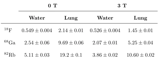
Table 1: Positron mean ranges (mm) for18F
(mean positron energy 250keV),68Ga (mean
positron energy 783 keV) and82Rb
(mean positron energy 1475 keV).
0 T 3 T
Water Lung Water Lung
18F 0.549±0.004 2.14±0.01 0.526±0.004 1.45±0.01
68Ga 2.54±0.06 9.69±0.06 2.07±0.01 5.25±0.04
82Rb 5.11±0.03 19.2±0.1 3.86±0.02 10.60±0.02
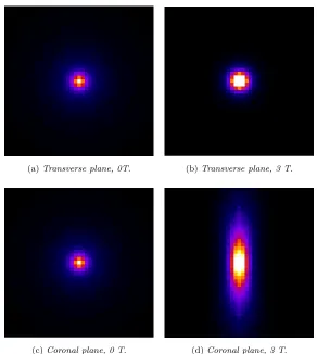
The kernels dimensions for68Ga in water are 21x21x21 and 41x41x41 for 0T
and 3T respectively, and in lung tissue 17x17x17 and 31x31x31 for 0T and 3T
respectively. Two-dimensional planes of the lung tissue kernel are illustrated in
(a)Transverse plane, 0T. (b)Transverse plane, 3 T.
[image:8.612.159.451.129.455.2](c)Coronal plane, 0 T. (d)Coronal plane, 3 T.
Figure 1: 2D planes of the kernels relative to68
Ga in lung tissue.
2.4. Method evaluation
Two versions of the proposed method were implemented:
• material-invariant correction: it only uses one kernel for the blurring of
the forward projected image, as if the imaged object were homogeneous;
• material-variant correction: the kernel is chosen depending on the
un-derlying voxel tissue, in order to consider the positron range effect with
surrounding material of different types. In this case a mask image is
2.4.1. Phantoms
The proposed method was evaluated using simulated data generated using
STIR software [16] of acquisitions of the HYPERImage preclinical PET/MR
scanner (diameter: 20.8 cm, length: 3.1 cm) [17].
Thematerial-invariant correction was applied on simulated acquisitions of
rectangular parallelepiped homogeneous phantoms made of water or lung
tis-sue containing two spherical hot spots of 3 mm and 5 mm radius respectively.
Simulations were done with and without additional Poisson noise. The activity
contrast ratio between the hot spots and the simulated background tissue was
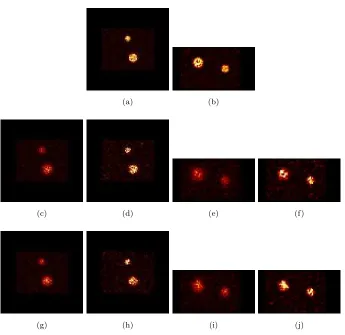
set to 11:1. The reconstructed images of the water phantom are shown in Figure
4. Lung and water phantoms are of equal shape.
Thematerial-variant correction was applied on a phantom composed of
wa-ter and lung tissue regions (activity contrast value respectively equal to 1 and
0.5). The lung tissue region, contained inside a rectangular parallelepiped of
water, is of cylindrical shape with radius 10 mm and height 17 mm and
con-tains a spherical hot spot of radius 5 mm (contrast value equal to 10). The
required mask image consisted in our study of a simulated transmission image,
with values equal to the Hounsfield Units relative to water and lung tissue.
In this experiment we investigated only the multi-material positron range
ef-fect therefore no Poisson noise, the efef-fect of which has been evaluated with the
material-invariant correction, was added to the ideal projection data.
The positron range effect was emulated by convolving the phantoms with
the 68Ga kernels for lung tissue or water, depending on the phantom
mate-rial, with or without the 3 T magnetic field. No random or scattered events
have been added to the simulated projections because at this stage we aimed
at highlighting the effects of the positron range correction within the iterative
reconstruction. The reconstructions were performed with and without
introduc-ing the correspondintroduc-ing filterintroduc-ing to the forward projected image with the OSEM
algorithm, with 7 subsets, 10 iterations and 0.7 mm isotropic voxel size.
images reconstructed with and without the use of the positron range correction
were compared to the reconstructed image of the simulated phantom, which has
no blurring related to the positron range effect.
3. Results
3.1. GATE simulations
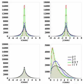
Figure 2 shows one-dimensional histograms illustrating the number of positron
annihilation events with respect to the distance from the origin, where the
ra-dioactive decay occurs. They show the effect of different magnetic field strengths
on the annihilation coordinates x,y,z and on the distribution of the range for
68Ga.
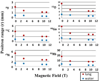
Figure 3 shows the mean positron range of the positron emitters as a function
of the magnetic field in all simulated media.
3.2. Impact of noise and material-invariant correction for homogeneous
phan-toms
The images resulted from the material-invariant kernel reconstruction with
the water phantom are shown in Figure 4. It can be seen that when the
correc-tion is applied there is improved recovery of the original radioisotope activity.
Axial and transversal line profiles drawn through the hot spots in the
re-constructed images (width: 2 voxels, centered in correspondence to the center
of the feature) illustrate that the use of the positron range correction yields
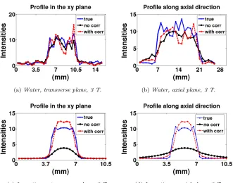
sharper boundary definition, albeit with noise enhancement. Figure 5 shows
the profiles for the water phantom with simulated Poisson noise and for the
lung tissue phantom without simulated Poisson noise. The line-profile referred
to as “true” is related to the standard OSEM reconstruction of the simulated
phantom, with no positron blurring, “no corr” stands for the standard OSEM
reconstruction (i.e. with no correction for positron range blurring) and “with
corr” is related to the positron range corrected reconstruction.
The over-shoots noticeable in the line-profiles of the “true” data are caused
Figure 2: Histograms of the positron range and of the annihilation coordinates for68 Ga in
water for different magnetic field strengths[14].
we are performing resolution modeling, correcting for the contribution of the
positron range to the overall degradation of the image), which is known to
pro-duce Gibbs artifacts at sharp intensity transitions inside the object. Typical
approaches to suppress these artifacts include post reconstruction smoothing
with a low-pass filtering or the use of penalties in a penalized likelihood
recon-struction algorithm [18, 19].
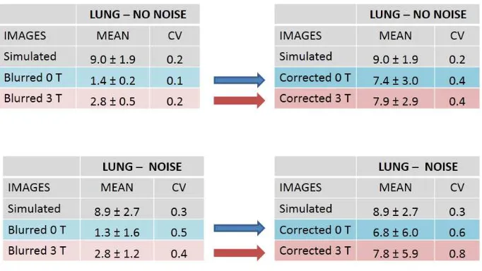
The values of the meanµand standard deviationσwere calculated relative
to the volumes corresponding to the simulated spherical hot spots. The related
Figure 3: Mean positron range versus magnetic field, for all simulated positron emitters, in
water and lung tissue phantoms [14].
variability of the values in relation to the mean (i.e. the standard deviation of
the selected region of interest over its mean) was also evaluated. The obtained
values are shown in Table 2 and Table 3.
3.3. Material-invariant versus material-variant correction for multi-material
phan-tom
In the analysis of the multi-material phantom, i.e. the water rectangular
parallelepiped with a cylindrical area made of lung tissue inside, in order to
compare the difference between the two methods the blurred images have been
reconstructed both with the material-invariant correction, using the kernel
re-lated to water, and with the material-variant correction, using in this case the
(a) (b)
(c) (d) (e) (f)
[image:13.612.135.480.117.452.2](g) (h) (i) (j)
Figure 4: Transverse (on the left) and sagittal plane (on the right) of the water phantom with
Poisson noise. (a) and (b): original phantom. (c) and (e): 0T, no correction. (d) and (f): 0T
with correction. (g) and (i): 3 T, no correction. (h),(j): 3 T with correction.
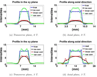
Line profiles were traced through the hot spot in the reconstructed images
(as previously described) and are illustrated in Figure 6. They highlight that
in both instances (displayed in the plot as “invar corr” and “var corr”) the use
of the positron range correction yields sharper boundary definition, although
resulting in edge artifacts. The mean, standard deviation and CV relative to
(a)Water, transverse plane, 3 T. (b)Water, axial plane, 3 T.
[image:14.612.138.469.130.390.2](c)Lung tissue, transverse plane, 3 T. (d)Lung tissue, axial plane, 3 T.
Figure 5: Line profiles: blue is the true phantom, black and red are relative to the blurred
non-corrected and corrected phantom respectively. (a) and (b): big spot in water phantom
with simulated noise. (c) and (d): small spot in the lung tissue phantom without simulated
noise.
4. Discussion
Analyzing the simulation results with respect to increasing magnetic field
strengths, it can be observed that there is a significant decrease in the mean
distance traveled by the positron in the x and y directions, as it can be seen
in Figure 2. In the absence of magnetic field the positron interactions result in
random direction changes of its path, whereas when a magnetic field is applied
the available direction change is reduced. This is as a consequence of the
con-finement of the trajectory in the transverse plane, and it is demonstrated by
the reduced distancer traveled by the positron as the magnetic field strength
Table 2: Mean and CV relative to the small hot spot volume in the case of the water
homo-geneous phantom.
Table 3: Mean and CV relative to the small hot spot volume in the case of the lung tissue
homogeneous phantom.
The consequence of the positron trajectory confinement is also visible from
the obtained kernels for68Ga: the dimensions in thexandydirections decrease
in the 3 T case, it can be observed that the distribution of the annihilation
[image:15.612.136.477.398.590.2](a)Transverse plane, 0 T. (b)Axial plane, 0 T.
[image:16.612.139.469.133.394.2](c)Transverse plane, 3 T. (d)Axial plane, 3 T.
Figure 6: Line profiles through the hot spot inside the lung region that is surrounded by
water (multi-material phantom). Top row is for 0 T and bottom row for 3 T. Blue line (true):
simulated phantom. Black line (no corr): non corrected blurred phantom. Red line (invar
corr): material-invariant correction. Green line (var corr): material-variant correction.
Table 4: Mean and CV values relative to the hot spot inside the lung region in the water and
lung tissue phantom.
and 1b 3T. In the 3T case, see Figure 1d, a condensing can be noticed in the
[image:16.612.135.479.513.622.2]due to the helicoidal motion of the positrons around the magnetic field more
annihilations are deposited along the direction of the field, as explained in [3].
No variation is observed in the axial direction profile, as it is showed in
Figure 2, as opposed to what has been reported in [20] where the mean of the
positron range in the z direction it was said to increase. From Figure 3 it can
also be observed that a trend common to all simulated materials is that the
reduction effect on the positron range is more dominant for lower (0T-3T) than
for higher magnetic field strengths (9.5T-11T). This is in agreement with what
has been reported in [3], where it is shown that the degree of reduction of the
path traveled by the positron is proportional to the positron range of the isotope
and to the magnetic field strength up to around 7T, where the extent of the
reduction saturates.
As a result of the application of material-invariant correction, the plots in
Figure 5 show that the filtering of the forward projected image yields sharper
boundary definition, albeit with noise enhancement, and the values in Tables
2 and 3 demonstrate that the correction results in the recovery of the activity
mean value compared to the simulated phantom. Without the magnetic field,
the achieved activity recovery ranges from 55% to 91% and from 54% to 90%
for the small hot spot without and with noise; with 3 T magnetic field it ranges
from 62% to 93% and from 61% to 91% for the small spot without and with
noise, respectively.
In the case of a multi-material object Figure 6 shows that both the
correc-tions (material-invariant and material-variant) yield sharper boundary
defini-tion, although resulting in edge artifacts. Moreover, when the material-variant
correction is applied, the recovered radioactivity mean value increases
consid-erably, as it can be observed by the values in Table 4, e.g. in the presence of
magnetic field it ranges from the 55% in the case of the material-invariant
cor-rection to 92%. Nevertheless our implementation does not take into account the
modification of the pathway of positrons that annihilate in a different medium
ap-the variation of ap-the positron range that might be caused if ap-the particle crossed
a different medium before annihilating. Various approaches to tackle this issue
have been presented in [7],[10],[21], [22].
Although this method implies the application of a non-convertible
projec-tor/backprojector pair (as the blurring is effective in the forward projection
only), it has been proven to be effective in [9] and [10]. Furthermore this
ap-proach allows to avoid a required re-evaluation of the system matrix for each
involved isotope given that the blurring with the chosen kernel is applied to the
image before the forward projection. The effect of including positron blurring in
both forward and backward projection operations is investigated in [23], where
it is shown that the use of positron blurring during backprojection has a
smooth-ing effect on the reconstructed image leadsmooth-ing to a delayed convergence, requirsmooth-ing
a large number of iterations to reach a comparable detail level. Furthermore
in [24] it is shown that MLEM algorithms demonstrate first a short convergent
trend but then deviates from the desired solution, independent from the
pro-jector/backprojector pair matching. The authors concluded that the concrete
choice for the backprojector may not be a very critical factor in a practical
image reconstruction problem. They suggest to choose a (maybe even
inconsis-tent) projector/backprojector pair, suited for rapid computation, supported by
further regularization methods to guide or stop the iteration process.
In the present study we only demonstrated the described method for68Ga
in water and lung tissue with no magnetic field and in the presence of a 3 T
magnetic field. However the same procedure can easily be carried out for other
radioisotopes, materials and for different magnetic field strengths, potentially
providing good results even in the presence of very strong magnetic fields, in
which case the anisotropy of the positron range distribution may affect the image
resolution in the axial direction.
With respect to the noise enhancement it must be underlined that in our
study no regularization was applied. To address this issue and simultaneously
the object dependency of the resolution properties when inter-iteration
aim of obtaining images with nearly object independent and uni-directional
resolution. A further development of our investigation would be the
considera-tion of simultaneous multi-isotope acquisiconsidera-tions, exploiting for example multiple
isotope-specific kernels.
In the future we plan to extend this method to take into account the positron
range behavior in correspondence to tissue borders and to incorporate the new
developments in STIR library.
5. Conclusions
We implemented a technique that accounts for the positron range effect in
iterative reconstruction in STIR library. The method is independent of the
ex-istence of a magnetic field once the blurring kernels have been chosen for the
correct combination of isotope, material and magnetic field strength. The
evalu-ation of the proposed correction method was performed on simulated phantoms,
in which a filtering process with the calculated kernels was used to emulate the
positron range blurring. The analysis on the reconstructed images shows that
the positron range correction is able to substantially restore the mean activity
values relative to high contrast regions and sharper boundaries, albeit resulting
in noise enhancement. When a 3 T magnetic field is present, the application
of the positron range correction can successfully correct for the non-isotropic
resolution along the axial plane. With regard to the use of a material-variant
kernel, the method performance in correspondence to the edges needs further
investigation.
Acknowledgment
We would like to thank Professor Alberto Del Guerra, Dr Georgios Loudos,
Dr Efstathios Stiliaris and Mr Georgios Soultanidis. This project was completed
with travel support from the EU COST Action (TD1007,
References
[1] Jan S, Santin G, Strul D, Staelens S, Assi K, et al., GATE: a simulation
toolkit for PET and SPECT, Phys Med Biol 2004; 49(19):4543.
[2] Iida H, Kanno I, Miura S, Murakami M, Takahashi K, Uemura K., A
Sim-ulation Study of a Method to Reduce Positron Annihilation Spread
Distri-butions Using a Strong Magnetic Field in Positron Emission Tomography,
IEEE Trans Nucl Sci 1986;33(1):597-600.
[3] Cheng JC, Boellaard R, Lafores, R.,Evaluation of the Effect of
Mag-netic Field on PET Spatial Resolution and Contrast Recovery Using
Clinical PET Scanners and EGSnrc Simulations, IEEE Trans Nucl Sci
2015;62(1):101-110.
[4] Shah NJ, Herzog H, Weirich C, Tellmann L, Kaffanke J, Caldeira L, et.
al.,Effects of Magnetic Fields of up to 9.4 T on Resolution and Contrast
of PET Images as Measured with an MR-BrainPET PLos ONE 2014;
9(4):e95250.
[5] Rahmim A, Qi J, Sossi V., Resolution modeling in PET imaging: Theory,
practice, benefits, and pitfalls, Med Phys 2013;40(6):064301.
[6] Haber SF, Derenzo SE, Uber D., Application of mathematical removal of
positron range blurring in positron emission tomography, IEEE Tran Nucl
Sci 1990;37(3):1293-1299.
[7] Bing B, Ruangma A, Laforest R, Tai YC, Leahy RM.,Positron range
mod-eling for statistical PET image reconstruction, IEEE NSS Conf Record
2003;4:2501-2505.
[8] Kotasidis FA, Angelis GI, Anton-Rodriguez J, Matthews JC, Reader AJ,
Zaidi H.,Isotope specific resolution recovery image reconstruction in high
[9] Cal-Gonzalez J, Perez-Liva M, Lopez-Herraiz J, Vaquero JJ, Desco M,
Udias J., Tissue-dependent and spatially-variant positron range correction
in 3D PET, IEEE Trans Med Imag 2015;34(11):2394-2403.
[10] Kraus R, Delso G, Ziegler SI., Simulation Study of Tissue-Specific Positron
Range Correction for the New Biograph mMR Whole-Body PET/MR
Sys-tem, IEEE Trans Nucl Sci 2012;59(5):1900-1909.
[11] Bertolli O, Cecchetti M, Camarlinghi N, Eleftheriou A, Belcari N,
Tsoumpas C., Iterative reconstruction incorporating positron range
cor-rection within STIR framework, EJNMMI Physics 2014;1(1):1-2.
[12] Chauvie S, Guatelli S, Ivanchenko V, Longo F, Mantero A, Mascialino B,
et al.. Geant4 low energy electromagnetic physics, IEEE NSS/MIC Conf
Record 2004;3:1881-1885.
[13] Agostinelli S, Allison J, Amako KA, Apostolakis J, Araujo H, Arce P,et al.,
GEANT4 a simulation toolkit, Nucl Instrum Methods A 2003;506(3):250–
303.
[14] Eleftheriou A, Tsoumpas C, Bertolli O, Stiliaris E., Effect of the
mag-netic field on positron range using GATE for PET-MR, EJNMMI Physics,
2014;1(1):1-2.
[15] Hudson HM, Larkin RS., Accelerated image reconstruction using ordered
subsets of projection data, IEEE Trans Med Imag 1994;13(4):601-609.
[16] Thielemans K, Tsoumpas C, Mustafovic S, Beisel T, Aguiar P, Dikaios N,
et al.,STIR: software for tomographic image reconstruction release 2, Phys
Med Biol 2012;57(4):867.
[17] Mackewn JE, Lerche CW, Sunassee K, de Rosales R, Phinikaridou A,
Sa-lomon A, et al., PET performance evaluation of a pre-clinical SiPM based
MR-compatible PET scanner, IEEE NSS/MIC Conf Record, 2012;
[18] Tong S, Alessio AM, Thielemans K, Stearns C., Ross S, Kinahan PE.,
Prop-erties and mitigation of edge artifacts in PSF-based PET reconstruction,
IEEE Trans Nucl Sci 2011;58(5):2264–2275.
[19] Nuyts J., Unconstrained image reconstruction with resolution modelling
does not have a unique solution, EJNMMI Physics, 2014;1(1):1-7.
[20] Soultanidis G, Karakatsanis N, Nikiforidis G, Loudos G., Study of the effect
of magnetic field in positron range using GATE simulation toolkit, Journal
of Physics: Conference Series, 2011;317(1):012021.
[21] Alessio AM, MacDonald L., Spatially variant positron range modeling
de-rived from CT for PET image reconstruction, IEEE NSS/MIC Conf Record
2008;3637–3640.
[22] Rahmim A, Tang J, Lodge MA, Lashkari S, Ay MR, Bengel FM.,
Reso-lution modeled PET image reconstruction incorporating space-variance of
positron range: Rubidium-82 cardiac PET imaging, IEEE NSS/MIC. Conf
Record 2008;3643–3650.
[23] Cal-Gonzalez J, Herraiz JL, Espaa S, Vicente E, Herranz E, Desco M,et
al., Study of CT-based positron range correction in high resolution 3D PET
imaging, Nucl Instrum Methods A 2011;648(1):S172 - S175.
[24] Zeng GL, Gullberg GT., Unmatched projector/backprojector pairs in an
iterative reconstruction algorithm, IEEE Trans Med Imag