A STUDY ON MUCIN HISTOCHEMISTRY AND p63 EXPRESSION IN BENIGN AND MALIGNANT PROSTATIC LESIONS
Dissertation submitted in
Partial fulfillment of the requirements for the degree of
M.D. PATHOLOGY BRANCH- III
INSTITUTE OF PATHOLOGY MADRAS MEDICAL COLLEGE
CHENNAI- 600003
THE TAMILNADU DR M.G.R. MEDICAL UNIVERSITY CHENNAI
CERTIFICATE
This is to certify that this Dissertation entitled “A STUDY ON
MUCIN HISTOCHEMISTRY AND p63 EXPRESSION IN BENIGN AND MALIGNANT PROSTATIC LESIONS” is the bonafide original work of DR.N.KIRUTHIKA, in partial fulfillment of the requirement
for M.D., (Branch III) in Pathology examination of the Tamilnadu Dr.M.G.R
Medical University to be held in May 2019.
Dr.K.INDUMATHI, M.D., DCP Prof.Dr.PAPPATHI, S., M.D., DCH Assistant Professor of Pathology Professor of Pathology
Department of Pathology, Department of Pathology,
Institute of child health, Institute of child health,
Madras Medical College, Madras Medical College,
Chennai- 600003 Chennai- 600003
Prof.Dr.Bharathi Vidhya Jayanthi, M.D., Prof.Dr.R.JEYANTHI, M.D., Director & Professor of Pathology, Dean,
Institute of Pathology, Madras Medical College
Madras Medical College Rajiv Gandhi Government Hospital
DECLARATION
I, Dr.N.KIRUTHIKA, solemnly declare that the dissertation entitled
“A STUDY ON MUCIN HISTOCHEMISTRY AND P63 EXPRESSION IN BENIGN AND MALIGNANT PROSTATIC LESIONS” is the bonafide work done by me at the Institute of Pathology, Madras Medical College under
the expert guidance and supervision of Prof.Dr.Pappathi.S, M.D., DCH,
Professor of Pathology and Dr.K.Indumathi, M.D., DCP, Assistant professor
of Pathology, Institute Of Pathology, Madras Medical College. The dissertation
is submitted to the Tamilnadu Dr.M.G.R Medical University towards partial
fulfillment of requirement for the award of M.D., Degree (Branch III) in
Pathology.
Place : Chennai
ACKNOWLEDGMENT
I express my sincere thanks to Prof.Dr.R.JEYANTHI, M.D., Dean,
Madras Medical College and Rajiv Gandhi Government General Hospital, for
permitting me to utilize the facilities of the Institution.
I take the opportunity to express my gratitude to Prof.Dr.BHARATHI
VIDHYA JAYANTHI, M.D., Director and Professor, Institute of Pathology, Madras Medical College, Chennai for her keen interest, constant
encouragement and valuable suggestions throughout the study.
I am extremely thankful to Dr.PAPPATHI.S, M.D., DCH, Professor of
Pathology and Dr.K.INDUMATHI, M.D.,DCP, Assistant professor of
Pathology, Department Of Pathology, Institute of child health, Madras
Medical College, for their valuable suggestions, constant support, advice and
encouragements throughout the study.
I am truly thankful to Prof.Dr.Sudha Venkatesh M.D., Prof.Dr.Geetha Devdas, M.D.,DCP., Prof.Dr.Padmavathi M.D.DGO, Prof.Dr.Ramamoorthi M.D., Prof.Dr.Rama M.D., Prof.Dr.M.P.Kanchana M.D., Prof. Dr.Rajavelu Indira M.D., Prof.Dr.Selvambigai M.D., for their valuable suggestions and encouragement throughout the study.
I express my heartfelt sincere thanks to all my Assistant Professors for
I am thankful to my colleagues, friends, technicians and staff of the
Institute of Pathology, Madras Medical College, Chennai for all their help and
support they extended for the successful completion of this dissertation.
My sincere thanks also go to all the patients and their families who were
co-operative during the course of this study.
Last but not the least, I am grateful to my family members and friends or
PLAGIARISM CERIFICATE
This is to certify that this dissertation work titled “A STUDY ON
MUCIN HISTOCHENISTRY AND p63 EXPRESSION IN BENIGN AND MALIGNANT PROSTATIC LESIONS” of the candidate Dr.N.KIRUTHIKA with registration Number 201513004 for the award of M.D PATHOLOGY (Branch-III). I personally verified the urkund.com website for the purpose of Plagiarism Check. I found that the uploaded thesis
file contains from introduction to conclusion and result shows 5 percentage of
plagiarism in the dissertation.
ABBREVIATIONS
AAH - atypical adenomatous hyperplasia PIN - prostatic intraepithelial neoplasia PAS - periodic acid schiff
BPH - benign prostatic hyperplasia LUTS - lower urinary tract symptoms PBCR - population based cancer registries AAR - age adjusted incidence rates
MAPC - mean annual percentage change EAPC - estimated annual percentage change OCP - organochlorine pesticides
HCH - hexachlorocyclohexane AR - androgen receptor HSP - heat shock protein DHT - dihydroxytestosterone
HRPC - hormone resistance in prostate cancer BCH - basal cell hyperplasia
CCCH - clear cell cribriform hyperplasia DRE - digital rectal examination
CONTENTS
S NO TITLE PAGE NUMBER
1 INTRODUCTION 1
2 AIMS AND OBJECTIVES 3
3 REVIEW OF LITERATURE 4
4 MATERIALS AND METHOD 63
5 OBSERVATION AND RESULTS 69
6 DISCUSSION 79
7 SUMMARY 85
8 CONCLUSION 86
9 BIBLIOGRAPHY
10 ANNEXURES
1
INTRODUCTION
Benign prostatic hyperplasia and prostatic carcinoma are the two most
common diseases involving men in older age. The prostatic cancer is the
second most frequently diagnosed cancer in men , sixth most common cause of
cancer death in males worldwide and fifth most common cancer overall[1]. As
prostatic cancer is a disease of older age,by 2030,the proportion of people
above 65 years will increase from 12.4% to 19.6% , the number of prostate
cancer cases will quadruple.
According to world cancer stat facts, the estimated new cases in 2018 is
about 164,690 with percentage of all new cancer being 9.5%. Estimated deaths
in 2018 is about 29,430 and percentage of all cancer deaths being 4.8%.The
percentage of overall survival rate is about 98.2% from 2008-2014. Prostatic
cancer is rare below 40 years and about 70% of cases occur after 65 years of
age. The lifetime risk of being affected by prostate cancer is 1. There is a
significant variation in geographic incidence with Asians having the lowest
incidence rates of prostate cancer at about 107.2 per 100,000.Incidence of
prostatic cancer in persons with family history increases about two to four
times higher than in control populations. Those with a family history of
prostate cancer tend to have earlier onset of disease about six or seven years
earlier than controls about 40% of those cancers diagnosed below the age of 55.
Charles C Huggins recieved a Nobel prize in 1941 for his noble work in
2
treatment of prostate cancer.
The diagnosis of limited well differentiated adenocarcinomas of prostate
is one of most difficult areas of surgical pathology. Benign hyperplasia can
sometimes mimic adenocarcinomas and differentiation between the two and
early diagnosis of prostatic carcinoma is crucial. It should also be differentiated
from benign lesions, mimickers and premalignant lesions such as prostatic
intraepithelial neoplasia(PIN) and atypical adenomatous hyperplasia (AAH).
Mucins are present in the tissues or are secreted by the glands. The
normal prostatic glands secrete neutral mucosubstance. Numerous reports have
claimed that acidic mucin is absent in benign prostatic glands and is present in
prostatic adenocarcinomas. Whereas PAS is positive in both benign prostatic
hyperplasia(BPH) and prostatic adenocarcinoma(Pca).
Prostatic adenocarcinomas are differentiated from benign hyperplasia of
prostate by absence of basal cell layer. Therefore basal cell marker (p63) is
useful in differentiating benign hyperplasia of prostate from prostatic
adenocarcinoma.
As special stains will be cost effective and simplicity of its procedure,it
can be used even in lower centres for diagnosis of prostatic adenocarcinoma
and its differentiation from benign prostatic hyperplasia.
This study is undertaken to demonstrate that acidic mucin maybe an
adjunctive aid in the diagnosis of prostatic adenocarcinomas from benign
3
AIMS AND OBJECTIVES
1. To study the mucin histochemistry in benign prostatic hyperplasia and
prostatic adenocarcinoma.
2. To study alcian blue staining among different grades of prostatic
adenocarcinoma.
3. To correlate and confirm with p63 expression in benign prostatichy
4
REVIEW OF LITERATURE
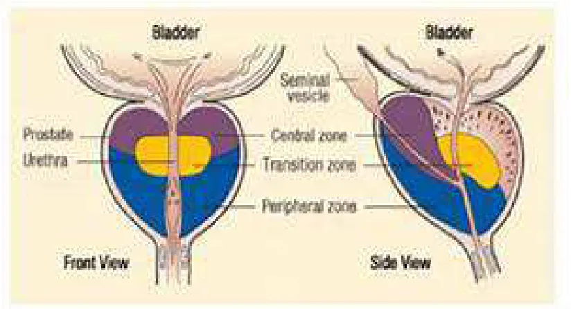
ANATOMY:
Prostate is a pear shaped encapsulated accessory sex gland situated at the
apex of urinary bladder in males. A healthy prostate in a normal adult weighs
about 20 grams and measures about 4x3x2 cm. It has a true internal connective
tissue capsule and a false external capsule, which derived from the pelvic fascia.
It can be broadly divided into an inner periurethral zone and an outer cortical
zone. This classification is important because, the outer zone is the most
common site for adenocarcinoma whereas the inner zone is the most common
site for benign prostatic hyperplasia.[55,56] It can be further classified based on
embryology and pathological features into peripheral, central,transitional and
periurethral regions.[57]
Transition zone
It is the portion of prostate surrounding the pre prostatic urethra and
constitutes about 5-10% of the prostate volume. A dramatic increase of the
transition zone volume is mainly due to benign prostatic hyperplasia and causes
lower urinary tract symptoms (LUTS)
Central zone:
The transition zone is surrounded by the conical central zone. It extends
from the base of the prostate gland to the verumontanum. This region forms a
5
present posterior to the pre prostatic urethra. It constitutes about 25% of the
prostate volume.
Peripheral zone
It is the outermost portion of the prostate gland surrounding the central
zone posteroanteriorly and most of the transition zone.
It constitutes about 75% of the total prostatic volume.
Anterior fibromuscular stroma:
It is present anterior to the urethra and extends into the transition zone.It
[image:18.595.113.525.441.664.2]constitutes about 5% of the total prostatic volume.
6
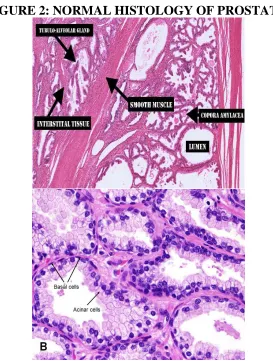
HISTOLOGY:[74]
It is a partially encapsulated organ with capsule covering posterior and
lateral aspects. The anterior and apical surfaces are covered by anterior
fibromuscular stroma which is part of the gland itself.
The prosate gland is composed of glands and stroma.The glands are of
branched tubule-acinar type embedded in a fibromuscular stroma.
The epithelium has a convoluted pattern and is thrown into folds,
sometimes into papillary pattern. The glands are lined by two layers of
epithelium. Luminal epithelium is tall columnar with prominent round basal
nuclei and pale staining eosinophilic cytoplasm. Other type is basal cells which
are stem cells and becomes prominent in prostatic hyperplasia.Some glands may
show inspissated secretions forming spherical concretions-corpora amylacea.
Corpora amylacea increase in number with increase in age and it may become
calcified.
The supporting stroma is composed of collagenous fibrous tissue and
7
FIGURE 2: NORMAL HISTOLOGY OF PROSTATE.
EPIDEMIOLOGY:
Prostate is the second leading site of cancer among males in large Indian
cities like Delhi, Kolkatta, Pune and Thiruvananthapuram and third leading
site of cancer in cities like Bangalore and Mumbai and it is among the top ten
leading sites of cancers in the rest of the population based cancer registries
(PBCRs) of India.[18] In Chennai, prostate cancer is 4th most common cancer.
According to GLOBOCON 2018, prostate cancer incidence is 7.1%
worldwide. Lathika et al [2]analyzed the time trends in the incidence of prostate
cancer for different age groups of the Indian population reported using relative
8
Bangalore, Chennai, Delhi, Mumbai, Karunagappalli, Nagpur, Pune, and
Thiruvananthapuram.
The estimated age-adjusted incidence rates (AARs) of prostate cancer in
India as a whole was 3.7/10 5 persons during the year 2008.
The mean annual percentage change (MAPC) in the crude incidence
rates ranged from 0.14 in Ahmedabad to 8.6 in Chennai. Peak incidence was
observed in the age group above 65 years, indicating that prostate cancer was a
cancer of the elderly. Chennai also recorded the highest MAPC of 5.66 in the
age group of patients above 65 years.
The estimated annual percentage change (EAPC) in the AAR ranged
0.8-5.8 in the various registries. Increase in the trend was seen in men aged
55-64 years in Bangalore, Chennai, and Mumbai during 1983-2002.
In other study by yoele et al, maximum increase in AAR was noted for
Chennai registry (4.95%) and the least for Mumbai registry (0.89%)[3]
Swaminathan et al. study showed that the average annual
age-standardized rate for prostate cancer had a significant increase by 47% during
the period of 2002-2006 in Chennai compared to the previous years. Their
study also showed that prostate cancer had become the ninth most common
cancer in Tamil Nadu.[4]
Herbert et al. compiled data available from various cancer registries and
9
India ranged 5.0-9.1 per 100,000/year. In India ,of all prostate cancers, 85%
were detected late (stages III and IV).A significant notable difference was also
observed between the rural and urban areas in India. [5]
RISK FACTORS:
1) AGE; In 2013,Singh et al. [9] studied the relationship of lifestyle, age,
and BMI with PSA levels in benign prostatic hyperplasia (BPH) and
prostate cancer in the North Indian population. They observed that
the mean age of prostate cancer patients (67.56 ± 5.72 years) was
significantly higher than that of BPH patients (63.56 ± 7.92 years).
2) HORMONAL STATUS: Prostatic cancer is hormone dependent that
is it develops in older men with circulating androgens.
Castration done before puberty protects against prostatic cancer.
Patients with hyperestrogenism due to liver cirrhosis have lower
incidence.
Therapeutic castration and antiandrogen treatment causes tumour
regression.
3) DIET: Increased consumption of fat and carcinogens in charred red
meat, lycophenes in tomatoes,soy products and vitamin D are
suspected to play a role.
In a study conducted by Terry et al. [7] had observed a reduced risk of
10
Heterocyclic amines produced during cooking of red meat and
pyrolysates produced during cooking of meat over charcoal/smoke had been
observed as a reason for increased prostate carcinogenesis in the non
vegetarians. [8]
4) SMOKING: In 2010, Huncharek et al., showed an increased risk of
prostate cancer in chronic smokers[6]
5) OBESITY: Amling et al. [10] and Freedland et al. [11] showed
positive correlation of obesity and BMI to prostate cancer.
6) ENVIRONMENTAL FACTORS: As India is an agricultural
country, exposure to pesticides and other agricultural chemicals is
inevitable. Banerjee et al[12] study reported that pesticides, mainly
organochlorine pesticides (OCPs), could be called as xenoestrogenic
pesticides as they possessed estrogenic properties. OCPs such as 1, 1,
1-hexachlorocyclohexane (HCH), dieldrin, and endosulfan are the
most commonly used xenoestrogenic OCPs in India. As prostate
cancer is an estrogen-dependent cancer, these pesticides might
increase the risk of prostate cancer incidence in the population
exposed to these carcinogenic agents.
7) PREMALIGNANT LESIONS: Nodular hyperplasia is not a
predisposing factor but both the conditions may occur
simultaneously.
High grade prostatic intraepithelial neoplasm is a premalignant
11
GENETICS:
Genetic association is seen in about 5-10% of prostatic cancers. The risk
is twice with single first degree relative with prostatic cancer and the risk
increases to five fold with two first degree relative with prostatic cancer.
It also occurs at an earlier age in patients with strong family history.
A 2 allele of the CYP17 polymorphism has also been reported to be associated
with an increased risk of prostate cancer in smokers and nonvegetarians. [13]
BRCA2 germline mutation is associated with 20 fold increased risk of
developing prostatic cancer.
Increased risk is associated with germline mutation of HOXB13,
chromosomal rearrangements in coding sequence of ETS family transcription
factor next to androgen regulated TMPRSS2 promoter.
Other genetic alterations include amplication of 8q24 locus(MYC
oncogene), deletions of PTEN tumour .
The commonest genetic alteration found in about half of all cases is
fusion of androgen responsive serine protease gene TMPRSS2 (21q22.2) with
one of the ETS transcription factor gene family members[30].
ETS transcription factor gene family includes ERG(21q22.2), ETV1
(7p21.2), ETV4 (17q21) , ETV5(3q27) with ERG accounting for about more
than 90% of cases. this molecular alteration seems to be an earlier event in
12
Expression of fusion transcript is downregulated as the tumour
progresses to become androgen resistant[31].
Advanced stage shows TP53 loss, deletion of RB gene and amplification
of androgen receptor gene locus.
Hypermethylation of glutathione s transferase (GSTP1) gene is the most
common epigenetic alteration which downregulates GSTP1 expression.
RB,CDKN2A,MLH1,MSH2 and suppression of Wnt pathway signaling
(APC) are other epigenetic modifications seen in prostatic cancers.
HER 2 gene amplication is seen in about one third of prostatic
adenocarcinomas and it correlates with tumour grade,stage and non diploid
DNA content[32].
PATHOGENESIS:
Androgen receptor (AR) signaling is important for prostate
differentiation, function as well as for prostate cancer growth and progression.
The human AR is encoded by a single copy gene on the X-chromosome
(Xq11.2-q12).
Although there is some evidence that the length of the poly-glutamine
repeat correlates with prostate cancer risk, there is no strong proven
relationship.[25]
In the absence of androgens, AR is present in the cytoplasm bound to
13
and protect it from degradation. AR activity is regulated by 2 major ligands,
testosterone and dihydrotestosterone (DHT). by in the prostate. Prostate
converts testosterone to DHT by 5α-reductase. DHT is more potent than
testosterone and has 10 times higher binding affinity for AR than testosterone
and is the primary androgen bound by AR. DHT binding to the AR results in
the recruitment of protein kinases, leading to phosphorylation of many serine
residues. Phosphorylation of the AR leads to many functions such as
protection from proteolytic degradation, stabilization, and transcriptional
activation.[26] The transactivation of AR involves several coregulatory proteins
that are able to differentially respond to a changing microenvironment to
regulate specific gene targets involved in cell growth and survival.[27]
In the normal prostate epithelium, there is a balance between the rate of
cell proliferation and the rate of apoptosis; which is lost in prostate cancer
14
FIGURE 3: MECHANISM OF PATHOGENESIS OF PROSTATIC
ADENOCARCINOMA:
Mechanism of ligand-dependent gene transactivation by the androgen receptor. Testosterone (T) inside the prostate epithelial cell is converted to dihydrotestosterone (DHT) by 5α-reductase. DHT binds to AR causes dissociation of the AR-heat shock protein (HSP) complex, dimerization, and translocation to the nucleus. AR binds to androgen response elements (ARE) and recruits multiple co-activators to enhance transcription.
Mechanisms of Hormone Resistance in Prostate Cancer
The mechanisms involved in the emergence of HRPC despite sustained
androgen ablation and/or the use of AR antagonists can be classified into
15
DNA-based alterations in the AR gene, such as amplification or point
mutations, AR- growth factors crosstalk, and activation of alternative pathways
of survival and proliferation.[29]
CLINICAL EXAMINATION:
Early localized cancer is asymptomatic.
Urinary symptoms such as hematuria , dysuria,increased frequency occur
in later stages because most of the tumour occurs in peripheral prostate.
Very advanced stage cancer may present as vertebral metastasis with
back pain. Digital rectal examination and PSA levels help in detection of early
prostatic cancers.
Skeletal surveys and radionucleotide bone scanning confirms
osteoblastic metastasis.
Diagnostic triad include serum PSA ,digital rectal examination and
transrectal ultrasonography for early prostatic carcinoma detection.[20]
DIGITAL RECTAL EXAMINATION AND ULTRASONOGRAPHY:
Early carcinomas cannot be distinguished from nodular hyperplasia,
granulomatous prostatitis, tuberculosis, infarct or lithiasis by rectal examination
alone. Pathological examination of prostatic tissue is confirmatory in such
16
As most of the prostatic carcinomas are located in the peripheral zone, it
may be detected by DRE when the volume is > 0.2 mL. In about approximately
18% of cases, carcinoma is detected by DRE alone, irrespective of PSA level.
DRE in patients with PSA level < 2 ng/mL has a positive predictive value of
about 5-30%. Abnormal DRE is associated with an increased risk of higher
Gleason score and is an indication for biopsy.
Transrectal ultrasound can detect tumours as small as 5 mm in
diameter.[19] However, 30% of prostatic tumours are missed on transrectal
ultrasound and hence it is not a valuable screening tool.
SERUM PSA LEVELS;
PSA has been widely used as a screening test for prostate cancer.
Prostatic epithelium synthesizes PSA which is a serine protease regulated by
androgen. It cleaves and liquefies coagulum formed after ejaculation.Though
elevated serum PSA is specific to prostate, it is not specific to prostatic tumour.
Although it is used as a screening test it lacks both specificity and
sensitivity.
It serum levels is also elevated in benign prostatic hyperplasia,infarction
of nodular hyperplasias, prostatitis, instrumentation of prostate and even after
ejaculation. A serum value of 4ng/mL is taken as normal cutoff in most
laboratories but in about 20-40% of early localized prostatic tumours the serum
value maybe 4ng/mL or even lower than that.
17
Serial assessment of serum PSA values is used in assessment of
response to therapy. Increase in serum PSA level following therapy for
localized tumour indicates recurrent or disseminated disease.
Immunohistochemical localization of PSA on tissue sections can be
used to find whether a metastatic tumour originated in prostate.
Modifications in PSA include PSA density,PSA velocity, ratio of free
and bound PSA in serum and age specific reference values.
AGE SPECIFIC SERUM PSA LEVELS:
In 2007,Ganpule et al. [14] study observations on age-specific PSA and
PSA density values in a community-based Indian population in Gujarat showed
that the mean PSA values increased from 2.1 ng/mL in the age group of 40-49
years to 5.0 ng/mL in the ag group of >70 years. Similarly, the mean PSA
density also increased from 0.15 to 0.2 ng/mL in the same age group of
patients.
Men with hyperplastic prostate produce more PSA than men with
smaller glands.
Older men have increased incidence of BPH and hence have more serum
PSA levels.
The upper age specific serum PSA values:
18
3.5ng/mL for men aged 50-59 years.
4.5ng/mL for men aged 60-69 years.
6.5ng/mL for men aged 70-79 years.
PSA DENSITY:
Serum PSA density identifies the contribution of benign prostatic tissue
to serum PSA level.
It is usually calculated by dividing the total serum PSA level by
estimated gland volume to calculate PSA produced per gram of prostate tissue.
Gland volume is estimated using transrectal ultrasound.
PSA VELOCITY:
It is the rate of change of PSA. More rapid increase in PSA level is seen
in men with prostatic cancer than in men without prostatic cancer. The rate of
change in PSA that distinguishes between prostatic cancer and without
prostatic cancer is 0.75 ng/mL per year.
For PSA velocity measurement, atleast three PSA measurements in
about 1.5 to 2 years should be done as there is about 20% variability between
19
RATIO OF FREE AND BOUND PSA:
Immunoreactive PSA exists in two forms-a major fraction bound to
alpha 1 antichymotrypsin and a minor free fraction. The percentage of free
PSA is lower in men with prostatic tumour than in men with benign prostatic
diseases.
In 2011,Shah et al. [15] in a hospital-based study found than the free PSA
(f PSA) levels correlated with the age of the patient. The mean f PSA levels
(ng/mL) among the four age categories (<45 years, 45-60 years, 60-75 years,
and >75 years) were 0.49 ± 0.13 ng/mL, 0.69 ± 0.10 ng/mL, 1.94 ± 0.04
ng/mL, and 2.33 ± 0.43 ng/mL, respectively.
In Chennai, a study conducted by Atish et al. [16] evaluated the
free-to-total PSA (f/t PSA) ratio to distinguish BPH and prostate cancer in the age
group of 40-75 years. They observed that f/t PSA ratio was decreased
significantly in prostate cancer compared to BPH.
One study has shown that a cutoff for biopsy in symptomatic men with
negative digital rectal examination (DRE) in India could safely be raised to 5.5
ng/mL, which could avoid about 10% of men unnecessarily subjected to
biopsy. [17]
ROLE OF OTHER GENES IN DIAGNOSIS OF PROSTATIC TUMOUR:
PCA3 is overexpressed in about 95% of prostatic tumours.it is a non
20
Urine PCA3 is used as an additional marker in patients with elevated
serum PSA levels but negative prostate biopsy. Elevated urine PCA3 levels is
associated with increased risk of positive repeat prostate biopsy.
Combination of urinary PCA3 and urinary TMPRSS2-ERG fusion DNA
have increased sensitivity and specificity than PSA screening alone.
CYTOLOGY:
According to Epstein et al,.[21]the accuracy of needle biopsy is 85.6%
and that of aspirates was 86.6%,together the accuracy is 95.8%. Inspite of this,
aspiration cytology has fallen into disuse because of large number of false
negative reports.
Poorly differentiated and moderately differentiated carcinomas are easy
to diagnose whereas well differentiated tumour diagnosis is difficult.
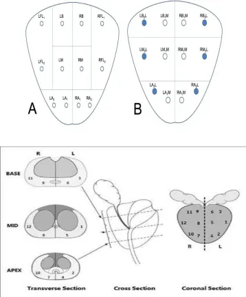
BIOPSIES:
Needle biopsy can be done either perineal or transrectal. Transrectal
route is more preferred.
Automated spring loaded 18 gauge biopsy gun is recently being used.
Six core technique (sextant biopsies) is used routinely[22]. However, 12 core
shows higher yield by 31% especially when the cores are taken from lateral
21
FIGURE 4 & 5: SITES OF PROSTATIC BIOPSY:
It has been found that if five blocks or 12 g of randomly selected tissue
submitted, the probability of detection of carcinoma is approximately 90% and
it rises to 98% with examination of eight blocks.[23]
HANDLING OF PROSTATE SPECIMENS:[24] RADICAL PROSTATECTOMY:
[image:34.595.137.495.114.546.2]22
Vas deferens and proximal bladder neck margins should be shaved.distal
apical margin is obtained by amputating the distal 1 cm of apex and sectioning
the so obtained cone perpendicularly to the cut edge.
Cut serial sections at 2-3 mm from apex to base.
Description:
Weight, dimensions and organs received should be noted. Location in
prostate, size,colour,borders,capsular and periprostatic involvement of tumour
should be noted. Whether urethra and seminal vescicle is involved by tumour
should be noted.
Any adjacent nodular hyperplasia should be noted.
Sections for microscopic examination;
Vas deferens margin.
Proximal bladder neck margin.
Right and left distal apical margin.
Proximal, mid and distal portions from each seminal vescicle.
Adequate sections from the tumour.
TRANSURETHRAL RESECTION (TURP);
Specimen should be weighed and examined carefully for hard yellow
areas which represents carcinomatous areas. Size ,shape and colour of
23
Sections:
If all fragments received in single container: all of specimen until four
cassettes. If excess, one additional cassette for each additional 10 g of tissue.
If received fragments are identified as from which lobe they were taken
then, all of specimen upto 4 cassettes. If excess, one cassette for each additional
10 g of tissue.
If carcinoma is identified microscopically in a lobe that was not entirely
submitted then the remainder of the tissue should be processed entirely
regardless of the amount.
SUPRAPUBIC PROSTATECTOMY FOR NODULAR HYPERPLASIA: Specimens should be sliced every 3mm after fixation and examined for
carcinomatous areas.
Sections:
Three sections from each left and right lobe.
One section from middle lobe.
PROSTATIC INTRAEPITHELIAL NEOPLASIA:
In 1926, Orteil[33] first described premalignant changes in prostate. In
1989, the term prostatic intraepithelial neoplasia (PIN) was coined and is
defined as a cytologic alteration in architecturally normal glands.[35] Prostatic
intraepithelial neoplasia is more common in the peripheral zone of the prostate
24
central zone (<5%).[76-78] The frequency of HGPIN in needle biopsy specimen
is about 5% to 16% and in transurethral resection of the prostate specimens it
is about 2.3% and 4.2%.[75]It comprises an intraluminal proliferation of the
secretory epithelium revealing a spectrum of atypical cytological changes
ranging from minimal changes to those that are indistinguishable from
carcinoma.[34]
According to McNeal Bostwick criteria,three grades of PIN were
identified.[36] Recently, PIN isdivided into two grades (low-grade and
high-grade) instead of the previous three-grade system. Low-grade includes PIN 1
and PIN 2; high-grade includes PIN 3.
McNeal,[37]also described atypical adenomatous hyperplasia (AAH). It is
characterized by an architectural alteration in cytologically unremarkable
glands[38] .High grade PIN is the most likely precursor of carcinoma prostate
because of its greater association with prostatic carcinoma. The other
premalignant lesion AAH and its more common association with nodular
hyperplasia than adenocarcinoma makes it a possible premalignant lesion to
transition zone adenocarcinoma.
Histological features of PIN:[83]
At low power:
• Ducts are lined by darker cells.
• The ducts are thicker than normal ducts.
25
At high power:
• nuclear enlargement and nuclear stratification.
• Hyperchromasia
• Prominent nucleoli.
Histological features of Low grade PIN:
• Epithelial proliferation with cellular crowding.
• Nuclear stratification and nuclear enlargement.
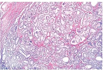
[image:38.595.119.512.374.607.2]• Nucleoli rare, if seen will be small.
FIGURE 6: HISTOLOGY OF LOW GRADE PIN
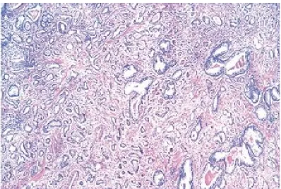
Histological features of High grade PIN:
• Nuclear enlargement and hyperchromasia
• One or more large nucleoli with clear halos.
26
• Common patterns of HGPIN: tufting pattern(87%),micropapillary
[image:39.595.130.505.176.416.2]pattern(85%),cribriform pattern(32%),flat pattern(28%).[79]
FIGURE 7: HISTOLOGY OF HIGH GRADE PIN:
other histologic variants of PIN:[80-82]
• signet ring variant.
• Mucinous variant.
• Small cell neuroendocrine variant.
• Foamy variant.
• Inverted variant.
27
BENIGN MIMICKERS OF PROSTATE CARCINOMA;
• Prostatic atrophy:
It is also known as simple or cystic atrophy and usually involves an
entire lobe. It is characterized by presence of both atrophic and hyperplastic
glands arranged in multiple lobules separated by fibrotic stroma.It is
differentiated from prostatic adenocarcinoma by presence of inflammatory
cells,corpora amylacea and discontinuous basal layer.
PROSTATIC HYPERPLASTIC LESIONS:
• Benign prostatic hyperplasia:
It is composed of glands varying from small and crowded glands to
large glands with cystic dilatation exhibiting complicated growth pattern, such
as papillary and branching. It is differentiated from adenocarcinoma by lacking
malignant nuclear features and presence of basal cell layer. Adenocarcinoma
shows the presence of luminal crystalloids or blue mucin at low power.
• Basal cell hyperplasia: BCH occurs in about 10% of peripheral zone by
needle core biopsy. It is associated with the androgen therapy-related atrophy.
It may present as glands with multilayered basal cells to solid basaloid nests
composed of hyperplastic basal cells having basophilic cytoplasm and bland
nuclear features without nucleoli. Morphologically, it can be florid,
pleomorphic or atypical. Atypical BCH is chararcterised by proliferation of
28
secretions, intracytoplasmic hyaline globules, and very few mitoses. IHC for
basal cells will help in distinguishing from prostatic adenocarcinoma.
• Clear cells cribriform hyperplasia (CCCH):
CCCH has been considered as a morphological variant of BPH. It
consists of enlarged glands composed of anastomosing clear cells forming a
cribriform growth pattern. It should be differentiated from high-grade PAA
with cribriform pattern and can be differentiated by the presence of the nodular
proliferation of cells with bland cytology in a cellular fibrous stroma, and the
presence of basal cell layer.
• Atypical adenomatous hyperplasia.:
It is also known as adenosis.It is a well circumscribed lesion with a
lobular appearance composed of small,round densely packed disorderly glands
with medium sized nucleoli and luminal crystalloids.Basal can be either present
or discontinuous.
• Sclerosing adenosis: It is similar to the lesion in breast. It is composed
of variable sized glands lined by clear secretory cells and dark staining basal
cells in a prominent sclerotic stroma. It may contain intraluminal mucin and
29
METAPLASTIC LESIONS: • Mucinous metaplasia:
It occurs in peripheral zone and composed of glands lined by tall
columnar cells with rich mucin and basal nuclei. It resembles Cowper glands
and positive for PAS, Alcian blue and Mucicarmine.
• Paneth cell metaplasia:
It can be neuroendocrine differentiation,exocrine differentiation or
intestinal Paneth cell differentiation. It is usually associated with HGPIN and
prostatic adenocarcinoma.
NON PROSTATIC LESIONS:
• Nephrogenic adenoma:
It is also known as adenomatoid or nephrogenic metaplasia.It is
metaplastic response of urothelium to injury and is composed of following
structural patterns- tubular, microcystic or papillary pattern exhibiting hobnail
30
Wolffian duct:
Wolffian duct remnants are very rare in adults and has a lobular
architecture composed of different sized glands without basal cells.It may also
show papillary hyperplasia, intraluminal eosinophilic secretion ,fibrocystic
change and sometimes infiltrative pattern. It can be differentiated from
prostatic adenocarcinoma by negativity for PSA and PSAP.
• Seminal colliculus:
It is present near the entrance of seminal vesicles known as posterior
urethral valves. They show nuclear pleomorphism and complex architecture
and can be differentiated from adenocarcinoma by presence of basal cells.They
contain cytoplasmic golden brown lipofuschin and luminal corpora amylacea.
• Rectal glands:
They are most likely to be found in TRUS guided prostatic biopsy. It is
chararcterised by presence of distorted,hyperplastic glands containing
intraluminal and extracellular mucin with prominent nucleoli and absence of
basal cells. It can be differentiated by absence of PSA and PSAP staining.
TYPES OF PROSTATIC CARCINOMA:
Prostatic carcinoma is mainly divided into two categories
1) adenocarcinoma of secondary ducts and acini.
31
Histologic features favouring Adenocarcinoma:[84]
CRITERIA FOR ADENO CARCINOMA
• Enlarged nuclei
• Nuclear hyperchromasia.
• Prominent nucleoli.
• Amphophilic cytoplasm.
• Sharp luminal border.
• Apoptotic bodies.
• Mitotic figures.
• Blue tinged mucinous material.
• Pink amorphous material.
• Crystalloids.
• Mucinous fibroplasias.
• Perineural invasion.
• Glomerulations.
ADENOCARCINOMA OF SECONDARY DUCTS AND ACINI:
SITE OF ORIGIN:
Most adenocarcinomas arise in the peripheral region of prostate[85].
Periurethral region is involved in later stages of the disease[86].Rarely
32
HISTOLOGIC FEATURES:
• There are four major cytological patterns[87]-medium sized glands, small
glands, diffuse individual cell infiltration and cribriform pattern.Gland forming
types are usually lined by single layer of epithelium but occasional stratified
epithelium may mimic late prostatic intraepithelial neoplasia. Most of the
carcinomas exhibit a combination of the above four cytological patterns either
synchronously or metachronously.
• The neoplastic cells show nuclear enlargement, irregular nuclear
contour, hyperchromasia and macronucleoli[88] measuring more than 1 micron
in diameter.
• Malignant glands contain intraluminal wispy blue mucin. Carcinomas
with medium sized glands appear as closely placed glands with irregular
outline and scanty intervening stroma under low power.
Carcinomas with small glands appear as expansive nodules with regular
round small sized individual glands.
Diffuse individual cell infiltration pattern resembles lobular carcinoma
of breast. Cribriform pattern is considered as intraductal carcinoma as the basal
layer is preserved[89].
Glomeruloid pattern is recently discovered which is characterized by
intraluminal ball like clusters of tumour cells[90].
Squamous metaplasia is uncommon and presents as high grade
33
Perinuerial involvement by the tumour cells is common. It is due to
spread of neoplastic glands along the planes of least resistance and its presence
in needle biopsy indicates Mucinous fibroplasias or collagenous micronodules
is deposition of basophilic ground substance in the surrounding stroma.[92]
PROTEIN CRYSTALLOIDS:[93]
10-23% of prostatic carcinomas especially the ones having medium
sized glands show protein crystalloids in their lumen.
Although it is seen in benign glands, its presence is an indicator of
malignancy. They predominantly contain inorganic sulfur.
VARIATIONS IN PROSTATIC ADENOCARCINOMA:
• Prostatic adenocarcinoma with atrophic features:
It is composed of tumour cells with scanty cytoplasm and the nuclei
occupies the entire cell. It mimics benign hyperplasia and is differentiated by
presence of infiltrative growth and cytological features of malignancy.
• Pseudohyperplastic variant:
It shows papillary folding, branching, corpora amylacea and microcystic
pattern, all resembling hyperplasia. It is differentiated by cytological features of
34
• Foamy gland variant:
Grossly the tumour has soft consistency and a bright yellow colour and
hence difficult to identify on digital examination.
The tumour cell cytoplasm has finely granular appearance or clear
foamy appearance due to accumulation of lipids.
It has a low gleason score but is an aggressive tumour.[94]
CARCINOMAS OF PRIMARY DUCTS:
It occurs in large ducts in periurethral region[96].
Grossly it is either polypoid villus or an infiltrative growth involving
urethra.
TYPES OF PRIMARY DUCT CARCINOMA:
Large duct adenocarcinoma;
Microscopically it shows large dilated ducts arranged in cribriform or
papillary pattern lined by columnar pseudostratified epithelium [97]with
occasional clear cell (mesonephroid).
Occasionally it shows pagetoid spread in the prostatic urethra.
It presents at more advanced stage and has higher short term survival
rate. Endometrioid(endometrial)type adenocarcinoma[98]:
35
It arises from prostatic utricle which is a mullerian remnant.
Primary urothelial carcinoma:[99]
It constitutes less than 2% of prostatic carcinomas.
It arises from the outer portion of periurethral ducts draining into urethra
which is lined by urothelium.
Primary urothelial carcinoma from urethra or bladder should be
excluded before making a diagnosis of primary urothelial prostatic carcinoma.
Mixed adenocacinoma and urothelial carcinoma of prostate:
It contains both glands and urothelial component in varying proportions.
OTHER MICROSCOPIC VARIANTS:
CARCINOMA WITH NEUROENDOCRINE FEATURES[100]:
About 80% of normal or hyperplastic prostate have endocrine cells with
argentaffin-argyrophil properties, serotonin, calcitonin,bombesin,somatostatin
and dense core granules on ultastructural examination.
About 10% to half of typical adenocarcinomas show endocrine features.
Prostatic carcinoma with neuroendocrine differentiation typically express
estrogen inducible pS2 protein.
Some prostatic carcinoma resemble typical or atypical carcinoid tumour.
36
component shows positivity for adrenocorticotropic hormone, beta endorphin
and calcitonin.
Small cell carcinoma: It presents as pure formor associated with
adenocarcinoma either synchronously or metachronously.
It causes cushing syndrome or inappropriate anti diuretic hormone
secretion.
It is an aggressive tumour and should be differentiated from high grade
prostatic adenocarcinoma.
Large cell neuroendocrine carcinoma: In most cases it arises after long
term hormonal therapy for prostatic adenocarcinoma.
MUCIN SECRETING ADENOCARCINOMA[101]:
This tumour contains large amounts of intracellular and extracellular
mucin comprising about 25% of tumour.
Microglandular, comedo,cribriform,hypernephroid and solid variants
can be seen.
The mucin secreted by well differentiated adenocarcinomas is
non-o-acylated sialomucins whereas poorly differentiated adenocarcinomas secrete
mono-o-acylated sialomucins but mucinous adenocarcinomas secrete mono- ,
di-, and tri-o-acylated sialomucins.
37
In contrast to usual adenocarcinomas, this type shows rare bone
metastasis, lesser response to radiation therapy and are not hormone dependent.
It should be differentiated from large bowel mucinous carcinoma
extension and Cowper’s gland carcinoma.
SQUAMOUS CELL CARCINOMA;
It is a rare carcinoma and it can occur either de nova or following
hormonal therapy. Grossly presents as a circumscribed nodule in transition
zone[102]. Closely related to adenosquamous carcinoma.
ADENOSQUAMOUS CARCINOMA:
It occurs either de nova or following radiation or hormonal therapy.
SIGNET RING CARCINOMA:
It is a highly malignant neoplasm composed predominantly of signet
ring cells.
It can have solid, acinar or Indian file pattern.
The cells contain microvilli lined intracytoplasmic lumen[103].
ADENOID BASAL CELL TUMOUR[104]:
Also known as basal cell carcinoma and adenoid cystic like tumour. It
resembles adenoid cystic carcinoma of salivary gland but has a more indolent
course. Microscopically,it presents as an expansile growth,multinodularity with
38
surrounding fibromyxoid stroma. Squamous differentiation and basal cell
hyperplasia can be seen.
PSA and PAP is usually negative or focally positive.
This variant should be differentiated from adenocarcinoma with
cribriform form of glands ,basal cell hyperplasia, basaloid carcinoma and true
adenoid cystic carcinoma.
BASALOID CARCINOMA:
It is a highly aggressive neoplasm and should be differentiated from
aenoid basal cell carcinoma. This carcinoma shows elevated expression of
BCL2 and high Ki 67 index.
SARCOMATOID CARCINOMA:
The epithelial component is mostly commonly adenocarcinoma but
squamous features can also be seen. Sarcomatoid element is either non specific
spindle cells or giant cell features showing differentiation toward cartilage ,
skeletal muscle or bone[105]. Pleomorphic giant cell adenocarcinoma is a
subtype of sarcomatoid carcinoma.
LYMPHOEPITHELIOMA LIKE CARCINOMA;
TUBULOCYSTICNCLEAR CELL ADENOCARCINOMA:
Rare cases similar to mullerian type clear cell adenocarcinoma and clear
39
TUMOUR METASTASIS:
Prostatic carcinoma initially spreads within the prostate,ducts and acini,
fibromuscular stroma , perineural spaces and blood vessels.[40] Invasion of
fibromuscular layer of prostate (capsule) is more common. It may also spread
to seminal vescicles, distal aspect of the gland, bladder and very rarely
prostatic urethra and rectum.
Seminal vescicle invasion should be diagnosed only when the muscular
wall of the organ is infiltrated by the tumour.[41]
Rectal invasion is rare because of Denonvillier’s fascia which covers the
posterior aspect of the prostate.[42] It can present as anterior rectal mass ,
subserosal implants or as circumferential infiltration causing annular rectal
stricture. Prostatic carcinoma most commonly metastasize to the skeletal
system and lymph nodes.
SKELETAL METASTASIS:
Skeletal metastasis can be either multiple or solitary. Most commonly
multiple. They are chararcteristically osteoblastic but can also be mixed or even
osteolytic. Lumbar spine, sacrum and pelvis are the more common sites. Spread
to these sites is via Batson’s vertebral plexus[43] .Metastasis to other bones is
through systemic circulation. Spinal metastasis may present as cord
compression due to epidural mass and base of skull metastasis may present as
40
Radiographically, osteoblastic metastasis should be differentiated from
Paget disease and osteosarcoma. Microscopic examination shows clusters of
malignant glands surrounded by abundant new bone formation. Patient may
present with hypocalcemia, hypophosphatemia and serum alkaline
phosphatase.
NODAL METASTASIS:
The tumour first spreads to pelvic group and later to retroperitoneal
nodes. If retroperitoneal nodal metastasis occurs in absence of pelvic nodal
group, then the patients are likely to have liver and lung metastasis.
Periprostatic, periseminal vescicle and perirectal nodes can also be involved[44].
Sometimes, left supraclavicular and mediastinal nodes can also be
involved. When these nodes are involved, the tumour is usually poorly
differentiated and immunohistochemical staining for PAP and PSA is used for
diagnosis.
OTHER METASTATIC SITES:
Lung metastasis present with massive pleural effusion and mostly
exhibit lymphangitic pattern of spread. Microacinar, tubulopapillary and
carcinoid like growth patterns can be seen. when large ducts are involved they
mimic metatatic colonic carcinoma. Other sites include testis, breast in patients
taking estrogens, liver, adrenal gland, dura, eye, skin, umbilicus, penis and
41
GLEASON MICROSCOPIC SCORING SYSTEM;
The Gleason scoring system is named after Donald Gleason, a
pathologist who developed it with his colleagues at that facility in the
1966s.[50]
The (2005 ISUP modified) [51,52]Gleason score of prostatic biopsy
includes the Gleason grade or the most extensive primary pattern plus the
second most common pattern that is the secondary pattern, if two are present.
If only one pattern is present then it should be doubled to yield the Gleason
score.
Gleason grading of prostatic adenocarcinoma should be typically
performed using the 4x objective, although in certain instances such as in
back-to-back glands arrangement and in fused glands require higher
magnification at 10x objective[54].
For three grades, the Gleason score is calculated by adding the most
common grade plus the highest grade, irrespective of its extent. When a
carcinoma is largely grade 4/5, identification of < 5% of Gleason grade 2 or 3
glands should not be included in the Gleason score.
In addition to reporting of the carcinoma features for each biopsy, an
overall Gleason score based on the carcinoma-positive biopsies shoud be given.
The 2014 ISUP Gleason grading conference of prostatic carcinoma has
introduced the concept of the grade groups of PCa, in order to:
42
2. eliminate the anomaly that the most highly differentiated PCas have a
Gleason score 6;
3. to further codify the clinically highly significant distinction between
Gleason score 7 (3 + 4) and 7 (4 + 3) PCa.
The ISUP 2015/2016 WHO prostate cancer grade groups therefore
[image:55.595.111.524.301.613.2]range from 1-5.
FIGURE 8: ISUP 2015/ WHO 2016 REVISED GLEASON SCORE:
Problems with the Current Gleason System[53]:
1) Scores 2-5 are currently no longer assigned and certain patterns that
43
contemporary Gleason score 6 cancers having a better prognosis than
historic score 6 cancers.
2) The combination of Gleason scores into a 3-tier grouping (6,7,8-10) is
used most frequently for prognostic and therapeutic purposes, despite
3+4=7 vs. 4+3=7 and 8 vs. 9-10 having very different prognoses.
3) In practice the lowest score is now assigned a 6, although it is on a scale
of 2-10. This leads to a logical yet incorrect assumption on the part of
patients that their cancer is in the middle of the scale, compounding the
fear of their cancer diagnosis with the belief that the cancer is serious,
thus leading to an expectation that treatment is necessary.
New Grading System[53]
The new 5 Grade Group system has been developed based on a study of
>20,000 prostate cancer cases treated with radical prostatectomy and >5,000
cases treated by radiation therapy.
Grade Group 1 (Gleason score ≤6) – Only individual discrete well-formed glands
Grade Group 2 (Gleason score 3+4=7) – Predominantly well-formed glands with a lesser component of poorly-formed/fused/cribriform glands
Grade Group 4 (Gleason score 8)
- Only poorly-formed/fused/cribriform glands or
- Predominantly well
- Predominantly lacking glands with a lesser component of well
Grade Group 5 (Gleason scores 9 necrosis) with or w/o poorly
For cases with >95% poorly
glands on a core or at Radical prostatectomy, the component of <5% well
formed glands is not factored
glands can also be a more minor
PATTERNS IN GLEASON SCORING:
44
(Gleason score 8)
formed/fused/cribriform glands or
Predominantly well-formed glands with a lesser component lacking glands or
Predominantly lacking glands with a lesser component of well
(Gleason scores 9-10) – Lacks gland formation (or with
necrosis) with or w/o poorly-formed/fused/cribriform glands
For cases with >95% poorly-formed/fused/cribriform g
or at Radical prostatectomy, the component of <5% well
formed glands is not factored into the grade. Poorly-formed/fused/cribriform
glands can also be a more minor component.
PATTERNS IN GLEASON SCORING:
formed glands with a lesser component lacking glands or
Predominantly lacking glands with a lesser component of well-formed glands
Lacks gland formation (or with
formed/fused/cribriform glands or lack of
or at Radical prostatectomy, the component of <5%
45
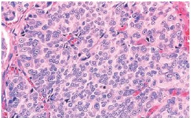
FIGURE 9: GLEASON PATTERN I
[image:58.595.116.519.434.707.2]46
FIGURE 11: GLEASON PATTERN III
[image:59.595.129.504.471.694.2]47
FIGURE 13 :GLEASON PATTERN V
GLEASON GRADE GROUPING:
GRADE GROUP GLEASON SCORE AND PATTERN
1 Grade 6(3+3)
2 Grade 7(3+4)
3 Grade 7(4+3)
4 Grade 8(4+4, 3+5 , 5+3)
48
DIFFERENTIAL DIAGNOSIS:
• Atypical adenomatous hyperplasia.
• Atrophy.
• Crowded benign glands.
• Basal cell hyperplasia.
• Sclerosing adenosis.
• Cribriform hyperplasia.
• Mesonephric hyperplasia.
• Nephrogenic adenoma.
• Squamous metaplasia.
• Transitional cell metaplasia.
• Veromontanum mucosal gland hyperplasia.
• Prostatitis.
• Radiation atypia.
• Malakoplakia.
• Endometriosis.
• Cowper glands.
• Paraganglia in prostate.
49
HISTOCHEMISTRY:
The normal prostatic secretion is a neutral mucosubstance. In about two
third of adenocarcinomas secrete acid mucosubstances.Acidic nature of the
mucin should be suspected when the luminal content of the gland is basophilic
in routine staining and it is confirmed with Alcian blue or colloidal iron
staining.
IMMUNOHISTOCHEMISTRY;
The two important prostatic epithelial markers are PAP and PSA.
PSA has more specificity than PAP.
They do not differentiate between benign and malignant lesions in
prostate but very useful in confirming prostatic origin in metastatic tumours.
They are absent in most undifferentiated tumours and in advanced cases
following hormonal therapy. They are useful in differentiating poorly
differentiated prostatic carcinoma and urothelial carcinomas.
PSA is localized to endoplasmic reticulum,vesicles,vacuoles and
50
PROSTATE SPECIFIC ANTIGEN:
It is a serine protease member of the family human glandular kallikrein.
It is a 34 kD glycoprotein of 237 amino acids.It is exclusively synthesized by
prostic acinar and ductal epithelium which is present in normal , hyperplastic
and as well as in malignant prostatic tissue[106].
OTHERS IMMUNO MARKERS:
PROSTATE SPECIFIC MEMBRANE ANTIGEN(PSMA):
It is a type II membrane glycoprotein.
It is expressed by both benign and malignant prostatic epithelium with
higher extent of staining in malignant epithelium.
It is also positive in high grade prostatic intraepithelial neoplasia (PIN)
and in hormone refractory prostatic carcinoma[107].
Its expression correlates with Gleason score and staging.[108]
In addition to prostatic tissue, it is expressed in lesser amount in central
and peripheral nervous system, small intestine and salivary gland.It is also
positive in endothelial cells of neovasculature of many solid tumours.
PROSTEIN/P501S:
It is localized in golgi complex and hence shows perinuclear
51
and metastatic prostatic carcinoma[109].It is positive even in PSA negative
metastatic tumours.
P504S/ALPHA METHYLACYL COENZYME A RACEMASE(AMACR):
It has 97% sensitivity and 92% specificity[110].It is localized to
peroxisomes. It I positive in high grade PIN, prostatic carcinoma as well as in
both untreated and hormone refractory prostatic carcinoma metastasis. As it is
positive in high grade PIN and benign mimics of prostatic carcinoma such as
glandular and partial atrophy and in adenosis, it is of limited value as an
individual marker. A panel of AMACR, HMWCK and p63 with negative basal
cell markers is used for identifying atypical prostatic glands.
HIGH MOLECULAR WEIGHT CYTOKERATINS[111]:
It is used in identifying the presence or absence of basal cells in atypical
prostate glands. 34BE12 is the most commonly used HMWCK. CK5/6 can be
used alternately.
P63:
It is expressed in basal cells and is absent in secretory cells and
neuroendocrine cells of prostate and hence is absent in prostatic carcinoma and
is expressed in basal cells of benign glands[45,46,47].
Signoretti et al reported that all basal cells express p63 and hence this
marker can be useful in distinguishing benign lesions from prostate malignancy
52
Person et al showed that p63 is expressed in normal basal cells and
benign prostate hyperplasia (BPH). It can be focally expressed in prostate
atrophy and HGPIN, but p63 expression is absent in the majority of prostate
adenocarcinomas.[49] Recent studies show that P63 gene is essential for normal
stem cell function in prostate.
NKX3-1:
It is a prostate specific androgen regulated homeobox gene involved in
tumour differentiation and its loss of function causes carcinogenesis. It is
superior to PSA in poorly differentiated prostatic carcinoma[112].
OTHER IHC MARKERS:
HER2/NEU protein is overexpressed in androgen independent prostatic
carcinoma.
CDX2 nuclear staining is occasionally present causing difficulty in
differentiating from intestinal adenocarcinoma.
They also show positivity for EMA , CEA , Leu7 , cathepsin D, B72.3,
parathyroid hormone related protein,gastric acid proteinase gastricism,
erythropoietin/erythropoietin receptor and glycoprotein A-80.
Protatic adenocarcinomas show reduced expression of E-cadherin and
53
PROGNOSIS:
In 1999,the College of American Pathologists(CAP) involved a group of
clinicians, pathologists and statisticians and established the following
categories for as prognostic indicators of prostatic carcinoma.[39]
Prognostic factors categorized by CAP:
I- Proven to be of prognostic importance and useful in clinical patient
management:
Preoperative serum PSA level
TNM stage grouping
Histologic grade as Gleason score
Surgical margin status.
II- Extensively studied but whose importance remains to be validated:
Tumour volume
Histologic type
DNA ploidy
III- Not sufficiently studied to demonstrate their prognostic value:
Perineurial invasion
Neuroendocrine differentiation
Microvessel density
Nuclear roundness
Chromatin texture
54
Proliferation markers
PSA derivatives
Other factors such as pncogenes , tumour suppressor genes , apoptosis
genes.
BENIGN PROSTATIC HYPERPLASIA
INTRODUCTION:
The term nodular hyperplasia as proposed by Moore is a more exact
designation. Benign prostatic hyperplasia (BPH) shows nodular enlargement
of gland and histologically shows unregulated proliferation of connective
tissue, smooth muscle and glandular epithelium within the prostatic transition
zone[113]. The weight of the increases above 20 grams which is considered as
normal for adult individuals.Prostate tissue is made up of two basic elements:
A glandular element composed of secretory ducts and acini and a stromal
element composed primarily of collagen and smooth muscle. In BPH, cellular
proliferation leads to increased prostate volume and increased stromal smooth
muscle tone.
McNeal [114]describes two phases of BPH progression. The first phase
shows an increase in BPH nodules in the periurethral zone and the second a
55
CLINICAL FEATURES:
BPH causes physical compression of the urethra resulting in anatomic
bladder outlet obstruction (BOO) through two distinct mechanisms[115]: First,
the static component which is associated with an increase in prostate volume;
second, the dynamic component, associated with an increase in stromal smooth
muscle tone. BOO may clinically present as lower urinary tract symptoms
(LUTS), urinary tract infections, acute urinary retention (AUR), renal failure
hematuria, and bladder calculi[116].
ETIOPATHOGENESIS:
Age
The prevalence of BPH rises markedly with increase in age. Autopsy
studies show a histological prevalence of 8%, 50% and 80% in the 4 th , 6 thand
9 th decades of life, respectively[116]. Krimpen and Baltimore Longitudinal
Study of Aging (BLSA) cohorts shows that Prostate volume also increases with
age, suggesting a prostate growth rate of 2.0% to 2.5% per year in older
men[117-119].
Geography
Several international studies have shown geographic heterogeneity in
prostate volume and LUTS prevalence. Significantly lower prostate volumes
have been observed in men from Southeast Asia when compared to western