This is a repository copy of
Characterisation of DOG-1 expression in salivary gland
tumours and comparison with myoepithelial markers
.
White Rose Research Online URL for this paper:
http://eprints.whiterose.ac.uk/150412/
Version: Published Version
Article:
Khurram, S.A. orcid.org/0000-0002-0378-9380 and Speight, P.M.
orcid.org/0000-0001-6459-1370 (2019) Characterisation of DOG-1 expression in salivary
gland tumours and comparison with myoepithelial markers. Head and Neck Pathology, 13
(2). pp. 140-148.
https://doi.org/10.1007/s12105-018-0917-3
eprints@whiterose.ac.uk https://eprints.whiterose.ac.uk/
Reuse
This article is distributed under the terms of the Creative Commons Attribution (CC BY) licence. This licence allows you to distribute, remix, tweak, and build upon the work, even commercially, as long as you credit the authors for the original work. More information and the full terms of the licence here:
https://creativecommons.org/licenses/
Takedown
If you consider content in White Rose Research Online to be in breach of UK law, please notify us by
https://doi.org/10.1007/s12105-018-0917-3
ORIGINAL PAPER
Characterisation of DOG-1 Expression in Salivary Gland Tumours
and Comparison with Myoepithelial Markers
Syed A. Khurram1 · Paul M. Speight1
Received: 31 January 2018 / Accepted: 13 April 2018 / Published online: 18 April 2018 © The Author(s) 2018
Abstract
DOG1 is an established diagnostic marker for gastrointestinal stromal tumours (GIST), but has been reported in salivary gland tumours (SGT) as an acinar and intercalated duct marker. However, its speciicity and distribution is not well established. The aim of this study was to evaluate the diagnostic utility of DOG-1 expression in SGT in addition to comparing it with myoepithelial markers. Normal salivary tissue and SGT (n = 184) were examined for expression of DOG1 and a range of myoepithelial markers. SGT included: acinic cell carcinoma (ACC, n = 15), secretory carcinoma (SC, n = 9), pleomorphic adenoma (PA, n = 49), carcinoma ex-PA (Ca ex-PA, n = 11), adenoid cystic carcinoma (AdCC, n = 20), polymorphous adeno-carcinoma (PAC, n = 6), myoepithelioma (n = 6), myoepithelial adeno-carcinoma (MC, n = 2), basal cell adenoma (BCA, n = 14), canalicular adenoma (CA, n = 19), mucoepidermoid carcinoma (MEC, n = 11), oncocytoma (n = 2), adenocarcinoma NOS (AdNOS, n = 4), basal cell adenocarcinoma (BCAC, n = 2), salivary duct carcinoma (SDC, n = 3) and papillary cystadeno-carcinoma (PCAC, n = 1). Normal acini and ACC (14/15) showed strong luminal DOG1 staining; SC were largely negative with only focal expression in 3/9 cases. Luminal staining was seen in PA (14/49), PAC (4/6), Ca ex-PA (4/11) and AdCC (6/20). 8/11 MEC showed luminal and/or mucous cell staining. No staining was seen in myoepithelioma, MC, CA, adNOS and BCAC. BCA showed strong staining of myoepithelial cells in some cases (5/14). Variable myoepithelial DOG1 staining was seen in PA, Ca ex PA, BCA, SDC and PCAC which was not as consistent as myoepithelial markers such as calponin, p63 and αSMA. Absence of DOG1 can diferentiate ACC from SC, but staining is variable in PA, PLGA and Ca ex-PA. Myoepi-thelial staining in some tumours but not in normal gland suggests a wider distribution in SGT than originally envisaged.
Keywords DOG-1 · Salivary gland tumours · Acinic cell carcinoma · Secretory carcinoma · Myoepithelial · Luminal
Introduction
Diagnosing salivary gland tumours can be challenging due to the heterogeneity of the cellular diferentiation, morpho-genesis and histological patterns. Many diferent tumour entities share similar histological patterns, which further complicates diagnosis.
In 2004, ‘Discovered on GIST-1’ (DOG1) was shown to be highly expressed in a high proportion of gastrointestinal stromal tumours (GISTs) [1–3]. Subsequently, a number of in vivo studies revealed DOG1 to be a calcium activated chloride channel expressed on secretory epithelium in mouse models [4, 5]. More recently, DOG1 expression has been
reported in salivary gland tumours [6–8], in particular, as a marker for acinic and intercalated duct cells. Some stud-ies have suggested that the DOG1 protein may be essential for salivary gland secretion with a possible role in salivary gland tumourigenesis [5, 9]. However, its pattern of expres-sion and speciicity in a range of tumours has not been fully established. Strong staining is seen at the luminal aspect of acinar cells in normal glands, and luminal staining has been shown in the acini in ACC [10], and in small ductal struc-tures in PAC and epithelial myoepithelial carcinoma (EMC) [6]. Luminal and abluminal staining has been described in BCA and AdCC [6, 11]. Our clinical experience has shown expression by myoepithelial cells in some instances, a ind-ing not reported to date.
The aim of this study was to study the expression pattern, speciicity and diagnostic potential of DOG-1 in salivary gland tumours.
* Syed A. Khurram
s.a.khurram@sheield.ac.uk
1 Unit of Oral and Maxillofacial Pathology, School of Clinical
141 Head and Neck Pathology (2019) 13:140–148
1 3
Methods
Normal parotid and submandibular gland tissue and SGT (n = 184) were examined for expression of DOG1 and a range of myoepithelial and cytokeratin markers using immunohistochemistry (IHC) on cases retrieved from the department archive. These included acinic cell carcinoma (ACC, n = 15), secretory carcinoma (SC, n = 9), pleomorphic adenoma (PA, n = 49), carcinoma ex-PA (Ca ex-PA, n = 11), adenoid cystic carcinoma (AdCC, n = 20), polymorphous adenocarcinoma (PAC, n = 6), myoepithelioma (n = 6), myoepithelial carcinoma (MC, n = 2), basal cell adenoma (BCA, n = 14), canalicular adenoma (CA, n = 19), mucoep-idermoid carcinoma (MEC, n = 11), oncocytoma (n = 2), adenocarcinoma NOS (AdNOS, n = 4), basal cell adenocar-cinoma (BCAC, n = 2), salivary duct caradenocar-cinoma (SDC, n = 3) and a papillary cystadenocarcinoma (PCAC, n = 1).
The diagnosis of the cases was conirmed by H&E stain-ing and examination under the light microscope. Tumours were classiied according to WHO 2017 guidelines [12] and current literature. FISH analysis for the ETV6
rearrange-ment was used as a gold standard for the diagnosis of all included SC.
Immunohistochemistry
IHC for DOG1 was performed on the entire cohort. For com-parison a range of other ‘myoepithelial markers’ were also studied in a proportion of tumours including S100, αSMA, p63, calponin and CK14 as previously described [8]. Multi-ple pilot assays were undertaken to determine the optimum dilution and conditions (Table 1).
4 µ thick sections from formalin-ixed, parain embed-ded tissue blocks were used for IHC staining. Sections were deparainised in xylene followed by incubation in ethanol for 5 min each. Endogenous peroxidase was blocked by incu-bation in 3% methanolic H2O2 blocking solution for 20 min followed by a wash in phosphate bufered saline (PBS). Antigen retrieval was carried out by placing the slides in a heat-resistant plastic container illed with citrate or EDTA bufer solution in a microwave for 10 min on high power. The slides were left to cool for 2 min, placed in PBS to
avoid dehydration and blocked with 100% normal serum for 30 min at room temperature (RT). Serum was removed followed by addition of the primary antibody overnight at 4 °C in a humidiied chamber. Omission of primary antibody served as the negative control.
After overnight incubation, unbound primary antibody was removed and the slides washed twice for 5 min with PBS. The secondary antibody and ABC solution were pre-pared according to the manufacturer’s instruction (Vec-tastain Elite kits, Vector Laboratories, Burlingame USA). Sections were covered with secondary antibody for 30 min followed by two washes in PBS and addition of avidin biotin complex (ABC) solution for 30 min at RT. After two further washes in PBS 3,3 -diaminobenzidine (DAB, Vector labo-ratories) was applied to the sections for 5–8 min and the colouring reaction stopped using distilled water. Sections were counterstained with Mayer’s haematoxylin, mounted in DPX mounting media and left to dry at RT.
IHC staining was assessed subjectively under standard light microscopy, taking into account the pattern and locali-sation of the staining. Stained sections were photographed using a digital imaging system (cell^D) and a digital camera attached to a light microscope (Olympus, UK). As previ-ously described, myoepithelial staining was considered posi-tive in the correct morphologic context to ensure exclusion of stromal cells [8].
Results
DOG‑1
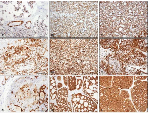
Strong apical/luminal DOG1 staining was seen in normal acini (10/10) (Fig. 1a), although occasional cells demon-strated lateral and basal expression. Staining was stronger in serous acini compared to mucous and focally interca-lated ducts showed positive luminal reactivity. A similar staining pattern was seen in ACC, with widespread lumi-nal DOG1 staining in 14 of the 15 cases (Fig. 1b). SC how-ever were largely negative (Fig. 1c) with only weak focal luminal staining in three cases with a microcystic archi-tecture (3/9) (Fig. 1d). SC with a papillary cystic architec-ture or clear cell change were negative. Variable luminal
Table 1 Details of primary
antibodies used in the study Antibody Clonality Dilution Retrieval Supplier
[image:3.595.176.546.619.715.2]staining was seen in PA (14/49) (Fig. 1d), PAC (4/6—not shown), Ca ex-PA (4/11) (Fig. 1i), AdCC (6/20) (Fig. 1e). MEC showed luminal and/or mucous cell staining in 8/11 cases (Fig. 1f). BCA showed strong staining of myoepi-thelial cells in 5/14 cases (Fig. 1h), and some myoepithe-lial staining was also seen in PA, Ca ex PA, BCA, SDC, AdCC and PCAC (Fig. 1; Table 2). No staining was seen in myoepitheliomas (0/6), MC (0/2), CA (0/19), AdNOS (0/4) and BCAC (0/2).
Myoepithelial DOG-1 staining was compared with a range of existing markers including αSMA, calponin, p63, S100 and CK14.
α‑Smooth Muscle Actin (αSMA)
Widespread αSMA staining was seen in myoepithelial cells surrounding the acini in normal salivary glands. The major-ity of the ACC and SC were negative for αSMA with focal staining seen in only 4/14 and 3/9 of cases. More consist-ent myoepithelial staining was seen in PA, Ca ex PA and myoepithelioma whereas PAC was negative.
Strong αSMA staining was also seen in abluminal cells of both tubular and cribriform AdCC (17/20 cases) consistent with myoepithelial cells. The three negative cases included two solid and one tubular AdCC.
Almost all cases of PAC were αSMA negative (not shown) whereas all basal cell adenomas demonstrated strong αSMA staining in the abluminal, myoepithelial cells. How-ever, areas lacking αSMA expression were also observed.
Calponin
ACCs and SC did not stain for calponin. Staining was seen in most cases of pleomorphic adenoma (44/47; 93.6%) (Fig. 2a). Staining was frequently observed in abluminal and spindled myoepithelial cells whereas plasmacytoid cells were always negative. Similar to PA, 90% (9/10) of Ca ex-PA showed Calponin staining in abluminal cells in addi-tion to scattered stromal cells (Fig. 2b). Calponin staining was seen in myoepithelial cells in 66.7% of myoepitheliomas (4/6) (Fig. 2c). Focal staining was seen in myoepithelial car-cinomas whereas PAC were largely negative with only one case showing some focal staining (Fig. 2d).
Calponin staining was seen in 16/20 (80%) AdCC with expression in myoepithelial cells in both cribriform (Fig. 2e) and tubular (Fig. 3f) variants whereas the three negative cases had solid and mixed patterns.
CK14
In normal salivary glands, CK 14 staining was seen in myoepithelial cells surrounding acini and ducts (Fig. 3a). Both ACC and SC were largely negative for CK14; however 4/16 ACC showed focal CK14 staining (Fig. 3b) with difuse staining seen in one SC (Fig. 3c).
For PA, 89.6% (43/48) of the cases were CK14 positive with staining of both ductal and myoepithelial cells. How-ever, staining was weak or absent in plasmacytoid cells, and cells in myxochondroid areas (Fig. 3d). CK 14 was difusely positive in 90.9% of Ca-ex-PA (10/11) with strong staining in myoepithelial and abluminal cells throughout the tumours (Fig. 3e).
Fig. 1 Representative photomicrographs showing DOG-1 staining
pattern in a normal gland; b acinic cell CA; c secretory carcinoma—
negative; d secretory carcinoma—focal positive, e pleomorphic
ade-noma; f adenoid cystic CA; g mucoepidermoid carcinoma; h basal
cell adenoma; i carcinoma in pleomorphic adenoma; j papillary
cys-tadenocarcinoma. g Solid arrow—mucous cells, dotted
[image:4.595.54.288.56.502.2]143 Head and Neck Pathology (2019) 13:140–148
1 3
In myoepithelioma, CK14 was variably positive in myoep-ithelial cells in 5/6 of the cases (83.3%), mainly in cells with
spindled morphology (Fig. 3f). Both cases of myoepithelial carcinoma showed focal staining of the tumour cells (Fig. 3g). Table 2 Summary of DOG1
staining in salivary gland neoplasms
For each tumour type the total number of positive cases is given in relation to the total number of the cases Site Cases +ve Pattern
Normal gland 10 10/10 Acini & ducts—luminal
Acinic cell carcinoma 15 14/15 Difuse luminal in acini and small ducts
Secretory carcinoma 9 3/9 Negative or weak/focal luminal in microcystic areas Pleomorphic adenoma 49 14/49 Focal luminal or myoepithelial (5/14)
Ca ex PA 11 4/11 Luminal and/or myoepithelial cells (2/4) Myoepithelioma 6 0/6 N/A
Myoepithelial carcinoma 2 0/2 N/A
Adenoid cystic carcinoma 20 6/20 Weak abluminal + luminal Polymorphous adenocarcinoma 6 4/6 Focal luminal
Basal cell adenoma 14 5/14 Luminal or abluminal/myoepithelial—variable (5/5) Canalicular adenoma 19 0/19 N/A
Mucoepidermoid carcinoma 11 8/11 Luminal + mucous cell brush borders Oncocytoma 2 0/2 N/A
Adenocarcinoma NOS 4 0/4 N/A Basal cell adenocarcinoma 2 0/2 N/A
Salivary duct carcinoma 3 1/3 Myoepithelial cells (1/1)
Papillary cystadenocarcinoma 1 1/1 Abluminal/myoepithelial cells (1/1) Total 184 70
Fig. 2 Calponin staining in a pleomorphic adenoma, b Ca
ex-pleo-morphic adenoma, c myoepithelioma and d PAC. Staining was
pre-dominantly seen in myoepithelial cells surrounding the ductal areas in (a, b). In c, scattered staining in spindle cells was seen throughout.
PAC were negative except with one case showing focal staining. d
[image:5.595.177.540.55.310.2] [image:5.595.51.544.350.600.2]CK 14 staining in AdCC was observed in 13 out of 14 examined cases with strong difuse staining in both lumi-nal and ablumilumi-nal cells (Fig. 3h). Variable reactivity of the tumour cells was seen in the tubular variant which also showed weaker staining intensity compared to tumours with a cribriform pattern. The only negative AdCC case had a solid architecture.
All cases of PAC showed difuse CK14 staining. The staining was strong and difuse throughout tumour cells (Fig. 3i). The variable expression proile of CK14 between diferent tumours indicates that it is not a reliable or spe-ciic myoepithelial marker.
S100
Difuse S100 staining was seen in myoepithelial cells in normal glands. Some ACC showed weak focal staining in acini, however most were negative. Cases with solid and papillary cystic patterns were completely negative for S100.
All cases of SC (9/9) were S100 positive and showed strong and diffuse staining of nuclei and cytoplasm of tumour cells (Fig. 4a). All PA (44/44; 100%) showed dif-fuse S100 staining including in spindle and plasmacytoid myoepithelial cells as well as cells within myxoid tissue (Fig. 4b). All cases of Ca-ex-PA (11/11) showed similar reactivity (Fig. 4c).
Fig. 3 CK14 staining in salivary gland tumours a in normal tissue
CK14 staining was mainly seen as a cytoplasmic staining of myoepi-thelial cells surrounding acini and in some basal cell in ducts, b
CK14 in ACC was variable with one case showing difuse ablumi-nal staining, c CK14 in one SC with difuse cytoplasmic staining
of abluminal cells, d CK14 staining in PA was predominantly seen
in the cytoplasm of most tumour cells, but plasmacytoid cells were
negative, e CK14 expression in CA ex-PA was mainly seen in the
cytoplasm of the abluminal type cells, f CK14 staining in
myoepithe-lioma was variably positive in the cytoplasm of the neoplastic myoep-ithelial cells, mainly spindle cells, g CK14 staining in MC was focal
with cytoplasmic staining of scattered tumour cells, i CK14 staining
in AdCC (cribriform variant) and j PAC—difuse staining was seen
[image:6.595.55.545.55.427.2]145 Head and Neck Pathology (2019) 13:140–148
1 3
There was strong and diffuse S100 reactivity in all myoepitheliomas (6/6) and myoepithelial carcinomas (2/2) including spindled and plasmacytoid cells (Fig. 4d, e). All examined cases of AdCC (14/14) were positive for S100. Staining was observed mainly in the luminal cells of the ducts or cystic structures in the tubular and cribriform cases with some showing both luminal and abluminal staining. Difuse staining was seen in the solid variant, but with less intensity. All ive cases of PAC (100%) were positive for S100, with all the neoplastic cells showing difuse strong to moderate cytoplasmic staining (Fig. 4f).
p63
In ACC, 14/16 (87.5%) of the examined cases were p63 negative, with only two cases showing focal staining. Stain-ing in SC was also variable with only 3/9 cases showStain-ing p63 positivity (not shown).
44/49 cases of PA (89.8%) showed p63 staining in ablu-minal and myoepithelial cells. Ca-ex-pleomorphic adenoma showed a similar staining distribution, pattern and intensity in 90.9% (10/11) of the cases. 100% of myoepitheliomas (6/6) showed strong nuclear staining in spindle and plas-macytoid cells. However, only one case of myoepithelial carcinoma (1/2) was positive, with focal areas of nuclear staining of the tumour cells.
In AdCC, 9/12 (75%) of the cases were p63 positive mainly in the abluminal cells, whereas luminal cells in ductal and tubular areas were negative. The negative cases showed a mostly solid pattern. All cases of PAC and BCA showed p63 staining in abluminal cells with stronger and more widespread staining seen in BCA.
Comparison with other myoepithelial markers showed that all cases with DOG-1 staining in myoepithelial cells were also positive for αSMA, calponin, CK14 and p63. In addition, only a limited number of PA, Ca ex PA and AdCC showed abluminal and/or myoepithelial DOG-1 positivity compared to other markers.
Discussion
DOG‑1 Expression
DOG1 has been reported as a marker for diferentiated acinic cells and intercalated duct cells [6], and is thought to be particularly useful for the diagnosis of ACC. In the current study, we examined a wide range of salivary neoplasms to determine the expression proile and distribution of DOG1 and report expression in myoepithelial cells for the irst time.
ACC showed difuse luminal DOG1 staining in 14/15 (93.3%) of the examined cases in agreement with the lit-erature and similar to the staining pattern seen in the acini Fig. 4 S100 staining in salivary gland tumours. a Nuclear and
cyto-plasmic staining in myoepithelial cells in PA whereas luminal cells were largely negative, b S100 expression in Ca ex-PA with difuse
staining in the cytoplasm of luminal, abluminal and scattered stromal
cells, c myoepithelioma showing difuse cytoplasmic S100 staining
in the neoplastic spindle cells, d S100 staining in MC with strong
cytoplasmic staining in the plasmacytoid cells, e PAC showed difuse
[image:7.595.53.546.52.305.2]of normal glands [6, 7, 10, 11, 13–17]. Staining of duct cells was patchy and weak but widespread luminal stain-ing was evident in all tumours. The majority of SC lacked DOG1 staining, similar to previous reports [6–8, 15, 18], conirming the diagnostic usefulness of DOG-1 in difer-entiating between ACC and SC.
28% of pleomorphic adenomas showed some DOG-1 staining with a predominantly apical/luminal pattern as previously reported [6, 17]. However, staining in myoepi-thelial cells was seen in 5 cases, which is a novel inding. A similar pattern was seen in carcinoma ex PA with apical/ luminal staining in addition to myoepithelial staining in two cases whereas no staining was evident in myoepithe-liomas or myoepithelial carcinomas.
Only two cases of AdCC showed weak focal reactivity for DOG-1. The irst had a tubular pattern with DOG1 staining of the luminal aspect of the tubules, while the second showed a mixture of solid and cribriform patterns with occasional cells staining towards the periphery of the solid tumour islands. These are somewhat diferent to pre-vious indings [6, 19] which reported consistent luminal staining within the cribriform areas. The reason for this diference is not entirely clear but lack of DOG1 staining in AdCC and diference between antibody clones has been reported by other groups [17].
Expression of DOG1 in PAC was only seen in two of ive cases and showed only focal luminal expression simi-lar to a previous report [6]. Somewhat similar to our ind-ings, Montalli et al. reported cytoplasmic DOG1 staining in a 4/21 PACs in their cohort with cytoplasmic or occa-sionally apical staining [11].
Five cases of basal cell adenoma showed strong and difuse staining for DOG1 in abluminal cells, which was similar to the pattern of αSMA and p63 staining, and was consistent with strong expression of DOG-1 on myoepi-thelial cells. Luminal staining was also seen focally in two cases. Montalli et al. suggested that staining was luminal in tubular and abluminal in non-tubular BCAs but we did not observe an obvious correlation between histological and staining patterns [20]. However, our BCA cohort was smaller including solid, tubular and non-tubular variants making it diicult to establish a relationship between mor-phology and staining.
MEC showed focal staining for DOG1 in 8 cases. In 3 this was weak luminal staining, and in the remainder there was weak or faint expression at the margins of mucous cells. This is somewhat similar to a previous study reporting weak staining in mucous and intermediate cells in MEC [19]. Can-berk et al. studied FNAs from salivary tumours reporting DOG-1 cytoplasmic staining in 14% of cases but did not describe the precise location of the staining [17]. Myoepi-thelial staining for DOG1 was also seen in one adenocarci-noma NOS and a papillary cyst adenocarciadenocarci-noma.
Expression of Myoepithelial Markers
Calponin appeared to the most specific and sensitive myoepithelial marker followed by αSMA and p63. In some AdCC, PA and BCA, DOG-1 variable staining in myoepi-thelial cells was seen. ACC and SC were largely negative for myoepithelial markers similar to previous reports [7, 21–23]. S100 was very useful in diferentiating between these two entities as strong difuse S100 staining was seen in all SC but ACC were negative except for occasional focal and weak staining. This supports previous reports in the literature [7, 14, 24–27] and further conirms the diagnostic usefulness of S100 in SC [8].
αSMA was expressed in the majority of PA in ablu-minal and myoepithelial cells whereas plasmacytoid cells were negative [28, 29]. Calponin staining was present in all analysed cases including plasmacytoid cells indicating higher sensitivity than αSMA [30]. Abluminal/myoepithe-lial staining for S100 and p63 was also seen in all cases and similar proile was exhibited by Ca ex-PA [26, 31, 32]. αSMA, calponin and p63 staining in AdCC was variable, but was mainly seen in myoepithelial cells surrounding tubular and cribriform structures [33, 34]. Almost all cases demonstrated difuse luminal and abluminal staining for CK14, suggesting a lack of speciicity. In PAC, staining for αSMA and calponin were largely negative, but difuse CK14 and S100 staining was seen in most tumour cells [35, 36] whereas p63 staining was limited to abluminal cells [37].
Conclusion
Absence of luminal DOG-1 staining can diferentiate ACC from SC, but variable staining is seen in PA, PLGA and Ca ex-PA. Myoepithelial staining in some tumours but not in normal gland is an interesting inding suggesting a wider distribution in SGT than originally envisaged. However, DOG-1 staining in myoepithelial cells appears inconsistent and not as reliable and sensitive as the existing markers, limiting its diagnostic utility.
Compliance with Ethical Standards
Conflict of interest No conlict of interest to disclose.
Ethical Approval All procedures performed in studies involving human
147 Head and Neck Pathology (2019) 13:140–148
1 3
Open Access This article is distributed under the terms of the
Crea-tive Commons Attribution 4.0 International License (http://creat iveco mmons .org/licen ses/by/4.0/), which permits unrestricted use, distribu-tion, and reproduction in any medium, provided you give appropriate credit to the original author(s) and the source, provide a link to the Creative Commons license, and indicate if changes were made.
References
1. Espinosa I, Lee CH, Kim MK, Rouse BT, Subramanian S, Mont-gomery K, Varma S, Corless CL, Heinrich MC, Smith KS, Wang Z, Rubin B, Nielsen TO, Seitz RS, Ross DT, West RB, Cleary ML, van de Rijn M. A novel monoclonal antibody against DOG1 is a sensitive and speciic marker for gastrointestinal stromal tumors. Am J Surg Pathol. 2008;32(2):210–8.
2. Miettinen M, Wang ZF, Lasota J. DOG1 antibody in the diferen-tial diagnosis of gastrointestinal stromal tumors: a study of 1840 cases. Am J Surg Pathol. 2009;33(9):1401–8.
3. Wada T, Tanabe S, Ishido K, Higuchi K, Sasaki T, Katada C, Azuma M, Naruke A, Kim M, Koizumi W, Mikami T. DOG1 is useful for diagnosis of KIT-negative gastrointestinal stromal tumor of stomach. World J Gastroenterol. 2013;19(47):9133–6. 4. Simon S, Grabellus F, Ferrera L, Galietta L, Schwindenhammer B,
Mühlenberg T, Taeger G, Eilers G, Treckmann J, Breitenbuecher F, Schuler M, Taguchi T, Fletcher JA, Bauer S. DOG1 regulates growth and IGFBP5 in gastrointestinal stromal tumors. Cancer Res. 2013;73(12):3661–70.
5. Yang YD, Cho H, Koo JY, Tak MH, Cho Y, Shim WS, Park SP, Lee J, Lee B, Kim BM, Raouf R, Shin YK, Oh U. TMEM16A confers receptor-activated calcium-dependent chloride conduct-ance. Nature. 2008;455(7217):1210–5.
6. Chênevert J, Duvvuri U, Chiosea S, Dacic S, Cieply K, Kim J, Shiwarski D, Seethala RR. DOG1: a novel marker of sali-vary acinar and intercalated duct diferentiation. Mod Pathol. 2012;25(7):919–29.
7. Urano M, Nagao T, Miyabe S, Ishibashi K, Higuchi K, Kuroda M. Characterization of mammary analogue secretory carcinoma of the salivary gland: discrimination from its mimics by the presence of the ETV6-NTRK3 translocation and novel surrogate markers. Hum Pathol. 2015;46(1):94–103.
8. Khurram SA, Sultan-Khan J, Atkey N, Speight PM. Cytogenetic and immunohistochemical characterisation of Mammary Ana-logue Secretory Carcinoma of Salivary glands. Oral Surg Oral Med Oral Pathol Oral Radiol. 2016;122(6):731–42.
9. Almaça J, Tian Y, Aldehni F, Ousingsawat J, Kongsuphol P, Rock JR, Harfe BD, Schreiber R, Kunzelmann K. TMEM16 proteins produce volume-regulated chloride currents that are reduced in mice lacking TMEM16A. J Biol Chem. 2009;284(42):28571–8. 10. Skálová A, Vanecek T, Majewska H, Laco J, Grossmann P,
Simp-son RH, Hauer L, Andrle P, Hosticka L, Branžovský J, Michal M. Mammary analogue secretory carcinoma of salivary glands with high-grade transformation: report of 3 cases with the ETV6-NTRK3 gene fusion and analysis of TP53, β-catenin, EGFR, and CCND1 genes. Am J Surg Pathol. 2014;38(1):23–33.
11. Montalli VA, Passador-Santos F, Martinez EF, Furuse C, Aguiar MC, Soares FA, Soares AB, Brown AL, de Araújo NS, de Araújo VC. Mammaglobin and DOG-1 expression in polymorphous low-grade adenocarcinoma: an appraisal of its origin and morphology. J Oral Pathol Med. 2017;46(3):182–7.
12. El-Naggar AK, Chan JKC, Grandis JR, Takata T, Slootweg PJ, editors. WHO classiication of head and neck tumours. Lyon: ARC Press; 2017.
13. Schmitt AC, Cohen C, Siddiqui MT. Expression of SOX10 in salivary gland oncocytic neoplasms: a review and a comparative
analysis with other immunohistochemical markers. Acta Cytol. 2015;59(5):384–90.
14. Pinto A, Nosé V, Rojas C, Fan YS, Gomez-Fernandez C. Search-ing for mammary analogue [corrected] secretory carcinoma of salivary gland among its mimics. Mod Pathol. 2014;27(1):30–7. 15. Naous R, Zhang S, Valente A, Stemmer M, Khurana KK. Util-ity of immunohistochemistry and ETV6 (12p13) gene rear-rangement in identifying secretory carcinoma of salivary gland among previously diagnosed cases of acinic cell carcinoma. Pathol Res Int. 2017;2017:1497023.
16. Said-Al-Naief N, Carlos R, Vance GH, Miller C, Edwards PC. Combined DOG1 and mammaglobin immunohistochemistry is comparable to ETV6-breakapart analysis for diferentiating between papillary cystic variants of acinic cell carcinoma and mammary analogue secretory carcinoma. Int J Surg Pathol. 2017;25(2):127–40.
17. Canberk S, Onenerk M, Sayman E, Goret CC, Erkan M, Atasoy T, Kilicoglu GZ. Is DOG1 really useful in the diagnosis of sali-vary gland acinic cell carcinoma?—a DOG1 (clone K9) analysis in ine needle aspiration cell blocks and the review of the litera-ture. CytoJournal. 2015;12:18.
18. Stevens TM, Kovalovsky AO, Velosa C, Shi Q, Dai Q, Owen RP, Bell WC, Wei S, Althof PA, Sanmann JN, Sweeny L, Car-roll WR, Siegal GP, Bullock MJ, Brandwein-Gensler M. Mam-mary analog secretory carcinoma, low-grade salivary duct carcinoma, and mimickers: a comparative study. Mod Pathol. 2015;28(8):1084–100.
19. Abd Raboh NM, Hakim SA. Diagnostic role of DOG1 and p63 immunohistochemistry in salivary gland carcinomas. Int J Clin Exp Pathol. 2015;8(8):9214–22.
20. Montalli VA, Martinez E, Tincani A, Martins A, Abreu Mdo C, Neves C, Costa AF, Araújo VC, Altemani A. Tubular vari-ant of basal cell adenoma shares immunophenotypical fea-tures with normal intercalated ducts and is closely related to intercalated duct lesions of salivary gland. Histopathology. 2014;64(6):880–9.
21. Zhu S, Schuerch C, Hunt J. Review and updates of immuno-histochemistry in selected salivary gland and head and neck tumors. Arch Pathol Lab Med. 2015;139(1):55–66.
22. Laco J, Švajdler M Jr, Andrejs J, Hrubala D, Hácová M, Vaněček T, Skálová A, Ryška A. Mammary analog secretory carcinoma of salivary glands: a report of 2 cases with expres-sion of basal/myoepithelial markers (calponin, CD10 and p63 protein). Pathol Res Pract. 2013;209(3):167–72.
23. Bishop JA, Yonescu R, Batista D, Begum S, Eisele DW, Westra WH. Utility of mammaglobin immunohistochemistry as a proxy marker for the ETV6-NTRK3 translocation in the diagnosis of
salivary mammary analogue secretory carcinoma. Hum Pathol. 2013;44(10):1982–8.
24. Sethi R, Kozin E, Remenschneider A, Meier J, VanderLaan P, Faquin W, Deschler D, Frankenthaler R. Mammary analogue secretory carcinoma: update on a new diagnosis of salivary gland malignancy. Laryngoscope. 2014;124(1):188–95. 25. Mariano FV, dos Santos HT, Azañero WD, da Cunha IW,
Coutinho-Camilo CM, de Almeida OP, Kowalski LP, Altemani A. Mammary analogue secretory carcinoma of salivary glands is a lipid-rich tumour, and adipophilin can be valuable in its identiication. Histopathology. 2013;63(4):558–67.
26. Luo W, Lindley SW, Lindley PH, Krempl GA, Seethala RR, Fung KM. Mammary analog secretory carcinoma of sali-vary gland with high-grade histology arising in hard palate, report of a case and review of literature. Int J Clin Exp Pathol. 2014;7(12):9008–22.
for the diagnosis of mammary analogue secretory carcinoma. Head Neck Pathol. 2015;9(1):85–95.
28. Furuse C, Sousa SO, Nunes FD, Magalhães MH, Araújo VC. Myoepithelial cell markers in salivary gland neoplasms. Int J Surg Pathol. 2005;13(1):57–65.
29. Santos EP, Cavalcante DR, Melo AU, Pereira JC, Gomes MZ, Albuquerque RL Jr. Plasmacytoid myoepithelioma of minor sali-vary glands: report of case with emphasis in the immunohisto-chemical indings. Head Face Med. 2011;7:24.
30. Cavalcante RB, Lopes FF, Ferreira AS, Freitas RDA, de Souza LB. Immunohistochemical expression of vimentin, calponin and HHF-35 in salivary gland tumors. Braz Dent J. 2007;18(3):192–7. 31. Zhao J, Wang J, Yu C, Guo L, Wang K, Liang Z, Lou J. Prognostic
factors afecting the clinical outcome of carcinoma ex pleomor-phic adenoma in the major salivary gland. World J Surg Oncol. 2013;11(1):180.
32. Di Palma S, Lambros MB, Savage K, Jones C, Mackay A, Dexter T, Iravani M, Fenwick K, Ashworth A, Reis-Filho JS. Oncocytic change in pleomorphic adenoma: molecular evidence in support of an origin in neoplastic cells. J Clin Pathol. 2007;60(5):492–9.
33. Namboodiripad PC. A review: immunological markers for malignant salivary gland tumors. J Oral Biol Craniofac Res. 2014;4(2):127–34.
34. Simpson RH, Skálová A, Di Palma S, Leivo I. Recent advances in the diagnostic pathology of salivary carcinomas. Virchows Arch. 2014;465(4):371–84.
35. Araújo V, Sousa S, Jaeger M, Jaeger R, Loyola A, Crivelini M, Araújo N. Characterization of the cellular component of polymor-phous low-grade adenocarcinoma by immunohistochemistry and electron microscopy. Oral Oncol. 1999;35(2):164–72.
36. Perez-Ordonez B, Linkov I, Huvos AG. Polymorphous low-grade adenocarcinoma of minor salivary glands: a study of 17 cases with emphasis on cell differentiation. Histopathology. 1998;32(6):521–9.