CASE REPORT
Leprosy in Denmark 1980–2010:
a review of 15 cases
Huma Aftab
1,2*, Susanne D. Nielsen
1and Ib C. Bygbjerg
1,2Abstract
Background: Leprosy, caused by Mycobacterium leprae, is a chronic and progressive granulomatous disease affecting mainly the skin and the peripheral nervous system. If left unrecognized, the infection can lead to permanent nerve damage and disability. The clinical presentation depends on the immune response of the patient and can result in a wide spectrum of symptoms. Leprosy is a rare encounter in Scandinavia but remains endemic in some parts of the world, with some areas reporting an increasing incidence. We performed a retrospective record review of leprosy cases in Denmark from 1980 to 2010 with the purpose of presenting the most common geographical, demographic and clinical findings and to discuss the diagnostic and therapeutic challenges of patients with leprosy.
Case presentation: In total 15 cases were reviewed. The majority (87 %) of leprosy patients in Denmark were born in South- and Southeast Asia, and were presumed to have contracted the infection in their countries of origin. Patients were predominately young males (mean age: 28.6 years). Anaesthetic skin lesion with or without nerve enlargement were the most common clinical presentations (73 %). Immunological leprosy reactions were seen in 40 % of the cases. Diagnoses were based on clinical findings and skin biopsies. Treatment length varied but all patients received multid-rug regimens.
Conclusion: Leprosy should be kept in mind when encountering patients with suspicious skin lesions originating from leprosy endemic areas or with history of travel or work in the tropics. Due to the long incubation period with symptoms presenting long after immigration or return, clinicians often do not have the diagnosis in mind. The wide spectrum of symptoms and immunological reactions further complicates the diagnostic process. Treatment of leprosy and the complicated immunological reactions, which frequently accompanies the infection, should be performed in collaboration with a specialist.
Keywords: Leprosy cases, Clinical findings, Immunology, Treatment
© 2016 Aftab et al. This article is distributed under the terms of the Creative Commons Attribution 4.0 International License (http://creativecommons.org/licenses/by/4.0/), which permits unrestricted use, distribution, and reproduction in any medium, provided you give appropriate credit to the original author(s) and the source, provide a link to the Creative Commons license, and indicate if changes were made. The Creative Commons Public Domain Dedication waiver (http://creativecommons.org/ publicdomain/zero/1.0/) applies to the data made available in this article, unless otherwise stated.
Background
Comparative genomics indicates that leprosy origi-nated in Eastern Africa or the Near East, and was spread by human migration to rest of the world. Leprosy was well-recognized in ancient India and China since 4000 BC with the first known written reference to the dis-ease 600 BC. Leprosy is believed to have arrived in Ire-land through trade and commerce with the Far East and travelled to Scandinavia through the Vikings; however, some believe that the crusaders brought the disease to
Europe from where it spread to the Americas [1]. Others believe that Alexander the Great’s Greek soldiers intro-duced it to Europe [2]. By the 12th century leprosy had become a widespread disease in Denmark giving rise to numerous leprosy hospitals/centres, demanding a large number of victims and inflicting a great deal of socioeco-nomic damage [3]. In Norway the disease raged well into the 19th century giving rise to the world’s first national patient registry. In 1873, driven by epidemiological stud-ies, the causative agent of leprosy was discovered by the Norwegian physician Gerard Armauer Hansen [4], but not until a century later, in 1982, an efficient multidrug therapy (MDT) was recommended by the World Health Organisation (WHO) [5]. The global prevalence rate has
Open Access
*Correspondence: humaftab@dadlnet.dk
1 Department of Infectious Diseases and Rheumatology, Rigshospitalet,
Copenhagen, Denmark
declined markedly following implementation of MDT. The WHO ‘elimination of leprosy’ goal is less than 1 per 10,000 persons in a given population receiving MDT. Elimination of leprosy at global level was achieved by the year 2000. At present, leprosy is endemic in countries such as India, Indonesia and Brazil, and these countries account for the bulk of newly detected cases. Thus, in 2013, a total of 180,618 cases were registered, and the prevalence rate was 0.32 in 10,000 worldwide [6]. How-ever, no decline was recorded and a rising incidence in some countries indicate continuous transmission. Some attribute this to the intensified leprosy control and new case finding strategies in endemic regions and not an increasing incidence per se [7, 8].
The vast majority of infected individuals never develop clinically detectable symptoms and signs. Age and sex are important risk factors for developing leprosy: adolescents aged 10–19 and persons aged 30 or above are the most susceptible. Adult men are twice as likely to develop the infection as adult women [9]. Although, household con-tacts of multibacillary (MB) cases have an increased risk of developing leprosy compared to the general popula-tion, infection acquired outside the household and sub-clinical infection is being recognized as an potential route of transmission, sustaining the high new detection rate of leprosy [10–13]; 90–95 % of spouses to infective leprosy patients did not acquire the infection in the pre-antibi-otic era. Children of leprosy patients have an increased risk of acquiring the infection compared to the spouse, indicating that genetic similarity to the infected parent makes one more susceptible to the infection [14]. Even though some studies have shown an association between susceptibility to leprosy and certain genes/chromosomal regions, more studies are needed to fully understand the genetics of susceptibility to leprosy [15–17].
Today, in Scandinavia, as in most of Europe, leprosy remains non- autochthonous, brought to the region by immigrants from endemic areas, thus explaining its rar-ity: In Denmark less than one case per year has been reported during the last 30 years, and the leprosy situa-tion in Denmark was last reviewed more than 20-years-ago [18].
The aim of our study was to give a brief overview of the history and of the current general knowledge on leprosy and conduct a retrospective review of leprosy cases from 1980 to 2010 in Denmark with emphasis on clinical char-acteristics and therapeutic challenges.
The causative agent
Leprosy is a chronic granulomatous infection caused by the intracellular acid fast bacilli Mycobacterium lep-rae (M. leprae) and is most likely transmitted through droplets from the upper respiratory tract of bacilliferous
patients. Although transmission through skin under con-trolled laboratory conditions has been reported, infection through intact skin is not considered possible. The myco-bacterium is difficult to study and has not successfully been cultured in vitro but only in armadillos and foot-pads of nude mice. The infection has low virulence and a mean incubation period of 4–5 years, but the incuba-tion period may be much longer [19]. Incubaincuba-tion time for paucibacillary (PB) infections is assumed to be shorter than MB.
The bacilli exhibit tropism for histiocytes and Schwann cells, causing anaesthetic skin lesions, and sensorimotor function loss [20], and also for cooler parts of the body, such as eyebrows, earlobes and the nose.
Even though M. leprae has been isolated from several environmental and animal reservoirs including armadil-los (10), most authors agree on humans being the main reservoir of infection [21].
The clinico‑pathological spectrum
The wide spectrum of clinical presentations reflects the complexity of the immune response towards the causa-tive agent and is graded according to the widely used Ridley–Jopling scheme, which is primarily based on the immunological characteristics. Clinical and histo-patho-logical characteristics are also considered, and delineated as two stable polar forms ranging from polar tuberculoid (TT) to polar lepromatous leprosy (LL), and in between the unstable borderline forms: borderline tuberculoid (BT), borderline leprosy (BB) and borderline leproma-tous leprosy (BL). Thus, tuberculoid leprosy (T) refers to TT and BT while lepromatous leprosy forms (L) to BB, BL and LL. Borderline types are more likely to down- or upgrade through immunological reactions; type 1 reac-tions [22]. Indeterminate leprosy, the earliest clinically detectable form of the disease is immunologically unsta-ble. Most cases can be cured spontaneously, but some cases may go on to develop one of the determinate forms later. Leprosy may even present with nerve affection as the only symptom: pure neural leprosy.
nerves close to the skin lesion is an important diagnostic clue.
T lesions are dominated by CD4+ cells, histologically characterized by well-defined granulomas consisting of lymphocytes, epitheloid and giant cells and virtually no bacilli [23], i.e. paucibacillary. The cellular infiltra-tion may extend up to the epidermal layer of the skin. The strong immune reaction in T is probably a result of switching towards a Th1 response. The Th1 type response in patients at the tuberculoid pole (TT and BT) results in abundance of interferon -γ (INF-γ), Tumor Necrosis Factor (TNF-α), interleukins (IL)-2 and IL-15 and a posi-tive skin test (Lepromin test). At the opposite pole (L), the clinical picture reflects a weak cell mediated immune response leading to disseminated infection with multiple skin lesions often distributed symmetrically on the body. In contrast to T, L patients have multiple erythematous or slightly hypopigmented skin lesions (macules, papules and nodules), with poorly defined borders and smooth shiny surfaces which are not necessarily anaesthetic in early stages of the disease. Nerve enlargement and palsy also develop later as compared to T: most common as multiple symmetric nerve affection. Progressive L cases can develop a disseminated infiltration of the skin caus-ing a waxy appearance and thickencaus-ing of the skin, most prominent in the face, facies leonidae. L cases may dis-play systemic symptoms affecting the eyes, testes, bones and liver.
L lesions are characterized by few T lymphocytes with a predominance of CD8+ cells, no granulomas, diffuse infiltration of undifferentiated ‘foamy’ macrophages and loads of bacilli, i.e. multibacillary. The epidermis stays intact until late stages of the disease. In contrast to the immune response seen in T, L lesions formed during the Th2 type response contain transcripts for IL-4, IL-5 and IL-10, are non-reactive to the Lepromin test and show high anti-M. leprae antibody titres which do not seem to convey any protection [24–26].
The unresponsiveness to M. leprae extracts in the Lep-romin test in L may be explained by immune deviation (the above described Th1 and Th2 immune responses), deletion (varying Th0 cell responsiveness to M. leprae)
and/or inhibition of T cell responses by regulatory T cells (Tregs) [27]. Some lepromatous patients are capa-ble of mounting a Th1 response upon stimulation with cytokines such as IL-12 secreted by activated dendritic cells and stimulate naïve T cells which are present in T but not L lesions. This observation has led some to con-clude that the Th2 immune status in L is reversible [28]. Findings from some studies though do not support the notion of immune deviation in leprosy [29] wherefore increasing interest in clarifying the role of Tregs in lep-rosy has evolved. However, studies of Tregs in leplep-rosy
have found conflicting results as both increased and decreased frequency of Tregs in L patients have been found [30–32]. Thus, more investigations into the role of Tregs in leprosy are needed [33].
Diagnosing leprosy
According to WHO diagnostic criteria, a person with one of the two cardinal signs: (1) positive skin smear or
(2) characteristic anaesthetic leprosy skin lesions with or
without nerve thickening/enlargement with sensory or motor loss, is regarded as having leprosy. WHO recom-mends a simpler classification scheme based on number of skin lesions and bacillary load in skin smears. This is often used to classify leprosy based on number of skin lesions alone, where Paucibacillary (PB) leprosy corre-sponds with 1-5 skin lesions (indeterminate leprosy, TT and BT) and MB leprosy with >6 skin lesions (BB, BL and LL) [34].
However, a considerable amount of patients will be misclassified with risk of both over- and undertreatment. Therefore, if not used in the field for operational pur-poses, experts recommend classifying patients according to the Ridley–Jopling scheme. Thus, whenever possible, a patient suspected of leprosy should undergo examina-tion for the three cardinal signs of leprosy: (1) thorough inspection of the skin for leprosy lesions (morphology described above) and anaesthesia (light touch, pin prick and temperature) of such, (2) enlargement with or with-out sensory or motor loss of most commonly affected peripheral nerves (predilection sites mentioned above), and (3) positive skin smear. A total of 3–6 skin smears should be performed from skin lesions and in L cases from cooler sites with a high probability of finding AFB (earlobes, the forehead, extensor surfaces of the forearms and knees). Skin smears are used to assess the bacillary load: the bacteriological index (BI) and the viability of the mycobacteria; the morphological index (MI). The BI indicating the bacterial load is expressed on a logarithmic scale from 1 + (at least 1 bacillus in every 100 fields) to 6 + (at least 1000 bacilli in every field). Leprosy patients with a BI < 2 are designated as PB while a BI > 2 as MB. Before WHO recommended the current operational method of counting skin lesions to classify a patient as PB or MB, a positive skin slit smear from any site designated the patient as MB. Skin slit smears show poor sensitivity (particularly in PB cases), but their high specificity is very useful for identifying MB, which is the most infectious.
and L are described above. According to WHO, only lep-rosy bacilli which appear as solid acid fast rods are viable while dead leprosy bacilli stain irregularly; this can aid in assessing the treatment effect.
The Lepromin test is analogous to the tuberculin test used in tuberculosis. It relies on the host’s ability to mount a delayed hypersensitivity reaction after an intra-dermal injection of M. leprae antigens. The Lepromin test has low sensitivity and specificity, and a long waiting period of 3 weeks before a reaction can be observed after injection; therefore the test is rarely used. It has no diag-nostic value, and was previously used to classify leprosy patients in tuberculoid when positive, and in leproma-tous when negative.
Serological tests are under development but are not used routinely due to their low sensitivity in patients at the tuberculoid pole, and have shown false positive reac-tions in non-leprosy patients [35]. Some studies indicate that antibodies specific M. leprae antigens may be useful for detection of early disease in house hold contacts and for monitoring treatment efficacy [36, 37].
PCR for M. leprae is sensitive and specific but not widely applied due to cost. Its use until now has been for epidemiological research purposes. In endemic areas it has a promising role for detection of early leprosy, PB cases with a low bacillary load and relapse [38–40].
Treatment
The WHO MDT for leprosy has proven to be highly effective and should be the first choice for all patients. Patients with up to five skin lesions and/or no bacilli (PB) are treated for 6 months with daily dapsone and monthly rifampicin, while multibacillary (MB) cases with more than six skin lesions and abundant bacilli should undergo a 12 months course of daily dapsone and clofazimine supplemented by monthly rifampicin and clofazimine [41]. Skin lesions in MB patients with a high BI may clear more slowly and have a high BI at the end of treatment compared to MB patients with a lower BI; if such patients do not show improvement after completion of 12 months of MDT, WHO recommends an additional 12 months of MDT.
Severe haemolysis can follow dapsone treatment in patients with Glucose-6-Phosphate Dehydrogenase (G6PD) deficiency. Dapsone should therefore be avoided in these patients. An adverse effect of clofazimine which doctors should be aware of, since it can lead to poor com-pliance, is reddish discoloration of the skin, conjunctivae and body excretions such as sputum and urine.
Second line agents, ofloxacin and minocycline can be used in a single dose regimen for PB cases; Rifampicin-Ofloxacin-Minocycline, ‘ROM,’ for patients with a sin-gle leprosy skin lesion. This regimen was introduced by
WHO in 1998 and should only be administered after careful examination of the skin making sure the patient only has one lesion and no nerve involvement. Since ROM has proven to be less effective than MDT for treat-ment of PB cases with more than one skin lesion, it is currently not recommended for PB cases presenting with 2–5 skin lesions [42].
Leprosy reactions
Around 30 % of borderline cases experience type 1 [43] reactions resulting in an upgrading of the cellu-lar immune response, shifting the patients towards the tuberculoid pole and a Th1 response. Leprosy type 1 reactions are type IV delayed hypersensitivity immune reactions. The reactions are most commonly observed at initiation of therapy or during the puerperium. How-ever, when the immune system fails to contain the infec-tion a downgrading towards the lepromatous pole is taking place. Distinction between up- and downgrad-ing may be difficult, and may require histo-pathological examination, although downgrading is usually observed prior to initiation of leprosy treatment, whereas upgrad-ing, often occurs as a response to treatment. Type 1 up- and downgrading reactions cause acute inflammation of existing skin (oedema and ulceration) and nerve (motor-sensory loss) disease, new lesions and nerve involve-ment can also appear. Prompt diagnosis and treatinvolve-ment is of utmost importance due to the risk of permanent nerve damage. Patients should undergo thorough exam-ination of nerves most commonly affected (as described earlier) and followed closely. Prednisolone starting at 30–40 mg tapered to 5 mg over 5–6 months under close observation of nerve function is generally recom-mended for type 1 reactions; there is however, currently no consensus on dosage and treatment duration [43]. Results are awaited from controlled trials looking into different treatment regimens with prednisolone for lep-rosy patients with nerve impairment, and the effect of prednisolone prophylaxis for subclinical nerve function impairment [44].
Severe ENL can be life threatening and require pro-longed immunosuppressive treatment. WHO rec-ommend corticosteroids 1 mg/Kg body weight for 12 weeks. In case of severe ENL not responding to cor-ticosteroid, clofazimine a corticosteroid sparing agents can be used; according to WHO guidelines clofazi-mine should be started at 100 mg, three times a day for maximum 12 weeks, where after it should be tapered to 100 mg twice a day for 12 weeks and then 100 mg once a day for 12–24 weeks. Clofazimine is more effective than thalidomide for prevention of ENL recurrences. Thalidomide in doses of 100–400 mg/day have shown to be effective in controlling the reaction, but more and larger trials are needed to prove its efficacy [47], and due to its teratogenic side effects, WHO does not rec-ommend its use for the treatment of ENL in women of childbearing age [48]. Despite this: thalidomide is by many practitioners considered to be the most effective drug against ENL. Pentoxifylline is less effective than thalidomide, but has shown to be effective in alleviating ENL symptoms, and could be used as an alternative to thalidomide against ENL when thalidomide is contrain-dicated [49].
The time frame is important when distinguishing between relapse and leprosy reactions: New elements or nerve affection appearing less than 3 years after a suc-cessful course of MDT is more likely to be a reaction. However, the final diagnosis will depend on presence or not of bacilli. According to WHO a relapse in a MB case is defined as “multiplication of M. leprae, suspected by the marked increase (at least 2+ over the previous value) in the BI at any single site, usually with evidence of clini-cal deterioration”. To distinguish between relapse and reversal reactions in PB cases a trial of prednisolone is recommended; improvement within 4 weeks favours the diagnosis of a reversal reaction.
Although co-infection with HIV does not seem to shift the patients towards the L pole, some studies indicate an increased incidence of type 1 reactions in patients receiving anti-retroviral treatment, probably as part of an immune reconstitution syndrome [50, 51].
Methods
Leprosy is a mandatory notifiable disease in Denmark. Based on social security numbers, and retrieved from the national surveillance service database at Statens Serum Institut (SSI) leprosy cases reported in the period Janu-ary, 1980 to December, 2010 were reviewed. Additionally, we searched the patient register at University Hospital of Copenhagen, Rigshospitalet which is the main referral hospital for tropical diseases in Denmark. For elabora-tion on medical history, clinical presentaelabora-tion, diagnostic procedures and treatment, medical records, photographs
and letters from these and other notifying hospitals were collected.
Permission to obtain information about leprosy cases was granted by the Danish National Board of Health (Additional file 1).
Case presentation
From 1980 to 2010, 17 cases of leprosy were reported to SSI and five additional cases were identified in the Rig-shospitalet patient register. Only 14 of 22 journals were available. One of the cases had been suspected for lep-rosy, but this was never confirmed. This left us with 13 available medical records of confirmed cases. For two patients, information was collected from letters, pho-tographs and personal correspondence with the attend-ing physician. Thus, in total, information on 15 leprosy patients was reviewed.
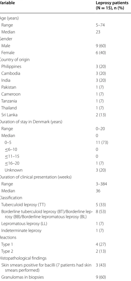
The available data were reviewed with focus on demo-graphic data, clinical findings, diagnostic procedures and treatment. The results of these findings are summarized in Table 1.
Leprosy was predominately imported to Denmark from South- and Southeast Asia (87 %), in particular India, The Philippines and Cambodia, all areas where leprosy is endemic; only two cases originated from African coun-tries. Age of patients ranged from 5 to 74 years, and 11 of 15 patients (73 %) debuted in their second or third dec-ade of life. The youngest patient (5-years-old) was a girl who had most probably contracted the infection from her father, who had been treated for leprosy in Nepal.
Three patients (20 %) recalled exposure to leprosy. Five patients (33 %) were diagnosed and had also undergone treatment for leprosy before migrating to Denmark, while one patient (7 %) was defined as a relapse after con-sulting a leprologist in London.
Diagnosis was based on travel history, clinical and path-ological findings. Most patients were classified according to the Ripley and Jopling scheme. Among the 15 patients 53 % were classified as borderline leprosy types evenly distributed along the spectrum, whilst 33 and 7 % were classified as TT and LL respectively. Duration of symp-toms varied from 3 weeks to 8 years, but it was only pos-sible to deduce the length of delay in diagnosis from a few medical records since the date of the first contact with a medical professional was rarely mentioned. For the few for whom the dates were available, the time period from contact with a doctor to suspicion of leprosy was only a few weeks.
presented with the two cardinal signs: anaesthetic skin lesions and peripheral nerve thickening. The most com-mon symptom bringing the patient in contact with health authorities was skin changes mimicking conditions such as sarcoidosis and pityriasis versicolor. Initially, two lep-rosy cases (13 %) were, based on the histo-pathological
finding of granulomatous skin changes, mistaken for sarcoidosis.
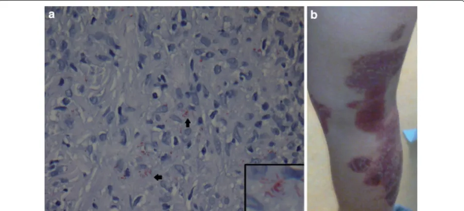
According to the records available, slit skin smears were performed in seven of the 15 patients (47 %), and the results recorded in six (40 %). The patients from whom skin smears were not obtained were all classified as TT/BT cases. Skin biopsies were obtained from all patients, often on the suspicion of other dermatologi-cal disease (Fig. 1). Bacilli were visualized in six of the 15 (40 %) skin biopsies, of which five were borderline lep-rosy types (BT, BB and BL) while one was a LL patient. Non-necrotizing granulomatous changes were found in nine biopsies (60 %). The Wade Fite stain was used on all biopsies but one. Histopathological description of biop-sies from patients with TT was only available for the one patient who experienced a relapse. This showed hyper-plasia of the epidermis, perivascular inflammation con-sisting of lymphohistiocytes, granulocytes and plasma cells and no granulomas. Biopsies from patients in the BT/BB spectrum showed epitheloid cells granulomas, giant cells and an unaffected epidermal zone, while BB/ BL was characterized by a more diffuse infiltration with or without lymphocytes, no giant cells and in some cases, ‘foamy’ macrophages and AFB. Confluent infiltrates of histiocytes with neutrophile granulocytes and loads of AFB was the main finding in the LL relapse patient. PCR for detection of M. leprae was not performed in any of the 15 cases.
Four of the 15 patients (27 %) experienced a type 1 reaction of which two were classified as borderline lep-romatous and diagnosed as downgrading reactions before initiation of leprosy treatment while the remain-ing presented with type 1 reaction symptoms shortly after initiation of leprosy treatment. The patients with downgrading type 1 reactions displayed the following symptoms: appearance of new anaesthetic skin elements and signs of increased inflammation of the existing ones. Both patients were feverish, one of them with increased C- reactive protein and neutrocytosis. Arthralgia and dactylitis were also present in both cases, while only one of them developed nerve palsy (drop foot). One patient experienced a type 1 reaction shortly after treatment initiation in Cameroun, before arriving in Denmark; the only symptom mentioned in the medical records regard-ing this was nerve palsy (peroneus paralysis). Half of the patients experiencing a type 1 reaction were treated with corticosteroids.
[image:6.595.59.289.128.620.2]The patients reacting with ENL (13 %) were BL/ LL patients experiencing fever, universal lymphad-enitis, flaring of skin elements in the form of inflamed noduli on the upper extremities. One had enlargement of the ulnar and auriuclaris magnus nerves, which was
Table 1 Summary of demographic and clinical features of leprosy patients in Denmark recorded from 1980 to 2010
Variable Leprosy patients
(N = 15), n (%)
Age (years)
Range 5–74
Median 23
Gender
Male 9 (60)
Female 6 (40)
Country of origin
Philippines 3 (20)
Cambodia 3 (20)
India 3 (20)
Pakistan 1 (7)
Cameroon 1 (7)
Tanzania 1 (7)
Thailand 1 (7)
Sri Lanka 2 (13)
Duration of stay in Denmark (years)
Range 0–20
Median 0
0–5 11 (73)
≤6–10 0
≤11–15 0
≤16–20 1 (7)
Unknown 3 (20)
Duration of clinical presentation (weeks)
Range 3–384
Median 36
Classification
Tuberculoid leprosy (TT) 5 (33)
Borderline tuberculoid leprosy (BT)/Borderline lep-rosy (BB)/Borderline lepromatous leplep-rosy (BL) 8 (53)
Lepromatous leprosy (LL) 1 (7)
Indeterminate leprosy 1 (7)
Reactions
Type 1 4 (27)
Type 2 2 (13)
Histopathological findings
Skin smears positive for bacilli (7 patients had skin
smears performed) 3 (43)
treated with a 1 week thalidomide course while the other received prednisolone due to systemic symptoms with haematuria.
Most common complication/sequelae were decreased sensibility, particularly on the peripheral extremi-ties, ulcers and secondary infections due to neuropa-thy, as well as keratosis and atrophy of fingers and toes. These patients were referred to an orthopaedist and/ or orthopaedic surgeon for evaluation and treatment. Two patients had undergone amputation of fingers/toes, in total grade 2 disability (G2D) was recorded for three patients (20 %). One of three patients with G2D had the amputations done in Cameroun, while another had received several years of leprosy treatment in Thailand, and had been living with G2D for some time when a new reaction (ENL) brought him to a physician in Denmark; bringing G2D to 6 % in our patient sample.
Some patients experienced improvement of their sen-sory disturbances during treatment but none had com-plete remission.
Skin lesion with findings of granulomatous changes in skin biopsies often led to the suspicion of sarcoidosis. Other differential diagnoses mentioned were mostly der-matological: acne, pityriasis versicolor, erythema multi-forme and Sweets syndrome.
All patients, diagnosed with leprosy after 1982, when WHO launched the use of MDT, received this regi-men. Duration of treatment varied and was not for all cases in accordance with WHO recommendations. Some received prolonged MDT (up to 12 years) due to
continued presence of bacilli in skin samples, despite clinical improvement.
Conclusion
Leprosy remains a sporadic disease imported to Den-mark. With less than one case detected per year it remains difficult for doctors in non-endemic countries to recognize or even to think of leprosy, which may lead to delay of diagnosis and treatment [52]. Delayed diagno-sis may lead to blindness, permanent nerve-damage and other disabilities. Even though anaesthesia of skin lesions is an important distinguishing feature it is often not rec-ognized by the patient.
According to some leprologists there is a predomi-nance of male leprosy patients after puberty, this was also the case in our patient sample. Skin smears were not per-formed in 53 % of the patients and although these were classified as TT/BT cases not expected to be skin smear positive, skin smears should be performed in all patients suspected for leprosy. Only one patient could recall close contact to an infectious leprosy patient in the household.
Skin biopsies, perhaps because of diagnostic uncer-tainty, were performed in all patients. Demonstration of acid fast bacilli was not possible in seven of the 15 (60 %) skin biopsies; therefore exposure, travel history and clini-cal presentation were essential to supplement histology. The more sensitive PCR for M. leprae could have proven helpful in these cases [53].
Classification of the disease is important for several reasons: borderline patients are unstable and at risk
Fig. 1 a 22-year-old Filipino woman with leprosy initially suspected for sarcoidosis. The patient was designated as a case of BT/BB with a type 1 reaction (a). WADE FITE special stain of skin biopsy showing the acid fast M. leprae bacilli. bSlightly elevated, scaly and light coppery coloured plaques
[image:7.595.60.538.90.307.2]of experiencing type 1 reaction that can lead to serious nerve damage, while lepromatous and borderline lepro-matous leprosy is associated with type 2 reactions which often are complicated by systemic effects. Both these conditions require corticosteroids and close monitoring of the clinical course due to the tendency to recur caus-ing further damage [54, 55]. Another aspect makcaus-ing clas-sification important is when choosing treatment regimen: cf. WHO’s recommendations which are only 6 months for paucibacillary against 12 months for multibacillary leprosy. Skin samples can be positive for non-viable-bacilli years after completion of MDT, and this is not an indication for prolongation of treatment. Unfortunately, viability may be difficult to determine, unless the bacilli are looking disintegrated/discoloured at microscopy. There are no standard international guidelines for how often and how long after completion of MDT a patient should be examined with skin smears. It is also unclear how long a patient should be followed after ending treat-ment. WHO recommends skin smears at the start of treatment and if relapse/deterioration is suspected. Lead-ing leprologists prefer to discharge patients if they dis-play no complications and are smear negative, except MB patients, who are followed for 2 years after completion of MDT, whilst patients with permanent eye and/or nerve damage are followed lifelong.
Due to the long incubation period, the diagnosis should be kept in mind when encountering patients from endemic areas displaying dermatological or neurological symptoms even years after immigration. Special atten-tion and care should be given to detecatten-tion and treatment of complications, in particular to those with neuropathy, which can be lasting. Anaesthetic skin lesions, which were the most common clinical finding in our review, should lead to a high suspicion of leprosy.
Consent
The Danish National Board of Health granted permis-sion to publish this case report. Written informed con-sent was obtained from the patient for publication of case reports and any accompanying images.
Abbreviations
AFB: acid fast bacilli; BB: borderline leprosy; BL: borderline lepromatous lep-rosy; BT: borderline tuberculoid leplep-rosy; ENL: erythema nodosum leprosum; G2D: grade 2 disability; IL: interleukin; INF-γ: interferon -γ; LL: lepromatous leprosy; MB: multibacillary; MDT: multidrug therapy; PB: paucibacillary; SSI: Statens Serum Institut; TT: tuberculoid leprosy; TNF-α: Tumor Necrosis Factor; WHO: World Health Organization.
Additional file
Additional file 1. CARE checklist was followed and used as a guideline for the development of the case report for this manuscript.
Authors’ contributions
HA conducted the literature search, interpreted the data and drafted the manuscript. SDN and ICB conceived of the study and revised the manuscript. All authors read and approved the final manuscript.
Author details
1 Department of Infectious Diseases and Rheumatology, Rigshospitalet,
Copenhagen, Denmark. 2 Global Health Section, Department of Public Health,
University of Copenhagen, Copenhagen, Denmark. Acknowledgements
The authors are very grateful to Professor Diana Lockwood for her insightful comments and recommendations on leprosy treatment and monitoring, and to Professor Sebastian Lucas for improving the histopathological diagnoses. Competing interests
The authors declare that they have no competing interests. Received: 1 September 2014 Accepted: 30 November 2015
References
1. Richards P. Leprosy in Scandinavia: a discussion of its origins, its survival, and its effect on Scandinavian life over the course of nine centuries. Centaurus. 1960;7:101–33.
2. Monot M, et al. On the origin of leprosy. Science. 2005;308(5724):1040–2. 3. Christensen VB, Boldsen JL. Medieval leprosy. Ugeskr Laeger.
2001;163(51):7248–50.
4. Vogelsang TM. Leprosy in Norway. Med Hist. 1965;9:29–35. 5. Waters MF. Chemotherapy of leprosy–current status and future
pros-pects. Trans R Soc Trop Med Hyg. 1993;87(5):500–3.
6. Global leprosy update. 2013; reducing disease burden. Wkly Epidemiol Rec. 2014;89(36):389–400.
7. Britton WJ, Lockwood DN. Leprosy. Lancet. 2004;363(9416):1209–19. 8. Talhari S, et al. Hansen’s disease: a vanishing disease? Mem Inst Oswaldo
Cruz. 2012;107(Suppl 1):13–6.
9. Bakker MI, et al. Risk factors for developing leprosy–a population-based cohort study in Indonesia. Lepr Rev. 2006;77(1):48–61.
10. Matsuoka M, et al. Genotyping of Mycobacterium leprae on the basis of the polymorphism of TTC repeats for analysis of leprosy transmission. J Clin Microbiol. 2004;42(2):741–5.
11. Beyene D, et al. Nasal carriage of Mycobacterium leprae DNA in healthy individuals in Lega Robi village, Ethiopia. Epidemiol Infect. 2003;131(2):841–8.
12. Salipante SJ, Hall BG. Towards the molecular epidemiology of Mycobac-terium leprae: strategies, successes, and shortcomings. Infect Genet Evol. 2011;11(7):1505–13.
13. Fine PE, et al. Household and dwelling contact as risk factors for leprosy in northern Malawi. Am J Epidemiol. 1997;146(1):91–102.
14. Joyce MP. Historic aspects of human susceptibility to leprosy and the risk of conjugal transmission. Mem Inst Oswaldo Cruz. 2012;107(Suppl 1):17–21.
15. Mira MT, et al. Chromosome 6q25 is linked to susceptibility to leprosy in a Vietnamese population. Nat Genet. 2003;33(3):412–5.
16. Mira MT, et al. Susceptibility to leprosy is associated with PARK2 and PACRG. Nature. 2004;427(6975):636–40.
17. Blackwell JM. Genetics of host resistance and susceptibility to intramac-rophage pathogens: a study of multicase families of tuberculosis, leprosy and leishmaniasis in north-eastern Brazil. Int J Parasitol. 1998;28(1):21–8. 18. Jensen AG, et al. Leprosy–still a possibility in Denmark. Ugeskr Laeger.
1992;154(24):1712–3.
19. Suzuki K, et al. Infection during infancy and long incubation period of leprosy suggested in a case of a chimpanzee used for medical research. J Clin Microbiol. 2010;48(9):3432–4.
• We accept pre-submission inquiries
• Our selector tool helps you to find the most relevant journal • We provide round the clock customer support
• Convenient online submission • Thorough peer review
• Inclusion in PubMed and all major indexing services • Maximum visibility for your research
Submit your manuscript at www.biomedcentral.com/submit
Submit your next manuscript to BioMed Central
and we will help you at every step:
21. Rodrigues LC, Lockwood D. Leprosy now: epidemiology, progress, chal-lenges, and research gaps. Lancet Infect Dis. 2011;11(6):464–70. 22. Ridley DS, Jopling WH. Classification of leprosy according to immunity.
A five-group system. Int J Lepr Mycobact Dis Off Organ Int Lepr Assoc. 1966;34(3):255–73.
23. Walker SL, Lockwood DN. The clinical and immunological features of leprosy. Br Med Bull. 2006;77–78:103–21.
24. Sieling PA, Modlin RL. Cytokine patterns at the site of mycobacterial infec-tion. Immunobiology. 1994;191(4–5):378–87.
25. Moraes MO, et al. Cytokine mRNA expression in leprosy: a possible role for interferon-gamma and interleukin-12 in reactions (RR and ENL). Scand J Immunol. 1999;50(5):541–9.
26. Madan NK, Agarwal K, Chander R. Serum cytokine profile in leprosy and its correlation with clinico-histopathological profile. Lepr Rev. 2011;82(4):371–82.
27. Britton WJ. Immunology of leprosy. Trans R Soc Trop Med Hyg. 1993;87(5):508–14.
28. de Jong R, et al. IL-2 and IL-12 act in synergy to overcome antigen-specific T cell unresponsiveness in mycobacterial disease. J Immunol. 1997;159(2):786–93.
29. Kaplan G, et al. An analysis of in vitro T cell responsiveness in lepromatous leprosy. J Exp Med. 1985;162(3):917–29.
30. Attia EA, et al. Circulating CD4 + CD25 high FoxP3 + T cells vary in differ-ent clinical forms of leprosy. Int J Dermatol. 2010;49(10):1152–8. 31. Palermo ML, et al. Increased expression of regulatory T cells and
down-regulatory molecules in lepromatous leprosy. Am J Trop Med Hyg. 2012;86(5):878–83.
32. Kumar S, et al. Th3 immune responses in the progression of leprosy via molecular cross-talks of TGF-beta, CTLA-4 and Cbl-b. Clin Immunol. 2011;141(2):133–42.
33. Polycarpou A, Walker SL, Lockwood DN. New findings in the pathogene-sis of leprosy and implications for the management of leprosy. Curr Opin Infect Dis. 2013;26(5):413–9.
34. WHO Expert Committee on Leprosy. World Health Organ Tech Rep Ser. 1998;874:1–43.
35. Kumar B, Sinha R, Sehgal S. High incidence of IgG antibodies to phenolic glycolipid in non-leprosy patients in India. J Dermatol. 1998;25(4):238–41. 36. Baeza I, et al. Lepromatous leprosy patients produce antibodies that
recognise non-bilayer lipid arrangements containing mycolic acids. Mem Inst Oswaldo Cruz. 2012;107(Suppl 1):95–103.
37. Spencer JS, et al. Identification of serological biomarkers of infection, dis-ease progression and treatment efficacy for leprosy. Mem Inst Oswaldo Cruz. 2012;107(Suppl 1):79–89.
38. Bang PD, et al. Evaluation of polymerase chain reaction-based detec-tion of Mycobacterium leprae for the diagnosis of leprosy. J Dermatol. 2009;36(5):269–76.
39. Banerjee S, et al. Diagnosing leprosy: revisiting the role of the slit-skin smear with critical analysis of the applicability of polymerase chain reac-tion in diagnosis. Int J Dermatol. 2011;50(12):1522–7.
40. Natrajan M, et al. Histological diagnosis of early and suspicious leprosy by in situ PCR. Indian J Lepr. 2012;84(3):185–94.
41. World Health Organization expert commitee on Leprosy. Summary of Current Recommendations for Management of Leprosy in the Field, 1998. WHO Technical Report Series.
42. Setia MS, et al. Is there a role for rifampicin, ofloxacin and minocycline (ROM) therapy in the treatment of leprosy? Systematic review and meta-analysis. Trop Med Int Health. 2011;16(12):1541–51.
43. Walker SL, Lockwood DN. Leprosy type 1 (reversal) reactions and their management. Lepr Rev. 2008;79(4):372–86.
44. Wagenaar I, et al. Two randomized controlled clinical trials to study the effectiveness of prednisolone treatment in preventing and restoring clinical nerve function loss in leprosy: the TENLEP study protocols. BMC Neurol. 2012;12:159.
45. Cuevas J, et al. Erythema nodosum leprosum: reactional leprosy. Semin Cutan Med Surg. 2007;26(2):126–30.
46. Kahawita IP, Lockwood DN. Towards understanding the pathol-ogy of erythema nodosum leprosum. Trans R Soc Trop Med Hyg. 2008;102(4):329–37.
47. Van Veen NH, et al. Interventions for erythema nodosum leprosum. A Cochrane review. Lepr Rev. 2009;80(4):355–72.
48. WHO guide to the management of ENL reactions. http://www.who.int/ lep/research/WHOenlguide.pdf?ua=1. Retrieved from the WHO topic page “Leprosy elimination” (http://www.who.int/lep/mdt/clofazimine/ en/).
49. Sales AM, et al. Double-blind trial of the efficacy of pentoxifylline vs thalidomide for the treatment of type II reaction in leprosy. Braz J Med Biol Res. 2007;40(2):243–8.
50. Ustianowski AP, Lawn SD, Lockwood DN. Interactions between HIV infec-tion and leprosy: a paradox. Lancet Infect Dis. 2006;6(6):350–60. 51. Menezes VM, et al. Epidemiological and clinical patterns of 92 patients
co-infected with HIV and Mycobacterium leprae from Rio de Janeiro State, Brazil. Trans R Soc Trop Med Hyg. 2014;108(2):63–70.
52. Forno C, et al. The difficulty in diagnosis and treatment of leprosy. J Travel Med. 2010;17(4):281–3.
53. Martinez AN, et al. PCR-based techniques for leprosy diagnosis: from the laboratory to the clinic. PLoS Negl Trop Dis. 2014;8(4):e2655.
54. Lockwood DN. Steroids in leprosy type 1 (reversal) reactions: mechanisms of action and effectiveness. Lepr Rev. 2000;71(Suppl):S111–4.