A CLINICAL STUDY OF ABDOMINAL WOUND
DEHISCENCE
DISSERTATION SUBMITTED TO
THE TAMILNADU DR.M.G.R. MEDICAL UNIVERSITY
CHENNAI
In partial fulfilment of
the requirements for the degree of
MASTER OF SURGERY
In
GENERAL SURGERY
DEPARTMENT OF GENERAL SURGERY
TIRUNELVELI MEDICAL COLLEGE
TIRUNELVELI
CERTIFICATE BY THE GUIDE
This is to certify that the dissertation entitled “A CLINICAL STUDY OF
ABDOMINAL WOUND DEHISCENCE” is a bonafide research work done by DR. A. CHUDAR, Post Graduate M.S student in Department of General Surgery, Tirunelveli medical college & Hospital, Tirunelveli, in fulfilment of the
requirement for the degree of Master of Surgery in General Surgery.
Date:
Place: Tirunelveli
Dr. CELINE FOUSTINA MARY M.S.,
Associate Professor of General Surgery, Tirunelveli Medical College & Hospital,
CERTIFICATE BY THE HEAD OF THE DEPARTMENT
This is to certify that the dissertation entitled “A CLINICAL STUDY OF
ABDOMINAL WOUND DEHISCENCE” is bonafide and genuine research
work carried out by DR. A. CHUDAR, Post Graduate M.S student in Department
of General Surgery, Tirunelveli medical college & Hospital, Tirunelveli under
the guidance of Dr. CELINE FOUSTINA MARYM.S., Associate Professor,
Department of General Surgery, Tirunelveli Medical College Tirunelveli in
partial fulfilment of the requirements for the degree of M.S inGENERAL
SURGERY.
Date:
Place: Tirunelveli
Prof. Dr. VARADARAJAN M.S.,
Professor and HOD, Department of General Surgery, Tirunelveli medical college & Hospital,
CERTIFICATE BY THE HEAD OF INSTITUTION
This is to certify that the dissertation entitled “A CLINICAL STUDY OF
ABDOMINAL WOUND DEHISCENCE” is abonafide and genuine research
work carried out by DR. A.CHUDAR, PostGraduate M.S student in Department
of General Surgery, Tirunelveli medicalcollege & Hospital, Tirunelveli under the
guidance of Dr.R. CELINE FOUSTINA MARYM.S M.S. Associate Professor,
Department of General Surgery, Tirunelveli Medical College, Tirunelveli in
partial fulfilment of the requirements for the degree of M.S in GENERAL
SURGERY.
Date:
Place: Tirunelveli
Dr. S. M. KANNAN M.S., M.Ch.,
The Dean,
DECLARATION BY THE CANDIDATE
I hereby declare that the dissertation entitled “A CLINICAL STUDY OF
ABDOMINAL WOUND DEHISCENCE” is abonafide and genuine research
work carried out by me under the guidance of Dr. CELINE FOUSTINA MARY
M.S.,Professor, Department of General Surgery,Tirunelveli Medical College, Tirunelveli.
Dr. A. CHUDAR,
Date: Postgraduate in General Surgery,
Place: Tirunelveli Tirunelveli Medical College & Hospital,
ACKNOWLEDGEMENT
I express my deep sense of gratitude and indebtedness to my respected
teacher and guide Dr. CELINE FOUSTINA MARY M.S. Associate Professor,
Department of General Surgery, Tirunelveli Medical College, Tirunelveli, whose valuable guidance and constant help have gone a long way in the preparation of this dissertation.
I am also thankful to Assistant Professors Dr. Karthiyayini M.S,
Dr. S. Amalan M.S, Dr. Deepan Karthik M.S., for their help.
I express my thanks to all of the staff members of the Department Of General Surgery and all my Postgraduates colleagues and friends for their help during my study and preparation of this dissertation and also for theirco-operation.
I always remember my family members for their everlasting blessings and encouragement.
Lastly, I express my thanks to my patients without whom this study would not have been possible.
Dr.A.CHUDAR, Postgraduate in General Surgery,
Date: Tirunelveli Medical College,
CERTIFICATE – II
This is certify that this dissertation work title “A CLINICAL STUDY OF
ABDOMINAL WOUND DEHISCENCE” of the candidate Dr. A. CHUDAR
with registration Number 221611353 for the award of M.S. Degree in the branch
of GENEARL SURGERY (I). I personally verified the urkund.com website for the purpose of plagiarism check. I found that the uploaded thesis file contains
from introduction to conclusion page and result shows 6 percentage of
plagiarism in the dissertation.
S.No TABLE OF CONTENTS Page No
1. INTRODUCTION 1
2. AIMS AND OBJECTIVES 3
3. REVIEW OF LITERATURE 4
4. MATERIALS AND METHODS 47
5. RESULTS 49
6. DISCUSSION 61
7. CONCLUSION 67
8. BIBLIOGRAPHY
9. ANNEXURE:
(a) PROFORMA
(b) MASTER CHART
1
INTRODUCTION
Abdominal wound dehiscence, also known as burst abdomen, acute
wound failure, wound disruption, evisceration or eventration, remains one
of the most dramatic and serious developments confronting the general
surgeon. Few postoperative events cause such morbidity, and when
accompanied by necrotizing fasciitis, none is as potentially disfiguring.
Abdominal wound dehiscence is defined as the postoperative separation of layers of a laparotomy wound, with or without eventration. Despite major advances in the preoperative care of surgical patients, including the introduction of broader spectrum antibiotics and an
improved understanding of the effects of systemic illness on wound
healing, the incidence of abdominal wound dehiscence has remained
constant at 0.4 to 3.0%.1, 2
Two general factors play contributory roles in causing wound
dehiscence - metabolic and local anatomic abnormalities and technical factors. Many aspects of the latter are within the surgeon’s control, such as
the site of the abdominal incision, technique of closure and type of suture
employed, the use of retention sutures, and the placement of drains and
enterostomies in relation to the wound. Metabolic abnormalities are
commonly corrected before elective operations, a factor which increases
2
unalterable variables of patient age, the procedure itself - whether it be
elective, emergent, clean, or contaminated, and associated systemic illness
have been shown to be contributory.3-7
Although specific guidelines describe the reoperative management of
abdominal wound dehiscence, more important is recognition at initial
operation of the patient who is at risk for wound separation as well as
3
AIMS AND OBJECTIVES
To study the clinical profile of patients with the diagnosis of
abdominal wound dehiscence with respect to:
age distribution
gender incidence
clinical presentation
nature of preceding surgery (elective or emergency)
contributing factors - local (type of incision, suture material used for
abdominal closure) and systemic (anemia, hypoproteinemia, postoperative
infection, postoperative pulmonary complications, obesity, comorbid
conditions, drug use)
management
4
REVIEW OF LITERATURE SURGICAL ANATOMY OF ABDOMINAL WALL7
The abdominal wall is a complex musculoaponeurotic structure that is
attached to the vertebral column posteriorly, the ribs superiorly, and the
pelvic bones below. The abdominal wall is composed of nine layers (Fig.1).
From without in, they are:
(1) Skin
(2) Tela subcutanea (subcutaneous tissue)
(3) Superficial fascia (Scarpa fascia)
(4) External abdominal oblique muscle
(5) Internal abdominal oblique muscle
(6) Transversus abdominis muscle
(7) Endoabdominal (transversalis) fascia
(8) Extraperitoneal adipose and areolar tissue
(9) Parietal peritoneum
The rectus muscles and rectus sheath require special description. The muscles are paired right and left, and they extend from the fifth rib
superiorly to the pubis inferiorly. They lie in apposition to each other,
separated only by the linea alba. The rectus muscles serve to support the
5
within a fascial sheath, the rectus sheath, which is derived from the
aponeuroses of the three flat abdominal muscles.
The relationship of the aponeuroses of the flat muscles is not
constant throughout the course of the rectus muscle. The relationship is
different above and below the semicircular line of Douglas, which is about
halfway between the umbilicus and the pubic symphysis (Fig.2). Above
the semicircular line, the rectus sheath is strong posteriorly. Here the
posterior sheath is composed of fascia from the internal oblique muscle,
the transversus abdominis muscle, and the transversalis fascia. Anteriorly,
above the semicircular line, the rectus sheath is composed of the external
oblique aponeurosis and the anterior lamella of the internal oblique
aponeurosis. Below the semicircular line, which is the point at which the
inferior epigastric artery enters the rectus sheath, the posterior rectus
sheath is lacking because the fasciae of the flat muscles pass anterior to the
rectus muscle. The muscle, below the semicircular line, is covered
posteriorly by a thin layer of transversalis fascia, which is usually
transparent when viewed from the inside at operation. The rectus
abdominis muscles are held close together near the anterior midline by the
linea alba. The linea alba itself has an elongated triangular shape and is
6
considerably below the umbilicus, so that the medial edge of one rectus
7
PHYSIOLOGY OF WOUND HEALING8
A wound initially is tissue that has lost normal structure and
functions as the result of internal or external forces. Wound healing is the
sequence of cellular and molecular events activated at the time of injury
resulting in a time-dependent pattern of tissue repair. Classically, the
phases of wound healing are described as hemostasis, inflammation,
fibroproliferation, and remodeling (maturation).
(a) Hemostasis:
Before a wound can heal it must stop bleeding. Therefore, the earliest
phase of wound healing following injury is characterized by the deposition
of fibrinogen, a soluble plasma protein synthesized by the liver and
secreted into the systemic circulation. Fibrinogen extravasates from
disrupted blood vessels and fills the gap of the wound.
(b) Inflammation:
The cellular and humoral inflammatory phase is induced next, and
an immune barrier is established against pathologic microorganisms.
Necrotic tissue locally releases cellular breakdown products capable of
maintaining and amplifying the early inflammatory response following
injury. The increased permeability of vessels adjacent to the injury
facilitates the migration of inflammatory cells into the wound which leads
8
microbes and release of cytochemoattractants. Hence if a wound infection
develops, healing will be delayed. Circulating monocytes enter the wound
in a second wave of inflammatory cells within 24 hours after the
appearance of neutrophils. Monocytes terminally differ into tissue
macrophages. Macrophages are vital for clearing the wound of microbes
and devitalized tissue, as well as for the production of a new connective
tissue matrix.
(c) Fibroproliferation and Remodeling:
Once hemostasis is achieved, ongoing injury has ceased, and an
immune barrier is in place, wound healing trajectories shift toward
fibroplasia and tissue repair. Scar tissue replaces normal tissue following
injury and is often a source of subsequent wound complications. Over
time, wound matrix cell number diminishes and collagen bundles are
increasingly organized during remodeling. This final phase of wound
healing can continue for years until a maximum wound strength plateau is
finally reached. In dermal wounds, overlying epidermal cells begin to migrate across the tissue defect at about this time to restore the skin’s
epithelial barrier function. Collagen is the major protein component of
wound connective tissue. Unwounded dermis contains approximately 80%
type I collagen and 20% type Ill collagen. Acute wound granulation tissue,
9
synthesis and secretion requires hydroxylation of lysine and proline
residues. The cofactors necessary for enzymatic collagen hydroxylation are
ferrous iron, molecular oxygen, a-ketoglutarate, and vitamin C. Impaired
wound healing results from deficiencies in any of these cofactors, as
during tissue hypoxia or with diets low in vitamin C. In acute wound
granulation tissue mature collagen fibers are oriented in overlapping arrays
parallel to the wound surface and usually along lines of maximum tension.
When the wound defect is filled, the maturing granulation tissue undergoes
remodeling. The density of macrophages and fibroblasts is reduced. There
is also no regeneration of lost sub epidermal appendages such as hair
follicles or sweat glands following skin healing.
The Lag Phase
The ―lag phase‖ of wound healing is defined as the earliest period
of time following wounding when hemostasis, inflammation, and early
fibroplasias are induced. It is during the lag phase of wound healing that
acute wounds are most vulnerable to mechanical failure (dehiscence). The
wound tensile strength is 0% to 30% of its maximum value during the first
7 days following wounding.
Wound failure occurs when there is an abnormality in the magnitude
or duration of the sequential components of tissue repair. Inadequate
10
hematoma formation with ensuing mechanical disruption of the provisional
wound matrix. Delayed or deficient inflammatory responses increase the
risk of wound contamination or infection. A prolonged inflammatory
response due to foreign material delays the progression of tissue repair into
the fibroproliferative phase in which rapid gains in breaking strength and
wound contraction should occur. Impaired fibroblast activation in turn
impedes the establishment of the early wound matrix and synthesis of
immature scar. Epithelialization requires an underlying functional bed of
granulation tissue. Obstacles to normal wound healing therefore shift the
11
EPIDEMIOLOGY SCOPE OF THE PROBLEM: INCIDENCE
Despite major advances in the perioperative care of surgical patients, the incidence of abdominal wound dehiscence has remained constant at 0.4 to
3.0%.1, 2
The comprehensive literature review by Poole9 in 1985 investigating
the incidence of AWD encompassed some 320,000 abdominal operations
performed during the past 35 years. Overall there were approximately 1900
dehiscences, yielding a mean incidence of 0.59%. Poole and others,
however, are critical of the accuracy of this low rate, citing as an
explanation the inclusion of muscle-splitting appendicectomy incisions,
herniorhaphies, and gynaecologic procedures in many of the trials. The
incidence of AWD with these incisions is sufficiently low that their
inclusion artificially lowers the incidence realized in major abdominal
surgery. Based on the above observation, Poole proposed that a more
accurate estimate in general abdominal surgery was 1 to 3%. To avoid
ambiguity, we define abdominal wound dehiscence as the postoperative
separation of all three layers of a laparotomy wound, with or without
12
Table 1: Incidence of AWD in various clinical trials:
Author Year No. of Dehiscences Primary variable patients (%)
Irvin et al10 1977 200 2(1.0) Closure(layered vs.
mass)
Greenall et al1 1980 577 2(0.4) Incision(midline vs.
transverse)
Guillou11 1980 207 1(0.5) Incision(midline vs.
paramedian)
Corman et al12 1981 161 1(0.6) Suture
Bucknall and 1981 210 2(0.95) Suture
Ellis13
Donaldson14 1982 231 0(0) Suture
Richards et al15 1983 571 8(1.0) Closure(interrupted vs.
continuous)
Gammelgaard 1983 306 1(0.33) Suture
and Jensen16
Ellis et al17 1984 175 1(0.6) Incision(transverse vs.
vertical)
Fagniez et al18 1985 3135 58(1.8) Closure(interrupted vs.
continuous)
13
RISK FACTORS DEFINING THE RISK : THE PATIENT
The seeds of wound disruption may be present before the patient goes to the
operating room. - W. I Wolff Patient related risk factors play an important role in the development of
AWD. Advancing age, poor vascular supply, male sex, preexisting
pulmonary disease, malnutrition and immunosuppression are the most
significant implicated factors today.
Advanced age is the single factor consistently emphasized by most
authors that coincides with a decreased ability to fight off infection. Wolff found the incidence of wound disruption in patients aged >45 years to be 4
times that in the younger age group (5.4% vs. 1.3%)3. Similar results were
noticed by McCallum and Link19, who demonstrated a threefold increase
in incidence in the older population (4.5% vs. 1.6%). The high incidence of
AWD noticed in the elderly is explained by a diminished rate of cell proliferation which has a detrimental impact on healing process.
Male gender is an additional predisposing factor emphasized in most
reviews. The ratio of males to females experiencing this complication
ranges from 1.6:1 to 3:1. The male predisposition to wound failure may be
14
abdominal wall in females following pregnancy, greater postoperative
physical activity in males, and a higher incidence of preoperative
pulmonary disorders in males resulting in excessive postoperative cough.
However, with the percentage of women smokers increasing each year, a
shift in these statistics is likely. All of these factors tend to increase wound
tension.
Preoperative pulmonary disease and subsequent postoperative
respiratory complications have a well-defined role in the development of
wound failure. Wolff reported severe paroxysmal coughing prior to wound
disruption in over 60% of cases3. Alexander and Pavdden20 and
Hampton21 also noted pulmonary complications to be the most frequent
event leading to postoperative wound disruption. With preoperative use of
bronchodilator agents, elimination of tobacco, treatment of bronchitis, and
respiratory therapy, optimal pulmonary function is achieved. Studies have
shown that cessation from smoking for as little as one week preoperatively
can lessen patients’ morbidity postoperatively and a diminished incidence
of postoperative wound failure.
Research implicating adequate nutrition in achieving secure surgical
wounds began 50 years ago with the important observations of Thompson
et al22 and Elman23, that hypoproteinemia leads to greatly impaired wound
15
experiencing postoperative wound disruption24, and reports of the
association have continued ever since. While protein deficiency is rare in the United States today, its prevalence in developing countries and occurrence in association with other diseases remains a continuing
problem. Reports by Wolff3, Alexander and Pavdden20 and Keill25 that
62%, 71% and 85% of their respective wound dehiscences were associated with hypoproteinemia emphasize the magnitude of the problem. Clearly every effort should be made to correct these protein deficiency states before elective surgery. Continued improvements in both enteral and parenteral formulas available today have made this feasible in even the most sever forms of malnourishment.
THE NATURE OF THE OPERATION
Emergency laparotomy (irrespective of the organ system involved),
gastric operations (particularly for peptic ulcer disease and haemorrhage),
and procedures involving the small and large bowel have increased rates of
dehiscence.
The rate of dehiscence following procedures on stomach, small and large bowel was found to be twice that after operations on the biliary
tree26. Whether anticipated or unexpected, the single common factor
involves some degree of peritoneal contamination and its subsequent
adverse impact on the abdominal wound6,25. Emergent laparotomy
16
failure is not well defined. Penninckx et al reported an incidence of dehiscence in emergent laparotomies of 6.7%, more than two-and- a-half
times that observed in elective cases27. This parallels the incidence
reported by Mendoza et al of 6.2% during emergent gastroduodenal
surgery for haemorrhage28. Presumably, similar factors are operative in
these emergent cases as in those involving the GIT, particularly some degree of peritoneal contamination. These emergencies may be associated with a break in technique as a result of hurried operation in an unstable patient or, as more frequently is the case, they may involve a procedure on an open unprepared bowel. One must also take into account the mechanism of the injury leading to the emergent laparotomy. Regardless of the cause, the common factor is wound contamination, and unless extra preventive measures are undertaken, dehiscence on the basis of infection may ensue.
Carcinoma leads to cachexia and marked anemia due to anorexia,
haemorrhage and bone marrow depression. The effect of cancer on wound
healing was studied by Wyatt et al68, who found that, although wound
healing may proceed in a relatively unimpeded manner for many patients
with cancer, there is a potential for wound failure due to the nature and
effects of the oncologic disease process and its treatments.
While pulmonary function should be optimized and nutritional
deficiencies corrected before elective surgery, little can be done
17
male sex, as well as the requirement for an emergency procedure or one
involving the gastrointestinal tract, are unalterable variables. Although the
presence of one of these alone may be relatively harmless, their presence in
combination should be considered an indication for measures to consider
dehiscence a realistic possibility.
LOCAL FACTORS CONTRIBUTING TO DEHISCENCE: THE INCISION
Deciding the most appropriate incision for a given patient is based a
variety of factors. Considerations, in order of importance, include
(1) Access afforded;
(2) Expediency of entry and closure (particularly in the unstable
trauma patient);
(3) Relative postoperative pain and pulmonary complications
(especially in the face of chronic pulmonary disease); and
(4) In the presence of multiple risk factors, the associated incidence
of postoperative wound disruption and incisional hernia formation.
The major controversy during the past 50 years has involved
transverse versus vertical abdominal incisions.
The musculoaponeurotic fibers of all three layers of the abdominal
wall run in a predominantly transverse direction. Active contraction of the
18
laterally away from the midline. These forces run perpendicular and in
opposing directions on either side of a vertical incision, thereby tending to
distract the wound edges. Transverse incisions are affected little by these
forces.
Sloan demonstrated a 30-fold increase in wound tension in vertical
as opposed to transverse incisions in lightly anesthetized patients29.
Subsequent publications by Hampton, McCallum, and Lehman and
Partington also cited excessive disruption rates with the vertical
wound19,21,30. Simultaneously several authors documented less
postoperative pain and concomitantly fewer postoperative pulmonary
complications when transverse incisions were used. The resurgence of
vertical incision popularity may be related to the exposure they provide
when aided by the new fixed retractors. In the face of trauma, the
expediency with which the abdomen may be opened and closed is of
primary importance, and continuing civilian abdominal trauma demands
this attribute of the vertical midline incision. More relevant to the
persistent popularity of the midline incision is the realization by many that
its use does not necessarily imply a sacrifice in terms of wound security.
From a strictly anatomic viewpoint, Tera and Aberg, using human
cadavers, demonstrated a clear superiority in the holding power of the
midline incision over the transverse when sutures were placed lateral to the
19
kPa)31. Similarly, Leaper et al also found the midline aponeurosis in
human cadavers to have the greatest suture-holding capacity32. Finally,
Higgins et al used a rabbit model to demonstrate that a vertical incision
closed with the Smead-Jones technique was far stronger at 7 days than a
transverse incision closed with the same suture technique33.
Currently, there is little evidence that incision orientation alone
plays a significant role in the etiology of abdominal wound dehiscence.
The nature of the operation itself, its attendant risk of wound infection, the
20
INTRAOPERATIVE PREVENTION Protecting the Wound
The time to think about and prepare for a possible wound separation is prior to and during operation, not after disruption has occurred.
- Lehman and Partington
Once the peritoneal cavity has been opened, every effort should be made to
prevent contamination of the wound with potential pathogens. This is
frequently more difficult in a reoperation than in a primary procedure.
Since dehiscence occurs far less frequently than wound sepsis, and often in
its absence, it is difficult extrapolate from wound infection to a specific
incidence of dehiscence. Suffice it to point out that wound infections were
noted in 72% of dehiscences reviewed by Keill et al, whereas the control
population of intact wounds had a 3% infection rate25. Thus, while local
mechanical factors contribute heavily to subsequent wound disruption, the
effect of infection on tissue strength cannot be underestimated.
The high dehiscence rates following gastric operations or those
involving large or small intestine were described earlier. The relationship
between intraoperative contamination of the peritoneal cavity and
subsequent wound infection is well established and continues to be
21
Any violation of the gastrointestinal tract, be it iatrogenic, for
decompressive purposes, or for resection of gangrenous bowel, will be
accompanied by significant wound contamination, and if skin is closed, the
risk of incisional infection is increased. Raahave et al have quantitatively
defined this risk by demonstrating an exponential relation ship between
intraoperative bacterial density within the wound and subsequent wound
infection37. The critical ―infective dose‖ observed in that trial was 4.6 x
105 colony-forming units per square centimeter (CFU/cm2).
In an attempt to diminish intraoperative wound involvement during
contaminated procedures, multiple different impermeable skin and wound
drapes have been introduced. Most prospective trials have been unable to
demonstrate a significant reduction in wound infections with their use38,39.
Nonetheless, the principle of avoiding wound and generalized peritoneal
contamination by isolating the pathologic area of the intestine should be
recognized as theoretically sound one to be adhered to at all times.
The use of drains in abdominal operations continues to be controversial.
Clear indications in the past have included40:
(1) anticipated leakage from an adjacent organ such as the
22
(2) isolated abscess cavities requiring drainage to achieve collapse
and progressive healing from the deepest portion outward
(3) a worrisome anastomosis as a result of tension or compromised
tissue at the suture line.
The currently accepted indication for abdominal drainage is a clear
recognition that the drain is essential to carry away infected material or
digestive enzymes, or other chemically irritating fluid, which will impair
wound healing. It is strongly recommended that drainage be performed
through a separate, more dependent stab wound in the abdominal wall,
well away from the operative incision)
Finally, an enteral stoma, be it from the stomach, small bowel, or
colon, should be considered a similar infectious hazard, particularly when
the wound is left open. This was emphasized by Wolff, who noted that as
many as 8 of 45 wound dehiscences reported were directly attributable to
bringing an enteral stoma through the operative incision3. This outcome
should be anticipated and avoided by extraincisional placement of the
stoma. Finally, the prevention of wound contamination during any
operation demands continuous attention to meticulous technique. The
delicate handling of tissue, removal of foreign material, debridement of
necrotic tissue and absolute hemostasis before closure all fall within this
23
Fascial Closure:
Choosing the appropriate suture:
Choosing the appropriate suture for a given situation requires little
more than a basic understanding of the materials available, their merits,
and their disadvantages. The effect of an inappropriate choice can be
considerable, resulting in unnecessary wound infection, draining sinuses,
incisional hernia, or dehiscence.
The ―ideal suture‖ should:
(1) have sufficient strength and maintain it until wound healing is complete,
(2) then disappear so as not to promote patient discomfort or suture
granulomas,
(3) have a low index of infectivity and reactivity so as not to promote wound
infection and inflammation,
(4) be easily handled and knot securely with minimal difficulty.
Such a suture does not exist. Yet the disadvantages of any one material
may be minimized if it is used appropriately.
For many years, the standard suture for fascial closure was alloy
steel wire. Its tensile strength was incomparable, and as a monofilament, its physical structure minimized foreign body reactivity, bacterial
24
recognized by Jones et al., who in 1941 reported a tenfold decrease in the
incidence of dehiscence (11% to 1.2%) and a pronounced reduction in the
incidence of wound infections (27.5% to 0.85%) when mass interrupted
alloy steel rather than layered continuous chromic catgut was used for
fascial closure41. Alloy steel, while preferable to catgut, is not without
problems. Most surgeons find it difficult to handle, and its propensity for
cutting through gloves is well established42. Tight knots are difficult to
achieve, and kinking may lead to fracture32,41. In addition, its permanent
nature may lead to palpable, uncomfortable knots or chronic suture sinuses
that eventually require extraction32,41,42.
It is generally accepted that healing of the midline aponeurosis, with a return of strength comparable to that of intact fascia, requires from 60 to
120 days43. Herein lies the problem with catgut. Because of its rapid
absorption, this material contributes little, if anything, to wound strength.
Catgut begins to weaken as early as 5 to 10 days postoperatively44,45.
Tagart has demonstrated that alter 5 days, in vivo catgut retains only 30% of its original strength and thereafter its support will be completely
unreliable44. The result is a wound that is unsupported and prone to
25
catgut41,42,44,46. Consequently, this material has largely been abandoned
for fascial closure.
In light of the above, research during the past years has focused on
the development of suture material that embodies the attributes of steel
(nonporous with lasting tensile strength and low infectivity) while avoiding
the poor handling characteristics and tendency toward sinus formation of
steel. Both monofilament nylon and polypropylene (Prolene) have been
developed as desirable substitutes. In surgical use, their inert,
nonabsorbable nature has demonstrated lasting strength and a low
incidence of dehiscence when used appropriately for abdominal closure.
Hermann, in 1974, first advocated the use of polypropylene after 250 consecutive mass closures with the material without a single
dehiscence47. Later, Knight and Griffen reported 1,000 consecutive
abdominal wound closures with polypropylene (including appendectomies) with an incidence of dehiscence and incisional hernia of only 0.4% and
0.7%, respectively48. Comparable results have been achieved with
monofilament nylon. The most dramatic of these was the report by Jenkins of 1,505 consecutive continuous, mass closures with nylon with only one
failure, an incidence of 0.07%49.23 Martyak and Curtis reported 280
consecutive midline wounds closed using monofilament nylon and
26
Finally, in two prospective, randomized clinical trials, Leaper et a1 and Pollock et al clearly demonstrated the equivalence of monofilament nylon and stainless steel in achieving minimal dehiscence rates of 0.56% and 0%,
respectively32. On the basis of these and other clinical trials, polypropylene
and monofilament nylon have largely supplanted the use of stainless steel in situations where the latter might be indicated, i.e., the patient at risk for dehiscence.
In general, however, most surgeons remain reluctant to use these
sutures routinely, and their reasoning is not unjustified. While more easily
handled than steel, both materials have perpetuated one of its lesser
qualities—the propensity for creating prolonged incisional discomfort and
suture granuloma or sinus formation. Despite ingenious methods bury the
knot, reports of these complications, particularly in the thin patient with
minimal subcutaneous fat, are numerous2,32,47,48.
Postlethwait et al, using histologic specimens of suture removed from
human tissue at reoperation, demonstrated the minimal reaction elicited by
permanent monofliament material in comparison to its multifilament
counterpart, silk40. While the monofilament was simply encapsulated by a
fine zone of fibrous tissue, the multifilament silk evoked a multicellular
reaction involving fibroblasts, giant cells, and lymphocytes, surrounding the
suture and within the interstices of the multiple filaments. The reason for this
27
More recently, scanning electron micrographs have been used by Bucknall to
demonstrate the intense inflammatory reaction elicited by multifilament silk
and other materials in both infected and noninfected states51. Katz et al, using
radiolabeled bacteria, have quantitated in vitro bacterial adherence to
different monofilament and multifilament sutures52. They found that
monofilament nylon bound the least bacteria while braided sutures (silk,
polyglycolic acid [Dexon, Tycron]) had bacterial adherence values fivefold
to eightfold higher. After implanting similar bacteria-coated sutures in
mice, they found that the inflammatory response or ―degree of infection‖
observed with the various sutures closely correlated with the adherence
characteristics noted in vitro. Monofilament nylon, even in the presence of
bacteria, consistently evoked a minimal, if even detectable, inflammatory
response. All of these observations give credence to the long-held concept
that synthetic, monofilament sutures, because of their structure and inert
chemistry, are relatively nonreactive and facilitate removal rather than the
harboring of bacterial organisms within the wound76.
Yet the fact that monofilament nonabsorbable sutures can result in
chronic sinuses is well established; this is evidently more due to their
permanence within the wound than to physicochemical properties. More
28
absorbable sutures, which theoretically support the wound long enough for
adequate healing and are then absorbed.
Both polyglycolic acid (Dexon), and polyglactin (Vicryl) were
introduced in the early 1970s, and each has been the subject of extensive
clinical trials since that time. Early concerns regarding the use of these
materials in abdominal wound closure focused on their absorption and
associated declining tensile strength profiles. Both materials are degraded
by hydrolysis, but while Vicryl is uniformly absent after 70 days, the
process takes somewhat longer for Dexon, which usually requires 3 to 4
months53. Loss of tensile strength, however, is rapid, with Dexon retaining
zero of its package strength after only 21 days and Vicryl similarly
retaining less than 10% of its original strength after 28 days53.
Yet despite the theoretical possibility of creating a weak wound susceptible to disruption, the use of Dexon and Vicryl has not led to an increased incidence of dehiscence. Murray and Blaisdell closed 650
consecutive abdominal and thoracic incisions54, and Bentley et al closed
814 consecutive laparotomy incisions with these sutures, with subsequent
dehiscence rates of less than 0.5% in each55. It appears unequivocal that
29
Several investigators have recently extended the concept of ―wound failure‖ to include incisional hernias, and they have considered
these a form of ―late dehiscence.‖ Given their time course of absorption
and diminution in strength, one might expect a higher incidence of this
complication with absorbable sutures. Bucknall et al, in a prospective trial
involving 1,129 major laparotomy wounds, reported a significantly higher
incidence of incisional hernias in wounds closed with mass polyglycolic
acid as opposed to mass nylon (11.5% vs. 7.2%)13. Similarly, Pollock et al,
in a prospective trial comparing steel, nylon, and polyglycolic acid, found
the highest incidence of incisional herniation (13%) when the polyglycolic
acid suture was used.56 Wissing and associates compared interrupted and
continuous closures with nylon, polydioxanone (PDS), and Vicryl.2 They
noted incisional hernias in 16.9% and 20.5%, respectively, of Vicryl
closures when they are used in an interrupted or continuous fashion. It appears, on the basis of these early results, that the ―long-term‖
absorbable sutures may simply alter the time course of wound failure such
that incisional hernia rather than dehiscence is the eventual outcome.
While the former is certainly preferable to dehiscence, it is an unfair
exchange for the occasional suture sinus that follows closure with the
30
Finally, the results of several recent trials indicate that these
absorbable sutures, despite their original intention, are not immune to the
complication of chronic sinus formation. Gammelgaard and Jensen noted
this complication in 6.5% and 11.3%, respectively, of Vicryl and Dexon
closures16. Similarly, Bucknall et al reported an identical incidence of
sinus formation (11.5%) when either nylon or polyglycolic acid was
used13. In summary, these absorbable materials, while potentially safe in
uncomplicated cases, may lead to a higher incidence of incisional hernias
and may result in suture sinus formation, a problem they were specifically
designed to avoid.
At present use of able sutures in patients at high risk for dehiscence
is sufficiently controversial so as not to be recommended. While
monofilament polypropylene and nylon may on occasion lead to chronic
suture sinuses, this problem can be virtually eliminated by an appropriate
continuous closure and adequate burying of the knots at both ends of the
incision. The result is an inherently strong wound, which should endure a
moderately stormy postoperative course without disruption.
Technique:
Many surgeons consider the technique of abdominal wound closure
to be the single most important factor in prevention of postoperative
31
techniques are ample evidence that the technical aspects of this ritual are
critical.
(a) Layer-by-layer vs. Mass Closure:
Little more than a decade ago, the first consideration at laparotomy closure would have been meticulous reapproximation of the peritoneum.
Numerous clinical trials have established that, contrary to previous thinking,
this maneuver is unnecessary and unrelated to secure wound healing25. Large
peritoneal defects heal rapidly with new serosa formation and without
increased adhesions57. Conversely, sutures that penetrate the peritoneum
elicit a substantial foreign body reaction
leading to excessive adhesions and potential intestinal obstruction57. Given
the obvious risk and doubtful benefit, a separate peritoneal closure should
be avoided. Further, fascial reapproximation should be achieved via a
preperitoneal technique in which sutures do not penetrate the peritoneum.
The layer-by-layer closure advocated by Halsted, though aesthetically
pleasing, fails to impart adequate strength to the wound. This method is
considerably more time-consuming, and it also adds significantly to the
amount of foreign material within the wound, neither of which will benefit
the patient.
The evolution of ―mass‖ closure began with the figure-of-eight
32
a subsequent clinical trial, has since been referred to as the ―Smead-Jones
far-and-near technique‖41. Strikingly impressive results were responsible
for the technique’s early popularity. Jones et al reported a tenfold
decrement in the incidence of wound disruption (11% to 1.2%) when this
method rather than the traditional layered closure was employed41.
In a more recent study from India by Sivam et al58, the early and
late results of the Smead-Jones (SJ) technique of closure of emergency
vertical midline laparotomies was compared with other conventional
methods of closure such as anatomical repair (AR), mass closure (MC) and
single layer (SL) closure. It was seen that the overall infection rate for SJ at
12.4% was significantly less than all other types of closure. The wound
dehiscence rate for SJ at 3.0% was the lowest. This protective effect of SJ
against dehiscence was also seen in the presence of post operative chest
infection and abdominal distension. The incisional hernia rate for SJ was
also lowest (4%). The appearance of the scar was comparable to the other
techniques of follow up. This study concluded that the Smead-Jones
techniques of laparotomy closure had very low incidence of early and late
complications and was superior to other conventional methods of closure.
In a similar study by Baggish et al59, a prospective study of 900
laparotomies utilizing polyglycolic acid suture material and the
Smead-Jones closure technique was carried out over a period of 1 year with a
33
Numerous prospective clinical trials using both the far-and-near as well as
the simple mass closure have shown that mass fascial closure results in
fewer dehiscences10,30,49.
(b) Interrupted vs. Continuous Sutures:
Traditionally, the interrupted mass closure using non-absorbable
sutures has been used for wounds prone to dehiscence. The trend in recent
years has been toward an increased use of the continuous suture. Its
advocates cite several advantages over the interrupted method. Chief
among these is a comparable, if not slightly lower, incidence of wound
disruption when the former was used. The dramatic results reported by
Jenkins49 (1 disruption in 1,505 continuous mass closures) were ample
evidence of the security of this technique. He emphasized that large tissue
bites, a small stitch interval, and appropriate wound tension were directly
responsible for the outcome. When performed correctly, the method uses a
length of suture four times as long as the wound. Tissue bite and stitch
interval being constant, it is this ratio (suture to wound length) that
determines wound tension at closure. The closure allows for potential
postoperative abdominal distention without sutures tearing through fascia.
This takes advantage of the accepted capability of continuous closures to
34
Poole et al noted that the continuous technique was not only
consistently stronger but that during increasing tension the suture line
would often ―shift‖ to accommodate increased stress9. In contrast,
interrupted closures would rupture suddenly, with initial disruption
occurring at the single suture under greatest stress. The authors emphasized
that fascial tearing due to wound tension is the primary mediator of
incisional dehiscence.
A review by Carlson on acute wound failure emphasized on taking
large bites of tissue during closure to prevent dehiscence60. Recent
randomized prospective clinical trials comparing interrupted and continuous
closure have confirmed the security of the latter technique15,18. In fact, in the
largest prospective trial to date (3,135 patients)18 the incidence of
dehiscence was actually higher (2.0% vs. 0.6%) when interrupted rather than continuous closure was used.
The advantages of a continuous, mass fascial closure include less
foreign material in the wound and expediency. The former assumes
increasing importance if nonabsorbable suture is used and sinus formation
about permanent suture knots is to be avoided. In regard to expediency,
most authors have shown that continuous closure reduces operative time
by about 20 minutes compared with the interrupted technique15,50. While
35
or critically ill patient who is most prone to dehiscence. Given these
advantages and the equal, if not greater, wound security provided, the
continuous mass closure with non- absorbable suture appears to be the
method of choice for wounds at high risk of dehiscence.
RETENTION SUTURES:
Most surgeons employ retention sutures in the presence of multiple risk
factors for dehiscence, or when a single risk factor is present in
combination with systemic disease sufficient to warrant concern about
adequate fascial healing. Irrespective of the cause, the intent of using
retention sutures is to hold cut fascial edges in apposition and thereby
reduce strain on the incisional suture line until adequate healing has taken
place. The ability of retention sutures to perform this function is entirely on
how they are placed in relation to the incision.
Based on findings at autopsy, Price61 demonstrated why
―conventional‖ retention sutures, which simply traversed all layers of the
abdominal wall, often failed:
(i) When sutures are place, there is a circumferential distribution of suture
tension with minimal support of the fascial aponeurosis.
(ii) With edema on the third day there is increased pressure on the soft
36
(iii) Increased suture tension causes pressure injury to tissues in skin and
fascial layers at about 7 days. The suture cuts through skin ad fascial
layers, reducing lateral support.
(iv) As edema subsides, more fascial support is lost, permitting wound
disruption.
Based on the above, Price advocated a retention suture designed
primarily to oppose the lateral distracting forces occurring at the incision.
(Fig.3, Fig.4) Large sutures traverse the midline to grasp a large bite of the
contralateral musculofascial layer; they are then brought back across to the
original side, emerging near the point of entry. Consecutive sutures are
placed on alternating sides of the wound and anchored either to an
overlying frame or to large buttons with tension appropriate to maintain
fascial apposition (Fig.5). This was followed by simple interrupted closure
of the anterior sheath.
More commonly used retention sutures of today are placed 4 to 5 cm
lateral to the incision, traverse all layers of the abdominal wall (with the
exception of peritoneum) and cross the midline beneath the mass fascial
closure just under the posterior sheath (Fig.6). The following principles
have to be adhered to during their placement:
(a)The suture should enter the skin closer to the incision than to the point at
37
be approximated without compression of the overlying skin and potential
necrosis.
(b)A suture that is large enough should be used, no. 2 polypropylene, to avoid
the tearing of fascia under considerable tension. The use of sterile
intravenous (IV) tubing has been advocated for this purpose.
(c)Peritoneal penetration has to be avoided so as to avoid the increased intra
abdominal adhesions provoked by this maneuver which can further lead to
small bowel obstruction or fistula.
If abdominal distention is present preoperatively, retention sutures that
are taut at the time of closure may become slack during convalescence,
leaving the wound susceptible to dramatic, episodic increase in tension as a
result of paroxysmal coughing or vomiting. Consequently, they should be
checked on a regular basis to ensure that tension is maintained. Depending
on the condition of the patient, it is generally safe to remove these sutures
between 14 and 21 days postoperatively.
PRESENTATION AND MANAGEMENT OF DEHISCENCE
Many a times wound disruption will occur despite extra preventive
measures taken at the initial operation. Early recognition and prompt
treatment are critical if the usual mortality of 30% is to be reduced.
In many instances, the time at which dehiscence occurs postoperatively
38
Partington30 have pointed out that wound disruptions occurring before the
fifth postoperative day cannot be attributed to poor wound healing. These
are usually the result of some technical error. This explanation accounts for
a minority of dehiscences.
The most frequent interval for dehiscence is between the seventh
and eighth postoperative days3,4,6,30,62, frequently following an episode of
severe coughing or vomiting, or progressive abdominal distention
secondary to ileus4,5,26,30,62. Its development under such circumstances
may be all too obvious and accompanied by eventration—the ―burst abdomen.‖ At other times, the only indication of a problem may be a
profuse, pink, serosanguinous incisional discharge that leads to the
removal of skin sutures revealing viscera in the wound. The pink
serosanguinous drainage is associated with dehiscence so often that it is
wise to take the patient to the operating room, do a sterile preparation of
the abdomen, drape off the wound, and then explore the wound. If a
dehiscence is found, the patient can be anesthetized and the wound can be
closed with minimal peritoneal contamination.
Irrespective of the presentation of dehiscence, once the diagnosis is
39
(1) If eventration has taken place, intestines are replaced in the peritoneal
cavity and covered with warm, saline-soaked dressings. Frequent
moistening of dressings will prevent the desiccation of involved bowel and
will minimize heat and evaporative fluid losses.
(2) A nasogastric tube is passed both for intestinal decompression purposes
and to empty the stomach in preparation for general anesthesia.
(3) Intravenous fluids are resumed at a rate that considers both
maintenance requirements and the additional losses due to drainage of
peritoneal fluid and evaporation from exposed bowel.
(4) After wound cultures have been obtained, broad-spectrum antibiotic
therapy is initiated.
Abdominal wound dehiscence is clearly a surgical emergency. Yet this
complication rarely necessitates that the patient be taken immediately to
the operating room. The resuscitative measures outlined above are initiated
to ensure that the patient is properly prepared for reoperation and that
additional risks, primarily related to anesthesia, are avoided. Consequently,
electrolytes and hemoglobin should be quickly determined, particularly in
patients with known derangements, so that corrective measures may be
40
Surgical Management:
1. Once the patient is fully anesthetized, the wound is reopened along its
entire length and the fascial suture line is inspected to determine the cause
of dehiscence. Slipped knots, broken sutures, and fascial tears are noted.
The fascia is carefully inspected to ensure that a necrotizing fascial
infection has not set in. All residual suture material and necrotic or
devitalized tissue are removed.
2. At this point formal exploratory laparotomy is performed. A diligent
search for intraabdominal abscesses is carried out, particularly if the patient’s clinical course
has been consistent with ongoing intraabdominal infection. Once
identified, these are drained through separate wounds in the abdominal
wall. If dehiscence has taken place during the course of an otherwise
uneventful recovery, relaparotomy provides an opportunity for simple
visual inspection of enteric anastomoses to assure that subsequent recovery
is imminent. In the absence of symptoms consistent with intestinal
obstruction, aggressive lysis of small bowel adhesions is not advocated,
because these will only reform with greater vigor, thereby increasing the
41
3. Before closure, the entire abdomen is irrigated with several liters of a
warm saline-antibiotic solution (commonly bacitracin or a cephalosporin).
This is particularly important following evacuation of an intraabdominal
abscess. Although the ability of peritoneal lavage to inhibit the reformation
of intraabdominal abscesses is theoretical, its favorable impact on
subsequent wound infection has been substantiated63.
4. Fascial closure is achieved, following principles outlined previously.
Evidence suggests that continuous reclosure using heavy nonabsorbable
suture material (0 polypropylene), with large tissue bites (1.5 cm), a small
stitch interval, and appropriate wound tension works best. Retention
sutures should be used in this setting. Both these and the continuous suture
line should be placed via a preperitoneal technique.
5. Finally, the wound is irrigated with an antibiotic- saline solution. In all
but extremely obese patients, a loose approximation of subcutaneous tissue
with absorbable suture may be performed. Primary skin closure is to be
avoided. According to work by Mendoza et al28, the wound infection rate
doubled (30% to 60%), following primary skin closure after disruptions.
The subcutaneous level is packed open with saline-moistened gauze
dressings.
6. Postoperatively, the wound is managed with twice-daily dressing
42
of healthy granulation tissue. The decision to close the skin after 5 to 7 days of such treatment depends on the surgeon’s preference but requires
sound clinical judgement. Meissner and Meiser36 reported no objective
differences between wounds that were closed primarily and healed by
primary intention and those managed openly as above. The incidence of
wound infection following delayed primary closure is roughly 10%.
7. Additional important postoperative measures include continued
nasogastric decompression until intestinal function has returned.
Nutritional support is maintained during this period of prolonged
postoperative ileus. Consideration should be given to central venous
hyperalimentation at this time if not initiated already. Finally, deep
breathing exercises via the hand-held spirometer will assist in removal of
tracheobronchial secretions and minimize postoperative coughing with its
detrimental effect on wound healing. A continuous epidural narcotic
infusion to control pain might also be a consideration.
8.In the rare event that a massive necrotizing fascial infection is present,
one must adopt a course that requires wide debridement of all involved
fascia muscle and soft tissue. The resulting defect is frequently large and
defies routine reclosure. If no closure is accomplished, massive dehiscence
43
overlap the defect margins by 1.5 inches. A plane is developed between the
peritoneum and the deepest fascia. Using 2-0
monofilament or braided coated synthetic suture the mesh is sutured
superficial to the peritoneal surface and deep to the fascia. The outer edge
of the mesh is folded over about 0.25 inches, and the cut edge is on the
outside so only a smooth mesh surface is on the bowel side. The
interrupted sutures are placed at intervals of 0.75 inches with both ends
going through the full thickness of the structures, subcutaneous tissues, and
the skin. They are tied through large plastic buttons. The mesh can be
tailored as the sutures are placed and tied. The abdomen can be washed through the resulting ―screen door‖. Wet antibiotic-saline dressings are
applied. If systemic infection is controlled and no fistulas are present, there
is a good chance of success. Granulation tissue develops through the mesh
and final coverage is achieved with meshed skin grafts with an expansion
ratio of 1.5:1.
A novel method of closure of a gaping abdominal wound was
described by Tripathy et al, where they used a radial artery pedicle flap to cover exposed mesh which had been used to cover a gaping abdominal
44
SURGICAL TECHNIQUES FOR PLANNED RELAPAROTOMY:
Planned reexploration of the abdomen following dehiscence is
indicated in circumstances like advanced peritonitis and ischemic bowel
necrosis. Re-exploration has been advocated to repeatedly clear the
abdominal cavity of pus and necrotic debris by warm irrigation and
debridement. It is also possible at such a reoperation to confirm the
viability of questionably revascularized bowel.
Staged Relaparotomy (STAR)
The most experience with planned reexploration has been obtained with
the relaparotomy technique of Whittmann et al65. Etappenlavage is a series
of planned multiple operative procedures performed at 24-hour intervals.
These stepwise procedures were conceived to treat patients at high risk for
diffuse advanced purulent peritonitis. The goal of these procedures is to
carefully remove as much purulent material as possible and allow the
abdomen to be left open enough to prevent tension or increased abdominal
pressure. The abdomen is temporally closed using one of four techniques:
(a)retention sutures (R-TAC)
(b)ordinary zippe (Z-TAC)
(c)plastic slide fastener or Glider (G-TAC), or
45
Of the four different methods employed, R-TAC and Z-TAC are
associated with the greatest incidence of complications because of the
inability to release the intraabdominal pressure, which resulted in severe
necrosis of the abdominal wall. G-TAC provided adequate intraabdominal
decompression, but it often opened, thus introducing more pathogens into
the abdominal cavity. These problems were avoided by using V-TAC,
which accomplished both decreasing intraabdominal pressure and
prevented visceral contents from escaping. The etappenlavage procedure
for diffuse peritonitis arguably ensures improved elimination of the
infectious source, better reduction of bacterial inoculum, and better
elimination of toxic necrotic material (Fig.9).
Laparoscopic Methods:
In a study by Eypasch et al, a laparoscopic indwelling cannula was
inserted at the time of initial laparotomy for peritonitis or bowel necrosis.
The goal of this procedure was to facilitate a relatively atraumatic
reexploration and preinsertion of a cannula during the primary operation;
this was presumed to greatly decrease the risk of bowel injury by Veress
needle66. The technique consisted of placing a 15-cm-long and
12-mm-diameter disposable cannula opposite the site of main interest in the
abdomen. The internal end was buried in a pocket of rectus muscle made
46
cannula was sutured to the skin, and on laparoscopic reexploration the
suture was cut, the cannula removed from the rectus pocket, and the
abdomen insufflated to 15 mm Hg. The study showed that no incisional
hernia resulted because insufflating the abdomen was well tolerated by the
abdominal wall. No survivor needed relaparotomy. The main drawback
was that pain was a problem in conscious patients because movement was
restricted as a result of the implantation of the cannula. In the future, a
more flexible cannula may facilitate this intriguing strategy of laparoscopic
reexploration.
Prognosis:
The incidence of incisional hernia following relaparotomy for
dehiscence is high, with most large series reporting rates upto 30%4,42.
Similarly, the reported mortality rate associated with dehiscence remains at
30%. The encouraging fact is that most deaths associated with dehiscence
today are the result of ongoing primary disease rather than being secondary
to peritonitis as a direct result of this complication. In the absence of
associated progressive disease, patients experiencing an uncomplicated
47
MATERIALS AND METHODS Source of Data:
After obtaining approval from the Hospital Ethical Committee (Ref.
No. 15806/E4/3/2010, copy enclosed in Annexure), patients admitted to
Tirunelveli medical college hospital between april 2017 and October 2018
diagnosed with dehiscence of abdominal wound after undergoing surgical
intervention in TVMCH were included in the study.
Type of study : Retrospective study
Sample size : 80 patients
Inclusion criteria:
All patients with the clinical diagnosis of abdominal wound
dehiscence, as evidenced by separation of layers of the abdominal wall
postoperatively.
Exclusion criteria:
All patients undergoing gynaecological procedures and relaparotomy
Data collection:
Data regarding following aspects were collected:
Age
Gender
Clinical presentation
48
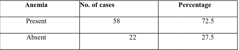
Presence of contributing factors -
1. Infection (local/systemic)
2. Anaemia (defined as blood haemoglobin <13g/dL in
males and <12g/dL in females)
3. Hypoproteinaemia (defined as serum total protein <6g/dL)
4. Postoperative cough or vomiting
5. Uremia (defined as serum urea >40mg/dL and/or serum
creatinine >1.4mg/dL)
6. Electrolyte abnormalities (normal serum sodium 135 -
145mEq/L, normal serum potassium 3.5 - 5mEq/L)
7. Ascites
8. Surgical technique (type of incision, suture material
used for abdominal closure)
9. Obesity (defined as Body Mass Index >30kg/m2)
10.Comorbid conditions if any
11.Drug use if any
Management
Outcome
Statistical analysis:
49
RESULTS
[image:59.595.99.573.160.324.2]AGE DISTRIBUTION Table 2: Age Distribution
Age No. of cases Percentage
15-30 8 10
31-45 22 25
46-60 42 52.5
>60 16 12.5
Maximum cases (52.5%) were found to be in the 46-60 years age
group (Graph 1). The youngest patient in this study was 15 years old and
the oldest was 74 years.
SEX DISTRIBUTION Table 3: Sex Distribution
Sex No. of cases Percentage
Male 55 68.75
Female 25 31.25
[image:59.595.97.57