Sim Yee Lean Paul Ellery Leesa Ivey Jim Thom Robert Oostryck Michael Leahy Ross Baker Murray Adams
The effects of tissue factor pathway inhibitor and
anti-ββ-2-glycoprotein-I IgG on thrombin generation
T
he antiphospholipid syndrome (aPS) is a systemic autoimmune disorder characterized by persistent moderate to high titers of antiphospholipid antibodies (aPL), with venous or arterial thrombosis, pregnancy morbidity and thrombocytope-nia.1 It is widely accepted that the pathogen-esis of this disorder is due to the contribu-tion of aPL, although the precise mecha-nisms are unknown. aPL are a family of het-erogeneous autoantibodies directed against phospholipid-binding proteins and include lupus anticoagulants (LA) and anticardiolipin antibodies (aCL).2,3The two most common phospholipid-binding proteins targeted are β-2-glycoprotein-I (β2GPI) andprothrom-bin.4-6They have higher affinities for anionic phospholipids upon binding with certain aPL7-9and therefore may interfere with phos-pholipid-dependent coagulation reactions and contribute to the prothrombotic state. The major pathway of blood coagulation,
the tissue factor (TF) pathway, is triggered by TF in complex with activated coagulation factor VII (FVIIa). Tissue factor pathway inhibitor (TFPI) is a natural anticoagulant that regulates TF: FVIIa proteolytic activity against coagulation factors X (FX) and IX (FIX), by either binding to activated FX (FXa), then to TF: FVIIa, or forming a com-plex directly with TF: FVIIa: FXa.10,11 The resulting quaternary TF: FVIIa: FXa: TFPI complex prevents further TF: FVIIa activity and consequentially decreases thrombin generation and fibrin formation. There is increasing evidence that antibodies with activity against TFPI have a significant role in the development of an increased throm-botic tendency observed in aPS patients.12-17 High titers of anti-TFPI autoantibodies have recently been detected with greater frequen-cy among aPS patients compared to patients with aPL only and healthy controls.12 Furthermore, an inhibitory component to From the Western Australian
Biomedical Research Institute, School of Biomedical Sciences, Curtin University of Technology, Perth, WA, Australia (SYL, PE, RO, MA); Department of Haematology, Fremantle Hospital, Perth, WA, Australia (LI, ML); Haematology Department, Royal Perth Hospital, Perth, WA, Australia (JT, RB).
Correspondence:
Murray Adams, Western Australian Biomedical Research Institute School of Biomedical Sciences Curtin University of Technology GPO Box U 1987, Perth WA 6845 Australia.
E-mail: M.Adams@curtin.edu.au
Background and Objectives. Recent evidence suggests that autoantibodies to tissue factor pathway inhibitor (TFPI) and/or antiphospholipid antibodies (aPL) may contribute to upregulation of the tissue factor (TF) pathway of blood coagulation and the develop-ment of thrombotic complications in the antiphospholipid syndrome (aPS). The aim of this study was to determine the influence of aPL e.g. anti-β-2-glycoprotein-I (anti-β2GPI) and anti-prothrombin, on in vitro TF-induced thrombin generation in the presence and absence of TFPI.
Design and Methods. IgG fractions were collected from subjects with aPL (n=21) and normal controls (n=36). Anti-TFPI activity was determined after incubation of IgG isolat-ed from control or subject plasma with poolisolat-ed normal plasma using an amidolytic assay for TFPI. The influence of IgG fractions and purified aPL (anti-β2GPI and anti-pro-thrombin) on TF-induced in vitro thrombin generation was determined using a chro-mogenic assay of thrombin activity.
Results. Patients with aPL had significantly elevated thrombin generation (median [interquartile range]) compared to normal controls (112.0 [104.0-124.0]% vs 89.9 [85.7-100.9]%, respectively;p<0.001). Thrombin generation was significantly correlat-ed with anti-TFPI activity in patients with aPL (rs=0.452; p=0.039). It was also
demon-strated that anti-β2GPI, but not anti-prothrombin IgG antibodies, significantly enhanced TF-induced thrombin generation in the presence of TFPI, using both purified and patients’ samples.
Interpretation and Conclusions. Our findings support the hypothesis that anti-β2GPI IgG antibodies accelerate thrombin generation in the presence of TFPI and may contribute to hypercoagulability in patients with aPS.
Key words: antiphospholipid antibodies, tissue factor pathway inhibitor, thrombin generation,β-2-glycoprotein-I.
TFPI has been identified in the IgG fraction of aPS patients,13which is associated with increased in vitro TF-induced thrombin generation.14 It is probable that this inhibitory component is an anti-TFPI antibody and/or another aPL with inhibitory activity against TFPI. Thus, inhibitors of TFPI or interference to TFPI activity, cou-pled with an aPL-induced increase in TF-like activity from monocytes and endothelial cells,18,19may upregu-late the TF pathway and contribute to hypercoagulabil-ity in aPS.
The aim of this study was to determine the influence of markers of aPS e.g. anti-β2GPI and anti-prothrombin
antibodies, on in vitroTF-induced thrombin generation. It was hypothesised that aPL would interfere with the control of the TF pathway and be enhanced by the pres-ence of TFPI.
Design and Methods
Ethics
This study was approved by the Human Research Ethics Committee of Curtin University of Technology (Approval Number: HR 238/2001).
Patients and controls
Plasma from 21 patients with aPL (mean age=57.4 years; age range=30-79 years) was collected from Fremantle Hospital (Fremantle, Australia) and Royal Perth Hospital (Perth, Australia). Plasma samples were aliquoted and stored at -80°C until assay. Normal con-trol plasma was collected from 36 volunteers (mean age=29.3 years; age range=19-60 years) with no appar-ent hemostatic abnormality from the School of Biomedical Sciences (Curtin University of Technology, Perth, Australia). Nine parts of venous blood was col-lected in one part of 0.109 M tri-sodium citrate antico-agulant and centrifuged for 15 minutes at 3000 g. Plasma was collected and centrifuged again for 15 min-utes at 3000 g to obtain platelet poor plasma. Equal vol-umes of platelet-poor plasma from 20 healthy male donors were pooled to prepare normal pooled plasma (NPP).
Plasmas, proteins and antibodies
NPP was used as the reference control in the anti-TFPI activity and TF-induced thrombin generation assays. TFPI-depleted plasma (TDP) and recombinant human TFPI (rTFPI) were purchased from American Diagnostica Inc. (Stamford, CT, USA). Purified human prothrombin, purified human β2GPI, affinity purified polyclonal rabbit anti-human β2GPI antibodies and affinity purified polyclonal rabbit anti-human pro-thrombin antibodies were purchased from Hyphen Biomed (Neuville, France). Polyclonal sheep anti-human TFPI IgG antibody was purchased from Hematologic
Technologies Inc. (Essex Junction, VT, USA). Rabbit anti-sheep IgG (Silenus, Hawthorn, Australia), sheep anti-rabbit IgG (Silenus, Hawthorn, Australia) and affin-ity purifed goat anti-human IgG, IgA, IgM and light chain (Biosource International, Camarillo, USA) were alkaline phosphatase-conjugated antibodies used in immunoblotting.
IgG isolation
IgG was isolated from plasma using a HiTrapTM
Protein G 1mL column (Amersham Pharmacia Biotech, Uppsala, Sweden) according to the manufacturer’s instructions and as described previously.14 Isolated IgG (2.5 mL) was buffer exchanged into 3.5mL of 0.9% NaCl or TFPI assay buffer pH 8.0 (0.05mol/L tris, 0.15mol/L NaCl, 0.01M tri-sodium citrate, 0.2g/L poly-brene and 0.1% normal serum albumin) using a PD 10 desalting column (Amersham Pharmacia Biotech, Uppsala, Sweden) equilibrated with 25 mL of PBS. The concentration of IgG was calculated by dividing the IgG eluate absorbance by an extinction coefficient of 1.43 and multiplying by the total volume (3.5mL). Aliquots of IgG were stored at -80°C.
TF-induced thrombin generation assay
In vitro thrombin generation in plasma was measured using a chromogenic assay20 recently modified by our laboratory.14 Prior to assay, platelet-poor plasma was heated for 15 minutes at 49° C, cooled on melting ice for 2 minutes and centrifuged for 15 minutes at 3000 g to remove fibrinogen. Defibrinated plasma diluted 1/2 in 0.9% NaCl, or protein (50 µL/well) was incubated with IgG or antibody (25 µL/well) for 20 minutes at room temperature in the well of a 96-well round bottomed microtiter plate. Thromboplastin (Innovin®, Dade
Behring, Marburg, Germany) diluted 1/20 in 0.9% NaCl was added (25 µL/well) and the mixture incubated for 10 minutes at 37°C. A chromogenic substrate specific for thrombin (Biophen CS-0138Thrombin Chromogenic Substrate, Hyphen Biomed, Neuville, France), diluted to 1 mmol/L in 0.9% NaCl was then added (50 µL/well) followed by 30 mmol/L CaCl2 (50 µL/well). The final
reaction volume was 200 µL. Using a Multiskan Ascent microtiter plate spectrophotometer the plate was read at 405nm immediately and every two minutes until peak absorbance was reached. Background absorbance was accounted for each sample by substituting throm-bin chromogenic substrate with 0.9% NaCl.
Anti-TFPI activity
Anti-TFPI activity was determined as previously described.14 Equal volumes of NPP and 25 µg/mL IgG or antibody were incubated for 30 minutes at 37°C and then assayed in the TFPI activity assay.21Anti-TFPI activity was the difference of TFPI activity between the IgG/NPP mix-ture and the 1U/mL control (buffer/NPP).
Immunoblotting
Immunoblotting (dot blot) involved immobilization of proteins on to a synthetic membrane support and recogni-tion of the proteins’ native epitopes by test antibodies. A polyvinylidene fluoride (PVDF) membrane (Hybond™-P, Amersham Biosciences, Buckinghamshire, UK) was pre-pared by immersion in 100% methanol and then tris-buffered saline (TBS) pH 8.0. Antigens (TFPI, β2GPI and prothrombin) (5 µg/mL) were blotted (0.5 µL per dot) onto the PVDF membrane and allowed to dry. Blocking solu-tion (5% skim milk in TBS buffer containing 0.05% Tween pH 8.0) was added and incubated with gentle agi-tation at room temperature for 30 minutes. The mem-brane was sequentially incubated with gentle agitation for 1 hour at room temperature or overnight at 4°C, in pri-mary antibody (0.5-10 µg/mL antibody in blocking solu-tion) and washed three times in TBS buffer containing 0.05% Tween pH 8.0 at 10-minute intervals. Secondary antibody (0.5 µg/mL alkaline phosphatase-conjugated antibody in blocking solution) was added and incubated with gentle agitation for 1 hour at room temperature or overnight at 4°C, followed by three washes in TBS buffer containing 0.05% Tween pH 8.0 at 10-minute intervals. Color development was generated using an alkaline phos-phatase-conjugate chromogenic substrate (Biorad, California, USA). Deionized water stopped the reaction and the membrane was dried at 37°C.
Statistical analysis
All experiments were performed in duplicate. Statistical analysis was performed using Superior Performance Statistical Software® (SPSS) for Mac® OS X (version
11.0.2.). Results for the effect of IgG on thrombin genera-tion were expressed as the median (interquartile range) as data was not normally distributed. Wilcoxon’s signed rank test was used to test for the differences between groups. Correlation between the effect of IgG on thrombin gener-ation and anti-TFPI activity was derived using Spearman’s rank correlation test (rs). All other results are reported as mean±standard deviation. Statistical significance was defined at pvalues of <0.05.
Results
Thrombin generation and anti-TFPI activity
Sheep anti-human TFPI antibody induced a dose-dependent increase in thrombin generation and normal control IgG had no significant effect on thrombin
gener-ation (Figure 1). The effect of IgG on thrombin genera-tion was significantly greater in patients [112.0 (104.0-124.0)%] than in controls [89.9 (85.7–100.9)%;
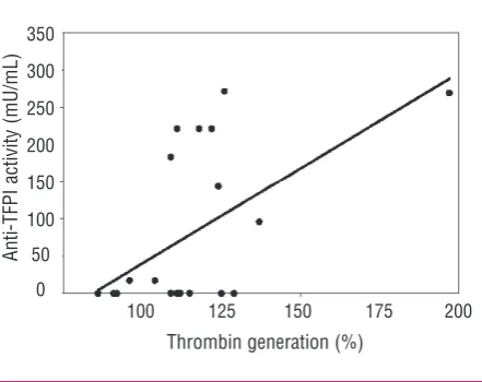
p<0.001]. Anti-TFPI activity was confirmed in 10/21 (47.6 %) of patients and was significantly correlated with the effect of IgG on thrombin generation (rs=0.452; p=0.039) (Figure 2). This relationship was reproducible on two separate occasions.
Interaction of purified protein and antibody markers
Thrombin generation in NPP (100%) was individual-ly reduced by rTFPI (73.1±3.8%), anti-β2GPI antibodies (74.9±10.1%) and anti-prothrombin antibodies (22.7±1.7%) (Figure 3). In the presence of additional TFPI, thrombin generation in NPP was increased by anti-β2GPI (110.0±2.1%) and decreased by
[image:3.595.301.519.64.232.2]anti-pro-thrombin (17.3±1.2%) (Figure 3). These results were
Figure 1. Effect of sheep anti-human TFPI control antibody (P)
and normal control IgG (p) on thrombin generation in NPP. Anti-human TFPI antibody induced a dose-dependent increase in thrombin generation and normal control IgG had no significant effect on thrombin generation.
Figure 2. Thrombin generation and anti-TFPI activity in IgG frac-tions of patients with aPL. The effect of patients’ IgG on thrombin generation was significantly correlated with anti-TFPI activity (rs=0.452; p=0.039).
0 50 100 150 200
100 125 150 175 200
Concentration (µg/mL)
A
n
ti
-T
FP
I
ac
ti
vi
ty
(
m
U
/m
L
)
Thrombin generation (%)
T
h
ro
m
b
in
g
en
er
at
io
n
(
%
)
150
100
50
0
350
300
250
200
150
100
50
[image:3.595.300.521.294.469.2]reproducible and enhanced in TDP. Compared to NPP, TDP had higher thrombin generation (139.9±13.5%), which was reduced with TFPI (55.3±10.6%), anti-β2GPI
(34.9±6.0%) and anti-prothrombin (21.3±0.9%) (Figure 4). Thrombin generation in TDP spiked with TFPI was markedly increased by anti-β2GPI (198.0±18.5%) and
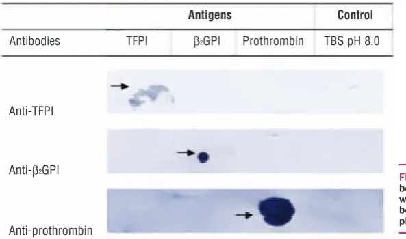
decreased by anti-prothrombin (18.5±0.2%) (Figure 4). Purified antibodies (anti-TFPI, anti-β2GPI and anti-prothrombin) demonstrated specificity for their target antigen with no cross-reactivity (Figure 5).
Interaction of patient IgG with purified protein and
antibody markers
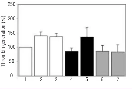
IgG fractions from ten patients with aPL were divided into two groups (five patients with and five without anti-TFPI activity) and assessed for their effect on thrombin generation in TDP, in the presence and
absence of TFPI. Thrombin generation in TDP was sim-ilarly decreased with IgG from patients with anti-TFPI activity (85.7±11.4%) and without anti-TFPI activity (85.9±19.9%) (Figure 6). Thrombin generation in TDP spiked with TFPI was significantly increased by IgG from patients with anti-TFPI activity (135.8±33.7%) compared to IgG from patients without anti-TFPI activ-ity (83.3±25.0%) (p=0.005) (Figure 6).
Discussion
[image:4.595.74.291.62.217.2]Despite its discovery more than two decades ago,22 aPS remains an enigma. Laboratory diagnosis and treat-ment of aPS is often difficult due to limited understand-ing of its pathophysiology. To date, no sunderstand-ingle pathogen-ic mechanism suffpathogen-iciently explains how aPL contribute
Figure 3. Effect of anti-β2GPI (100 µg/mL) and anti-prothrombin
(100 µg/mL) antibodies on thrombin generation in NPP in the
presence and absence of rTFPI (100 ng/mL). Thrombin generation
was enhanced by anti-β2GPI, but not anti-prothrombin, in NPP
spiked with rTFPI. 1: NPP;2: TDP;3: NPP+TFPI;4:
NPP+anti-β2GPI;5:NPP+anti-β2GPI+TFPI;6: NPP+anti-prothrombin;7:
NPP+anti-prothrombin + TFPI.
Figure 4. Effect of anti-β2GPI (100 µg/mL) and anti-prothrombin
(100 µg/mL) antibodies on thrombin generation in TDP in the
presence and absence of rTFPI (100 ng/mL). Thrombin
genera-tion was enhanced by anti-β2GPI, but not anti-prothrombin, in the
presence of rTFPI in TDP. 1: NPP;2: TDP;3: TDP+TFPI;4:
TDP+anti-β2GPI;5: TDP+anti-β2GPI+TFPI; 6:
[image:4.595.316.534.68.216.2]TDP+anti-prothrom-bin;7: TDP+anti-prothrombin+TFPI.
Figure 5. Antigen specificity of purified anti-bodies by immunoblotting. No cross-reactivity was demonstrated between antigens and anti-bodies. Arrows indicate positive reactions. TBS pH 8.0 was a negative control.
1 2 3 4 5 6 7 1 2 3 4 5 6 7
T
h
ro
m
b
in
g
en
er
at
io
n
(
%
)
250
200
150
100
50
0
T
h
ro
m
b
in
g
en
er
at
io
n
(
%
)
250
200
150
100
50
0
Antigens Control
Antibodies TFPI β2GPI Prothrombin TBS pH 8.0
Anti-TFPI
Anti-β2GPI
[image:4.595.77.374.548.723.2]to the development of thrombosis in aPS and account for all clinical manifestations observed in patients. It may be that an array of pathogenic mechanisms con-tributes to an ongoing hypercoagulable state. Recently, interference to the control of the TF pathway of blood coagulation has been implicated in the pathogenesis of thrombotic aPS.12-17,23,24Therefore, the aim of the present study was to determine the influence of anti-β2GPI and
anti-prothrombin aPL on the TF pathway in the pres-ence and abspres-ence of TFPI. It was clearly demonstrated that anti-β2GPI, but not anti-prothrombin IgG
antibod-ies interfere with TFPI-dependent inhibition of TF-induced thrombin generation.
Anti-β2GPI antibodies, anti-prothrombin antibodies
and rTFPI reduced thrombin generation in both NPP (Figure 3) and TDP (Figure 4). TFPI is a natural anticoag-ulant and inhibits thrombin generation through its inhibitory activity against FXa and the TF: FVIIa com-plex. In contrast, anti-β2GPI and anti-prothrombin are
acquired antibodies with an anticoagulant effect on in vitroTF-induced thrombin generation.20,25 Their antico-agulant activities may be mediated by binding with their co-factors as it has been demonstrated that the anti-β2GPI: β2GPI complex7,9,26 and anti-prothrombin:
prothrombin complex8,27 have increased affinities for anionic phospholipids and thereby may compete with other coagulation factors for the binding to anionic phospholipids. Thus, in vitro thrombin generation may be inhibited by anti-β2GPI and prothrombin
anti-bodies due to decreased catalytic phospholipid surfaces for assembly and activation of coagulation factors. Despite the demonstrated reduction of in vitrothrombin generation, anti-β2GPI and anti-prothrombin antibodies
are associated with thrombotic complications in aPS.
The reason for this paradox is presently unclear. Thrombin generation in both NPP and TDP spiked with rTFPI was increased by anti-β2GPI antibodies and
decreased by anti-prothrombin antibodies (Figures 3 and 4). This suggests that anti-β2GPI, but not
anti-pro-thrombin antibodies have inhibitory activity towards TFPI. Anti-β2GPI anti-TFPI-like activity has been
previ-ously reported.16Using a FXa generation assay initiated with low concentrations of recombinant TF (rTF), phatidylserine (anionic phospholipids) and phos-phatidylcholine (neutral phospholipids), it was demon-strated that anti-β2GPI IgG antibodies isolated from aPS
patients required TFPI and β2GPI to enhance FXa
gener-ation. The authors of this study subsequently hypothe-sized that the anti-β2GPI: β2GPI complex interfered with
TFPI dependent inhibition of FXa by competing with the TFPI: FXa complex for the same phospholipid bind-ing sites to therefore increase FXa generation. Although this hypothesis was not tested, it could have been sup-ported if the stimulatory effect of anti-β2GPI IgG
anti-bodies on FXa generation was neutralized in presence of high concentrations of phospholipids. In the present study, the stimulatory effect of anti-β2GPI and TFPI was
demonstrated by increased thrombin generation with the reaction initiated by high concentrations of throm-boplastin (rTF and synthetic phospholipids). Therefore, the mechanism by which anti-β2GPI inhibits TFPI
activ-ity is probably not related to the competition of proteins for binding to catalytic phospholipid surfaces. Instead, the current study proposes a novel mechanism whereby anti-β2GPI binds directly or indirectly to TFPI. This
binding may then completely or partially abolish TFPI-dependent inhibition of blood coagulation resulting in accelerated thrombin generation.
Immunoblotting was used to determine whether anti-β2GPI IgG antibody binds directly to TFPI. The
interac-tion between aPL and their target proteins is currently not well understood, with two models of interaction proposed in the literature. The first model proposed that aPL may recognize and bind epitopes on the conforma-tionally altered protein structure resulting from interac-tions with an oxidized surface or anionic phospho-lipids.28 The other model describes aPL as low-affinity antibodies that require their target antigen in high den-sities to form divalent complexes.26,29No cross-reactivity was demonstrated between anti-β2GPI IgG and TFPI
(Figure 5). Other possible mechanisms may include anti-β2GPI antibody binding a neo-epitope on TFPI when
bound to FXa or the anti-β2GPI: β2GPI complex blocking
TFPI binding to FXa.
Anti-β2GPI in the presence of TFPI has inhibitory
activity against the regulation of TF-induced blood coagulation. Additions of anti-β2GPI and TFPI in TDP
[image:5.595.62.282.64.213.2]resulted in elevated thrombin generation, compared to TDP alone (Figure 4). This inhibitory component may be another natural anticoagulant e.g. protein C,
Figure 6. Mean effect of IgG (25 µg/mL) from patients with (n=5) and without (n=5) anti-TFPI activity on thrombin generation in TDP, in the presence and absence of rTFPI (50 ng/mL). Mean thrombin generation was significantly enhanced by IgG from patients with anti-TFPI activity, but not with IgG from patients without anti-TFPI
activity, in the presence of rTFPI, in TDP (p=0.005). 1: NPP;2: TDP;
3: TDP+TFPI;4: TDP+patient IgGs with anti-TFPI activity;5:
TDP+patient IgGs with anti-TFPI activity+TFPI;6: TDP+patient IgGs
without anti-TFPI activity;7: TDP+patient IgGs without anti-TFPI
activity TFPI.
1 2 3 4 5 6 7
250
200
150
100
50
0
T
h
ro
m
b
in
g
en
er
at
io
n
(
%
antithrombin, or β2GPI. Although the physiological role
of β2GPI has not been established, it has been reported
to have procoagulant and anticoagulant effects in blood coagulation.30In keeping with the mechanisms suggest-ed earlier between anti-β2GPI and TFPI, it is possible
that this inhibitory component is β2GPI. Anti-β2GPI may
preferentially bind to TFPI in the presence of increased concentrations of TFPI compared to β2GPI, thus
simul-taneously inhibiting the anticoagulant functions of TFPI and β2GPI, resulting in enhanced thrombin generation.
Thrombin generation in TDP was reduced in groups of patients with and without anti-TFPI activity (Figure 6), reflecting anticoagulant activities of anti-β2GPI and
anti-prothrombin IgG antibodies. In comparison, thrombin generation in TDP spiked with TFPI was sig-nificantly enhanced by IgG fractions from the group with anti-TFPI activity and essentially unchanged by the group without anti-TFPI activity (Figure 6). These findings demonstrate that the entity in IgG fractions from patients with aPL responsible for the increase in thrombin generation compared to IgG fractions from normal controls, has anti-TFPI activity. It is probable that this entity is an anti-TFPI IgG and/or anti-β2GPI IgG
antibody. Isolation of these entities from IgG fractions will help elucidate the antibodies involved.
This study represents the first work to provide evi-dence that anti-TFPI-like activity expressed by both anti-TFPI and anti-β2GPI IgG antibodies is associated
with increased in vitroTF-induced thrombin generation. The precise relationship and/or interactions between anti-β2GPI antibodies, TFPI and potentially other as yet unidentified entities, remains to be determined. The findings from this study will provide better understand-ing of how interactions within the TF pathway may contribute to thrombotic complications in aPS. Additionally, these results may have implications for the use of rTFPI as an antithrombotic agent in patients with aPL.
SYL: laboratory work, writing and review of manuscript; PE: laboratory work, writing and review of manuscript; LI: sample col-lection, laboratory work, writing and review of manuscript; JT: sample collection, laboratory work, writing and review of manu-script; RO: project design, writing and review of manumanu-script; ML: sample collection, writing and review of manuscript; RB: sample collection, writing and review of manuscript; MA: study design, laboratory work, sample collection, writing and review of manu-script.
The authors wish to acknowledge the School of Biomedical Sciences for funding this study and the excellent technical support of Mr Jeffrey Jago and Dr Gerardine Pinto.
Manuscript received April 5, 2006. Accepted August 8, 2006.
References
1. Wilson WA, Gharavi AE, Koike T, Lockshin MD, Branch DW, Piette J-C, et al. International consensus statement on preliminary classification criteria for definite antiphospholipid syndrome: report of an international workshop. Arthritis Rheum 1999; 42:1309-11. 2. Roubey RAS. Immunology of the
antiphospholipid antibody syndrome. Arthritis Rheum 1996; 39:1444-54. 3. Roubey RAS. Autoantibodies to
phos-pholipid-binding plasma proteins: a new view of lupus anticoagulants and other "antiphospholipid" autoantibod-ies. Blood 1994;84:2854-67.
4. McNeil HP, Simpson RJ, Chesterman CN, Krilis SA. Anti-phospholipid anti-bodies are directed against a complex antigen that includes a lipid-binding
inhibitor of coagulation: β2
-glycopro-tein I (apolipopro-glycopro-tein H). Proc Natl Acad Sci USA 1990;87:4120-4. 5. Galli M, Comfurius P, Maassen C,
Hemker HC, de Baets MH, van Breda-Vriesman PJC, et al. Anticardiolipin antibodies (ACA) directed not to cardi-olipin but to a plasma protein cofactor. Lancet 1990;335:1544-7.
6. Matsuura E, Igarashi Y, Fujimoto M, Ichikawa K, Koike T. Anticardiolipin cofactor(s) and differential diagnosis of autoimmune disease. Lancet 1990; 336: 177-8.
7. Willems GM, Janssen MP, Pelsers MMAL, Comfurius P, Galli M, Zwaal. RFA, et al. Role of divalency in the high-affinity binding of anticardiolipin
antibody-β2-glycoprotein I complexes
to lipid membranes. Biochemistry
1996;35:13833-13842.
8. Rao LVM, Hoang AD, Rapaport SI. Mechanism and effects of the binding of lupus anticoagulant IgG and pro-thrombin to surface phospholipid. Blood 1996;88:4173-82.
9. Takeya H, Mori T, Gabazza EC, Kuroda K, Deguchi H, Matsuura E, et
al. Anti-β2-glycoprotein I (β2GPI)
mon-oclonal antibodies with lupus
anticoag-ulant-like activity enhance the β2GPI
binding to phospholipids. J Clin Invest 1997;99:2260-8.
10. Broze GJ Jr, Warren LA, Novotny WF, Higuchi DA, Girard JJ, Miletich JP. The lipoprotein-associated coagulation inhibitor that inhibits the factor VII-tis-sue factor complex also inhibits factor Xa: insight into its possible mechanism of action. Blood 1988;71:335-43. 11. Rao LVM, Rapaport SI. Studies of a
mechanism inhibiting the initiation of the extrinsic pathway of coagulation. Blood 1987;69:645-51.
12. Forastiero RR, Martinuzzo ME, Broze GJ, Jr. High titers of autoantibodies to tissue factor pathway inhibitor are associated with the antiphospholipid syndrome. J Thromb Haemost 2003; 1: 718-24.
13. Adams M, Donohoe S, Mackie IJ, Machin SJ. Anti-tissue factor pathway inhibitor activity in patients with pri-mary antiphospholipid syndrome. Br J Haematol 2001;114:357-9.
14. Adams M, Breckler L, Stevens S, Thom J, Baker R, Oostryck R. Anti-tissue fac-tor pathway activity in subjects with antiphospholipid syndrome is associat-ed with increasassociat-ed thrombin generation. Haematologica 2004;89:985-90. 15. Jacobsen EM, Sandset PM, Finn W. Do
antiphospholipid antibodies interfere with tissue factor pathway inhibitor?
Thrombosis Research 1999;94:213-220. 16. Salemink I, Blezer R, Willems GM, Galli M, Bevers E, Lindhout T.
Anti-bodies to β2-glycoprotein I associated
with antiphospholipid syndrome sup-press the inhibitory activity of tissue factor pathway inhibitor. Thromb Haemost 2000;84:653-6.
17. Amengual O, Atsumi T, Khamashta MA, Hughes GRV. The role of the tis-sue factor pathway in the hypercoagu-lable state in patients with the antiphospholipid syndrome. Thromb Haemost 1998;79:276-81.
18. Branch DW, Rodgers GM. Induction of endothelial cell tissue factor activity by sera from patients with antiphospho-lipid syndrome: a possible mechanism of thrombosis. Am J Obstet Gynecol 1993;168:206-10.
19. Kornberg A, Blank M, Kaufman S, Shoenfeld Y. Induction of tissue factor-like activity in monocytes by anti-car-diolipin antibodies. J Immunol 1994; 153:1328-32.
20. Sheng Y, Hanly JG, Reddel SW, Kouts S, Guerin J, Koike T, et al. Detection of ‘antiphospholipid’ antibodies: a single chromogenic assay of thrombin gener-ation sensitively detects lupus antico-agulants, anticardiolipin antibodies,
plus antibodies binding β2-glycoprotein
and prothrombin. Clin Exp Immunol 2001;124:502-8.
21. Sandset PM, Larsen ML, Abildgaard U, Lindahl AK, Odegaard OR. Chromo-genic substrate assay of extrinsic path-way inhibitor (EPI): levels on the nor-mal population and relation to choles-terol. Blood Coagul Fibrinol 1991;2: 425-33.
23. Martinuzzo M, Varela Iglesias ML, Adamcuk Y, Broze GJ, Forastiero R. Antiphospholipid antibodies and anti-bodies to tissue factor pathway inhibitor in women with implantation failures or early and late pregnancy losses. J Thromb Haemost 2005;3:1-3. 24. Roubey RAS. Tissue factor pathway
and the antiphospholipid syndrome. J Autoimmun 2000;15:217-220.
25. Hanly JG, Smith SA. Anti-β
(2)-glyco-protein I autoantibodies, in vitro thrombin generation, and the anti-phospholipid syndrome. J Rheumatol 2000;27:2152-9.
26. Roubey RAS, Eisenberg RA, Harper MF, Winfield JB. "Anticardiolipin"
autoantibodies recognize β
2-glycopro-tein I in the absence of phospholipid: importance of antigen density and bivalent binding. J Immunol 1995; 154:954-60.
27. Simmelink MJA, Horbach DA, Derksen RHWM, Meijers JCM, Bevers EM, Willems GM, et al. Complexes of anti-prothrombin antibodies and pro-thrombin cause lupus anticoagulant activity by competing with the bind-ing of clottbind-ing factors for catalytic phospholipid surfaces. Br J Haematol 2001;113:621-9.
28. Matsuura E, Igarashi Y, Yasuda T, Triplett DA, Koike T. Anticardiolipin
antibodies recognize β2-glycoprotein I
structure altered by interacting with an oxygen modified solid phase surface. J
Exp Med 1994;179:457-62.
29. Tincani A, Spatola L, Prati E, Allegri F, Ferremi P, Cattaneo R, et al. The
anti-β2-glycoprotein I activity in human
anti-phospholipid syndrome sera is due to monoreactive low-affinity autoantibodies directed to epitopes
located on native β2-glycoprotein I
and preserved during species' evolu-tion. J Immunol 1996;157:5732-8. 30. Miyakis S, Giannakopoulos B, Krilis
SA. β2glycoprotein I-function in health