UNCORRECTED PROOF
0203 04 05 06 07 08 09 10 11 12 13 14 15 16 17 18 19 20 21 22 23 24 25 26 27 28 29 30 31 32 33 34 35 36 37 38 39 40 41 42 43 44 45
Cyclic AMP Signalling in Pancreatic Islets
Brian Furman, Wee Kiat Ong, and Nigel Pyne
Abstract Cyclic 3’5’AMP (cAMP) is an important physiological amplifier of
glucose-induced insulin secretion by the pancreatic islet
β-cell, where it is formed
by the activity of adenylyl cyclases, which are stimulated by glucose, through
elevation in intracellular calcium concentrations, and by the incretin hormones
(GLP-1 and GIP). cAMP is rapidly degraded in the pancreatic islet
β
-cell by
var-ious cyclic nucleotide phosphodiesterase (PDE) enzymes. Many steps involved in
glucose-induced insulin secretion are modulated by cAMP, which is also
impor-tant in regulating pancreatic islet
β
-cell differentiation, growth and survival. This
chapter discusses the formation, destruction and actions of cAMP in the islets with
particular emphasis on the
β-cell.
Keywords Cyclic AMP
·
Adenylyl cyclase
·
Phosphodiesterase
·
Insulin
secretion
·
Protein kinase A
·
Epac
·
GLP-1
13.1 Introduction
Interest in the role of cyclic 3’5’ AMP (cAMP) in regulating insulin secretion dates
back more than 40 years, since Turtle and Kipnis [1] showed increases in cAMP in
isolated islets in response to glucagon. Increases in islet
β-cell cyclic AMP occur
in response to nutrients, especially glucose. Glucose has been widely shown to
increase intracellular levels of cAMP in islets and various insulin-secreting cell
lines [2–6]. Although cyclic AMP does not appear to be essential for
glucose-induced insulin secretion [3, 7–9], it is established as an important intracellular
amplifier of this process [10–12]. Several hormones exert their effects on insulin
secretion through increased
β-cell cAMP levels. These include glucose-dependent
B. Furman (
B
)Strathclyde Institute of Pharmacy and Biomedical Sciences, University of Strathclyde, Glasgow G4ONR, Scotland, UK
279 Md.S. Islam (ed.), The Islets of Langerhans, Advances in Experimental Medicine and
Biology 654, DOI 10.1007/978-90-481-3271-3_13, C
UNCORRECTED PROOF
4647 48 49 50 51 52 53 54 55 56 57 58 59 60 61 62 63 64 65 66 67 68 69 70 71 72 73 74 75 76 77 78 79 80 81 82 83 84 85 86 87 88 89 90
insulinotropic polypeptide (GIP) and glucagon-like peptide 1 (GLP-1) which are
collectively referred to as the incretins, and which are also secreted in response
to nutrients [13–16]. GLP-1 and GIP serve to augment meal-related insulin
secre-tion [17]. Their physiological importance is evident from observasecre-tions that mice
lacking receptors for both incretin hormones show marked glucose intolerance
and impairment of insulin secretion [18]. This chapter focuses largely on cAMP
in the
β-cell. Much less is known about the role of cAMP in other islet cells,
although there is some information on this in relation to glucagon and
somato-statin secretion/synthesis and these aspects will be addressed briefly at the end of
the chapter.
13.2 Control of cAMP Levels in the
β
-Cell
The level of cyclic AMP in the
β
-cell depends on the balance between its
forma-tion through the activity of adenylyl cyclases (ACs) and its destrucforma-tion by cyclic
nucleotide phosphodiesterases (CN-PDEs). This is summarized in Fig. 13.1 and
discussed below.
13.2.1 Formation of Cyclic AMP in the
β
-Cell
Glucose-induced elevations in intracellular cAMP are probably secondary to
changes in the concentration of calcium, which is itself elevated as a result of
a number of mechanisms but primarily by Ca
2+influx through voltage-sensitive
Ca
2+channels in response to membrane depolarization, following closure of
ATP-sensitive potassium channels. Hormone-induced formation of cAMP results from
stimulation of seven transmembrane G-protein-coupled receptors (GPCRs), leading
to activation of the Gs
protein and dissociation of the G
αβγ
heterotrimeric complex
and sequential activation of adenylyl cyclases [19]. The
β
-cell expresses several
GPCRs coupled to Gs, stimulation of which leads to elevation in the
β
-cell level
of cAMP. These include receptors for GLP-1, GIP, PACAP as well as the receptor
GPR119 (see below). On the other hand, reductions in cAMP occur in response
to several agents that activate GPCRs coupled to Gi, for example adrenaline [20],
PGE2
[21] and NPY (Y1) [22]. There is also evidence for the role of the pertussis
toxin-insensitive G-protein Gz
in the reduction of cAMP and inhibition of insulin
secretion in response to prostaglandin E
1[23].
UNCORRECTED PROOF
9192 93 94 95 96 97 98 99 100 101 102 103 104 105 106 107 108 109 110 111 112 113 114 115 116 117 118 119 120 121 122 123 124 125 126 127 128 129 130 131 132 133 134 135
Fig. 13.1 Summary of the mechanisms for the formation and destruction of cAMP in the
pan-creatic isletβ-cell. Glucose is transported into theβ-cell using GLUT2 and is then metabolized generating ATP. This results in closure of the KATPchannel, membrane depolarization and calcium influx through voltage-sensitive calcium channels. Calcium is also mobilized from intracellu-lar stores by Ca2+(calcium-induced calcium release – not shown). The increased cytosolic-free Ca2+ triggers exocytosis. These processes are amplified through increases in cAMP effected both through activation of adenylyl cyclases by glucose itself (through calcium-activated adeny-lyl cyclase – type VIII- AC VIII) and by the incretin hormones GLP-1 and GIP, acting through G-protein-coupled receptors in theβ-cell membrane. Endogenous agonists for the G-protein-coupled receptor GPR119 include oleoylethanolamide (OEA). Activation of GLP-1 receptors acts synergistically with glucose in activating AC VIII and also activates other adenylyl cyclases, including soluble adenylyl cyclase (not shown). Activation of adenylyl cyclases increases the for-mation of cAMP which activates PKA and Epac which mediate the actions of cAMP in the cell. PKA/Epac facilitates calcium-induced calcium release which in turn may also activate AC VIII. The destruction of cAMP is effected through various phosphodiesterases (PDEs). Ca2+activates PDE1 whereas PKA activates PDE3B, which is also activated by other signals generated through the IGF-1 and leptin receptors, as well as, possibly, the insulin receptor. On the other hand, PDE3B may be inhibited by increases in cGMP, allowing cross-talk between cGMP and cAMP signalling. Roles for other PDEs (PDE4, 8B and 10A) have been proposed (modified from [54])
adenylyl cyclase in INS-1 cells, thereby increasing cAMP and stimulating insulin
secretion.
[image:3.439.35.387.60.328.2]UNCORRECTED PROOF
136137 138 139 140 141 142 143 144 145 146 147 148 149 150 151 152 153 154 155 156 157 158 159 160 161 162 163 164 165 166 167 168 169 170 171 172 173 174 175 176 177 178 179 180
islet
β-cell GIP receptor is down-regulated by exposure to high concentrations of
glucose, which prevents the GIP-induced elevation in intracellular cAMP [31]. This
is hypothesized to explain the lack of response of diabetic patients to the peptide.
PACAP is expressed in nerve fibres and the pancreatic islets and is a potent
stimulator of insulin secretion [32, 33] through activation of adenylyl cyclase [34].
There are several receptors for PACAP, with the PAC1 receptor (PAC1-R) and
VPAC2 receptor (VPAC2-R) thought to be the most important in relation to insulin
secretion [35].
GPR119 is a Class I GPCR, the expression of which is restricted largely to
pancreatic islets, although lesser amounts of message are detected in the human
gastrointestinal tract and in the rodent brain [36–38]. The potential endogenous
ligands for this receptor so far identified are oleoyl lysophosphatidylcholine and
oleoylethanolamide, although there is as yet no evidence that they are available in
sufficient concentrations in the blood to stimulate the
β
-cell GRP119 receptor in
vivo. The receptor is coupled through Gs
to adenylyl cyclase, and its activation
produces an increase in cAMP and stimulation of insulin secretion.
13.2.1.1 Adenylyl Cyclases in the Pancreatic Islet
β
-Cell
There are at least nine different membrane-bound isoforms of AC, described as
AC I–AC IX and expressed in mammalian cells [39, 40]. An additional, soluble
form is also expressed in certain mammalian cells [41]. RT-PCR studies, as well as
immunohistochemical staining, using rat and human islets, rat
β-cells, and clonal
β-cell lines have shown expression of AC II [42] and III, IV, V, VI, VII and VIII
[5, 43–45]. All isoforms of adenylyl cyclase, apart from ACIX, are activated by the
diterpene forskolin, which produces marked increases in cAMP in numerous cell
types [46, 47]. There are three calcium-activated ACs (AC1, ACIII and ACVIII),
and the presence of calcium–calmodulin-activated ACVIII probably explains
activa-tion of cyclic AMP formaactiva-tion in response to glucose, which rapidly elevates [Ca
2+]i.
This AC isoform is synergistically activated by both Gsα
and calcium/calmodulin
[48]. Thus, the combination of glucose and GLP-1 increases cAMP accumulation in
rat isolated primary
β
-cells or clonal
β
-cell lines more markedly than either alone,
the effect being reduced if calcium entry through voltage-sensitive L-type channels
is prevented using verapamil [45]. The expression of type VI (but not types II, III or
V) adenylyl cyclase was increased along with the expression of the GLP-1 receptor
rat pups fed a high-carbohydrate diet for 12 days [42]. These findings provide some
circumstantial evidence that the type VI adenylyl cyclase may be associated with
GLP-1 signalling. More recently, a role for soluble AC was proposed to explain
the different kinetics of cAMP formation in response to glucose and GLP-1 in
INS-1E cells. GLP-1 produced a rapid increase as a result of activation of
transmem-brane AC, whereas the increase in cAMP in response to glucose was delayed and
was attributed to activation of the calcium, bicarbonate and ATP-sensitive soluble
AC [6].
UNCORRECTED PROOF
181182 183 184 185 186 187 188 189 190 191 192 193 194 195 196 197 198 199 200 201 202 203 204 205 206 207 208 209 210 211 212 213 214 215 216 217 218 219 220 221 222 223 224 225
also activated adenylyl cyclases and elevated cAMP content in islets from
GK-diabetic rats [49]. The insulin secretory response to acetylcholine in these islets was
blocked by inhibitors of adenylyl cyclase or PKA inhibitors. The abnormal nature of
the islet in these rats may somehow has facilitated cross-talk resulting in activation
of a calcium-sensitive adenylyl cyclase, or a PKC-sensitive adenylyl cyclase, e.g.
ACII [40], in response to acetylcholine.
13.2.2 Destruction of cAMP in the Pancreatic Islet
β
-Cell -Cyclic
Nucleotide Phosphodiesterases
Cyclic nucleotide phosphodiesterases (CN-PDEs) provide the only known means
for the rapid inactivation of the cyclic nucleotides cAMP and cGMP in most cells.
There are now known to be at least 100 PDE enzymes derived from 11 known
gene families (PDE1-11). The enzymes show differences in their tissue distribution,
substrate selectivities (cGMP vs cAMP), kinetics, regulation, and susceptibility to
pharmacological inhibition. There are several excellent reviews [50–53], and the
properties of those PDE enzymes present in pancreatic islets have been reviewed
elsewhere [54, 55]. The key observations are summarized in this chapter, together
with more recent findings.
Several PDE isoforms, including PDE1 [56–61], PDE3B [59–67], PDE4 [59, 60,
64] and PDE8B [68], contribute to the total
β-cell PDE activity, and several of these
isoforms regulate glucose-induced insulin secretion and other cAMP-mediated
β-cell functions in islets and in β-cell lines [see 54, 55 for references]. There is much
evidence from RT-PCR, immunostaining, siRNA and biochemical and functional
studies using selective inhibitors that PDE3B plays a key role in both islets and
insulin-secreting cell lines in terms of regulating insulin secretion [54, 55, 61, 63–
66]. Additional evidence for the role of PDE3B in regulating
β
-cell cAMP and
insulin secretion was obtained by over-expressing PDE3B in the INS-1
β
-cell line
and in islets and by using transgenic animals over-expressing PDE3B in the
β
-cell. These in vitro and in vivo studies clearly showed that glucose-induced, as
well as GLP-1-induced, insulin secretion was impaired by PDE3B over-expression.
Interestingly, both endogenous and over-expressed PDE3B was found to be located
in insulin granules and the plasma membrane [67]. In vitro, the over-expression of
PDE3B markedly reduced cAMP-induced exocytosis and animals over-expressing
PDE3B in islets showed markedly impaired glucose tolerance [65–67]. In addition,
activation of PDE3B appears to mediate the effect of IGF-1 [63] and leptin [69] in
inhibiting insulin secretion.
UNCORRECTED PROOF
226227 228 229 230 231 232 233 234 235 236 237 238 239 240 241 242 243 244 245 246 247 248 249 250 251 252 253 254 255 256 257 258 259 260 261 262 263 264 265 266 267 268 269 270
Although evidence for the importance of PDE3B is widely supported there is also
evidence, but no consensus, for roles for other PDEs. Roles for PDE1C and PDE4
have been suggested on the basis of the use of either selective inhibitors [59, 64] or
siRNA [64]. Depletion of PDE8B using siRNA produced a marked enhancement of
glucose-induced insulin secretion from INS-1E cells [64, 68] and rat islets [68]. A
role for PDE10A has been proposed and selective inhibitors have been patented for
the treatment of diabetes [74], but there is no consensus on the expression of this
PDE in the
β-cell, and in one study [64] selective knockdown of PDE10A failed to
modify glucose-induced insulin secretion in INS-1 cells.
13.2.3 Dynamics of cAMP Formation and Destruction
UNCORRECTED PROOF
271272 273 274 275 276 277 278 279 280 281 282 283 284 285 286 287 288 289 290 291 292 293 294 295 296 297 298 299 300 301 302 303 304 305 306 307 308 309 310 311 312 313 314 315
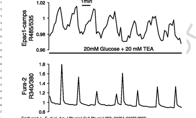
Fig. 13.2 Ca2+and cAMP oscillations in glucose-stimulated MIN6 cells. Simultaneous imaging of cytosolic cAMP concentration ([cAMP]i; top trace, R485/535) and cytosolic Ca2+concentration ([Ca2+]i; bottom trace, R340/380) in a single MIN6 cell stimulated with 20 mM glucose and 20 mM tetraethylammonium chloride (TEA). Note that second messenger oscillations were out of phase, with each [Ca2+]ispike coupled to a rapid and transient reduction in [cAMP]i. (Reproduced from Fridlyand LE, Harbeck MC, Roe MW, Philipson LH. Regulation of cAMP dynamics by Ca2+and G protein-coupled receptors in the pancreatic beta-cell: a computational approach. Am J Physiol Cell Physiol 293: C1924–33, 2007 [75] with permission)
(see next section). On the other hand, translocation of the catalytic subunit of PKA to
the nucleus occurred relatively slowly and only in response to sustained increases in
cAMP. Glucose also induced oscillations of intracellular cAMP levels in MIN6 and
mouse primary
β-cells. These oscillations correlated with pulsatile insulin secretion
and both cAMP oscillations and pulsatile insulin release were reduced by inhibiting
adenylyl cyclases [78]. Forskolin, glucagon and IBMX all augmented the frequency
of glucose-induced oscillations in [Ca
2+]i
in mouse pancreatic islets [79]
13.2.4 Intracellular Compartmentalization of cAMP Formation,
Action and Degradation
[image:7.439.38.380.61.267.2]UNCORRECTED PROOF
316317 318 319 320 321 322 323 324 325 326 327 328 329 330 331 332 333 334 335 336 337 338 339 340 341 342 343 344 345 346 347 348 349 350 351 352 353 354 355 356 357 358 359 360
hydrolysis and activity of cAMP are ensured by spatial distribution into
compart-ments, or signalling complexes, of adenylyl cyclases, PDEs and effector proteins,
as well as phosphatases that terminate the activity of various kinases (e.g. 80, 81).
This spatial anchoring of signalling complexes is effected by a family of A-kinase
anchoring proteins (AKAPs). Recent work has suggested the importance of AKAPs
in the insulin-secreting
β-cell. Peptides that competitively inhibit the interaction
between the regulatory subunit of PKA and the AKAP inhibited GLP-1-induced
insulin secretion from rat islets without modifying its ability to elevate
intracellu-lar cAMP [9]. Expression of this inhibitory peptide in the clonal rat
β-cell line,
RINm5F, resulted in a redistribution of the PKA regulatory subunit and
inhib-ited elevations in [Ca
2+]i
and insulin secretion in response to a cAMP analogue.
Expression of an AKAP (AKAP18) in clonal insulin-secreting cells (RINm5f)
aug-mented GLP-1-induced insulin release, whereas expression of a mutant form in
these cells was inhibitory [82]. These findings were supported by others [83] who
used a cell-permeable peptide (TAT-AKAPis) to competitively inhibit PKA–AKAP
interactions in INS-1 cells. This peptide disrupted PKA–AKAP interactions and
inhibited both glucagon-induced augmentation of insulin secretion and
phosphory-lation of p44/p42 MAPKs and cAMP response element binding protein. While
rela-tively little is known about the role of phosphatases in terminating
phosphorylation-mediated actions of cAMP in the pancreatic islet
β-cell [84], there is evidence that
the AKAP AKAP79 (the human homologue of AKAP150) is important in targeting
the serine–threonine phosphatase PP2B to PKA-sensitive target proteins [85].
13.3 Functions of Cyclic AMP in the Pancreatic Islet
β
-Cell
cAMP modulates a number of
β-cell functions including insulin secretion, insulin
synthesis,
β
-cell replication, and
β
-cell apoptosis. Actions of cAMP in general
are mediated by at least two distinct mechanisms. The first of these is through
protein kinase A (PKA)-mediated phosphorylation [86]. However, a second, and
PKA-independent, effect of cAMP on insulin secretion [87–88] is mediated by the
cyclic AMP-binding proteins known either as cAMP-regulated guanine nucleotide
exchange factors (GEFs) or as exchange proteins activated by cAMP (Epacs)
which target the small G-protein Rap1 [86]. Interestingly, most of the
β-cell
Rap1, at least in MIN6 cells, appears to be co-localized with insulin secretory
granules [89]. When activated by cAMP, Epac, which exists as two isoforms
(Epac1 and Epac2) exchanges GDP for GTP and activates downstream
sig-nalling. The pancreatic islet
β-cell expresses both Epac1 and Epac2 [90]. Antisense
oligodeoxynucleotides against Epac reduced the effect of a permeant cAMP
ana-logue in augmenting glucose-induced insulin secretion in pancreatic islets [91].
Studies using selective inhibitors/activators of PKA, selective activators of Epac
or the use of dominant-negative forms of Epac are revealing the roles of Epacs
vs PKA in the
β
-cell. Novel cAMP analogues, such as 8-(4-chlorophenylthio)-2
UNCORRECTED PROOF
361362 363 364 365 366 367 368 369 370 371 372 373 374 375 376 377 378 379 380 381 382 383 384 385 386 387 388 389 390 391 392 393 394 395 396 397 398 399 400 401 402 403 404 405
much more cell-permeant acetoxy methyl ester [92] activate Epac but not PKA,
having a 100-fold lower affinity for PKA relative to Epac [86]. Similarly, cAMP
analogues such as N6-Bnz-cAMP selectively activate PKA relative to Epac. Both
Epac and PKA mediate the effects of cAMP on insulin secretion. However, at least
in INS-1 cells, PKA-mediated effects account for the greater proportion of cAMP
effects [92]. There is evidence for interaction between PKA-mediated and
Epac-mediated effects in augmenting insulin secretion in native
β-cells [93]. Some of
the reported discrepancies may be explained by the poor cell permeability of some
Epac-selective cAMP analogues [92].
The cyclic AMP-mediated effects of GIP and GLP-1 on insulin secretion involve
both PKA [24] and PKA-independent actions. The latter are probably mediated
through Epac, as evidenced by the comparative effects of the PKA inhibitor H89
and antisense oligodeoxynucleotides (ODNs) against Epac in reducing
incretin-augmented insulin secretion [91, 94]. Interestingly, Epac-dependent effects of
cAMP on insulin release are impaired in islets from mice lacking the SUR subunit
of the KATP
channel [94, 95].
13.3.1 Insulin Secretion
Malaisse’s group was the first to systematically examine the actions of cAMP
on insulin secretion [96, 97]. Elevations in cAMP in the
β-cell augment
glucose-induced insulin secretion at several sites in the secretory pathway.
13.3.1.1 Effects on the
β
-Cell ATP-Sensitive Potassium Channel
The
β-cell ATP-sensitive potassium channel (KATP
channel) plays a fundamental
role in glucose-induced insulin secretion. Elevation of cAMP in the
β
-cell using
GLP-1, forskolin, or the non-selective PDE inhibitor IBMX inhibits the
β
-cell KATP
channel promoting depolarization of the cell [98–103]. This effect was reported to
be mediated via PKA in INS-1 cells [101] through phosphorylation of the SUR1
subunit. On the other hand, Epac was found to inhibit this channel in both human
β-cells and INS-1 cells, producing a leftward shift in the ATP-concentration–effect
curve [102, 103]. The same study [103] suggested a PKA-mediated activation of
the ATP-sensitive K channel.
13.3.1.2 Voltage-Sensitive Potassium Channels
UNCORRECTED PROOF
406407 408 409 410 411 412 413 414 415 416 417 418 419 420 421 422 423 424 425 426 427 428 429 430 431 432 433 434 435 436 437 438 439 440 441 442 443 444 445 446 447 448 449 450
13.3.1.3 Elevations in Intracellular Calcium [Ca
2+]
iIncreases in [Ca
2+]i
can be effected through two main mechanisms, namely influx
through voltage-sensitive Ca
2+channels and mobilization of Ca
2+from intracellular
stores and cAMP influences both these mechanisms in the
β
-cell.
Voltage-Sensitive Ca
2+Channels
Entry of Ca
2+through L-type voltage-sensitive calcium channels in response to
membrane depolarization is an important trigger for exocytosis. Agents elevating
cAMP as well as cAMP itself augment the opening of channel and increase calcium
influx [99, 107–109] through PKA-dependent mechanisms. This is consistent with
observations that forskolin and IBMX were shown to produce phosphorylation of
the cardiac-type alpha 1 subunit of the voltage-sensitive calcium channel in a mouse
β-cell line
βTC3 [110].
Mobilization of Ca
2+from Intracellular Stores
Calcium-Induced Calcium Release
In addition to facilitating calcium entry, agents that elevate
β-cell cAMP also
promote calcium-induced Ca
2+release [111–116]. For example, the uncaging of
calcium from a membrane-permeable caged calcium (NP EGTA) produced a large,
transient increase in [Ca
2+]i
but only in the presence of the GLP-1 mimetic exendin 4
or the adenylyl cyclase activator forskolin. This could be replicated by non-selective
cAMP analogues or those that selectively activated either PKA or Epac. The effects
of exendin-4 were relatively insensitive to the PKA inhibitor H89 but were inhibited
by expression of a dominant-negative Epac2 [116], suggesting an important role
of Epac2 in the sensitizing effect of cAMP on calcium-induced Ca
2+release. The
importance of non-PKA-dependent effects of GLP-1 in elevating [Ca
2+]i
was also
reported previously [117].
The mechanism whereby cAMP promotes calcium-induced Ca
2+release may be
through activation of the ryanodine channel in the ER [93, 112, 113] and/or through
phosphorylation of the IP3
receptor [118]. The interaction of cAMP, via PKA, with
IP3
receptors is supported by the finding that 2-aminoethoxydiphenyl borate, a
cell-permeable IP3-receptor antagonist, blocked the PKA-mediated cAMP amplification
of calcium-induced Ca
2+release [119].
Generation of Ca
2+-Mobilizing Second Messengers
UNCORRECTED PROOF
451452 453 454 455 456 457 458 459 460 461 462 463 464 465 466 467 468 469 470 471 472 473 474 475 476 477 478 479 480 481 482 483 484 485 486 487 488 489 490 491 492 493 494 495
relative role of cyclic ADPR and NAADP in producing cAMP-mediated increases
in [Ca
2+]i
remain to be determined.
13.3.1.4 Direct Effect on Exocytosis
Ammala et al. [107] and Gillis and Misler [121] were the first to demonstrate that
cAMP produced direct effects on exocytosis. This effect was suggested to
repre-sent the most important effect of cAMP on insulin release [107]. Both GIP and
GLP-1 promote PKA-dependent and PKA-independent exocytosis, independently
of changes in calcium entry [87, 99, 122]. Moreover, photo release of caged cAMP
produces a marked increase in granule exocytosis that is independent of changes
in [Ca
2+]i
[87, 99, 123, 124]. GLP-1 and cAMP augmented depolarization-induced
exocytosis, and the effects of cAMP were mediated through both PKA-dependent
and PKA-independent, Epac-mediated effects [95]. cAMP also enhanced
exocyto-sis in single INS-1 cells, the effect being augmented by inhibition of PDE3 [65].
In permeabilized rat islets cAMP enhanced calcium-induced insulin secretion,
inde-pendently of changes in [Ca
2+]i; this effect was largely dependent on Epac as it
was mimicked by an Epac-selective, but not by a PKA selective, cAMP analogue
and was unaffected by a PKA inhibitor [125]. Use of two-photon extracellular polar
tracer (TEP) imaging and electron microscopy showed different roles of PKA or
Epac in the enhancement by cAMP of calcium-evoked exocytosis of small compared
with large, secretory vesicles [124]. Effects of cAMP on large vesicle exocytosis
appeared to be PKA dependent, whereas effects on small vesicles were mediated
via Epac.
There are different pools of insulin secretory granules in the
β-cell. The first
phase of glucose-induced insulin secretion is due to the release of granules docked at
the membrane in a readily releasable pool and the second phase is dependent on the
mobilization of granules to refill this readily releasable pool. The effects of cAMP,
which augments both first and second phases of insulin secretion, are at least partly
attributable to an expansion and refilling of the readily releasable pool [126–128].
Knockout of Epac2 specifically blocks the first phase of glucose-induced granule–
plasma membrane fusions, suggesting the importance of cAMP signalling through
Epac2 in this phase [89]. This supports earlier findings that the augmentation by
cAMP of short depolarizations was Epac dependent, whereas the effect on longer
depolarizations was largely PKA dependent and was more sensitive to cAMP [95].
The second phase of exocytosis appears to be mediated via both PKA and Epac
[95, 127, 128], although a PKA dependency of the first phase of glucose-induced
exocytosis has also been reported [123].
13.3.1.5 Activation of Protein Kinase C
UNCORRECTED PROOF
496497 498 499 500 501 502 503 504 505 506 507 508 509 510 511 512 513 514 515 516 517 518 519 520 521 522 523 524 525 526 527 528 529 530 531 532 533 534 535 536 537 538 539 540
mimicked by forskolin. This activation was Ca
2+dependent, and it was
hypothe-sized that it was effected through mobilization of Ca
2+as a result, for example, of
PKA sensitization of the IP3
channel and consequent Ca
2+-mediated activation of
phospholipase C [129].
13.4 Role of cAMP in Insulin Synthesis and in
β
-Cell
Differentiation, Proliferation, and Survival
The incretin GLP-1, acting to an important extent through cAMP effector
mecha-nisms, increases insulin synthesis, promotes
β
-cell proliferation and inhibits
β
-cell
apoptosis [25], although there is evidence for cAMP-independent effects [130].
Indeed much of the evidence for the importance of cAMP in these processes is
derived from studies using GLP-1 and exendin-4. The finding that mice with a
β-cell-specific deficiency in the
α
subunit of Gs
showed reduced
β-cell mass, reduced
islet content of insulin, reduced
β-cell proliferation, and increased
β-cell apoptosis,
and marked hyperglycaemia suggests the fundamental importance of responsiveness
to incretin hormones [131] in
β-cell homeostasis.
Glucose-mediated increases in insulin synthesis involve the phosphorylation of
the transcription factor pancreatic duodenal homeobox-1 (PDX-1) and its
transloca-tion to the nucleus [132]. There is strong evidence for the importance of cAMP,
acting through PKA-dependent mechanisms, in mediating the ability of GLP-1
to increase
β-cell levels of PDX-1, stimulate its translocation to the nucleus and
consequently activate the insulin gene promoter [133]. PDX-1 expression is itself
required for the generation of cAMP in response to exendin-4 through controlling
the expression of the GLP-1 receptor and the Gs
protein a subunit [134].
CREB (cAMP response element binding protein) is the key transcriptional
acti-vator that mediates the effects of cAMP on gene regulation and its effects in
regulating islet
β-cell proliferation and survival. cAMP, through a PKA-dependent
mechanism, and glucose act synergistically to regulate CREB activation in MIN6
or INS-1 cells [135, 136]. This appears to involve cAMP/PKA and glucose-induced
modulation of the phosphorylation status of TORC2, a key co-activator of CREB,
and the stimulation of its translocation to the nucleus [135, 136].
13.4.1 Immediate Early Response Genes
UNCORRECTED PROOF
541542 543 544 545 546 547 548 549 550 551 552 553 554 555 556 557 558 559 560 561 562 563 564 565 566 567 568 569 570 571 572 573 574 575 576 577 578 579 580 581 582 583 584 585
identified among the targets that were sequentially induced in primary cells from
rat islets. In the same context, cAMP also amplifies the effect of glucose in
stimulating the MAPK/ERK pathway [6, 140–142]. The augmentation of
glucose-induced activation of ERK in response to GLP-1 required both influx of Ca
2+through voltage-dependent calcium channels and was PKA dependent [143] and
GIP activates this kinase pathway through cyclic AMP and PKA [144].
13.4.2 Protection Against
β
-Cell Apoptosis and Stimulation
of
β
-Cell Proliferation
There is abundant evidence for suppression of
β-cell apoptosis by agents that elevate
cAMP, including GLP-1, GIP, exendin-4, ghrelin and obestatin [135, 145–151]. This
appears to be PKA mediated [148, 149]. Paradoxically, some
β-cell lines were made
more susceptible to apoptosis following exposure to dibutyryl cyclic AMP [152] or
the cyclic AMP-elevating agent forskolin [153]. The anti-apoptotic effects of cAMP
are mediated, in part, by increased expression of the anti-apoptotic proteins Bcl-2
and Bcl-xL [135, 146], and are PKA dependent [135, 146, 151]. The anti-apoptotic
effects also involve caspase inhibition [147]. Inhibition of cytokine-mediated nitric
oxide production by
β
-cells [154] may also be implicated.
In addition to preventing apoptosis of
β
-cells, the incretin hormones and other
agents elevating cAMP promote
β
-cell proliferation through PKA-dependent
mech-anisms [134, 155, 156]. This effect appears to involve expression of cyclin D1 [155,
157] and cyclin A2 [134]. In this context, there may be an interaction of cAMP with
Wnt signalling, which plays an important role in
β-cell proliferation and survival
with upregulation of cyclins D1 and D2 [158]. Thus, GLP-1 and exendin-4
acti-vated Wnt signalling in INS-1 cells and in isolated islets [159]. Exendin-induced
β-cell proliferation was inhibited by blocking
β-catenin or the transcription factor
TCF7L2, critical mediators of Wnt signalling [159].
An additional mechanism whereby cAMP modulates
β-cell proliferation may
be through regulation of the CREB antagonists cAMP response element modulator
CREM-α
and ICERI and the dual specificity phosphatase DUSP14, a negative
reg-ulator of the MAPK/ERK1/2 pathway. Thus, genes for these proteins were rapidly
and strongly upregulated by GLP-1 in a
β-cell line and in rat primary
β-cells, an
effect that was mimicked by forskolin and blocked by the PKA inhibitor H89 but
not by an Epac inhibitor. shRNA-mediated knockdown of CREM-
α
or DUSP14,
or expression of a dominant-negative DUSP14, augmented GLP-1-induced
β
-cell
proliferation [156].
13.5 Possible Roles of cAMP in Other Islet Cell Types
UNCORRECTED PROOF
586587 588 589 590 591 592 593 594 595 596 597 598 599 600 601 602 603 604 605 606 607 608 609 610 611 612 613 614 615 616 617 618 619 620 621 622 623 624 625 626 627 628 629 630
cells. Forskolin was shown to stimulate glucagon secretion from rat islets [160].
GLP-1 (and GIP) augmented depolarization-evoked exocytosis from rat
α-cells;
this effect was accompanied by elevations in intracellular cAMP, increases in Ca
2+currents and was mediated by PKA [161]. Exposure of an
α-cell line
(INRl-G9) expressing recombinant GLP-1 receptors to GLP-1 increased the formation
of cAMP and elevated free cytosolic [Ca
2+] [162]. In the same cell line, an
Epac-selective cAMP analogue stimulated the expression of the glucagon gene
pro-moter and stimulated glucagon production, although not glucagon secretion [163].
Moreover, a dominant-negative Epac-2 attenuated forskolin-stimulated expression
of the glucagon gene promoter in the InR1-G9 cells [163]. While these data
indi-cate a stimulatory effect of GLP-1 on glucagon synthesis and secretion, GLP-1 is
known to inhibit glucagon secretion, an action likely to contribute to its therapeutic
effect in the treatment of diabetes [164]. The inhibition of glucagon secretion by
GLP-1 is thus likely to be mediated by a paracrine action in the islets, for example,
through stimulation of somatostatin secretion, which markedly inhibits glucagon
release [165]. In this context, GLP-1, oxyntomodulin and glucagon were shown
to potently stimulate somatostatin secretion from somatostatin-secreting cell lines
(RIN T3; RIN 1048-38) and to stimulate the accumulation of cAMP [166, 167].
Increases in cAMP levels in response to forskolin, theophylline or dibutyryl cAMP
were shown to be associated with increased somatostatin release from isolated islets
[168].
Glucagon itself stimulates glucagon release by activating glucagon, rather than
GLP-1, receptors, through cAMP-dependent mechanisms involving both PKA and
Epac [169].
Adrenaline, or isoprenaline, acting through
β-adrenoceptors, augmented
depolarization-evoked glucagon secretion from rat primary
α-cells [170]. This effect
was mimicked by forskolin and was PKA dependent. As in the
β
-cell the
PKA-dependent effects appear to involve more than one mechanism, including increased
Ca
2+entry and augmentation of the effects of Ca
2+. Photo release of caged cAMP
increased exocytosis even when intracellular [Ca
2+] was clamped [170]. These data
were supported by observations using mouse primary
α-cells, in which
adrenaline-induced increases in
α-cell [Ca
2+]i
were mediated, in part, by elevations in cAMP
and activation of PKA [171].
13.6 Conclusion
UNCORRECTED PROOF
631632 633 634 635 636 637 638 639 640 641 642 643 644 645 646 647 648 649 650 651 652 653 654 655 656 657 658 659 660 661 662 663 664 665 666 667 668 669 670 671 672 673 674 675
development [37]. A number of non-peptide agents that act both as direct agonists
and allosteric modulators of the GLP-1 receptor are also being examined [172].
References
1. Turtle J, Kipnis D. An adrenergic receptor mechanism for the control of cyclic 35adenosine monophosphate synthesis in tissues. Biochem Biophys Res Commun 1967;28:797–802. 2. Charles M, Fanska R, Schmid F, Forsham P, Grodsky G. Adenosine 3,5-monophosphate in
pancreatic islets: glucose-induced insulin release. Science 1973;179:569–571.
3. Grill V, Cerasi E. Activation by glucose of adenyl cyclase in pancreatic islets of the rat. FEBS Lett 1973;33:311–4.
4. Kim J, Roberts C, Berg S, Caicedo A, Roper S, Chaudhari N. Imaging cyclic AMP changes in pancreatic islets of transgenic reporter mice. PLoS ONE 2008;3:e2127.
5. Landa LJ, Harbeck M, Kaihara K, Chepurny O, Kitiphongspattana K, Graf O, Nikolaev V, Lohse M, Holz G, Roe M. Interplay of Ca2+and cAMP signaling in the insulin–secreting MIN6 beta-cell line. J Biol Chem 2005;280:31294–302.
6. Ramos L, Zippin J, Kamenetsky M, Buck J, Levin L. Glucose and GLP-1 stimulate cAMP production via distinct adenylyl cyclases in INS-1E insulinoma cells. J Gen Physiol 2008;132:329–38.
7. Sharp G. The adenylate cyclase-cyclic AMP system in islets of Langerhans and its role in the control of insulin release. Diabetologia 1979;16:287–96.
8. Persaud S, Jones P, Howell S. Glucose-stimulated insulin secretion is not dependent on activation of protein kinase A. Biochem Biophys Res Commun 1990;173:833–9.
9. Lester L, Langeberg L, Scott J. Anchoring of protein kinase A facilitates hormone-mediated insulin secretion. Proc Natl Acad Sci U S A 1997;94:14942–7.
10. Holz G, Habener J. Signal transduction crosstalk in the endocrine system: pancreatic beta-cells and the glucose competence concept. Trends Biochem Sci 1992;17:388–93.
11. Howell S, Jones P, Persaud S. Regulation of insulin secretion: the role of second messengers. Diabetologia 1994;37 Suppl 2:S30–5.
12. Braun M, Ramracheya R, Johnson P, Rorsman P. Exocytotic properties of human pancreatic beta-cells. Ann N Y Acad Sci 2009;1152:187–93.
13. MacIntosh C, Horowitz M, Verhagen M, Smout A, Wishart J, Morris H, Goble E, Morley J, Chapman I. Effect of small intestinal nutrient infusion on appetite, gastrointestinal hor-mone release, and gastric myoelectrical activity in young and older men. Am J Gastroenterol 2001;96:997–1007.
14. Brubaker P, Anini Y. Direct and indirect mechanisms regulating secretion of glucagon-like peptide-1 and glucagon-like peptide-2. Can J Physiol Pharmacol 2003;81:1005–12. 15. Feinle C, Chapman I, Wishart J, Horowitz M. Plasma glucagon-like peptide-1
(GLP-1) responses to duodenal fat and glucose infusions in lean and obese men. Peptides 2002;23:1491–95.
16. Wolfe M, Zhao K, Glazier K, Jarboe L, Tseng C. Regulation of glucose-dependent insulinotropic polypeptide release by protein in the rat. Am J Physiol Gastrointest Liver Physiol 2000;279:G561–6.
17. Thorens B. Expression cloning of the pancreatic beta cell receptor for the gluco-incretin hormone glucagon-like peptide 1. Proc Natl Acad Sci U S A 1992;89:8641–5.
18. Preitner F, Ibberson M, Franklin I, Binnert C, Pende M, Gjinovci A, Hansotia T, Drucker D, Wollheim C, Burcelin R, Thorens B. Gluco-incretins control insulin secretion at multiple levels as revealed in mice lacking GLP-1 and GIP receptors. J Clin Invest 2004;113:635–45. 19. Selbie L, Hill S. G protein-coupled-receptor cross-talk: the fine-tuning of multiple
UNCORRECTED PROOF
676677 678 679 680 681 682 683 684 685 686 687 688 689 690 691 692 693 694 695 696 697 698 699 700 701 702 703 704 705 706 707 708 709 710 711 712 713 714 715 716 717 718 719 720
20. Yamazaki S, Katada T, Ui M. Alpha 2-adrenergic inhibition of insulin secretion via interfer-ence with cyclic AMP generation in rat pancreatic islets. Mol Pharmacol 1982;21:648–53. 21. Robertson R, Tsai P, Little S, Zhang H, Walseth T. Receptor-mediated adenylate
cyclase-coupled mechanism for PGE2 inhibition of insulin secretion in HIT cells. Diabetes 1987;36:1047–53.
22. Morgan D, Kulkarni R, Hurley J, Wang Z, Wang R, Ghatei M, Karlsen A, Bloom S, Smith D. Inhibition of glucose stimulated insulin secretion by neuropeptide Y is mediated via the Y1 receptor and inhibition of adenylyl cyclase in RIN 5AH rat insulinoma cells. Diabetologia 1998;41:1482–91.
23. Kimple M, Nixon A, Kelly P, Bailey C, Young K, Fields T, Casey P. A role for Gz in pancreatic isletβ-cell biology. J Biol Chem 2005;280:31708–13.
24. Drucker D, Philippe J, Mojsov S, Chick W, Habener J. Glucagon-like peptide I stimulates insulin gene expression and increases cyclic AMP levels in a rat islet cell line. Proc Natl Acad Sci U S A 1987;84:3434–8.
25. Doyle M, Egan J. Mechanisms of action of glucagon-like peptide 1 in the pancreas. Pharmacol Ther 2007;113:546–93.
26. Maida A, Lovshin J, Baggio L, Drucker D. The glucagon-like peptide-1 receptor agonist oxyntomodulin enhances beta-cell function but does not inhibit gastric emptying in mice. Endocrinology 2008;149:5670–8.
27. Sonoda N, Imamura T, Yoshizaki T, Babendure J, Lu J, Olefsky J. Beta-Arrestin-1 mediates glucagon-like peptide-1 signaling to insulin secretion in cultured pancreatic beta cells. Proc Natl Acad Sci U S A 2008;105:6614–9.
28. Amiranoff B, Vauclin-Jacques N, Laburthe M. Functional GIP receptors in a hamster pan-creatic beta cell line, In 111: specific binding and biological effects. Biochem Biophys Res Commun 1984;123:671–6.
29. Siegel E, Creutzfeldt W. Stimulation of insulin release in isolated rat islets by GIP in physiological concentrations and its relation to islet cyclic AMP content. Diabetologia 1985;28:857–61.
30. Wheeler M, Gelling R, McIntosh C, Georgiou J, Brown J, Pederson R. Functional expres-sion of the rat pancreatic islet glucose-dependent insulinotropic polypeptide receptor: ligand binding and intracellular signaling properties. Endocrinology 1995;136:4629–9.
31. Zhou J, Livak M, Bernier M, Muller D, Carlson O, Elahi D, Maudsley S, Egan J. Ubiquitination is involved in glucose-mediated downregulation of GIP receptors in islets. Am J Physiol Endocrinol Metab 2007;293:E538–47.
32. Yada T, Sakurada M, Ihida K, Nakata M, Murata F, Arimura A, Kikuchi M. Pituitary adeny-late cyclase activating polypeptide is an extraordinarily potent intra-pancreatic regulator of insulin secretion from islet beta-cells. J Biol Chem 1994;269:1290–3.
33. Ahrén B. Role of pituitary adenylate cyclase-activating polypeptide in the pancreatic endocrine system. Ann N Y Acad Sci. 2008;1144:28–35.
34. Borboni P, Porzio O, Pierucci D, Cicconi S, Magnaterra R, Federici M, Sesti G, Lauro D, DAgata V, Cavallaro S, Marlier L. Molecular and functional characterization of pitu-itary adenylate cyclase-activating polypeptide (PACAP-38)/vasoactive intestinal polypeptide receptors in pancreatic beta-cells and effects of PACAP-38 on components of the insulin secretory system. Endocrinology 1999;140:5530–7.
35. Yamada S, Komatsu M, Sato Y, Yamauchi K, Kojima I, Aizawa T, Hashizume K. Time-dependent stimulation of insulin exocytosis by 3,5-cyclic adenosine monophosphate in the rat islet beta-cell. Endocrinology 2002;143:4203–9.
36. Soga T, Ohishi T, Matsui T, Saito T, Matsumoto M, Takasaki J, Matsumoto S, Kamohara M, Hiyama H, Yoshida S, Momose K, Ueda Y, Matsushime H, Kobori M, Furuichi K. Lysophosphatidylcholine enhances glucose-dependent insulin secretion via an orphan G-protein-coupled receptor. Biochem Biophys Res Commun 2005;326:744–51.
UNCORRECTED PROOF
721722 723 724 725 726 727 728 729 730 731 732 733 734 735 736 737 738 739 740 741 742 743 744 745 746 747 748 749 750 751 752 753 754 755 756 757 758 759 760 761 762 763 764 765
of a G protein-coupled receptor for oleoylethanolamide and its use in the discovery of small-molecule hypophagic agents. Cell Metab 2006;3:167–75.
38. Chu Z, Jones R, He H, Carroll C, Gutierrez V, Lucman A, Moloney M, Gao H, Mondala H, Bagnol D, Unett D, Liang Y, Demarest K, Semple G, Behan D, Leonard J. A role for beta-cell-expressed G protein-coupled receptor 119 in glycemic control by enhancing glucose-dependent insulin release. Endocrinology 2007;148:2601–9.
39. Hanoune J, Defer N. Regulation and role of adenylyl cyclase isoforms. Annu Rev Pharmacol 2001;Toxicol.;41:145–74.
40. Willoughby D, Cooper D. Organization and Ca2+regulation of adenylyl cyclases in cAMP microdomains. Physiol Rev 2007;87:965–1010.
41. Kamenetsky M, Middelhaufe S, Bank E, Levin L, Buck J, Steegborn C. Molecular details of cAMP generation in mammalian cells: a tale of two systems. J Mol Biol 2006;362:623–39. 42. Srinivasan M, Aalinkeel R, Song F, Lee B, Laychock S, Patel M. Adaptive changes in insulin
secretion by islets from neonatal rats raised on a high-carbohydrate formula. Am J Physiol Endocrinol Metab 2000;279:E1347–57.
43. Leech C, Castonguay M, Habener J. Expression of adenylyl cyclase subtypes in pancreatic beta-cells. Biochem Biophys Res Commun 1999;254:703–6.
44. Guenifi A, Portela-Gomes G, Grimelius L, Efendi´c S, Abdel-Halim S. Adenylyl cyclase isoform expression in non-diabetic and diabetic Goto-Kakizaki (GK) rat pancreas. Evidence for distinct overexpression of type-8 adenylyl cyclase in diabetic GK rat islets. Histochem Cell Biol 2000;113:81–9.
45. Delmeire D, Flamez D, Hinke S, Cali J, Pipeleers D, Schuit F. Type VIII adenylyl cyclase in rat beta cells: coincidence signal detector/generator for glucose and GLP-1. Diabetologia 2003;46:1383–93.
46. Seamon K, Daly J. Forskolin: its biological and chemical properties. Adv Cyclic Nucleotide Protein Phosphorylation Res 1986;20:1–150.
47. Insel P, Ostrom R. Forskolin as a tool for examining adenylyl cyclase expression, regulation, and G protein signaling. Cell Mol Neurobiol 2003;23:305–14.
48. Cali J, Zwaagstra J, Mons N, Cooper D, Krupinski J. Type VIII adenylyl cyclase. A Ca2+/calmodulin-stimulated enzyme expressed in discrete regions of rat brain. J Biol Chem 1994;269:12190–5.
49. Dolz M, Bailbé D, Giroix M, Calderari S, Gangnerau M, Serradas P, Rickenbach K, Irminger J, Portha B. Restitution of defective glucose-stimulated insulin secretion in diabetic GK rat by acetylcholine uncovers paradoxical stimulatory effect of beta-cell muscarinic receptor activation on cAMP production. Diabetes 2005;54:3229–37.
50. Perry M, Higgs G. Chemotherapeutic potential of phosphodiesterase inhibitors. Curr Opin Chem Biol 1998;2:472–81.
51. Soderling S, Beavo J. Regulation of cAMP and cGMP signaling. new phosphodiesterases and new functions. Curr Opin Cell Biol 2000;12:174–9.
52. Mehats C, Andersen C, Filopanti M, Jin S, Conti M. Cyclic nucleotide phosphodiesterases and their role in endocrine cell signaling. Trends Endocrinol Metab 2002;13:29–35. 53. Conti M, Beavo J. Biochemistry and physiology of cyclic nucleotide phosphodiesterases:
essential components in cyclic nucleotide signaling. Annu Rev Biochem 2007;76:481–511. 54. Pyne N, Furman B. Cyclic nucleotide phosphodiesterases in pancreatic islets. Diabetologia
2003;46:1179–89.
55. Furman B, Pyne N, Flatt P, OHarte F. Targeting beta-cell cyclic 35adenosine monophos-phate for the development of novel drugs for treating type 2 diabetes mellitus. A review. J Pharm Pharmacol 2004;56:1477–92.
56. Sugden M, Ashcroft S. Cyclic nucleotide phosphodiesterase of rat pancreatic islets. Effects of Ca2+, calmodulin and trifluoperazine. Biochem J 1981;197:459–64.
UNCORRECTED PROOF
766767 768 769 770 771 772 773 774 775 776 777 778 779 780 781 782 783 784 785 786 787 788 789 790 791 792 793 794 795 796 797 798 799 800 801 802 803 804 805 806 807 808 809 810
58. Lipson L, Oldham S. The role of calmodulin in insulin secretion: the presence of a calmodulin-stimulatable phosphodiesterase in pancreatic islets of normal and pregnant rats. Life Sci 1983;32:775–80.
59. Han P, Werber J, Surana M, Fleischer N, Michaeli T. The calcium/calmodulin-dependent phosphodiesterase PDE1C down-regulates glucose-induced insulin secretion. J Biol Chem 1999;274:22337–44.
60. Ahmad M, Flatt P, Furman B, Pyne N. The role of the cyclic GMP-inhibited cyclic AMP-specific phosphodiesterase (PDE3) in regulating clonal BRIN-BD11 insulin secreting cell survival. Cell Signal 2000;12:541–48.
61. Shafiee-Nick R, Pyne N, Furman B. Effects of type-selective phosphodiesterase inhibitors on glucose-induced insulin secretion and islet phosphodiesterase activity. Br J Pharmacol 1995;115:1486–92.
62. Parker J, VanVolkenburg M, Ketchum R, Brayman K, Andrews K. Cyclic AMP phospho-diesterases of human and rat islets of Langerhans: contributions of types III and IV to the modulation of insulin secretion. Biochem Biophys Res Commun 1995;217:916–23. 63. Zhao A, Zhao H, Teague J, Fujimoto W, Beavo J. Attenuation of insulin secretion by
insulin-like growth factor 1 is mediated through activation of phosphodiesterase 3B. Proc Natl Acad Sci U S A 1997;94:3223–28.
64. Waddleton D, Wu W, Feng Y, Thompson C, Wu M, Zhou Y, Howard A, Thornberry N, Li J, Mancini J. Phosphodiesterase 3 and 4 comprise the major cAMP metabolizing enzymes responsible for insulin secretion in INS-1 (832/13) cells and rat islets. Biochem Pharmacol 2008;76:884–93.
65. Härndahl L, Jing X, Ivarsson R, Degerman E, Ahrén B, Manganiello V, Renström E, Holst L. Important role of phosphodiesterase 3B for the stimulatory action of cAMP on pancreatic beta-cell exocytosis and release of insulin. J Biol Chem 2002;277:37446–55.
66. Härndahl L, Wierup N, Enerbäck S, Mulder H, Manganiello V, Sundler F, Degerman E, Ahrén B, Holst L. Beta-cell-targeted overexpression of phosphodiesterase 3B in mice causes impaired insulin secretion, glucose intolerance, and deranged islet morphology. J Biol Chem 2004;279:15214–22.
67. Walz H, Härndahl L, Wierup N, Zmuda-Trzebiatowska E, Svennelid F, Manganiello V, Ploug T, Sundler F, Degerman E, Ahrén B, Holst L. Early and rapid development of insulin resis-tance, islet dysfunction and glucose intolerance after high-fat feeding in mice overexpressing phosphodiesterase 3B. J Endocrinol 2006;189:629–41.
68. Dov A, Abramovitch E, Warwar N, Nesher R. Diminished phosphodiesterase-8B potentiates biphasic insulin response to glucose. Endocrinology 2008;149:741–8.
69. Zhao A, Bornfeldt K, Beavo J. Leptin inhibits insulin secretion by activation of phosphodi-esterase 3B. J Clin Invest 1998;102:869–73.
70. Grapengiesser E, Gylfe E, Dansk H, Hellman B. Nitric oxide induces synchronous Ca2+ transients in pancreatic beta cells lacking contact. Pancreas 2001;23:387–92.
71. Smukler S, Tang L, Wheeler M, Salapatek A. Exogenous nitric oxide and endoge-nous glucose-stimulated beta-cell nitric oxide augment insulin release. Diabetes 2002;51: 3450–60.
72. Kaneko Y, Ishikawa T, Amano S, Nakayama K. Dual effect of nitric oxide on cytosolic Ca2+ concentration and insulin secretion in rat pancreatic beta-cells. Am J Physiol Cell Physiol 2003;284:C1215–22.
73. Sunouchi T, Suzuki K, Nakayama K, Ishikawa T. Dual effect of nitric oxide on ATP-sensitive K+channels in rat pancreatic beta cells. Pflugers Arch 2008;456:573–9.
74. Cantin L, Magnuson S, Gunn D, Barucci N, Breuhaus M, Bullock W, Burke J, Claus T, Daly M, Decarr L, Gore-Willse A, Hoover-Litty H, Kumarasinghe E, Li Y, Liang S, Livingston J, Lowinger T, Macdougall M, Ogutu H, Olague A, Ott-Morgan R, Schoenleber R, Tersteegen A, Wickens P, Zhang Z, Zhu J, Zhu L, Sweet L. PDE-10A inhibitors as insulin secretagogues. Bioorg Med Chem Lett 2007;17:2869–73.
UNCORRECTED PROOF
811812 813 814 815 816 817 818 819 820 821 822 823 824 825 826 827 828 829 830 831 832 833 834 835 836 837 838 839 840 841 842 843 844 845 846 847 848 849 850 851 852 853 854 855
76. Dyachok O, Isakov Y, Sågetorp J, Tengholm A. Oscillations of cyclic AMP in hormone-stimulated insulin-secreting beta-cells. Nature 2006;439:349–52.
77. Dyachok O, Sågetorp J, Isakov Y, Tengholm A. cAMP oscillations restrict protein kinase A redistribution in insulin-secreting cells. Biochem Soc Trans 2006;34:498–501.
78. Dyachok O, Idevall-Hagren O, Sågetorp J, Tian G, Wuttke A, Arrieumerlou C, Akusjärvi G, Gylfe E, Tengholm A. Glucose-induced cyclic AMP oscillations regulate pulsatile insulin secretion. Cell Metab 2008;8:26–37.
79. Baltrusch S, Lenzen S. Regulation of [Ca2+]i oscillations in mouse pancreatic islets by adrenergic agonists. Biochem Biophys Res Commun 2007;363:1038–43.
80. Jarnaess E, Taskén K. Spatiotemporal control of cAMP signalling processes by anchored signalling complexes. Biochem Soc Trans 2007;35:931–37.
81. Dodge-Kafka K, Kapiloff M. The mAKAP signaling complex: integration of cAMP, calcium, and MAP kinase signaling pathways. Eur J Cell Biol 2006;85:593–602.
82. Fraser I, Tavalin S, Lester L, Langeberg L, Westphal A, Dean R, Marrion N, Scott J. A novel lipid-anchored A-kinase Anchoring Protein facilitates cAMP-responsive membrane events. EMBO J 1998;17:2261–72.
83. Faruque O, Le-Nguyen D, Lajoix A, Vives E, Petit P, Bataille D, Hani e-H. Cell-permeable peptide-based disruption of endogenous PKA-AKAP complexes: a tool for studying the molecular roles of AKAP-mediated PKA subcellular anchoring. Am J Physiol Cell Physiol 2009;296:C306–16.
84. Jones PM, Persaud SJ. Protein kinases, protein phosphorylation, and the regulation of insulin secretion from pancreaticβ-cells. Endocr Rev. 1998;429–461.
85. Lester LB, Faux MC, Nauert JB, Scott JD. Targeted protein kinase A and PP-2B regulate insulin secretion through reversible phosphorylation. Endocrinology. 2001;142(3):1218–27. 86. Kopperud R, Krakstad C, Selheim F, Døskeland S. cAMP effector mechanisms. Novel twists
for anoldsignaling system. FEBS Lett 2003;546:121–6.
87. Renström E, Eliasson L, Rorsman P. Protein kinase A-dependent and independent stimula-tion of exocytosis by cAMP in mouse pancreatic B-cells. J Physiol 1997;502:105–18. 88. Seino S, Shibasaki T. PKA-dependent and PKA-independent pathways for cAMP-regulated
exocytosis. Physiol Rev 2005;85:1303–42.
89. Shibasaki T, Takahashi H, Miki T, Sunaga Y, Matsumura K, Yamanaka M, Zhang C, Tamamoto A, Satoh T, Miyazaki J, Seino S. Essential role of Epac2/Rap1 signaling in regula-tion of insulin granule dynamics by cAMP. Proc Natl Acad Sci U S A; 2007;104:19333–193. 90. Holz G. Epac: A new cAMP-binding protein in support of glucagon-like peptide-1
receptor-mediated signal transduction in the pancreatic beta-cell. Diabetes 2004;53:5–13.
91. Kashima Y, Miki T, Shibasaki T, Ozaki N, Miyazaki M, Yano H, Seino S. Critical role of cAMP-GEFII––Rim2 complex in incretin-potentiated insulin secretion. J Biol Chem 2001;276:46046–53.
92. Chepurny O, Leech C, Kelley G, Dzhura I, Dzhura E, Li X, Rindler M, Schwede F, Genieser H, Holz G. Enhanced Rap1 activation and insulin secretagogue properties of an acetoxymethyl ester of an Epac-selective cyclic AMP analog in rat INS-1 cells: Studies with 8-pCPT-2-O-Me-cAMP-AM. J Biol Chem, 2009.
93. Liu G, Jacobo S, Hilliard N, Hockerman G. Differential modulation of Cav1.2 and Cav1.3-mediated glucose-stimulated insulin secretion by cAMP in INS-1 cells: distinct roles for exchange protein directly activated by cAMP 2 (Epac2) and protein kinase A. J Pharmacol Exp Ther 2006;318:152–60.
94. Nakazaki M, Crane A, Hu M, Seghers V, Ullrich S, Aguilar-Bryan L, Bryan J. cAMP-activated protein kinase-independent potentiation of insulin secretion by cAMP is impaired in SUR1 null islets. Diabetes 2002;51:3440–9.
95. Eliasson L, Ma X, Renström E, Barg S, Berggren P, Galvanovskis J, Gromada J, Jing X, Lundquist I, Salehi A, Sewing S, Rorsman P. SUR1 regulates PKA-independent cAMP-induced granule priming in mouse pancreatic B-cells. J Gen Physiol 2003;121:181–97. 96. Malaisse W, Malaisse-Lagae F, Mayhew D. A possible role for the adenyl cyclase system in
UNCORRECTED PROOF
856857 858 859 860 861 862 863 864 865 866 867 868 869 870 871 872 873 874 875 876 877 878 879 880 881 882 883 884 885 886 887 888 889 890 891 892 893 894 895 896 897 898 899 900
97. Brisson G, Malaisse-Lagae F, Malaisse W. The stimulus-secretion coupling of glucose-induced insulin release. VII. A proposed site of action for adenosine-3,5-cyclic monophos-phate. J Clin Invest 1972;51:232–41.
98. Holz G, Kühtreiber W, Habener J. Pancreatic beta-cells are rendered glucose-competent by the insulinotropic hormone glucagon-like peptide-1(7-37). Nature 1993;361:362–5. 99. Gromada J, Bokvist K, Ding W, Holst J, Nielsen J, Rorsman P. Glucagon-like peptide 1
(7-36) amide stimulates exocytosis in human pancreatic beta-cells by both proximal and distal regulatory steps in stimulus-secretion coupling. Diabetes 1998;47:57–65.
100. He L, Mears D, Atwater I, Kitasato H. Glucagon induces suppression of ATP-sensitive K+ channel activity through a Ca2+/calmodulin-dependent pathway in mouse pancreaticβ-cells. J Membr Biol 1998;166:237–44.
101. Light P, Manning Fox J, Riedel M, Wheeler M. Glucagon-like peptide-1 inhibits pancreatic ATP-sensitive potassium channels via a protein kinase A- and ADP-dependent mechanism. Mol Endocrinol 2002;16:2135–44.
102. Kang G, Chepurny O, Malester B, Rindler M, Rehmann H, Bos J, Schwede F, Coetzee W, Holz G. cAMP sensor Epac as a determinant of ATP-sensitive potassium channel activity in human pancreaticβcells and rat INS-1 cells. J Physiol 2006;573:595–609.
103. Kang G, Leech C, Chepurny O, Coetzee W, Holz G. Role of the cAMP sensor Epac as a determinant of KATP channel ATP sensitivity in human pancreaticβ-cells and rat INS-1 cells. J Physiol 2008;586:1307–19.
104. Kim S, Choi W, Han J, Warnock G, Fedida D, McIntosh C. A novel mechanism for the suppression of a voltage-gated potassium channel by glucose-dependent insulinotropic polypeptide: protein kinase A-dependent endocytosis. J Biol Chem 2005;280:28692–700. 105. MacDonald P, Salapatek A, Wheeler M. Glucagon-like peptide-1 receptor activation
antag-onizes voltage-dependent repolarizing K+currents inβ-cells: a possible glucose-dependent insulinotropic mechanism. Diabetes 51 Suppl 2002;3:S443–47.
106. MacDonald P, Wang X, Xia F, El-Kholy W, Targonsky E, Tsushima R, Wheeler M. Antagonism of ratβ-cell voltage-dependent K+ currents by exendin 4 requires dual acti-vation of the cAMP/protein kinase A and phosphatidylinositol 3-kinase signaling pathways. J Biol Chem 2003;278:52446–53.
107. Ammälä C, Ashcroft F, Rorsman P. Calcium-independent potentiation of insulin release by cyclic AMP in single beta-cells. Nature 1993;363:356–58.
108. Kanno T, Suga S, Wu J, Kimura M, Wakui M. Intracellular cAMP potentiates voltage-dependent activation of L-type Ca2+ channels in rat islet beta-cells. Pflugers Arch 1998;435:578–80.
109. Suga S, Kanno T, Nakano K, Takeo T, Dobashi Y, Wakui M. GLP-I (7-36) amide augments Ba2+ current through L-type Ca2+ channel of rat pancreaticβ-cell in a cAMP-dependent manner. Diabetes 1997;46:1755–60.
110. Leiser M, Fleischer N. cAMP-dependent phosphorylation of the cardiac-type alpha 1 sub-unit of the voltage-dependent Ca2+ channel in a murine pancreaticβ-cell line. Diabetes 1996;45:1412–8.
111. Gromada J, Dissing S, Bokvist K, Renström E, Frøkjaer-Jensen J, Wulff B, Rorsman P. Glucagon-like peptide I increases cytoplasmic calcium in insulin-secreting beta TC3-cells by enhancement of intracellular calcium mobilization. Diabetes 1995;44:767–74.
112. Islam M, Leibiger I, Leibiger B, Rossi D, Sorrentino V, Ekström T, Westerblad H, Andrade F, Berggren P. In situ activation of the type 2 ryanodine receptor in pancreaticβcells requires cAMP-dependent phosphorylation. Proc Natl Acad Sci U S A 1998;95:6145–50.
113. Holz G, Leech C, Heller R, Castonguay M, Habener J. cAMP-dependent mobilization of intracellular Ca2+stores by activation of ryanodine receptors in pancreatic beta-cells. A Ca2+ signaling system stimulated by the insulinotropic hormone glucagon-like peptide-1-(7-37). J Biol Chem 1999;274:14147–56.
UNCORRECTED PROOF
901902 903 904 905 906 907 908 909 910 911 912 913 914 915 916 917 918 919 920 921 922 923 924 925 926 927 928 929 930 931 932 933 934 935 936 937 938 939 940 941 942 943 944 945
115. Kang G, Joseph J, Chepurny O, Monaco M, Wheeler M, Bos J, Schwede F, Genieser H, Holz G. Epac-selective cAMP analog 8-pCPT-2-O-Me-cAMP as a stimulus for Ca2+-induced Ca2+release and exocytosis in pancreaticβ-cells. J Biol Chem 2003;278:8279–85. 116. Kang G, Chepurny O, Rindler M, Collis L, Chepurny Z, Li W, Harbeck M, Roe M, Holz G.
A cAMP and Ca2+coincidence detector in support of Ca2+-induced Ca2+release in mouse pancreaticβcells. J Physiol 2005;566:173–88.
117. Bode H, Moormann B, Dabew R, Göke B. Glucagon-like peptide 1 elevates cytoso-lic calcium in pancreatic beta-cells independently of protein kinase A. Endocrinology 1999;140:3919–27.
118. Tsuboi T, da Silva Xavier G, Holz G, Jouaville L, Thomas A, Rutter G. Glucagon-like peptide-1 mobilizes intracellular Ca2+and stimulates mitochondrial ATP synthesis in pancreatic MIN6β-cells. Biochem J 2003;369:287–99.
119. Dyachok O, Gylfe E. Ca2+-induced Ca2+release via inositol 1,4,5-trisphosphate receptors is amplified by protein kinase A and triggers exocytosis in pancreaticβ-cells. J Biol Chem 2004;279:45455–61.
120. Kim BJ, Park KH, Yim CY, Takasawa S, Okamoto H, Im MJ, Kim UH Generation of nico-tinic acid adenine dinucleotide phosphate and cyclic ADP-ribose by glucagon-like peptide-1 evokes Ca2+signal that is essential for insulin secretion in mouse pancreatic islets. Diabetes 2008;57:868–78.
121. Gillis K, Misler S. Enhancers of cytosolic cAMP augment depolarization-induced exo-cytosis from pancreatic B-cells: evidence for effects distal to Ca2+ entry. Pflugers Arch 1993;424:195–7.
122. Ding W, Gromada J. Protein kinase A-dependent stimulation of exocytosis in mouse pancre-atic beta-cells by glucose-dependent insulinotropic polypeptide. Diabetes 1997;46:615–21. 123. Hatakeyama H, Kishimoto T, Nemoto T, Kasai H, Takahashi N. Rapid glucose sensing
by protein kinase A for insulin exocytosis in mouse pancreatic islets. J Physiol 2006;570: 271–82.
124. Hatakeyama H, Takahashi N, Kishimoto T, Nemoto T, Kasai H. Two cAMP-dependent pathways differentially regulate exocytosis of large dense-core and small vesicles in mouse β-cells. J Physiol 2007;582:1087–98.
125. Hashiguchi H, Nakazaki M, Koriyama N, Fukudome M, Aso K, Tei C. Cyclic AMP/cAMP-GEF pathway amplifies insulin exocytosis induced by Ca2+and ATP in rat islet beta-cells. Diabetes Metab Res Rev 2006;22:64–71.
126. Kwan E, Gaisano H. Glucagon-like peptide 1 regulates sequential and compound exocytosis in pancreatic isletβ-cells. Diabetes 2005;54:2734–43.
127. Kwan E, Xie L, Sheu L, Ohtsuka T, Gaisano H. Interaction between Munc13-1 and RIM is critical for glucagon-like peptide-1 mediated rescue of exocytotic defects in Munc13-1 deficient pancreatic beta-cells. Diabetes 2007;56:2579–88.
128. Kwan E, Gao X, Leung Y, Gaisano H. Activation of exchange protein directly activated by cyclic adenosine monophosphate and protein kinase A regulate common and distinct steps in promoting plasma membrane exocytic and granule-to-granule fusions in rat islet beta cells. Pancreas 2007;35:e45–54.
129. Suzuki Y, Zhang H, Saito N, Kojima I, Urano T, Mogami H. Glucagon-like peptide 1 activates protein kinase C through Ca2+-dependent activation of phospholipase C in insulin-secreting cells. J Biol Chem 2006;281:28499–507.
130. Chepurny O, Hussain M, Holz G. Exendin-4 as a stimulator of rat insulin I gene promoter activity via bZIP/CRE interactions sensitive to serine/threonine protein kinase inhibitor Ro 31-8220. Endocrinology 2002;143:2303–13.
131. Xie T, Chen M, Zhang Q, Ma Z, Weinstein L.β-cell-specific deficiency of the stimulatory G proteinα-subunit Gsαleads to reducedβ-cell mass and insulin-deficient diabetes. Proc Natl Acad Sci U S A. 2007;104:19601–6.
UNCORRECTED PROOF
946947 948 949 950 951 952 953 954 955 956 957 958 959 960 961 962 963 964 965 966 967 968 969 970 971 972 973 974 975 976 977 978 979 980 981 982 983 984 985 986 987 988 989 990
133. Wang X, Zhou J, Doyle M, Egan J. Glucagon-like peptide-1 causes pancreatic duodenal homeobox-1 protein translocation from the cytoplasm to the nucleus of pancreaticβ-cells by a cyclic adenosine monophosphate/protein kinase A-dependent mechanism. Endocrinology 2001;142:1820–27.
134. Song W, Schreiber W, Zhong E, Liu F, Kornfeld B, Wondisford F, Hussain M. Exendin-4 stimulation of cyclin A2 inβ-cell proliferation. Diabetes 2008;57:2371–81.
135. Kim S, Nian C, Widenmaier S, McIntosh C. Glucose-dependent insulinotropic polypeptide-mediated up-regulation of beta-cell antiapoptotic Bcl-2 gene expression is coordinated by cyclic AMP (cAMP) response element binding protein (CREB) and cAMP-responsive CREB coactivator 2. Mol Cell Biol 2008;28:1644–56.
136. Jansson D, Ng A, Fu A, Depatie C, Al Azzabi M, Screaton R. Glucose controls CREB activity in islet cells via regulated phosphorylation of TORC2. Proc Natl Acad Sci U S A 2008;105:10161–66.
137. Jonas J, Laybutt D, Steil G, Trivedi N, Pertusa J, Van de Casteele M, Weir G, Henquin J. High glucose stimulates early response gene c-Myc expression in rat pancreatic beta cells. J Biol Chem 2001;276:35375–81.
138. Susini S, Roche E, Prentki M, Schlegel W. Glucose and glucoincretin peptides synergize to induce c-fos, c-jun, junB, zif-268, and nur-77 gene expression in pancreatic beta(INS-1) cells. FASEB J 1998;12:1173–82.
139. Glauser D, Brun T, Gauthier B, Schlegel W. Transcriptional response of pancreatic beta cells to metabolic stimulation: large scale identification of immediate-early and secondary response genes. BMC Mol Biol 2007;8:54.
140. Frödin M, Sekine N, Roche E, Filloux C, Prentki M, Wollheim C, Van Obberghen E. Glucose, other secretagogues, and nerve growth factor stimulate mitogen-activated protein kinase in the insulin-secreting beta-cell line, INS-1. J Biol Chem 1995;270:7882–89. 141. Benes C, Roisin M, Van Tan H, Creuzet C, Miyazaki J, Fagard R. Rapid activation
and nuclear translocation of mitogen-activated protein kinases in response to physio-logical concentration of glucose in the MIN6 pancreatic beta cell line. J Biol Chem 1998;273:15507–13.
142. Benes C, Poitout V, Marie J, Martin-Perez J, Roisin M, Fagard R. Mode of regulation of the extracellular signal-regulated kinases in the pancreatic beta-cell line MIN6 and their implication in the regulation of insulin gene transcription. Biochem J 1999;340 (Pt 1): 219–25.
143. Gomez E, Pritchard C, Herbert T. cAMP-dependent protein kinase and Ca2+influx through L-type voltage-gated calcium channels mediate Raf-independent activation of extracellular regulated kinase in response to glucagon-like peptide-1 in pancreaticβ-cells. J Biol Chem 2002;277:48146–51.
144. Ehses J, Pelech S, Pederson R, McIntosh C. Glucose-dependent insulinotropic polypep-tide activates the Raf-Mek1/2-ERK1/2 module via a cyclic AMP/cAMP-dependent protein kinase/Rap1-mediated pathway. J Biol Chem 2002;277:37088–97.
145. Drucker D. Glucagon-like peptides: regulators of cell proliferation, differentiation, and apoptosis. Mol Endocrinol 2003;17:161–71.
146. Hui H, Nourparvar A, Zhao X, Perfetti R. Glucagon-like peptide-1 inhibits apoptosis of insulin-secreting cells via a cyclic 5-adenosine monophosphate-dependent protein kinase A-and a phosphatidylinositol 3-kinase-dependent pathway. Endocrinology 2003;144:1444–55. 147. Ehses J, Casilla V, Doty T, Pospisilik J, Winter K, Demuth H, Pederson R, McIntosh C. Glucose-dependent insulinotropic polypeptide promotes beta-(INS-1) cell survival via cyclic adenosine monophosphate-mediated caspase-3 inhibition and regulation of p38 mitogen-activated protein kinase. Endocrinology 2003;144:4433–45.
148. Ranta F, Avram D, Berchtold S, Düfer M, Drews G, Lang F, Ullrich S. Dexamethasone induces cell death in insulin-secreting cells, an effect reversed by exendin-4. Diabetes 2006;55:1380–90.