City, University of London Institutional Repository
Citation
:
Haenschel, C. and Linden, D. (2011). Exploring intermediate phenotypes with EEG: Working memory dysfunction in schizophrenia. Behavioural Brain Research, 216(2), pp. 481-495. doi: 10.1016/j.bbr.2010.08.045This is the accepted version of the paper.
This version of the publication may differ from the final published
version.
Permanent repository link:
http://openaccess.city.ac.uk/12840/Link to published version
:
http://dx.doi.org/10.1016/j.bbr.2010.08.045Copyright and reuse:
City Research Online aims to make research
outputs of City, University of London available to a wider audience.
Copyright and Moral Rights remain with the author(s) and/or copyright
holders. URLs from City Research Online may be freely distributed and
linked to.
City Research Online: http://openaccess.city.ac.uk/ [email protected]
1 2 3 4 5 6 7 8 9 10 11 12 13 14 15 16 17 18 19 20 21 22 23 24 25 26 27 28 29 30 31 32 33 34 35 36 37 38 39 40 41 42 43 44 45 46 47 48 49 50 51 52 53 54 55 56 57 58 59
Exploring intermediate phenotypes with EEG: Working memory dysfunction in schizophrenia
Corinna Haenschel and David Linden
Corinna Haenschel and David Linden
Wales Institute of Cognitive Neuroscience,
School of Psychology
Adeilad Brigantia
Bangor University,
Gwynedd LL57 2AS, UK.
Tel: + 44 (0) 1248 388835
Fax: + 44 (0) 1248 382599
Email: [email protected]
Abstract: 135 words Article: 10789 words Figures: 4
*Manuscript
1 2 3 4 5 6 7 8 9 10 11 12 13 14 15 16 17 18 19 20 21 22 23 24 25 26 27 28 29 30 31 32 33 34 35 36 37 38 39 40 41 42 43 44 45 46 47 48 49 50 51 52 53 54 55 56 57 58
1. Introduction
2. Perceptual and encoding deficits
3. Synchronous oscillatory activity during working memory
4. Abnormal neural Synchronisation in Schizophrenia and Working Memory
5. Neuropharmacological mechanisms underlying WM and oscillatory activity
1 2 3 4 5 6 7 8 9 10 11 12 13 14 15 16 17 18 19 20 21 22 23 24 25 26 27 28 29 30 31 32 33 34 35 36 37 38 39 40 41 42 43 44 45 46 47 48 49 50 51 52 53 54 55 56 57 58 59
Abstract:
This review brings together two strands of investigation in the neuropsychology and
neurophysiology of schizophrenia that have been particularly productive over the last 20
years. We review the literature on working memory deficits, particularly in the visual
domain, and changes in oscillatory neural activity as measured with electroencephalography
(EEG) and magnetoencephalography (MEG). We argue that recent results suggest a link
between these two phenomena, in that altered oscillations underlie some of the working
memory deficits. We furthermore argue that early sensory mechanisms contribute more to
working memory (and other) deficits than previously thought. The final part of our review
suggests links between working memory, oscillations, and their alterations in schizophrenia
and the dopamine, GABA, glutamate and acetylcholine system. These links have already
resulted in the development of new remediation strategies, which have some translational
1 2 3 4 5 6 7 8 9 10 11 12 13 14 15 16 17 18 19 20 21 22 23 24 25 26 27 28 29 30 31 32 33 34 35 36 37 38 39 40 41 42 43 44 45 46 47 48 49 50 51 52 53 54 55 56 57 58
Introduction
Schizophrenia is a severe mental disorder affecting approximately 1% of the population with
an equal gender distribution. The typical age of onset is between 16 and 35 years, but
retrospective studies have shown milder cognitive and psychopathological abnormalities
during a prodromal period, which lasts on average for five years before the fully fledged
clinical picture. Minor neurological and behavioural abnormalities can occur even earlier
[129]. Core clinical symptoms include auditory hallucinations and bizarre delusions, illogical
thinking and incoherent conversation, affective flattening, poverty of speech and lack of
drive. The heritability has been estimated at around 50%, and genome-wide association
studies have yielded the first replicated genetic risk variants [154]. Treatment with
antipsychotic agents is often effective but only brings symptomatic relief. The observation
that all antipsychotic agents are dopamine receptor antagonists has led to the dopamine
hypothesis of schizophrenia. However, negative symptoms and cognitive deficits do not
respond well, and therefore more research is needed into their mechanisms.
Many current models ascribe schizophrenia to cortical circuit abnormalities resulting from
the interplay of genetic risk factors and environmental influences [82, 134]. The models
generally propose that dysfunctional coordination of distributed neural activity leads to the
psychopathology and neuropsychology of schizophrenia. Cognitive deficits are often present
before the onset of clinical symptoms. Working memory impairments, particularly in the
visuospatial and verbal domain, are amongst the most consistent cognitive deficits in the
schizophrenia prodrome. Prodromal patients perform about one standard deviation below the
norm on standardised tests of working memory [193]. Furthermore, the magnitude of
cognitive impairments following disease onset is associated with poor functioning and lower
quality of life [72, 122]. Working memory deficits may in themselves lead to poorer outcome
(for example because patients are cognitively less able to cope with psychotic symptoms) or
they may reflect neurophysiological processes that lead to a more chronic course of
schizophrenia. In either case, it will be important to identify the underlying
neurophysiological mechanisms, and the hope is that these will inform future prevention and
rehabilitation programmes.
Working memory encompasses processes that form, maintain, and manipulate short-term
1 2 3 4 5 6 7 8 9 10 11 12 13 14 15 16 17 18 19 20 21 22 23 24 25 26 27 28 29 30 31 32 33 34 35 36 37 38 39 40 41 42 43 44 45 46 47 48 49 50 51 52 53 54 55 56 57 58 59
tasks [9]. The content of WM stores can be derived from sensory (e.g. visual, auditory, tactile
etc.) information or internal processes (e.g. retrieving information from long-term memory).
Due to its ubiquity WM has been selected as a promising measure to improve quality of life
in schizophrenia (Cognitive Neuroscience Treatment Research to Improve Cognition in
Schizophrenia, CNTRICS). WM spans a variety of processing levels (perceptual to cognitive)
and is comprised of a number of component tasks. First, the information needs to be
accurately perceived and encoded, second, the internal representation needs to be precisely
maintained and defended against interference, and third it needs accurately to be compared
with the relevant information in the probe. Additionally tasks may also involve the
manipulation of items in WM (reordering of letters, spatial or object transformations) [e.g.
146].
Deficits in WM task performance can potentially arise from impairments in any of these
phases irrespective of sensory modality or information source. In fact, patients with
schizophrenia show deficits in fundamental processes during the early stages of sensory
processing (e.g., lateral inhibition) up to deficits in higher cognitive processing in prefrontal
areas. Nonetheless, many studies of WM impairments in schizophrenia have focussed on the
later stages of processing (i.e. memory maintenance and retrieval) and particularly the
contribution of prefrontal cortex (PFC) dysfunction. This bias is attributable both to the well
documented physiological effects of schizophrenia on PFC function and Goldman-Rakic‟
[66] highly influential theory regarding the importance of the DLPFC in WM, which suggests
that the sustained delay-period activity of neurons in PFC provides a neurophysiological
substrate of the storage buffer proposed by the standard WM cognitive model [9]. Although
this approach has been extremely fruitful it is becoming increasingly apparent that it cannot
account for all empirical findings and that other approaches are needed [164]. Indeed, due to
the traditional disciplinary divisions, executive and perceptual dysfunctions in patients with
schizophrenia have largely been studied separately. In this review we argue that the study of
visual working memory (WM) provides a natural opportunity for bridging the gap between these approaches because it inherently involves basic sensory and higher strategic processes.
However, it is important to emphasise that WM deficits in schizophrenia have been reported
from a wide variety of tasks that span across modalities some of the mechanisms described
1 2 3 4 5 6 7 8 9 10 11 12 13 14 15 16 17 18 19 20 21 22 23 24 25 26 27 28 29 30 31 32 33 34 35 36 37 38 39 40 41 42 43 44 45 46 47 48 49 50 51 52 53 54 55 56 57 58
Neurophysiological and functional neuroimaging studies of visual WM in normal participants
emphasise its dependence upon an extended network of neural areas including the prefrontal,
parietal, primary and higher sensory cortices [133, 242]. For example, Pasternak and
Greenlee [159] emphasise the nature of early (visual) areas contributing to WM. Importantly,
areas within the WM network may be differentially associated with the various components
of WM processing, but there may also be considerable interactions between areas
contributing to these component processes. For example, top-down processes may contribute
to the fidelity of visual representation during encoding.
In recent years there has been considerable interest in the dynamics of network interactions
through analysis of functional connectivity and the time course of network activity. One
proposed mechanism is that functional networks are created transiently by the
synchronisation of periodic firing of groups of neurons (termed oscillatory activity) within
and between cortical areas [194, 195, 225]. Oscillatory activity has been shown to occur at a
wide range of temporal frequencies (3 to over 100 Hz) and associated with a variety of
cognitive functions [194]. These include a number of processes likely to be critical for WM
performance including visual perception, object recognition, attention and memory
maintenance [98, 208].
This raises the possibility that abnormalities in synchronous oscillatory activity [121, 218]
may provide a parsimonious account for many of the cognitive deficits observed in
schizophrenia [163]. This is in line with the observation that the pathological process in
schizophrenia does not respect regional (and functional) boundaries. For example, Selemon et
al. [188] reported post-mortem changes in regions as widespread and functionally diverse as
the primary visual and prefrontal cortices in schizophrenia [188]. As a consequence a number
of researcher propose that core aspects of the pathophysiology of the disorder arise from a
dysfunction in the integration and coordination of distributed neural activity [4, 57, 163]. This
disconnection hypothesis is supported by reports of altered functional connectivity in
schizophrenia during WM particularly between prefrontal and parietal areas [104, 144, 212]
which indicates that perceptual deficits may in part result from impairments in reciprocal
interactions between sensory and higher cortical areas [54]. Our own recent results confirm
the important contribution of early visual processes to successful WM performance in healthy
1 2 3 4 5 6 7 8 9 10 11 12 13 14 15 16 17 18 19 20 21 22 23 24 25 26 27 28 29 30 31 32 33 34 35 36 37 38 39 40 41 42 43 44 45 46 47 48 49 50 51 52 53 54 55 56 57 58 59
Nonetheless, only a few studies investigating WM impairment in schizophrenia have
addressed the effects of early perceptual encoding problems or the interaction between
sensory and prefrontal processing. This is surprising given the evidence (i) for patients‟
deficits in basic sensory processing [27, 28, 34, 94] for review), (ii) impaired encoding as a
major contributor to WM deficits [120] and (iii) the link between WM and sensory processes
[159]. Additionally, impairments in both perception and WM have been associated with
deficits in synchronous oscillatory activity [14, 29, 76, 115, 132, 200, 219, 239] and at the
neurochemical level both are associated with an imbalance between excitatory and inhibitory
transmitters (glutamate and GABA) [130, 134]. The fractionation of working memory
functions in the study of neurocognitive mechanisms of schizophrenia is also important for
the molecular genetics approach. Working memory, like schizophrenia, has a heritability of
about 50%, and some genetic variants have been implicated as risk factors for both
schizophrenia and working memory dysfunction. Examples include genes for proteins
involved in neurodevelopment, neurotransmission and neuroplasticity such as
catechol-O-methyltransferase, dysbindin-1 and neuregulin-1. Any genetic variant, unless it affected
neuronal functioning so severely as to lead to global cognitive impairment, will only act on
one or several of the component processes of WM, but not affect them indiscriminately.
Thus, a better understanding of the physiology of the perceptual and executive processes
needed for working memory will also aid the translation of genetic research. Studies on
perceptual [43] and working memory [42, 233] effects of variations in the dysbindin-1 gene
are a case in point.
---
Figure 1 about here
---
The current review will argue that WM deficits in schizophrenia can be best understood by
considering the interactions of distributed neural populations associated with a variety of
1 2 3 4 5 6 7 8 9 10 11 12 13 14 15 16 17 18 19 20 21 22 23 24 25 26 27 28 29 30 31 32 33 34 35 36 37 38 39 40 41 42 43 44 45 46 47 48 49 50 51 52 53 54 55 56 57 58
of neurotransmitter receptors and other synaptic proteins create abnormalities in cortical
networks that underlie the core perceptual and cognitive deficits contributing to WM
dysfunctions (Fig. 1). We will argue that synchronous oscillatory activity dynamically
instantiates networks of neural activity and provides a substrate for linking WM deficits in
schizophrenia across behavioural, neurophysiological, neurochemical and genetic levels.
Synchronous oscillatory activity may be considered an intermediate phenotype or
endophenotype, which are more closely correlated with the fundamental abnormalities
underlying schizophrenia than the cognitive and behavioural symptoms themselves.
Perceptual and encoding deficits contributing to WM in schizophrenia
The earliest stages of WM involve extracting the critical information from a transient sensory
input to form a more durable WM representation. This necessitates a variety of processes,
collectively described under the term encoding, which if impaired can potentially contribute
to impaired WM performance. Since the first evidence of WM impairments in patients with
schizophrenia [158], several behavioural studies have shown impairments associated with
WM encoding performance [60, 95, 120, 123], but the underlying causes remain to be fully
understood.
A WM encoding deficit may be attributable to documented disturbances in basic visual
processing in schizophrenia. In a number of studies Butler et al. [25, 27, 94 for review] have
provided evidence for a specific impairment in the magnocellular pathway. This is a visual
pathway which specialises in the processing of luminance information, while chromatic
information is subserved by parvo- and koniocellular pathways [112]. Interestingly, the rapid
and transient signal transmission in the magnocellular pathway makes it ideal for quick links
between early visual and higher cognitive areas. In fact, recent models of visual perception
posit that the efficiency and speed of everyday vision largely rely on early
bottom-up/top-down interactions between occipital and prefrontal areas of the cortex which are driven by
magnocellular projections from early visual areas [12, 114]. As a consequence magnocellular
pathway deficits might contribute to encoding problems by causing basic difficulties in
perceptual discriminability or top-down enhancement of earlier representations. Further
fundamental perceptual difficulties that might affect WM have been reported by Dakin et al
[34]. They showed that individuals with schizophrenia are less prone to the visual „contrast–
1 2 3 4 5 6 7 8 9 10 11 12 13 14 15 16 17 18 19 20 21 22 23 24 25 26 27 28 29 30 31 32 33 34 35 36 37 38 39 40 41 42 43 44 45 46 47 48 49 50 51 52 53 54 55 56 57 58 59
resulting from impaired lateral inhibition. This might result in difficulties encoding stimuli
into WM due to either reduced perceptual salience or increased interference from competing
stimuli. However, the consequences of these very basic effects on WM encoding have not
been considered.
---
Figure 2 about here
---
Patients‟ WM performance may improve with increasing stimulus presentation duration or
decreasing sensory discrimination thresholds, which points to an underlying encoding
problem [8, 83, 123, 213]. Tek et al. [213] demonstrated that impaired perceptual processing
in schizophrenia patients contributes to visuospatial working memory deficits. Patients
showed impaired performance compared to controls for both an object and spatial perceptual
discrimination task. The authors then adjusted the target exposure duration for participants to
reach 80-90% performance on the task and used these stimuli in a working memory task
where either the object or location had to be matched. After controlling for the perceptual
performance patients still showed impaired performance for the spatial but not the object
working memory task. These results suggest that both perceptual impairments and
maintenance processes contribute to WM impairments in patients. Hartman et al. [83] asked
participants to encode a set of 3 colours as part of a delayed match to sample task. They first
adjusted stimulus presentation time to reach 80% correct performance level under a 0-delay
condition for all participants. Patients needed longer stimulus presentation times to achieve
such a performance level. However, there was no group difference in the delay conditions.
These studies support the view that slowed or impaired visual processing may contribute to
WM deficits, and that they can be remediated by longer presentation times or presentation at
higher contrast.
Visual WM is generally considered to comprise an initial iconic representation [202], from
which critical information is extracted and consolidated for longer durations [100] into a
1 2 3 4 5 6 7 8 9 10 11 12 13 14 15 16 17 18 19 20 21 22 23 24 25 26 27 28 29 30 31 32 33 34 35 36 37 38 39 40 41 42 43 44 45 46 47 48 49 50 51 52 53 54 55 56 57 58
iconic memory is a high-capacity, but rapidly decaying (after approx. 100 ms) memory trace,
more or less like a fleeting (and degraded) internal snapshot [202]. In contrast, short-term
visual memory contains a more abstract representation [162] held for a brief time span and
either consolidated for retention [165] or not retained. Elements seem to be transferred from
this store sequentially [162], and its limited capacity of around four [137] means that many
details are not encoded. Importantly, both iconic and short-term memory can be driven by
low level perceptual processes as well being subject to the effects of strategic and other
attentional factors [64, 120].
Results from studies investigating iconic and short-term memory in patients with
schizophrenia have been mixed. Backward masking tasks have been used to investigate
iconic memory formation. Presenting a mask within the first 100 ms following stimulus
presentation supposedly interrupts processing within iconic memory, whereas presenting
masks at longer intervals affects short-term memory. Patients typically show deficits in
backward masking with mask presentations within 100 ms of stimulus presentation [20, 70,
176]. This has usually been interpreted as an indication of a slow transfer from iconic to
vSTM under the assumption that stimulus duration without a mask would be unchanged
[153]. Knight et al. [107] have questioned the specificity of masks used to probe iconic
memory. They argue that patients may impose meaning upon random pattern masks and
consequently their disruptive effects may not be restricted to iconic processing, but instead
may be found in more cognitive processing of representations into short-term memory. The
authors compared patients‟ and controls‟ performance for masks that were either meaningless
random patterns or real world photographs (cognitive mask). Although controls showed
greatest interference for the cognitive mask, patients were equally affected by both, and the
magnitude of the effect was similar to that of the cognitive mask in controls. In addition,
Green and Walker [71] suggested that masking interrupts stimulus classification processes
and that the masking deficit in patients with schizophrenia may reflect a slowing in the
classification process. Hahn et al. [81] investigated whether patients‟ iconic memory was
subject to a faster decay rate than that of controls. The authors used a partial report procedure,
where patients had to memorise up to six letters arranged in a circle with a variable delay
interval until a cue appeared indicating the target. Patients showed a similar decay rate to
controls and the speed of cue processing and attention shifting was also unimpaired.
1 2 3 4 5 6 7 8 9 10 11 12 13 14 15 16 17 18 19 20 21 22 23 24 25 26 27 28 29 30 31 32 33 34 35 36 37 38 39 40 41 42 43 44 45 46 47 48 49 50 51 52 53 54 55 56 57 58 59
with patients with schizophrenia, it remains to be investigated whether there is a deficit in the
formation of iconic memory.
Several studies have argued that patients have a deficit in short term visual memory, but not
in the iconic store [107, 168, 238]. One argument is that patients‟ performance is more
disrupted than that of controls by masks presented at longer intervals [59, 60]. In these
studies patients with schizophrenia were vulnerable to interference from mask for an
abnormally prolonged duration, providing evidence for impaired WM consolidation into a
more durable representation. Wynn et al. [238] used the attentional blink (AB) paradigm to
dissociate masking effects at early (iconic) and later (STM) stages. They found that patients
exhibited prolonged AB compared to controls, but that varying the mask strength specifically
to target iconic representations had little effect on the patients‟ performance (no interaction
between the groups). Thus the authors concluded that the abnormalities are specifically owed
to deficits in short-term memory. However, the extent to which AB changes in schizophrenia
are owed to perceptual masking or problems in selective attention has not been determined.
WM encoding deficits may in principle arise from impairments in attention. Specifically,
attention is important for the selection and transfer of task-relevant perceptual representations
into WM. In particular patients may find it difficult to selectively encode task-relevant
information and gate access to WM storage [64, 136]. However, attentional modulation of
WM seems to be at least partially preserved in patients. In five change detection task
experiments Gold et al. [64] demonstrated that patients with schizophrenia are able to use
selective attention to guide WM encoding (this included both bottom-up and top-down cues
for a subset of the stimuli and the ability to select these for WM encoding). These findings do
not support a generalised attentional deficit, but there may still be specific impairments, e.g.
whenever tasks require a high degree of top down control and rule selection. Patients with
schizophrenia are consistently impaired in cognitive control (i.e. the ability to adjust
strategies flexibly in accordance with one‟s intentions and goals) and n-back paradigms [see
for instance 65, 136 for reviews]. In a N-back task participants are typically required to
monitor a series of numbers or letters and to respond whenever a stimulus occurs that is the
same as the one presented 1 or 2 trials previously (1 or 2 N-back). However, these tasks
conflate the different stages of WM and thus make it difficult to assess their specific affects
1 2 3 4 5 6 7 8 9 10 11 12 13 14 15 16 17 18 19 20 21 22 23 24 25 26 27 28 29 30 31 32 33 34 35 36 37 38 39 40 41 42 43 44 45 46 47 48 49 50 51 52 53 54 55 56 57 58
In summary, patients with schizophrenia exhibit fundamental perceptual deficits as well as
higher level cognitive impairments. As a consequence patients exhibit deficits in encoding
sensory information into WM. It is unclear whether initial iconic representations are affected,
but there is good evidence for impairment in short-term memory. Furthermore, studies have
indicated that although not generally impaired, bottom-up and top-down attentional processes
can modulate (both positively and negatively) the encoding process in patients. To further
understand the nature of these encoding deficits it is necessary to elucidate the underlying
neurophysiological mechanisms. The available evidence for both perceptual and higher level
contribution to WM encoding processes suggests that they are likely to require coordinated
contribution from a network of processing areas. In the next section we will review whether
impaired neural synchrony (as an index of network activity) may provide a parsimonious
explanation for deficits during WM encoding.
Synchronous oscillatory activity during cognitive tasks
To form networks for any cognitive task it is necessary to link together the activity of groups
of neurons involved in that particular task. There is a considerable body of evidence
supporting the hypothesis that functional networks may be instantiated by groups of neurons
repeatedly synchronising their firing at different frequencies in time [195, 225]. This elegant
proposal enables neurons to flexibly contribute to many different networks implementing a
variety of cognitive tasks. Synchronous oscillatory activity can be measured in a broad range
of frequencies (theta: 3-7 Hz; alpha: 8-12 Hz, beta: 12-30 Hz, gamma: >30 Hz) by examining
power and phase on a trial by trial basis. The last decade has demonstrated that such
measures are related to a multitude of cognitive tasks including working memory and a
variety of processes which may contribute to WM, e.g. feature binding in perception, object
representation and attention [e.g. 6, 23, 38, 39, 52, 53, 74, 86, 90, 97, 99, 101, 105, 116, 138,
140, 156, 157, 171, 194, 208-210, 220]. There is considerable evidence that synchronous
oscillatory activity is impaired in schizophrenia and its widespread functional significance
may provide a neurophysiological mechanism to help explain the range of deficits
demonstrated by patients in WM tasks.
Theorists distinguish between three main forms of synchronized oscillatory activity: evoked
activity, induced activity and long-range synchrony. Evoked oscillatory activity is tightly
1 2 3 4 5 6 7 8 9 10 11 12 13 14 15 16 17 18 19 20 21 22 23 24 25 26 27 28 29 30 31 32 33 34 35 36 37 38 39 40 41 42 43 44 45 46 47 48 49 50 51 52 53 54 55 56 57 58 59
stimulus-driven encoding processes, which is commonly measured by averaging the response
across all trials and then examining power in a specific frequency band. It can also be
measured by examining the variability in the phase of a stimulus elicited response at a
specific electrode across individual trials (termed inter-trial phase locking or inter-trial
coherence (ITC)).
Although induced activity is also elicited in direct response to the appearance of the stimulus
its timing reflects internal network processes and is therefore less tightly linked to stimulus
onset, which is commonly measured by examining the power in each frequency band for
individual trials and then averaging this power across trials1. Finally, long-range synchrony
measures phase coupling between electrodes, which reflects the degree to which activity at
those sites form part of a common functional network of activity. In general long-rang
synchrony is considered to depend upon coordination in the lower frequency ranges (theta,
alpha, beta) [185, 226] because synchronization at lower frequencies tolerates longer
conduction delays [108] necessary for forming temporal synchronised networks over large
distances. All these forms of synchronised oscillatory activity provide mechanisms to
integrate neural activities that instantiate the stable, salient and coherent representations
required for WM even when information is no longer available in the environment [98].
Additionally, they provide mechanisms for understanding the fundamental basis of WM
processes.
The relationship between synchronised oscillations at different frequencies has been
proposed to explain WM capacity limitations. More specifically, Lisman and Idiart [135]
used computer simulations to demonstrate that working memory capacity can in principle be
explained by the number of gamma cycles (where each cycle represents an individual
memory item) per theta cycle. Subsequent physiological studies provided evidence to support
such a relationship between memory capacity and activity in the gamma and theta frequency
range [5, 182]. Axmacher et al [5] showed that cross-frequency coupling between the phase
of the theta activity and gamma band amplitude in the human hippocampus accompanies
1
1 2 3 4 5 6 7 8 9 10 11 12 13 14 15 16 17 18 19 20 21 22 23 24 25 26 27 28 29 30 31 32 33 34 35 36 37 38 39 40 41 42 43 44 45 46 47 48 49 50 51 52 53 54 55 56 57 58
WM maintenance of multiple items. Importantly, a recent physiological study has
demonstrated that the order and segregation of items may be encoded by the phase of the
theta wave in relation to each gamma peak [189, see 223 for comment].
A recent study has attempted to understand the dynamic functions of the entire WM
network by examining synchronised oscillatory activity across the whole of the cortex during
the performance of a delayed discrimination task, which enabled segregation of the distinct
processes underlying WM [156]. They concluded that the maintenance of object
representations in WM is implemented by interareal phase synchrony in the alpha, beta and
gamma band -frequency bands within and between fronto-parietal and visual areas.
In summary, synchronized oscillatory activity provides a mechanism for dynamic
formation of networks during cognitive tasks. More specifically, the interaction of oscillatory
activity across different frequencies may be the missing (physiological) link between the
processes underlying WM and may explain the limitations in WM capacity. In the next
section we review the evidence that patients with schizophrenia exhibit impairments in
oscillatory activity during a variety of cognitive tasks associated with WM.
Abnormal Neural Synchronisation in Schizophrenia and Working Memory
Recent models of cognitive deficits and a substantial body of findings from EEG
studies have emphasized the potential role of neural synchrony as a pathophysiological
mechanism underlying impaired perceptual [e.g. 132, 200, 219] and cognitive processes [e.g.
7, 14, 29, 76, 184], which may consequently explain deficits across processes associated with
WM [79].
Evidence for early visual deficits in synchronised oscillatory activity have been
provided by studies investigating steady-state response, visual binding and object
representation and backward masking [see 218 for a recent review]. Visual steady-state
evoked potential (SSVEP) paradigms are used to probe the ability of cortical networks to
generate and maintain oscillatory activity in patients with schizophrenia. A stimulus is
flickered at a specific temporal frequency and modulates neural activity in early visual areas
to produce the SSVEPs. These are synchronized to the flicker in frequency and phase.
Krishnan et al. [111] showed significantly reduced SSVEPs in patients with schizophrenia
compared to controls at high (17 Hz, 23 Hz, and 30 Hz) but not at low flicker frequencies.
1 2 3 4 5 6 7 8 9 10 11 12 13 14 15 16 17 18 19 20 21 22 23 24 25 26 27 28 29 30 31 32 33 34 35 36 37 38 39 40 41 42 43 44 45 46 47 48 49 50 51 52 53 54 55 56 57 58 59
schizophrenia have a specific deficit in the magnocellular pathway [e.g. 26, 183, but see
Skottun BC, Skoyles J. for an alternative view], which is associated with high temporal
frequency visual responses. Furthermore, Butler et al. [25, 28] demonstrated a correlation
between impaired SSVEP generation and reduced integrity of the optic radiation in patients
with schizophrenia.
There is also evidence for reduced evoked oscillatory activity in response to stable
non-flickering visual stimuli [200, 201, 219]. Spencer et al. [200] examined responses to
illusory Kanizsa triangles, which evoked gamma frequency oscillations and a high-degree of
inter-trial phase locking at electrodes associated with visual processing in healthy
participants, but these responses were considerably attenuated in patients. Finally, patients
show reduced evoked [239] and induced [73] gamma band oscillations in response to
backward visual masking. In comparison to controls patients showed a specific deficit in
evoked gamma-band activity for masked targets but not unmasked targets.
Steady-state evoked potentials can also be elicited by periodic auditory stimulation.
Light et al. [132] showed impairments at 30 and 40 Hz for the auditory steady state response
and demonstrated and an association with reduced working memory performance (measured
with the Letter-Number Sequencing test). This suggests that deficits at early
sensory-perceptual stages of processing may contribute to WM encoding difficulties across sensory
modalities. A more global deficit in thalamocortical projections, perhaps mediated through
dysfunction in one of the key neurotransmitter systems (glutamatergic, GABAergic,
cholinergic) may thus underlie these impairments in stimulus-driven oscillations. However,
reduced gamma oscillations in schizophrenia are not confined to situations where the
oscillations are driven by external stimuli (and reduced power could thus be an effect of
impaired sensory input channels), but also occurred with direct transcranial cortical
stimulation [50].
In a recent series of studies we have directly examined the effects of WM encoding
deficits in schizophrenia, which are assumed to arise in large part due to the visual processing
difficulties described above. We first measured neural activity with event-related potentials
(ERPs) during WM encoding of up to three abstract test shapes that were presented
sequentially and followed by a probe shape, which was either drawn from the test shapes
(50%) or was a non match (a visual delayed discrimination task). For control participants, but
not patients, a prominent early P100 (related to stimulus encoding) increased with WM load
1 2 3 4 5 6 7 8 9 10 11 12 13 14 15 16 17 18 19 20 21 22 23 24 25 26 27 28 29 30 31 32 33 34 35 36 37 38 39 40 41 42 43 44 45 46 47 48 49 50 51 52 53 54 55 56 57 58
mirrored by reduced activation of visual areas in fMRI [77]. A reduced P100 during WM
encoding has recently also been reported in participants with high schizotypy compared to
low schizoptypy [109]. Together with the P100 reduction observed in relatives of patients
with schizophrenia these results point to a role of such neurophysiological changes as trait
markers of schizophrenia, or indeed of a psychosis spectrum [32], considering that similar
effects have also been reported in bipolar disorder [240].
---
Figure 3 about here
One possible explanation for this P100 deficit is an increase in neuronal response
variability (“cortical noise”) in patients. This would lead to a higher trial to trial variation of
the P100 response and thus the average would be reduced compared to controls. In a
subsequent paper we examined these deficits in patients with schizophrenia in greater detail
by looking at the effects of oscillatory activity in a broad frequency range. We demonstrated
that patients show reduced evoked theta, alpha, and beta oscillatory activity during WM
encoding [76] (Fig.3B). Importantly, in contrast to ERPs and evoked oscillatory activity,
induced oscillatory activity can be used to assess directly the processes occurring during the
maintenance period. In our study, induced gamma activity increased monotonically across all
tested loads in controls, but reached an asymptote at load 2 in patients, reflecting a greater
impact of task difficulty in patients.
Although most neurophysiological studies of cognitive tasks in patient groups focus
on the mechanisms of cognitive deficit, it is of equal interest to investigate the mechanisms
that support the cognitive abilities that are preserved or provide some compensation. One
possibility is that patients use selective attention to enhance the salience of items to be
encoded into WM, because this function is largely unaffected in patients with schizophrenia
[64, 65]. Consistent with this proposal we recently demonstrated that alpha phase-locking
during encoding correlates with working memory performance in patients (but not controls)
1 2 3 4 5 6 7 8 9 10 11 12 13 14 15 16 17 18 19 20 21 22 23 24 25 26 27 28 29 30 31 32 33 34 35 36 37 38 39 40 41 42 43 44 45 46 47 48 49 50 51 52 53 54 55 56 57 58 59
Interestingly, physiological evidence from animal models indicates that attention can
directly enhance the temporal precision with which networks of oscillatory activity are
formed [117] and suggests a physiological mechanism for its putative compensatory role in
the above studies. Furthermore, Lakatos et al [117] link the specific pattern of evoked and
phase reset responses to the specific (parvalbumin-expressing neurons) and nonspecific
(calbindin expressing neurons) thalamocortical pathways, respectively. Given that mainly the
former have been related to deficits in oscillatory activity in schizophrenia [130], this may
further support the notion that some subprocesses of WM are preserved (see below for
details). In summary, oscillatory activity can be used to identify both the impaired
components of the WM network and the compensatory mechanisms.
Several studies have now shown abnormal oscillatory activity in response to working
memory and executive function in schizophrenia [14, 29]. There is also evidence from animal
models of schizophrenia for reduced prefrontal-hippocampal synchronization as a substrate
for impaired working memory [190].
In addition to the delayed discrimination studies described above that looked at the
different component processes separately, there are a few studies that investigated the
relationship between reduced oscillatory activity and tasks that require a high degree of top
down control [136] such as N-back paradigms and other tasks involving executive function
[14, 29, 184]. For example, Cho et al. [29] used a stimulus-response compatibility task where
patients with schizophrenia showed a higher behavioral cost for incongruent compared to
congruent trials. Controls, but not patients, showed increased induced gamma band activity
for the incongruent condition, which correlated with performance. The authors linked this
induced gamma band activity to the need to override the automatic pre-potent response. This
aspect of cognitive control was impaired in the patients, which could be explained by the
reduced oscillatory activity.
Basar-Eroglu et al. [14] and Schmiedt et al. [184] used an N-back task in which the
stimuli were selected from three possible numbers. In addition they manipulated demands on
executive function by having one number require a response with the opposing hand. In
conditions requiring high cognitive control (respond with opposing hand) they found evoked
frontal theta (not apparent in other conditions) and evoked gamma activity that increased with
1 2 3 4 5 6 7 8 9 10 11 12 13 14 15 16 17 18 19 20 21 22 23 24 25 26 27 28 29 30 31 32 33 34 35 36 37 38 39 40 41 42 43 44 45 46 47 48 49 50 51 52 53 54 55 56 57 58
attenuated evoked theta activity and high gamma band activity but neither increased with
WM load.
In addition to these studies, which focused on impairments in measures of power,
there is also EEG and some MEG evidence for impairments in functional connectivity during
WM tasks. Measures of functional connectivity in neural networks can be obtained through
correlation analysis [160] or graph theory [15, 36, 145, 155]. Networks with “small-world
properties” are characterised by a combination of local clustering of activity and a short
characteristic path length as an index of global integration and cost efficiency. Patients with
schizophrenia exhibited dysfunctional organization of neuronal networks during WM. Bassett
et al [15] used small-world properties to demonstrate that working memory performance in
the N-back task was associated with greater cost efficiency in the beta frequency band in
controls than in patients with schizophrenia, indicating cortical inefficiency within these
networks. In addition to reduced efficiency, De Vico Fallani et al. [36] also reported an
increase in cortical synchronization in the high alpha (11-13 Hz) frequency range in a group
of high functioning patients with schizophrenia who were able to perform the N-back task as
well as controls, which was interpreted as a compensatory mechanism (see also Haenschel et
al., 2010 for findings of relationship between alpha phase locking and performance in
patients with schizophrenia).
In summary, the reviewed studies suggest a relationship between impairments in
synchronized oscillatory activity and perceptual and higher-level cognitive processing
contributing to WM deficits in schizophrenia. Additionally, correlations in synchronized
activity and performance may also indicate the use of compensatory mechanisms to perform
the task, e.g. increased alpha phase locking as an indicator of increased attention. In the next
section we review the evidence for a link between impairments in oscillatory activity and
neurotransmitter dysfunctions.
Neuropharmacological mechanisms underlying WM and oscillatory activity
Contemporary models understand WM dysfunctions as a result of cortical circuit
abnormalities. One way of gaining insights into the neural circuits in which the WM
dysfunction is embedded it to investigate the actions and interaction of neurotransmitters
involved in the disorder [16, 134]. The relationship between the dopaminergic, GABAergic,
1 2 3 4 5 6 7 8 9 10 11 12 13 14 15 16 17 18 19 20 21 22 23 24 25 26 27 28 29 30 31 32 33 34 35 36 37 38 39 40 41 42 43 44 45 46 47 48 49 50 51 52 53 54 55 56 57 58 59
human studies [170] also point to effects on WM-related brain activation. However, we only
start to understand the relationship between these neurotransmitters and impaired oscillatory
activity contributing to WM deficits.
Dopamine
First evidence for a relationship between neuromodulators and WM came from a study
showing that dopamine depletion in the monkey DLPFC markedly impaired WM [22].
Sawaguchi & Goldman-Rakic [179] reported selective impairment of working memory
(delayed saccades) after local infusion of D1, but not D2 antagonists; further studies showed
that response-related but not mnemonic activation is D2 receptor-dependent [228], which
provides insight into the specific pharmacology of WM subprocesses. [179, 180]. Because
administration of dopaminergic drugs in humans has not consistently resulted in memory
improvement, it is likely that any WM-enhancing effects of dopamine will depend on the
specific receptor and postsynaptic signaling cascade and/or the homoestatic state of
dopamine. Regarding the latter, it has been proposed that dopamine promotes cognitive
function during hypodopaminergic states but can disrupt it during hyperdopaminergic states.
Functional polymorphisms of genes related to the dopamine system may provide a
non-invasive way of measuring these states. For example, the Val(108/158)Met polymorphism on
the gene for catechol-O-methyltransferase (COMT), a dopamine-degrading enzyme,
influences dopamine concentration. Val-carriers, who have reduced prefrontal dopamine
levels, show slightly reduced performance on the n-back WM task [211] and higher noise
levels in prefrontal activity, measured by ERPs [232]. These effects may interact with genetic
variants that influence the concentration of postsynaptic dopamine receptors [203]. Such
interactions may explain why low intrinsic dopamine levels alone are not sufficient for
dopaminergic medication to enhance working memory. A recent study on emotional face
working memory in Parkinson‟s disease found changing emotion biases (from sad to angry)
after dopaminergic medication, but no overall improvement [206in revision]. Furthermore,
recent evidence for epistatic effects between variants on the genes for the dopamine D2
receptor and the alpha-4 subunit of the nicotinic acetylcholine receptor on WM capacity at
higher WM loads suggests an interplay of multiple neuromodulatory systems [139].
It has further been suggested that dopamine/D1 signaling modulates the cortical
1 2 3 4 5 6 7 8 9 10 11 12 13 14 15 16 17 18 19 20 21 22 23 24 25 26 27 28 29 30 31 32 33 34 35 36 37 38 39 40 41 42 43 44 45 46 47 48 49 50 51 52 53 54 55 56 57 58
[67] and it has been shown that reduced D1-receptor signaling on prefrontal pyramidal cells
attenuates GABAA and NMDA- receptor induced currents [45, 186, 187]. Dopamine may
thus modulate frequency-dependent signal transmission and thereby adjust oscillations in
cortical networks [91]. Recent evidence for the importance of dopaminergic modulation of
parietal in addition to prefrontal areas [141] suggests that similar mechanisms may also apply
to other parts of the cortical WM. Dopaminergic input can have inhibitory or excitatory
effects on pyramidal neurons, depending on which group of dopamine receptors (D2, D3, D4
or D1, D5) are activated, and thus may reduce or enhance oscillatory activity. A study of the
effects of functional polymorphisms in the dopamine transporter and D4 receptor genes has
provided first EEG evidence for such divergence [40], but further receptor-specific studies
are needed to determine the direction of the effects of dopamine on gamma oscillations.
Dopamine modulates glutamatergic and GABAergic transmission, and is also under
the influence of the same synaptic proteins. Dystrobrevin-binding protein-1 (dysbindin-1)
regulates both dopamine and glutamate release and trafficking. Genetic variability in
dysbindin-1 contributed to interindividual differences in spatial working memory in schizophrenia patients [42] and in working memory for emotional faces in healthy controls
[233]. The neurophysiological effects of dysbindin-1 variants have been investigated with both early/ perceptual ERPs (P100) [43] and indices of (prefrontal) cognitive control [48].
These findings underline the importance of looking beyond the classical neurotransmitter
pathways of synthesis, release, receptor binding, transport and degradation and assess
functional differences in the synaptic apparatus, which is likely to be crucial for the
maintenance of oscillatory activity as well.
GABA
The work on the link between dopamine and WM was later complemented by studies
showing that in monkeys‟ activity of gamma-aminobutyric acid (GABA) neurons in DLPFC
are essential for normal WM function [169, 181]. Interestingly, this work was based on
previous results showing that GABAA mediated inhibition plays an important role in the
generation of spatial selectivity in the primary sensory areas of cortex. For instance, in the
primary visual cortex both broadening of orientation tuning [47, 178, 191] and reduction of
directional selectivity [151] have been observed with the application of bicuculline
methiodide, a GABAA receptor antagonist. Rao et al. [169] showed that GABAA mediated
1 2 3 4 5 6 7 8 9 10 11 12 13 14 15 16 17 18 19 20 21 22 23 24 25 26 27 28 29 30 31 32 33 34 35 36 37 38 39 40 41 42 43 44 45 46 47 48 49 50 51 52 53 54 55 56 57 58 59
working memory in the dPFC as well, improving spatial selectivity and possibly playing
critical roles in the attentional control mechanisms of central executive function.
Interestingly, Yoon et al. [241] showed a reduced GABA concentration in visual cortex in
patients with schizophrenia. They found a correlation with GABA concentration and
orientation-specific surround suppression as further evidence for impaired lateral inhibition
[34] and thus a deficit in early visual processing.
GABAergic inhibition has also been functionally linked to the generation of cortical
oscillations within different frequency bands [130, 134, 217, 231]. For instance, gamma band
oscillations can be produced and propagated intracortically by network interactions among
large groups of inhibitory and excitatory neurons. These networks consist of interconnected
inhibitory interneurons that are coupled to each other and shaped into a rhythmic pattern
through their mutual connections. When this network of inhibitory neurons is tonically
excited by excitatory pyramidal neurons, the interneurons entrain each other and impose a
synchronised inhibition across the population. When the synchronised inhibition decays, the
neurons will enable and determine when a pyramidal cell to which they project will fire
[231]. In summary the inhibitory network receives a steady tonic drive, which makes the
network oscillate. It is thus providing a clock, which determines when pyramidal cells can
fire, if they receive suprathreshold, excitatory afferent inputs (Jefferys, Traub & Whittington,
1996) and thus generates oscillation in different frequency bands.
Inhibition from subclasses of GABA neurons has been shown to be important for
synchronised oscillatory activity. For instance, whereas selective activation of the
interneurons containing the Ca2+-binding protein parvalbumin (PV) is sufficient to generate
gamma oscillations in mice in vivo [197], the multipolar GABA neurons that express both
PV and calbindin may give rise to theta frequency (4–7 Hz) oscillations [19].
Muthukumaraswamy et al. [152] provided further support for a relationship between GABA
concentration and gamma band frequency. The authors showed a correlation between the
individual gamma frequency and the GABA concentration measured in visual cortex with
MR-spectroscopy [46, 152].
PV neurons are characterized by a fast-spiking pattern and control the excitability of
pyramidal neurons [58]. The release of GABA from PV neurons is controlled by the growth
factor neuregulin 1 (NRG1) through its ErbB4 receptor. Selective ablation of these receptors
in mice resulted in a phenotype with schizophrenia-like features, including impaired working
1 2 3 4 5 6 7 8 9 10 11 12 13 14 15 16 17 18 19 20 21 22 23 24 25 26 27 28 29 30 31 32 33 34 35 36 37 38 39 40 41 42 43 44 45 46 47 48 49 50 51 52 53 54 55 56 57 58
after treatment with diazepam, a positive allosteric modulator of the GABAA receptor. Thus,
reduced GABAergic activity can lead to schizophrenia-like phenotypes in experimental
animals. It is interesting that both the NRG1 and the ErbB4 gene have been suggested to be
susceptibility genes for schizophrenia [13, 229].
In ErbB4 knockout mice both the number of PV-interneurons and induced gamma
oscillations have been shown to be reduced [51]. Furthermore, blocking GABAA receptors
alters the dynamic profile of gamma oscillatory activity to changes in the network drive [216,
231]. Genetically modified mice, in which GABAA receptor-mediated synaptic inhibition
onto PV-interneurons was removed exclusively, exhibited altered theta oscillations and
altered coupling between theta and gamma oscillations [235]. In contrast, gamma oscillations
were not changed, indicating that mutual inhibition between PV interneurons is not necessary
for the generation of oscillations in this frequency range [68 for review, 235].
However, a direct link between alterations in glutamatergic (using ketamine) onto
GABAergic neurotransmission and gamma oscillatory activity has been found in animal
models of schizophrenia [33, 174], demonstrating a deficit in rhythmogenesis. It has therefore
been suggested that the abnormalities of GABAergic networks in schizophrenic patients may
lead to reduced oscillatory activity and thus to WM deficits.
Thus it is not surprising that alterations in the GABA system have been suggested to
underlie WM deficits. Several studies have suggested alterations of the GABA system in the
brains of patients with schizophrenia [17, 126, 130] providing evidence of a dysfunction of
inhibitory interneurons in schizophrenia (Fig. 4). Postmortem studies have shown that the
67-kiloDalton isoform of glutamic acid decarboxylase (GAD67) responsible for the synthesis of
GABA is reduced in patients with schizophrenia [224]. In general, the GABAergic system is
vulnerable to changes and can be modified by a variety of factors [246]. For instance, there is
some evidence that GAD67 can be reduced by sensory deprivation [31, 84].
Using network simulation Vierling-Classen et al. [222] showed that increasing the
time the decay time at the GABAA synapse from interneuron to pyramidal neuron can model
the gamma band deficits found in schizophrenia. Increasing the decay time of the extended
inhibitory postsynaptic current (IPSCs) resulted in longer inhibition and a reduced probability
of pyramidal cell spiking for a longer duration. As expected from these simulations, patients
showed pronounced 20 Hz (beta band), but reduced 40 Hz activity in an auditory steady state
paradigm. Interestingly, these authors noted that the fidelity of the networks is not only
1 2 3 4 5 6 7 8 9 10 11 12 13 14 15 16 17 18 19 20 21 22 23 24 25 26 27 28 29 30 31 32 33 34 35 36 37 38 39 40 41 42 43 44 45 46 47 48 49 50 51 52 53 54 55 56 57 58 59
to the network. If the drive is too strong, it will overrule any extended inhibition. This raises
the question whether a sufficiently salient or motivating stimulus may also be enough to
overcome extended inhibition and enhance weak synchronization, in this instance the
diminished gamma activity.
Finally, it has been proposed that WM impairments in schizophrenia might be
improved with GABAA agonists [130]. The suggestion is that these drugs may alleviate WM
dysfunctions by increasing the synchronization of pyramidal cell firing at gamma frequencies
attributable to an enhancement of the chandelier neuron inhibition of DLPFC pyramidal
neurons [131]. Indeed, there is now some evidence that MK-0777, a relatively selective
agonist at GABAA receptors containing α2 subunits, improves performance in a cognitive
control task and increases the power of gamma band oscillations in individuals with
schizophrenia [127]. Lewis et al. note that “the adverse cognitive effects and sedation
associated with the benzodiazepines currently available (which are attributable to their
activity at GABAA receptors containing alpha1 and alpha5 subunits) are likely to mask the
hypothesized cognitive benefits associated with alpha2 selectivity”.
In addition GABAB receptor activity may also be impaired in schizophrenia. GABAB
receptor agonists such as baclofen had ameliorating effects in several animal models of
schizophrenia. Using a paired pulse TMS paradigm Daskalakis et al. [35] combined
interleaved transcranial magnetic stimulation and EEG to measure long interval cortical
inhibition, which has been suggested to be related to GABAB receptor mediated
neutransmission [177]. They first reported a selective inhibition of DLPFC (middle frontal
gyrus) but not of motor cortex in healthy participants and now extended this finding by
reporting that patients with schizophrenia exhibit deficits in inhibition of DLPFC [49, see
also 50]. In summary, both deficits in inhibition (both GABAA & B) may result in a lack of
precision that is necessary to ensure multiple item coding by specific phase codes and thus
disrupt the functional connectivity necessary to ensure functioning of the WM network.
NMDA
Evidence for an involvement of N-methyl-d-aspartate (NMDA) receptor activity in WM has
come from studies with NMDA receptor antagonists in rats [221]. A study that applied an
NMDA receptor antagonist directly to the DLPFC also reported impaired working memory
performance in monkeys [44]. A role for NMDA receptor activity in schizophrenia is based
1 2 3 4 5 6 7 8 9 10 11 12 13 14 15 16 17 18 19 20 21 22 23 24 25 26 27 28 29 30 31 32 33 34 35 36 37 38 39 40 41 42 43 44 45 46 47 48 49 50 51 52 53 54 55 56 57 58
positive and negative symptoms of schizophrenia [96]. The NMDA receptor has consistently
been related to the cognitive symptoms of schizophrenia. Several studies investigated the
effects of acute ketamine on different aspects of working memory tasks [147]. NMDA
receptor antagonists have been shown to disrupt encoding processes [30] and to have an
effect on manipulation but not on maintenance in frontal-parietal regions measured with
fMRI [88]. Furthermore, in an N-back study ketamine was associated with decreased scores
on the one-back and two-back, but not the zero-back condition [2, 148], indicating stronger
effects with higher WM loads.
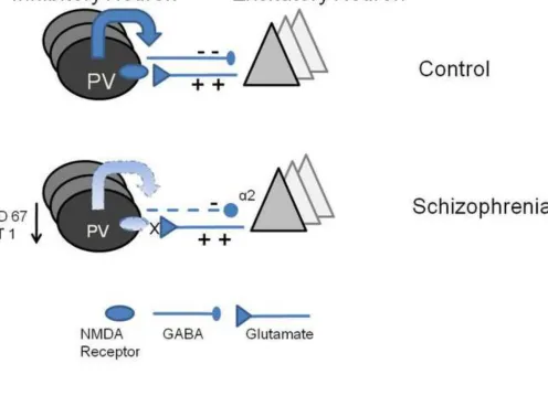
NMDA receptors play a crucial role in neuronal communication. Blocking NMDA
does not only interfere with excitatory transmission and synaptic plasticity, but it also reduces
the drive to inhibitory interneurons [230] (see Figure 3). Several recent studies demonstrate
that altered GABA neurotransmission may be secondary to abnormalities in NMDA receptor
functioning [87, 246]. This is based on the finding that ketamine reduces the activity of
GABA interneurons and thus disinbibits pyramidal neurons [75]. In addition to the effect of
acute intake, chronic ketamine intake has been shown to result in a reduction in the number
of parvalbumin-containing axoaxonic cartridges (see Fig 4). These are synaptic terminals of
inhibitory chandelier cells [149]. The reduced input on parvalbumin- containing interneurons
(indicating low pyramidal activity) have been suggested to not only reduce parvalbumin, but
to also downregulate GAD67, the principal synthesizing enzyme for GABA [149, 246]. This
can be seen as an maladaptive attempt to restore pyramidal cell activity to the correct levels
[134]. Taken together, these abnormalities may interfere with the generation of oscillatory
activity and may lead to the observed changes in schizophrenia.
---
Figure 4 about here
There is evidence both from in vivo recordings from mouse hippocampus [119] and
from human EEG [89] using the auditory paired-click gating paradigm that ketamine
increases gamma oscillatory activity and reduces slow frequency activity. Interestingly, Hong
1 2 3 4 5 6 7 8 9 10 11 12 13 14 15 16 17 18 19 20 21 22 23 24 25 26 27 28 29 30 31 32 33 34 35 36 37 38 39 40 41 42 43 44 45 46 47 48 49 50 51 52 53 54 55 56 57 58 59
withdrawal–retardation symptoms measured using the Brief Psychiatric Rating Scale (BPRS)
directly following the EEG recording.
There is however in-vitro evidence for reduced oscillatory high-frequency activity in
response to ketamine as well, which fits better with the cellular models of interneuron
inhibition discussed above [33, 41, 174]. Roopun et al. [174] showed that ketamine can have
region-specific effects with an increase in gamma in one region and reduced or no effect in a
different region. They argued that reduced power in one region may lead to phase delays
between oscillating networks across the cortex and as a consequence changes in long-range
synchrony may occur. In addition, this would explain a reduction in lower frequencies, but
also an increase in phase variability. Any of these changes may result or contribute to WM
deficits in schizophrenia. These results emphasize the importance of understanding the time
course of differential contributions of areas comprising the WM-network.
Alternatively, differential effects of acute or chronic ketamine have to be taken into
account when using this as a model for schizophrenia [see 89, 166]. Whereas acute ketamine
augments glutamate concentration measured with MR-spectroscopy in humans [175], it is the
chronic use of ketamine that results in NMDA receptor hypofunction. Indeed, glutamate
concentration has been shown to be higher in patients with recent onset of schizophrenia [24]
but reduced in chronic schizophrenia [215].
Chrobak et al [30] tested the effects of ketamine on the encoding, retrieval and
retention of memory in a delayed-match-to-place radial water maze task and showed
impairment in encoding of new location information because of an increase of proactive
interference. The authors suggested that the strength of the encoded representation is
weakened with ketamine administration, which is in line with previous results of
hippocampal place cells [103]. They also suggest that ketamine produces changes in theta and
gamma power and coherence and that it decouples the phase relationship between the two
frequencies, which may contribute to reduced WM capacity found in individuals with
schizophrenia.
In addition to the abnormalities of excitatory NMDA-receptor transmission on
shaping the inhibitory GABAergic transmission, these functional impairments may also
interact with structural abnormalities [21], such as reductions in amount of cortical neuropil,
axon terminals and dentritic spines density on cortical pyramidal neurons in schizophrenia
[62, 126]. Dendritic spines are the principal targets of excitatory synapses to pyramidal