A STUDY ON ASSOCIATION OF SUBCLINICAL
HYPOTHYROIDISM WITH ACUTE ISCHEMIC
STROKE
Dissertation submitted to
THE TAMILNADU DR. M.G.R MEDICAL UNIVERSITY
CHENNAI
In partial fulfillment of regulations
For award of the degree of
M.D (GENERAL MEDICINE)
BRANCH – 1
KILPAUK MEDICAL COLLEGE
CHENNAI
BONAFIDE CERTIFICATE
This is to certify that dissertation named “A STUDY ON ASSOCIATION OF SUBCLINICAL HYPOTHYROIDISM WITH
ACUTE ISCHEMIC STROKE” is a bonafide work performed by Dr. M. Saranya, post graduate student, Department of Internal Medicine,
Kilpauk Medical College, Chennai-10, under my guidance and supervision in fulfillment of regulations of the Tamilnadu , Dr. M.G.R Medical University for the award of M.D. Degree Branch I (General Medicine) during the academic period from May 2012 to April 2015.
Prof. Dr.R.Sabaratnavel M.D., Prof. Dr.Mayilvahanan M.D.,
Professor and Head Professor and Unit Chief Department of Medicine Department of Medicine
Kilpauk Medical College, Government Royapettah Hospital Chennai- 10 Chennai-14
Prof. Dr. N. Gunasekaran M.D., DTCD., The DEAN
DECLARATION
I solemnly declare that this dissertation “A STUDY ON ASSOCIATION OF SUBCLINICAL HYPOTHYROIDISM WITH ACUTE ISCHEMIC
STROKE” was prepared by me at Government Kilpauk Medical College and Hospital, Chennai, under the guidance and supervision of Dr. R.Sabaratnavel M.D., Professor, Department of Internal Medicine, Government Royapettah Hospital, Chennai.
This dissertation is submitted to The Tamil Nadu Dr. M.G.R. Medical University, Chennai in partial fulfillment of the University regulations for the award of the degree of M.D. Branch I (General Medicine).
Place: Chennai
ACKNOWLEDGEMENT
At the outset, I would like to thank my beloved Dean, Kilpauk Medical College Prof. Dr. N. Gunasekaran M.D., DTCD, for his kind permission to conduct the study in Kilpauk Medical College. I would like to express my special thanks to Medical Director and Superintendent, Govt. Royapettah Hospital, and my unit chief Prof. Dr. S. Mayilvahanan M.D., for permitting to conduct this study.
I would like to thank wholeheartedly, Prof. Dr. R.Sabaratnavel M.D., Head of Department and Professor of Medicine for his encouragement and guidance during the study.
I am extremely thankful to Assistant Professors of Medicine Dr. S. Kalaichelvi M.D., Dr. G. Ranjani M.D., Dr. Jayakumar M.D. and
Dr. T. Balaji for their assistance and guidance.
I would always remember with extreme sense of thankfulness, the co-operation and constructive criticism shown by my fellow post graduate colleagues and friends.
Finally, I thank all my patients for their active co-operation in this study, without which this would not have become a reality.
CONTENTS
SL.NO TITLE PAGE NO.
1. INTRODUCTION 1
2. AIMS AND OBJECTIVES 3
3. REVIEW OF LITERATURE 4
4. MATERIALS AND METHODS 58
5. RESULTS 61
6. DISCUSSION 96
7. CONCLUSION 103
8. LIMITATIONS 104
9. BIBLIOGRAPHY 106
10.
ANNEXURES
ABBREVATIONS PROFORMA MASTER CHART
ABSTRACT
BACKGROUND
Stroke is one of the leading causes of morbidity and mortality in the
general population. Apart from conventional risk factors, identifying potential
modifiable risk factors can play a significant role in stroke prevention and
outcome.
AIMS AND OBJECTIVES
This study aims to determine the frequency of subclinical hypothyroidism
in patients presenting with acute ischemic stroke, its association with altered
lipid metabolism and its role as an independent, modifiable risk factor in stroke
prevention.
MATERIALS AND METHODS
85 patients presenting with acute ischemic stroke are taken as cases and
85 patients without stroke as controls matched for age,sex and risk factors. CT
brain is done to confirm the diagnosis and thyroid profile measured.
Patients with normal free T3, T4 and serum TSH above upper limit of
normal are diagnosed to have subclinical hypothyroidism.
The presence of subclinical hypothyroidism in both the cases and controls
OBSERVATION AND RESULTS
Among the 85 patients with acute ischemic stroke, data analysis showed
that 8.23% (7 out of 85) of them had subclinical hypothyroidism which was
statistically significant with a p value of 0.007. Subjects in the control group
had no subclinical hypothyroidism. Among the 7 cases with subclinical
hypothyroidism, 6 (85.7 %) of them had positive correlation with serum total
cholesterol which was statistically significant.
CONCLUSION
The study results show a significant association of subclinical
hypothyroidism with acute ischemic stroke and signifies its potential role as an
1
INTRODUCTION
Stroke or cerebrovascular accident is defined as an abrupt onset of a neurological deficit, either focal or global that is attributable to a vascular etiology. Definition of stroke is thus clinical and laboratory studies including brain imaging are used to support the diagnosis.
The term cerebrovascular disease defines any brain lesion resulting from a pathologic process of the blood vessels such as occlusion of the lumen by embolus or thrombus, rupture of a vessel, an altered permeability of the vessel wall or a change in the quality of the blood flowing through the cerebral vessels. These are two main types—ischemia, with or without infarction and hemorrhage.
There are two main types—ischemia, with or without infarction and hemorrhage. Stroke can also be classified in terms of the underlying pathogenesis involved, i.e., atherosclerosis, arteriosclerotic changes developing secondary to Hypertension, arteritis, aneurysmal dilatation and developmental vascular malformations of the brain.
2
tissue. Diffusion weighted imaging methods have been proved invaluable in stroke workup.
In recent population based studies, the rate of incidence of stroke in India is found to be 119-145/100,000. The estimated adjusted prevalence rate of stroke ranges 84-262/100,000 in rural and 334-424/100,000 in urban areas.
There are several risk factors for stroke which includes hypertension, diabetes mellitus, atrial fibrillation, cigarette smoking, and dyslipidemia. Other causes like hypercoagulable states and medications like birth control pills also contribute but only in special circumstances. In addition, hypothyroidism has been studied as an independent risk factor for causing ischemic stroke by accelerating the process of atherosclerosis.1
Hypothyroidism is linked to altered lipid metabolism and lower levels of HDL cholesterol and higher levels of total cholesterol/HDL-C and LDL-C/HDL-C ratios and also associated with elevated homocysteine levels.
3
AIMS AND OBJECTIVES OF THE STUDY
1. To determine the frequency of subclinical hypothyroidism in patients presenting with acute ischemic stroke.
2. To study the association of subclinical hypothyroidism with altered lipid metabolism and its role in atherosclerosis.
3. To study the role of subclinical hypothyroidism as an, independent, modifiable risk factor in stroke prevention.
4
REVIEW OF LITERATURE
Definition of Stroke
The World Health Organization (WHO) definition of stroke is: “rapidly developing clinical signs of focal or global disturbance of cerebral function, with symptoms lasting 24 hours or longer or leading to death, with no apparent cause other than of vascular origin ”.
Types of stroke
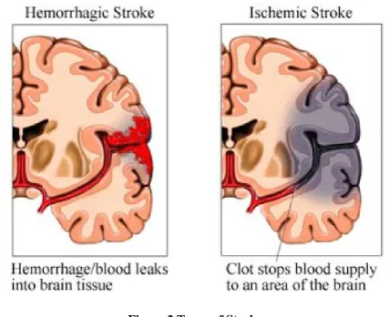
Stroke can either be ischemic (with or without infarction) or hemorrhagic (intracerebral or subarachnoid).
Ischemic stroke
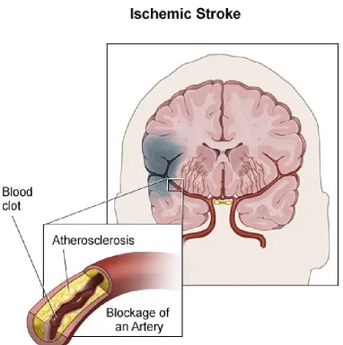
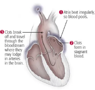
Ischemic stroke results from occlusion of the cerebral arteries by an atherosclerotic thrombus or from an emboli arising from the proximal part of the arterial system or the heart. Cardiac rhythm disturbances like atrial fribrillation which results in stasis and blood clot formation, forms an important source of embolic stroke.
5
Figure 1 Atherosclerosis and acute ischemic stroke
Hemorrhagic stroke
Besides trauma as an etiology, spontaneous intracerebral hemorrhages are mainly due to arteriolar hypertensive disease, and rarely due to coagulation disorders and vascular malformation within the brain.
6
[image:15.595.83.511.198.546.2]hemorrhagic stroke.Hemorrhagic stroke either by its mass effect on the surrounding structures or by raising the intracranial pressure produces significant neurological deficit.
Figure 2 Types of Stroke
7
Subarachnoid hemorrhage results from rupture of aneurysms present at the branching points of the large arteries of the circle of Willis, the blood is contained within the subarachnoid spaces and therefore, causes little focal effect on the brain. However, when there is severe bleeding it causes a delayed cerebral ischemia through a mechanism of constriction of the vessels of the circle of Willis and their primary branches (vasospasm).
The Stroke Syndrome
Stroke in its mildest form may consist of a trivial and transient neurologic disorder insufficient for the patient even to seek medical attention whilst in the most severe form the patient becomes hemiplegic or even comatose.
However, the essential feature in all forms of stroke is the temporal profile of neurologic events irrespective of the grading of severity between these two extremes. The abruptness with which the neurologic deficit develops usually in a matter of seconds helps in making a diagnosis of vascular origin.
Based on Time of onset
8 Based on pattern of involvement
The second essential feature is the focal signature of stroke. The neurologic deficit helps in localizing the anatomical site inolved and also in estimating the size of the infarct or hemorrhage. Hemiplegia stands as the classic sign of all cerebrovascular diseases, whether the lesion is restricted to the cerebral hemisphere or when it also involves the brainstem with accompanying other diverse manifestations but in highly recognizable patterns. These include paralysis, numbness and sensory deficits on one side of the body, aphasia, visual field defects, diplopia, dizziness and dysarthria. The different patterns of involvement enable the physician to localize the lesion at times so precisely to even specify the culprit artery involved.
The territory of any artery, large or small, deep or superficial, may be
9
Based on sequence of resolution of neurological deficit
Excluding fatal strokes another important aspect in the temporal profile of stroke evolution is the arrest and then partial regression of the neurologic deficit in almost all cases. A focal syndrome of rapid regression that reverses itself entirely and dramatically over a period of minutes or up to an hour has been defined as the "transient ischemic attack" (TIA). Often, an extensive deficit from embolism also partially reverses itself within a few hours or days. In most cases of thrombotic strokes, improvement occurs gradually over weeks and months, and there is considerable residual disability.
10
Figure 3 CT Brain Showing Infarct
Pathophysiology of Cerebral Ischemia
The brain derives its nourishment from a constant and adequate supply of oxygenated blood and this is maintained by a series of baroreceptors and vasomotor reflexes under the control of centers in the lower brainstem. So any disturbance in this hemostasis for eg the complete stoppage of blood flow for longer than 5 min produces irreversible brain damage. At autopsy, this damaged brain tissue which undergoes ischemic necrosis or infarction is seen as a zone of softening or encephalomalacia. Focal ischemic damage usually results from occlusion of an artery by a thrombus or embolus, but cardiac failure resulting in
Area of Infarction with
11
hypotension when severe and prolonged enough, can be the cause of focal as well as diffuse ischemic changes.
Several additional ischemia-modifying factors may influence the extent of necrosis. The speed of occlusion has a significant effect- with gradual narrowing of a vessel allowing time for collateral channels to open. Hypoxia and hypercapnia have additional deleterious effects. Altered viscosity and osmolality of the blood and hyperglycemia are other important factors. Finally, vascular anomalies (of neck vessels, circle of Willis) and the presence of previous vascular occlusions also influence the outcome.
Cerebral infarcts vary greatly depending on the amount of congestion and hemorrhage found within the softened tissue. Pale infarcts are devoid of blood; few infarcts show mild congestion (dilatation of blood vessels) at their margins while hemorrhagic infarcts show an extensive extravasation of blood from many small vessels in the infarcted area.
12
also show a mixed type and has special characteristics of not conforming to the territories of the main cerebral arteries and usually contains substantial hemorrhage within the infarcted zone.
Figure 4 Infarct with hemorrhagic transformation
Acute Infarct
13
RISK FACTORS FOR CEREBROVASCULAR DISEASE
Important risk factors for stroke have been identified as age, sex, blood pressure, hyperglycemia, dyslipidemia, smoking, alcoholism, sedentary lifestyle and dietary habits. Besides the above mentioned factors, ischemic and valvular heart disease, atrial fibrillation and history of similar events in the past also increase the risk of stroke.
Patient’s socioeconomic condition, ethnicity, educational status and environmental factors like physical (temperature, altitude), geographical, or psychosocial issues also seem to play a role.4
Exposure to environmental tobacco smoking have been studied as an independent risk factor for stroke. As in coronary artery disease, the level of low-density lipoprotein (LDL) cholesterol has the greatest impact on the incidence of stroke with elevated triglycerides also conferring additional risk. Other factors, such as low potassium intake and reduced serum potassium are also associated with an increased stroke rate in several studies, but the mechanism of this effect is obscure with a detrimental effect on blood pressure being a likely possibility.
HYPERTENSION AND STROKE
14
lacunar stroke. A transient rise in blood pressure is common in acute stroke and in cases with pre-existing chronic hypertension. Based on major controlled trials, no evidence exists for acutely lowering blood pressure in such patients and suggests allowing blood pressures in acute stroke to settle spontaneously unless very high.
PATHOPHYSIOLOGY
High blood pressure results in endothelial injury which increases blood-brain barrier permeability leading to blood-brain oedema6 and mass effects due to raised intra cranial pressure. Intraparenchymal haemorrhages results from smooth muscle cell and endothelial degeneration.
1. In addition, it increases cerebral artery stenosis and increased incidence of embolism arising from the heart and great vessels by accelerating the process of arteriosclerosis.
2. The structural derangements in the resistance vessels leads to increase in peripheral vascular resistance thereby increasing the risk for ischaemic events by compromising on the collateral blood supply.
15
central role in atherogenesis and plaque formation and occlusion of the vessel lumen resulting in reduced blood supply distal to the site of occlusion.
16
Figure 6 Vascular Changes in Hypertension
INDICATIONS FOR ANTI-HYPERTENSIVE THERAPY IN ACUTE
STROKE
For patients with stroke who are not candidates for thrombolytic therapy, the recommended guideline7 to initiate anti-hypertensive therapy is when blood pressure is above 220/130 mm Hg and if thrombolytic therapy8 is to be used, the goal blood pressure is <185/110 mm Hg.
For patients presenting with hemorrhagic stroke, therapy can be initiated at blood pressures above 180/130 mm Hg.
In subarachnoid hemorrhage, cautious reduction of blood pressure is indicated when mean arterial pressure is >130 mm Hg.
17 DIABETES AND STROKE
Diabetes has been proven to be a significant independent risk factor9 for stroke; the risk in diabetics being 2 to 4 fold greater than in normal population. It is also an important risk factor for morbidity and mortality associated with stroke, with increased incidence of severe neurological deficits and disability10, higher incidence of stroke recurrence11-12 and a poorer long-term prognosis.
Diabetes is associated with both microvascular and macrovascular complications affecting several organs including heart,brain, kidneys, skin and muscle. Impaired glucose metabolism in diabetics may favour the initiation of vascular complications through structural derangements mediated by advanced glycation end products and production of reactive oxygen species.
The role of diabetes in stroke has been attributed to the pathophysiological changes seen in the cerebral vessels of patients which includes
1. The pathologic effects of advanced glycation end product accumulation
2. Nitric oxide inhibition leading to impaired vasodilation, 3. Chronic inflammation
4. Smooth muscle cell proliferation,
18 7. Enhanced platelet aggregation
Apart from diabetes being an independent risk factor for stroke, it also increases the incidence of other conventional risk factors which includes hypertension, heart failure, dyslipidemia, and atrial fibrillation.13
PATHOGENESIS
Endothelial injury marks the initiation of macrovascular complications in diabetes, with other factors super imposed on it.
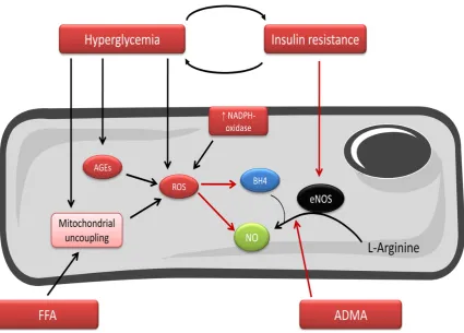
Chronic hyperglycemia by inhibiting nitric oxide reduces the vasodilatory response14 through inhibition of endothelial nitric oxide synthase (eNOS) and increases the production of reactive oxygen species,that leads to further inhibition of eNOS.15-16.
19
Figure 7 Mechanism of Endothelial dysfunction in diabetes
Impaired platelet function leading to rupture of atherosclerotic plaque17 and thrombus formation may underlie the pathogenesis of macrovascular complications in diabetes.
20
Smooth muscle cell dysfunction contributes to the structural vessel wall abnormalities seen in diabetes. Following endothelial injury, smooth muscle cells in the tunica media migrate to the intimal layer producing solidification of the atherosclerotic fatty streaks and resulting in an extensive extracellular matrix, thereby weakening the tunica media and resulting in plaque rupture.
A unique effect of diabetes on cerebral vasculature is its effect on neurons and glial cells which occurs during ischemia. This has been explained through the relationship between hyperglycemia and increased intracellular acidosis.145
Anaeroic metabolism that occurs during an ischemic event, for instance as in stroke produces neuronal intracellular acidosis which causes nerve cell damage through the mechanism of Reactive Oxygen Species (ROS) production.19
21
Figure 8 Role of inflammation in atherosclerosis
STROKE AND DYSLIPIDEMIA
22
According to recent studies ,apart from elevated LDL other lipids like the reduced levels of HDL-c, increased triglycerides and Apolipoprotein B are also found contributing to the stroke risk. Atherogenic dyslipidemia comprising of reduced HDL-c and an elevated TGL has been proved to be an independent risk factor of increased vascular risk. The MRFIT study conducted on 3,50,977 individuals has proved a positive correlation between total cholesterol levels and risk of ischaemic stroke.5 Furthermore , its proved that the relative risk for stroke doubles every decade after the age of 55.8,9
High density lipoprotein (HDL–c) in reduced concentration ,an independent risk factor of atherosclerosis is also considered a risk for stroke. Numerous prospective studies have proved a significant reduction in non fatal stroke in subjects with an elevated or normal HDL-c levels10. The levels of HDL-c were inversely correlated with atheroma burden. The plasma levels of triglyceride has no independent predictive value in stroke risk.
Lipoproteins- no concrete evidence exists pointing to the connection between stroke risk and lipoprotein levels. There are controversial studies denoting the association between lipoprotein–a and stroke incidence20.the association between the levels of Apo E and the cerebrovascular risk is also under study. Few other studies indicate the positive predictive capacity of Apo B , and Apo A1 in cerebrovascular events.
23
Further, treatment of dyslipidemia is associated with significant risk reduction in stroke. According to the CARE25 ,PROSPER ,ASCOT trials there is a proven in reduction in stroke risk by 31 %, on treatment with statins aimed at reducing LDL levels.
STROKE AND HEART DISEASES
Cardioembolic stroke results when a thrombus within the heart chambers breaks and dislodges embolic fragments into the vessel wall. Sometimes an emboli may originate from the distal end of a thrombus intraarterially, in a severely stenosed or occluded carotid or vertebral artery or from large atheromatous plaques in the aorta.
Arrhythmias such as atrial fibrillation, secondary to valvular heart diseases or ischemic heart disease and atrial myxoma also predispose to embolic stroke. Thrombotic or infected material e.g. as in infective endocarditis which adheres to the aortic or mitral heart valves and clots originating on prosthetic heart valves also break free to produce embolic stroke.
Ischemic infarction results when the embolus becomes arrested at a
24
block large vessels like the carotids in the neck, while tiny fragments may reach vessels as small as 0.2 mm in diameter, usually with minimal effects.
As embolic strokes develops rapidly, collateral blood supply does not become established and hence sparing of the brain territory distal to the site of occlusion as seen in slowly developing thrombotic strokes is usually not evident.
[image:33.595.132.465.374.688.2]The most common cause of an embolic stroke being a chronic or recent atrial fibrillation, it usually arises from a mural thrombus within the atrial appendage.
25
Based on the Framingham Heart Study, patients with chronic atrial fibrillation are approximately 6 times more prone to stroke than an age-matched population with normal cardiac rhythm (Wolf et al, 1983) and the risk is higher if rheumatic valvular heart disease is also present.
Furthermore, the risk for stroke in patients with atrial fibrillation varies with age, being 1 percent per year in persons younger than age 65 years, and as high as 8 percent per year in those older than age 75 years with additional risk factors. These play a significant role in determining the benefit of chronic anticoagulation therapy.
Embolism also occurs in cases of paroxysmal atrial fibrillation or flutter. Post myocardial infarction, mural thrombus formed on the damaged endocardium in the left ventricle, particularly if there is an aneurysmal sac, is an important source of cerebral emboli.
26 STROKE AND TOBACCO USE
Smoking as an independent risk factor has been known to double the risk of ischemic stroke. Cigarette smoke is generally divided into two phases which include a tar phase (particulate phase) and a gas phase. When passed through the Cambridge glass-fiber filter, the material that gets trapped is the tar phase with a particulate material of size >0.1µm. While the material that passes through the filter comprises the gas phase.
The particulate phase of cigarette smoke contains more than 1017 free radicals/g, and gas phase contains more than 1015 free radicals/puff .The radicals related to the particulate phase have a longer life span ( hours to months) where as those associated with gas phase are short lived(seconds). It is found that amidst all the identified constituents, nicotine found in the particulate phase, is the addictive substance in cigarette smoke.
27
Figure 10 Role of Nitric Oxide in Endothelial vasodilation
SMOKING AND INFLAMMATION
The inflammatory response plays a central role in the instigation and progression of atherosclerosis. Based on various studies, evidence exists about smoking causing a significant increase in the peripheral leukocyte count. Inflammatory markers like C-reactive protein, interleukin-6, and tumor necrosis factor- alpha(TNF-α) have also been found to be significantly increased in smokers 22.
28
E-Selectin levels were higher in smokers than non smokers. It also activates proatherogenic molecules resulting in alteration in cell-cell interactions and helps in hastening the process of atherosclerosis.
Smokers have higher serum total cholesterol, LDL and triglyceride levels and lower levels of HDL. The triglyceride and high-density lipoprotein abnormalities have been found to be linked to insulin resistance.
It increases oxidation of LDL molecules by decreasing the activity of paraoxonase, an enzyme that inhibits LDL oxidation.
TREATMENT: ACUTE ISCHEMIC STROKE
After the clinical diagnosis of stroke is made, an orderly process of evaluation and treatment should follow. The first goal is to prevent or reverse brain injury. Attend to the patient's airway, breathing, circulation (ABC's). Perform an head CT scan to differentiate between ischemic stroke and hemorrhagic stroke.
Medical Support
29
Because collateral blood flow within the ischemic brain is blood pressure dependent, there is controversy about whether blood pressure should be lowered acutely. Blood pressure should be lowered if there is malignanthypertension or if blood pressure is >185/110 mm Hg and thrombolytic therapy is anticipated. Fever is detrimental and should be treated with antipyretics and surface cooling. Serum glucose should be monitored and kept at <6.1 mmol/L (110 mg/dL) using an insulin infusion if necessary.
The larger the infarct, the greater the likelihood that clinically significant edema will develop. Water restriction and IV mannitol may be used to raise the serum osmolarity, but hypovolemia should be avoided as this may contribute to hypotension and worsening infarction.
Intravenous Thrombolysis
The NINDS study used IV rtPA (0.9 mg/kg to a 90-mg max, 10% as a bolus, then the remainder over 60 minutes) versus placebo in patients with ischemic stroke within 3 hours of onset. One-half of the patients were treated within 90 minutes. Thus, despite an increased incidence of symptomatic intracerebral hemorrhage, treatment with IV rtPA within 3 hours of the onset of ischemic stroke improved clinical outcome.
30
stroke and is cost-effective and cost-saving. The time of stroke onset is defined as the time the patient's symptoms began or the time the patient was last seen as normal. Patients who awaken with stroke have the onset defined as when they went to bed.
Antithrombotic Treatment
Platelet Inhibition
Aspirin is the only antiplatelet agent that has been proven effective for the acute treatment of ischemic stroke; there are several antiplatelet agents proven for the secondary prevention of stroke. Use of aspirin within 48 hours of stroke onset reduced both stroke recurrence risk and mortality minimally. The glycoprotein IIb/IIIa receptor inhibitor abciximab was found to cause excess intracranial hemorrhage and should be avoided in acute stroke. Clopidogrel is being tested as a way to prevent stroke following TIA and minor ischemic stroke.
Lipid Lowering drugs
31
HYPOTHYROIDISM
THYROID: ANATOMY AND DEVELOPMENT
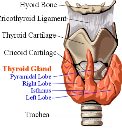
The thyroid gland derives its origin from the greek word ‘thyreos’ denoting shield and it comprises of two lobes joined by an isthmus. It lies in front of the trachea extending from the cricoid cartilage to the suprasternal notch. The gland is highly vascular, soft in consistency and weighs around 12– 20 g. The thyroid gland develops from the floor of the pharynx and migrates along the thyroglossal duct to reach its final location in the neck. Thyroid hormone synthesis begins as early as around 11 weeks gestation. The hormones secreted by the thyroid gland play a key role in cell differentiation during development and helps to maintain thermogenic and metabolic homeostasis in the adult
32
[image:41.595.92.511.180.623.2]though in most cases,etiology of congenital hypothyroidism remains unknown. The thyroid gland consists of numerous spherical follicles that contain colloid rich in thyroglobulin, which is essential for thyroid hormones synthesis.
33 REGULATION OF THYROID AXIS
Thyroid stimulating hormone( TSH), secreted by the thyrotrope cells of the anterior pituitary is a 31-kDa hormone composed of α and β subunits; the α subunit which shares similarity to many other glycoprotein hormones [luteinizing hormone, follicle-stimulating hormone, human chorionic gαonadotropin ], whereas the β subunit is unique to TSH.
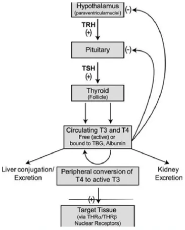
The thyrotropin releasing hormone (TRH) from the hypothalamus stimulates the pituitary production of TSH which stimulates thyroid gland to produce the thyroid hormones T3 and T4. Thyroid hormones, acting
predominantly through thyroid hormone receptor β 2, sends feed back to pituitary and hypothalamus to inhibit TSH and TRH respectively.
Like other pituitary hormones, TSH is the main regulator of this endocrine feedback loop mechanism. TSH is measured using immunoradiometric assays which are highly sensitive and specific. And TSH can be used for the diagnosis of hyperthyroidism (low TSH) as well as hypothyroidism (high TSH). Drugs like dopamine, glucocorticoids and somatostatin when administered exogenously suppress TSH function.
Reduced levels of T3 and T4 enhance TSH production whereas higher
34
35
THYROID HORMONE SYNTHESIS AND FUNCTION
Iodine Metabolism and Transport
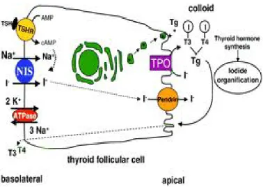
The first critical step in thyroid hormone synthesis is Iodine uptake23. Iodine from diet is carried by serum binding proteins, particularly albumin from where it is taken into thyroid follicular cells by NIS( sodium iodide symporter) expressed at its basolateral membrane.
Adaptations to variations in dietary intake of iodine is highly regulated by this symporter function and expression. One of the rare causes of congenital hypothyroidism is mutation of the NIS gene.
36
Organification, Coupling, Storage, Release
Following uptake into follicular cells, iodine is oxidized in an organification reaction catalysed by TPO and hydrogen peroxide and is added to selected tyrosyl residues within Tg to form diiodotyrosine(DIT) and monotyrosine(MIT).These are further coupled via an ether linkage involving thyroperoxidase(TPO) with coupling of two molecules of DIT or one of DIT and one of MIT leading to formation of T4 and T3, respectively. Reuptake of Tg
into the thyroid follicular cell allows proteolysis and the release of newly synthesized T4 and T3.
Uncoupled mono- and diiodotyrosines (MIT, DIT) are deiodinated by the enzyme dehalogenase, thereby recycling any iodide that is not converted into thyroid hormones.
37
Figure 13 Thyroid Hormone Synthesis TSH Action
TSH regulates thyroid gland function through the TSH-R, a seven-transmembrane G protein–coupled receptor (GPCR) that is coupled to the subunit of stimulatory G protein (Gs), which activates adenylyl cyclase, leading
38
Other Factors that Influence Hormone Synthesis and Release
Apart from TSH being the prime hormonal regulator of thyroid function, a variety of growth factors also influence thyroid hormone synthesis. These include insulin-like growth factor I (IGF-1), epidermal growth factor, transforming growth factor (TGF-), endothelins, and various cytokines. In individuals with a normal thyroid, the gland escapes from inhibitory effect of excess iodide, however the suppressive action of high iodide may persist in patients with underlying autoimmune thyroid disease.
Serum Binding Proteins
T4 is secreted in about twentyfold excess over T3 and.both hormones are
39
Characteristics of Circulating T4 and T3
Hormone Property T4 T3
Serum concentrations
Total hormone 8 g/dL 0.14 g/dL
Fraction of total hormone in the free form 0.02% 0.3% Free (unbound) hormone 21 × 10–12M 6 × 10–12M
Serum half-life 7 d 0.75 d
Fraction directly from the thyroid 100% 20% Production rate, including peripheral conversion 90 g/d 32 g/d
Intracellular hormone fraction 20% 70%
Relative metabolic potency 0.3 1
Receptor binding 10–10M 10–11M
Table 1: Characteristics of Circulating T4 and T3s
Despite its low serum concentration,TBG carries about 80% of the bound hormones due to its high affinity for thyroid hormones. Whereas albumin with a high plasma concentration has relatively low affinity for thyroid hormones and it binds up to 10% of T4 and 30% of T3. TTR carries about 10% of T4 but
little T3.
Approximately 99.98% of T4 and 99.7% of T3 are protein-bound and
40
unbound T3 > unbound T4, but there is less unbound T3 in the circulation as it is
produced in smaller amounts and cleared more rapidly than T4
Nuclear Thyroid Hormone Receptors
Thyroid hormones24 bind with high affinity to nuclear thyroid hormone receptors (TRs) α and β..The receptors bind as heterodimers with retinoic acid
X receptors (RXRs) and then to specific DNA sequences, termed thyroid response elements (TREs), in the promoter regions of target genes. The activated receptor then in turn can stimulate gene or inhibit transcription depending on the nature of the regulatory elements in the target gene.
Though T4 is produced in excess of T3, receptors are occupied mainly by
T3 owing to their high affinity and greater plasma availability resulting in
maximum hormonal potency. After binding to TRs, thyroid hormone induces conformational changes in the receptors leading to alteration in gene expression.
Laboratory Evaluation
Measurement of Thyroid Hormones
41
by measurements of serum T3 and T4 levels to make a diagnosis of hypo (with
elevated TSH) or hyperthyroidism (with suppressed TSH levels).
As total T4 and T3 are highly protein-bound and influenced by numerous
factors like illness, medications and genetic factors it is useful, therefore, to measure the free, or unbound, hormone levels, which reflects the biologically active hormone pool.
Total T4 and T3 levels are elevated when TBG is increased due to
estrogens (pregnancy, oral contraceptives, hormone therapy, tamoxifen), and decreased when TBG binding is reduced (androgens, nephrotic syndrome). As total T4 and T3 are highly protein-bound and influenced by numerous factors
like acute illness, genetic disorders and various medications (phenytoin, carbamazepine, salicylates, and NSAIDs)which can interfere with thyroid hormone binding, it is preferable to measure the free, or unbound, hormone levels, which remains normal under such situations.
42
on exposure to certain drugs like dopamine or high doses of steroids.
The most common antibodies detected in patients with auto immune thyroid disease is antibodies against TPO and Tg.
Hypothyroidism
Iodine deficiency remains the most common cause of hypothyroidism worldwide. In areas of iodine sufficiency, autoimmune disease and iatrogenic causes are the most common causes.
Symptoms
Tiredness, weakness Dry skin
Feeling cold Hair loss
Difficulty concentrating and poor memory
Constipation
Weight gain with poor appetite Dyspnea
Hoarse voice
Menstrual disturbances
Signs
Puffy face, hands, and feet (myxedema)
Diffuse alopecia Dry coarse skin Decreased heart rate Carpal tunnel syndrome
Delayed tendon reflex relaxation Serous cavity effusions
43 Autoimmune Hypothyroidism
Classification
Autoimmunity is the most common cause of hypothyroidism which may be either associated with a goiter (Hashimoto's thyroiditis) or resulting in atrophy of the thyroid gland. As auto immunity progresses and reduces thyroid function, there is a compensatory rise in TSH levels to maintain normal thyroid hormone levels. With most of the patients being asymptomatic, this is termed subclinical hypothyroidism. It progresses to overt hypothyroidism when patients become symptomatic with reduced T4 levels, and a compensatory rise in TSH levels >10 mIU/L.
Prevalence
44 Pathogenesis
In Autoimmune thyroiditis25, there is lymphocytic infiltration of the gland, by CD4+ and CD8+ T cells resulting in atrophy and fibrosis of thyroid gland. CD8+ cytotoxic T cells mediate destruction by either perforin-induced cell necrosis or granzyme B–induced apoptosis.
Genetic and environmental factors contribute to the development of autoimmune thyroid disorders, of which HLA-DR polymorphisms are the most widely recognized.
By its direct toxic effects or through immune mediated mechanisms, high iodine intake increases the risk of autoimmune hypothyroidism.
45 Clinical Manifestations
The skin is dry, coarse with decreased sweating, thinning of the epidermis and hyperkeratosis of the stratum corneum. Increased dermal glycosaminoglycan content traps water, giving rise to skin thickening without pitting (myxedema). A puffy face with edematous eyelids and nonpitting pretibial edema gives a typical picture of hypothyroid patients. The hair becomes dry, brittle and falls out easily resulting in diffuse alopecia, with thinning of the outer third of the eyebrows, though not specific for hypothyroidism. Nail growth retardation is also a common finding.
Other common features include constipation and increase in weight which is due to fluid accumulation in the subcutaneous tissues. Decreased libido, reduced fertility and increased incidence of miscarriage is common which may be due to increased Prolactin levels. There may be oligomenorrhea or amenorrhea in long-standing disease, but menorrhagia is also common.
46
remains normal, dyspnea may be caused by pleural effusion, impaired respiratory muscle function, diminished ventilatory drive, or sleep apnea.
On examination, there may be slow relaxation of tendon reflexes and pseudomyotonia. Cognitive disturbances include memory disturbances and poor concentration.
DEFINITION OF SUBCLINICAL AND OVERT HYPOTHYROIDISM
Overt hypothyroidism is diagnosed in patients who have symptoms of hypothyroidism, reduced levels of thyroid hormones T3, T4 and elevated TSH (>
10µIU/ml) whereas a diagnosis of subclinical hypothyroidism is made when T3,
T4 levels are within the normal range with mild elevations of TSH levels
(<10µIU/ml) which act to maintain normal thyroid hormones but can eventually progress to overt hypothyroidism, when the compensatory mechanism is overwhelmed by the auto immune process.
HYPOTHYROIDISM AND ENDOTHELIAL DYSFUNCTION
The vascular homeostasis is maintained by the endothelium by a balance in the production of vasoconstrictor and vasodilator substances26. Nitric oxide, the most important vasodilator substance is produced by the endothelium during conversion of L-arginine into citrulline catalysed by the enzyme nitric oxide synthase.
47
In presence of increased serum cholesterol levels, due to increased oxidative stress the endothelium causes breakdown of nitric oxide production.In addition, endothelial dysfunction is also influenced to a major extent by other risk factors like aging, hypertension, smoking and post menopausal status.So dyslipidemia acts as an independent risk factor for altering endothelial function and in patients with subclinical hypothyroidism (SH) having abnormal lipid profile, this could play a role in mediating endothelial dysfunction.
Evidence from a study conducted on patients with SH studied the vasodilating effect in response to acetylcholine and it was found to be significantly reduced in patients with SH compared to euthyroid controls. And it was also found to be impaired in patients with SH whose total serum cholesterol levels were in the normal range.This clearly indicates the direct effect of thyroid hormones as an independent risk factor in producing endothelial dysfunction in SH patients apart from the contributory effects of lipid abnormalities on endothelial function and NO production.
48
hypothesis that inflammation present in autoimmune thyroid disorders could also play a part in mediating impaired endothelial vasodilation.
But more specific inflammatory markers like C-Reactive Protein and interleukins need to be studied to confirm this hypothesis as ESR is a non specific inflammatory marker found to be raised in various other pathologies.
In patients with SH, lipoprotein a values were also found to be elevated and has a significant co-relation with endothelial dysfunction in coronary circulation thereby contributing to coronary artery disease.
So all these factors in patients with subclinical hypothyroidism including thyroid dysfunction, chronic inflammatory state seen in autoimmune thyroid disorders and altered lipid metabolism leading to hypercholesterolemia could jointly contribute to the impaired endothelium dependent vasodilation seen in these patients.
49
So to conclude, subclinical hypothyroidism represents an early, independent, reversible risk factor in causing endothelial dysfunction in addition to the co-existing lipid abnormalities seen in SH patients. This highlights the importance of early replacement therapy in not only delaying the progression to overt hypothyroidism but also in restoring endothelium dependent vasodilation, reversal of lipid abnormalities and slowing down the atherogenic process and its associated complications.
50
HYPOTHYROIDISM AND DYSLIPIDEMIA:
Conventionally, subclinical and overt hypothyroidism is associated with elevation of total serum and low density lipoprotein (LDL) cholesterol levels and normal or elevated high density lipoprotein (HDL) cholesterol levels.27 Overt and subclinical hypothyroidism is associated with reduced levels of total LDL and HDL cholesterols.28 These alterations in lipid metabolism are mainly due to the effect of thyroid hormones in regulating the activity of the enzymes involved in metabolism of the lipoproteins.
ACTION OF THYROID HORMONES IN LIPID METABOLISM
To be more specific, the thyroid hormones regulates the activity of the enzyme 3 - hydroxyl – 3 – methyl glutaryl – coenzyme A (HMG – CoA) which forms the first step in cholesterol synthesis by catalyzing the conversion of HMG - CoA to mevalonate and resulting in elevation of cholesterol levels.
The thyroid hormones have a key role in activating the LDL receptors, the binding of the thyroid hormones to the promoter region of the LDL receptor gene which contains a thyroid hormone responsive element (TRE) that allow the T3 levels in upregulating the gene expression of LDL receptors.
29
51
and very low density lipoproteins (VLDL) in the opposite direction.30 In addition, these hormones also stimulate the enzyme lipoprotein – lipase (LPL) that is involved in the metabolism of TGL rich lipoproteins and hepatic lipase (HL) an enzyme which converts HDL2 to HDL3.
31
Therefore disturbancess in thyroid function can result in changes in composition and transport of lipoproteins.32 With declining thyroid function, the levels of total serum and LDL cholesterol are elevated, thereby making hypothyroidism an important cause of secondary dyslipidemia.33
LIPID PROFILE IN OVERT HYPOTHYROIDISM
Despite lower cholesterol synthesis due to reduced activity of HMG – COA reductase enzyme in hypothyroidism, there is an increase in total serum cholesterol levels due to raised levels of LDL and intermediate density lipoprotein (IDL) cholesterol.
The main reason for elevated cholesterol levels i.e, the increase in LDL and IDL cholesterol concentration in hypothyroidism is mainly due to down regulation of LDL receptors leading to decreased receptor mediated catabolism of LDL and IDL.34
52
hence associated with increased TGL and VLDL levels. The VLDL and IDL particles in hypothyroid patients are rich in cholesterol and apolipoprotein E resembling the lipid profile in type III hyperlipoproteinemia.35 Hence in patients who are homozygous for apolipoprotein E 2 allele if super imposed with an insult to the thyroid may develop full blown features of type III hyperlipoproteinemia.
Mechanism of increase in HDL Cholesterol levels
There is an increase in HDL cholesterol levels mainly due to increased concentration and also due to decreased catabolism of of HDL2 particles36
owing to the decreased activity of HL enzyme. There is reduced transfer of cholestryl esters from HDL to VLDL due to decreased activity of CETP also increasing the HDL cholesterol levels.37
HYPOTHROIDISM AND ATHEROSCLEROSIS
There are a number of mechanisms leading to development of atherosclerosis in hypothyroidism
1. T4 has three specific binding sites on apolipoprotein B and inhibits LDL
oxidation invitro.38 So hypothyroidism favors LDL oxidation39.
53
3. Through sympathetic and adrenaline activation41 and increased peripheral resistance leading to vessel wall (aortic) stiffness produces diastolic hypertension.42
4. The decrease in thyroid hormones also favors a hypercoagulable state.43
EVIDENCE SUPPORTING THE ROLE OF HYPOTHYROIDISM IN
DYSLIPIDEMIA
The lipid abnormalities seen in hypothyroidism has been found to be significantly reduced with L thyroxine (T4) supplementation and also increases
previously low biliary cholesterol excretion.44 It usually takes around 4 to 6 weeks of thyroxine supplementation to correct lipid abnormalities in overt hypothyroidism. Despite restoring an euthyroid status, the failure of substitution therapy to normalise the lipid abnormalities should take into account any superimposed dyslipidemia45.
1. According to a recent meta analysis after initiating substitution therapy there is a decrease in serum total and LDL cholesterol levels to 7.9mg/dl and 10mg/dl respectively.46 In patients with higher cholesterol levels prior to treatment and in hypothyroid patients taking sub optimal T4 doses
this reduction in cholesterol levels was significantly high.47
54
3. Though a less consistent finding serum HDL cholesterol levels were also found to decrease with L – thyroxine (T4) therapy.48
4. So in total there is a significant decrease in serum total and LDL cholesterol, lipoprotein a, apolipoprotein B but such significant changes were not seen with HDL cholesterol, triglycerides and apolipoprotein AI.49
In patients with dyslipidemia prevalence of overt hypothyroidism is not low.50
LIPID PROFILE IN SUBCLINICAL HYPOTHRYOIDISM
Normal levels of T3 and T4 with mildly elevated serum TSH levels up to
10µIU/ml defines subclinical hypothyroidism (SH). In comparison to overt hypothyroidism subclinical hypothyroidism is more common and has a higher prevalence in women and elderly.51 It was found that in patients with positive anti thyroid anti bodies but with high normal TSH levels (2-4µIU/ml) there was increase in total serum cholesterol.
55
In patients with antibodies to thyroid peroxidase (TPO) as well as in smokers, subclinical hypothyroidism has been associated with increased risk of coronary events owing to the lipid abnormalities seen in SH.54 Subclinical hypothyroidism impairs endothelial vasodilation by decreasing heart rate variability, ventricular function as well as cardio vascular adaptation to effort. In women with SH, smoking acts as an additional risk factor in altering the lipid profile and aggravating thyroid failure jointly contributing to the development of atherosclerosis.55
ROLE OF THERAPY IN SUBCLINICAL HYPOTHYROIDISM
There has been conflicting results regarding the effect of L thyroxine substitution therapy in reversing the lipid abnormalities in patients with subclinical hypothyroidism.56
56
Though many studies support the beneficial role of thyroid replacement in reducing dyslipidemia and risk of CAD in overt hypothyroidism, studies concerning the treatment of subclinical hypothyroidism is still not conclusive.
Hence it is evident that thyroid substitution would play a significant beneficial role in patients with prominent thyroid dysfunction (TSH>10µIU/ml), high pretreatment cholesterol levels and in patients with smoking as an additional risk factor.
However, the beneficial effect of lowering total and LDL cholesterol level in SH patients is outweighed by thyroid substitution-induced decrease in HDL cholesterol levels. Risk of exacerbating angina or inducing cardiac arrhythmia by thyroxine therapy ensures cautious use of supplementation therapy when treating people with a previous history of angina.
57
AUTOIMMUNE THYROID DISEASE AND LIPOPROTEIN (a)
In recent studies, increased levels of lipoprotein (a) was found in euthyroid males and females with evidence of auto immune thyroid disease (increased titers of TPO and/or Thyroglobulin antibodies).62
However in another study, which compared the levels of lipoprotein (a) in euthyroid patients with positive thyroid auto antibodies with age and sex matched controls showing no evidence of thyroid auto immunity, the results showed no significant difference in lipid profiles including lipoprotein (a) between the two groups.63In the above mentioned study, apolipoprotein (a) which is known to influence Lp(a), chronic renal failure were also taken into account.
Hence the presence of auto immune thyroid disorders does not have a significant role in reversing the lipid abnormalities in patients with subclinical hypothyroidism.64
EVALUATION
58
MATERIALS AND METHODS
The study was conducted at Govt. Royapettah Hospital, Chennai between April 2014 to September 2014.
The study was performed after getting written consent from all the atients chosen for the study. Clearance was obtained from ethical committee of Govt. Kilpauk College and Hospital, Chennai.
STUDY DESIGN AND PATIENT SELECTION
This is a cross sectional, case control study in which 85 patients presenting with acute ischemic stroke are taken as cases and 85 people not suffering from stroke were selected as controls matched for age, sex and risk factors.
Inclusion Criteria:
1. patients with acute ischemic stroke 2. no previous H/O thyroid abnormality
3. no H/O any chronic drug intake known to interfere with thyroid function.
Exclusion criteria:
1. Patients with previous H/O thyroid dysfunction. 2. Cardiac dysfunction leading to embolic stroke.
59 4. Severe renal function impairment. 5. Severe liver function impairment.
6. Patients taking drugs known to interfere with thyroid function. METHODOLOGY
A total no. of 85 patients presenting with acute ischemic stroke from both sexes (51 Males & 34 Females) with age range from 45 to 80 years were selected as cases, diagnosis confirmed by CT brain and thyroid profile (free T3,
T4, TSH) measured in these patients.
A total no. of 85 controls (not suffering from stroke) matched for age, sex and risk factors were selected from outpatient department, Govt. Royapettah Hospital.
Patients and controls taking drugs that interfered with thyroid function were excluded from the study.
MATERIALS
Thyroid function was tested by quantitative measurements of free triiodothyronine (T3), free Thyroxine (T4) and Thyroid Stimulating hormone
60 Range of Expected Values:
Free T3 – 1.71-3.70 pg/ml
Free T4- 0.89- 1.76 ng/dl
TSH- 0.35- 5.50 µIU/ml
So patients, with normal T3, T4 levels and elevated TSH levels (< 10 µIU/ml)
were diagnosed to have subclinical hypothyroidism. Normal TSH levels (0.35-5.50 µIU/ml).
STATISTICAL ANALYSIS:
Data was entered in Windows Excel format, data analysis was carried out using statistical package of SPSS 15 for determination of statistical significance among different variables. Co-relation was assessed using chi-square test.
61
RESULTS AND OBSERVATIONS
DISTRIBUTION OF AGE AMONG CASES
Observ
ed N Expected N Residual
P Value
<= 50 5 21.3 -16.3
51-60 18 21.3 -3.3 0.000
61-70 35 21.3 13.8
71-80 27 21.3 5.8
Total 85
Table 3: Distribution Of Age Among Cases
In the study, 62 were above 60 years and 23 people below 60 years. The frequency of ischemic stroke was more in the age group > 60 years 6(73%) compared with age group <60 years. The p value of the study which is <0.05 is significant.
62
Figure 16: Distribution Of Age Among Cases
Figure 17: Distribution Of Age Among Cases
5
18
35
27
<= 50 51-60 61-70 71-80
Observed N
Observed N
35
27
0 5 10 15 20 25 30 35 40
63
AGE DISTRIBUTION AMONG CASES AND CONTROLS
Diagnosis Total P
value
Cases Control
Age in years
<= 50
Count
5 1 6
% within Age in
years 83.30% 16.70% 100.00%
% within
Diagnosis 5.90% 1.20% 3.50%
51-60
Count
18 16 34
% within Age in
years 52.90% 47.10% 100.00%
% within
Diagnosis 21.20% 18.80% 20.00%
61-70
Count
35 46 81
% within
Age in
years 43.20% 56.80% 100.00%
% within
Diagnosis 41.20% 54.10% 47.60%
71-80
Count
27 22 49
0.188
% within Age in
years 55.10% 44.90% 100.00%
% within
Diagnosis 31.80% 25.90% 28.80%
Total Count 85 85 170
% within
Age in
years 50.00% 50.00% 100.00%
% within
64
Table 4: Distribution Of Age Among Cases and Controls The age distribution among cases and controls was equal with a p value of 0.188 which is not statistically significant.
Figure 18: Age Distribution Between Case And Control Groups
Age in years
71-80 61-70
51-60 <= 50
C
ount
50
40
30
20
10 0
Diagnosis
65
GENDER DISTRIBUTION BETWEEN CASES AND CONTROLS
Diagnosis
Total Value P Cases Control
Male
Count 51 51 102
1.000 % within
Sex 50.00% 50.00% 100.00%
% within
Diagnosis 60.00% 60.00% 60.00%
Female
Count 34 34 68
% within
Sex 50.00% 50.00% 100.00%
% within
Diagnosis 40.00% 40.00% 40.00%
Total
Count 85 85 170
% within
Sex 50.00% 50.00% 100.00%
% within
Diagnosis 100.00% 100.00% 100.00%
Table 5: Gender Distribution Between Cases And Controls
66
Figure 19: Gender distribution between case and control groups
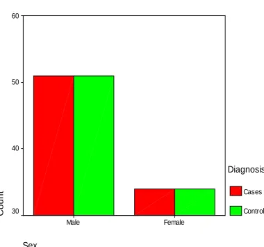
In this study, the frequency of ischemic stroke was found to be higher in males (51) than females (34) but there was no correlation between gender distribution among cases and controls with a p value of 1.000 which showed no statistical significance.
Sex
Female Male
C
ount
60
50
40
30
Diagnosis
67
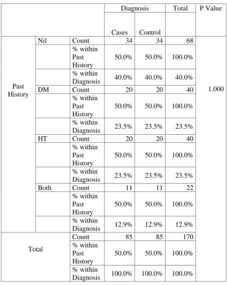
RISK FACTOR DISTRIBUTION BETWEEN CASES AND CONTROLS
Diagnosis Total P Value
Cases Control
Past History
Nil Count 34 34 68
1.000
% within
Past History
50.0% 50.0% 100.0%
% within
Diagnosis 40.0% 40.0% 40.0%
DM Count 20 20 40
% within
Past History
50.0% 50.0% 100.0%
% within
Diagnosis 23.5% 23.5% 23.5%
HT Count 20 20 40
% within
Past History
50.0% 50.0% 100.0%
% within
Diagnosis 23.5% 23.5% 23.5%
Both Count 11 11 22
% within
Past History
50.0% 50.0% 100.0%
% within
Diagnosis 12.9% 12.9% 12.9%
Total
Count 85 85 170
% within Past History
50.0% 50.0% 100.0% % within
[image:78.595.70.525.129.699.2]Diagnosis 100.0% 100.0% 100.0%
68
[image:79.595.113.496.227.540.2]The frequency of risk factors i.e. HT, DM or both was found to be equally distributed between cases and controls in the study with a p value of 1.000 which showed no statistical significance.
Figure 20: Risk Factor distribution between case and control groups
Past History
Both HT
DM Nil
C
ount
40
30
20
10
0
Diagnosis
69
DISTRIBUTION OF SMOKING/ALCOHOLISM AMONG CASES AND CONTROLS
Diagnosis Total P Value Cases Control
Smoker/ Alcoholic
Yes
Count 29 29 58
1.000 % within
Smoker /Alcoholic
50.0% 50.0% 100.0% % within
Diagnosis 34.1% 34.1% 34.1%
No
Count 56 56 112
% within Smoker/ Alcoholic
50.0% 50.0% 100.0% % within
Diagnosis 65.9% 65.9% 65.9%
Total
Count 85 85 170
% within Smoker/ Alcoholic
50.0% 50.0% 100.0% % within
[image:80.595.78.518.119.452.2]Diagnosis 100.0% 100.0% 100.0%
Table 7: Distribution Of Smoking/Alcoholism Among Cases And Controls
70
Figure 19: Distribution Of Smoking/Alcoholism Among Cases And Controls
The frequency of smoking / alcoholism was found to be equally distributed between cases and controls in the study with a p value of 1.000 which showed no statistical significance.
Smoker/Alcoholic
No Yes
C
ount
60
50
40
30
20
Diagnosis
Cases
71
DISTRIBUTION OF RT/LT MCA INFARCT AMONG CASES AND CONTROLS
Diagnosis Total P
Value
Cases Control
CNS Left
Hemiplegi a
Count
60 0 60
% within
CNS 100.0% .0% 100.0%
0.000
% within
Diagnosis 70.6% .0% 35.3%
Right
Hemiplegi a
Count
25 0 25
% within
CNS 100.0% .0% 100.0%
% within
Diagnosis 29.4% .0% 14.7%
NFND Count 0 85 85
% within
CNS .0% 100.0% 100.0%
% within
Diagnosis .0% 100.0% 50.0%
Total Count 85 85 170
% within
CNS 50.0% 50.0% 100.0%
% within
[image:82.595.71.505.165.595.2]Diagnosis 100.0% 100.0% 100.0%
72
Figure 20: Distribution Of Rt/Lt Mca Infarct Among Cases And Controls
The distribution of rt MCA infarct was more common than lt MCA infarct among cases with a p value of 0.000 which is significant.
CNS
NFND Right Hemiplegia
Left Hemiplegia
C
ount
90
80
70
60
50
40
30
20
10
Diagnosis
Cases
73
COMPARISON OF RENAL AND LIVER FUNCTION TESTS AMONG CASES AND CONTROLS
Diagnosis N Mean
Std. Deviation
Std. Error Mean
P Value
Serum Urea Cases 85 30.89 4.816 .522
>0.05
Control 85 31.42 4.910 .533
Serum creatinine
Cases
85 .787 .0949 .0103
Control 85 .791 .0908 .0098
Serum Total Protein
Cases
85 6.761 .2875 .0312
Control 85 6.776 .2918 .0317
Serum Albumin
Cases
85 4.256 .2233 .0242
[image:84.595.78.520.134.384.2]Control 85 4.269 .2361 .0256
Table 9: Comparison Of Renal And Liver Function Tests Among Cases And Controls
The mean serum urea in cases was 30.89 and in controls was 31.42 with a p value of 0.479 which is statistically not significant.
74
Figure 21: Comparison Of Renal Function Tests Among Cases And Controls
The mean total protein value in cases was 6.761 and in controls was 6.776 with a p value of 0.731 which is statistically not significant.
30.89
0.787 31.42
0.791 0
5 10 15 20 25 30 35
75
Figure 22: Comparison of Liver Function Tests Among Cases and Controls
The mean serum albumin value in cases was 4.256 and in controls was 4.269 with a p value of 0.714 which is statistically not significant.
6.761
4.256 6.776
4.269
0 1 2 3 4 5 6 7 8
76
COMPARISON OF SERUM TRIGLYCERIDE LEVELS AMONG CASES AND CONTROLS
Diagnosis N Mean
Std. Deviation
Std. Error Mean
P Value Serum
Triglyceride (mg/dl)
Cases
85 133.92 16.810 1.823 0.761
Control 85 134.71 16.896 1.833
Table 10: Comparison Of Serum Triglyceride Levels Among Cases And Controls
77
Figure 23: Serum Triglyceride distribution between case and control groups
The mean triglyceride level in cases was 133.92 and in controls was 34.71 and there was no significant difference in serum triglyceride levels between cases and controls with a p value of 0.761 that is not statistically significant.
Serum Triglyceride (mg/dl)
Abnormal Normal
C
ount
80 70 60 50 40 30 20 10 0
Diagnosis
78
COMPARISON OF BLOOD SUGAR VALUES AMONG CASES AND CONTROLS
Diagnosis N Mean
Std. Deviation
Std. Error Mean
P Value
Random Blood Sugar (mgs%)
Cases
85 115.98 25.429 2.758 0.953
[image:89.595.130.466.469.680.2]Control 85 116.21 26.015 2.822
Table 11: Comparison Of Blood Sugar Values Among Cases And Controls
The mean random blood sugar value in cases was 115.98 and 116.21 in controls and with a p value of 0.953 which also shows no statistical significance.
Figure 24: Comparison of Blood Sugar Values Among Cases And Controls
115.98
116.21
115.85 115.9 115.95 116 116.05 116.1 116.15 116.2 116.25
Cases Controls
79
COMPARISON OF BLOOD PRESSURE MEASUREMENTS AMONG CASES AND CONTROLS
Diagno
sis N Mean
Std. Deviation
Std. Error Mean
P Value
SBP Cases 85 165.46 10.403 1.128
Control 85 131.61 18.505 2.007
DBP Cases 85 99.98 5.778 .627 .000
Control 85 82.94 6.828 .741
Table 12 : Comparison Of Blood Pressure Measurements Among Cases And Controls
The mean systolic blood pressure in cases was 165.46 and in controls was 131.61 with a p value of 0.000 which is statistically highly significant.
The mean diastolic blood pressure in cases was 99.98 and in controls was 82.94 with a p value of 0.000 which is statistically highly significant.
Figure 25: Comparison of Blood Pressure Measurements Among Cases And Controls
165.46
10.403 131.61
18.505 0
20 40 60 80 100 120 140 160 180
SBP DBP
80
COMPARISON OF DIASTOLIC BLOOD PRESSURE AMONG EUTHYROID AND HYPOTHYROID PATIENTS
TSH N Mean
Std. Deviation
Std. Error Mean
P VALU
E DBP Norm
al 78 99.62 5.730 .649
Abnor
mal 7 104.00 5.033 1.902
0.048
Table 13 : Comparison of diastolic blood pressure among Euthyroid and hypothyroid patients
In the study, among cases 78 people with normal TSH values had a mean DBP of 99.62 and 7 with elevated TSH levels had a mean DBP of 104.00 which is significantly higher with a p value of 0.048 which is statistically significant.
Figure 26: Comparison of diastolic blood pressure among Euthyroid and hypothyroid patients
99.62
104
97 98 99 100 101 102 103 104 105
78 7