A COMPARATIVE STUDY BETWEEN
ENDOSCOPIC ASSISTED AND MICROSCOPIC
ASSISTED MYRINGOPLASTY
DISSERTATION SUBMITTED FOR
MASTER OF SURGERY
BRANCH IV(OTO – RHINO – LARYNGOLOGY)
THE TAMILNADU
DR.M.G.R. MEDICAL UNIVERSITY
CERTIFICATE FROM HOD
This is to certify that the dissertation entitled “A Comparative study between
endoscopic assisted and microscopic assisted myringoplasty” is the bonafide
work of DR.VIJAYABHARATHI.D, in partial fulfilment of university regulation of the Tamil Nadu Dr. M.G.R. Medical University, Chennai, MASTER
OF SURGERY BRANCH IV (OTORHINOLARYNGOLOGY) examination
to be held in April 2015.
DR.N.DHINAKARAN.M.S.,
Place:Madurai
Professor and HOD
Date: Department of ENT
CERTIFICATE FROM DEAN
This is to certify that the dissertation entitled “A Comparative study between
endoscopic assisted and microscopic assisted myringoplasty” is the bonafide
work of DR.VIJAYABHARATHI.D, in partial fulfilment of university regulation of the Tamil Nadu Dr. M.G.R. Medical University, Chennai, MASTER OF
SURGERY BRANCH IV (OTORHINOLARYNGOLOGY) examination to be
held in April 2015.
Place:Madurai
Date : Captain. Dr.B. SANTHAKUMAR,
M.Sc (F.Sc), M.D (F.M),PGDMLE, DNB (F.M).,
DEAN,
CERTIFICATE FROM GUIDE
This is to certify that the dissertation entitled “A Comparative study between
endoscopic assisted and microscopic assisted myringoplasty” is the bonafide
work of DR.VIJAYABHARATHI.D, in partial fulfilment of university regulation of the Tamil Nadu Dr. M.G.R. Medical University, Chennai, MASTER OF
SURGERY BRANCH IV (OTORHINOLARYNGOLOGY) examination to be
held in April 2015.
NAME & DESIGNATION OF THE GUIDES:
1. PROF. Dr.N.DHINAKARAN M.S.,
HOD, DEPARTMENT OF OTORHINOLARYNGOLOGY, MADURAI MEDICAL COLLEGE
2. Dr.RAJAGANESH M.S ENT., ASSISTANT PROFESSOR,
DEPARTMENT OF OTORHINOLARYNGOLOGY, MADURAI MEDICAL COLLEGE,
CO-ORDINATOR
1. PROF Dr. N.DHINAKARAN M.S.,
DECLARATION
I hereby solemnly declare that the dissertation titled “A Comparative study
between endoscopic assisted and microscopic assisted myringoplasty” has been prepared by me under the guidance and supervision of Dr.N.Dhinakaran M.S.,
Professor and HOD Department of ENT diseases, Government Rajaji Hospital, Madurai. This is submitted to The TamilNadu Dr. M.G.R. Medical University Chennai, in partial fulfillment of the regulations for the award of MS Degree (Branch IV) in OTO RHINO LARYNGOLOGY.
Place: Madurai
Date : DR.D.VIJAYABHARATHI
ACKNOWLEDGEMENT
I have great pleasure in expressing my deep sense of gratitude to Prof. Dr. N. Dhinakaran M.S.,D.L.O., Professor and Head of the department of ENT diseases , Government Rajaji Hospital and Madurai Medical College, Madurai for their kind encouragement and valuable guidance during the period of this study, without their continuous encouragement and guidance this dissertation would not have materialized.
I gratefully acknowledge and sincerely thank THE DEAN, Government Rajaji Hospital, Madurai for granting the permission to utilise the resources of this institution for my study.
I am very much obliged to the ASSISTANT PROFESSORS of Department of ENT, Madurai medical college, Madurai, who helped in preparing and bringing a shape to this work.
CONTENTS
S. No TITLE PAGE NO
1 INTRODUCTION 1-3
2 AIM OF THE STUDY 4
3 REVIEW OF LITERATURE 5-71
4 MATERIALS AND METHODS 72-76
5 OBSERVATION AND RESULTS 77-95
6 DISCUSSION 96-102
7 SUMMARY 103-104
8 CONCLUSION 105
9 ANNEXURES
a) BIBLIOGRAPHY
b) PROFORMA
c) MASTER CHART
d) ETHICAL COMMITTEE APPROVAL
LETTER
ABSTRACT
INTRODUCTION:
Myringoplasty is repair of the perforation of tympanic membrane when middle ear space, its mucosa and ossicle are free of disease. Myringoplasty is commonly done under microscopy; Major disadvantage of operating microscope is that it provides a magnified image along a straight line. Now a day’s Endoscope is widely used to perform various surgeries and very useful while operating in cavities. Therefore in the present study we correlate the outcome of myringoplasty by microscopic method and endoscopic method.
AIM AND OBJECTIVES:
To compare the percentage of graft uptake and improvement in A-B gap between microscopic and endoscopic group
MATERIALS AND METHODOLOGY:
The graft uptakewas assessed and postoperative pure tone audiograms were compared with preoperative pure tone audiogram
RESULTS:
At end of 6 month post op period endoscopic group, out of 20cases, 85% graft uptake. Microscopic group 80% graft uptake. Student’s t- test( P value 0.915) shows there is no significant difference in graft uptake in both group. Hearing improvements are similar in both groups. Student's t-test was used for statistical analysis for comparing the results (gain in A-B gap) of two groups, P value was 0.514. Hence there is no significant difference in the gain in A-B gap between the two groups.
CONCLUSION:
The present study, graft uptake rate in endoscopic and microscopic myringoplasty are similar ,but in terms of minimal invasive surgery, proximity vision provided , wide angle view, visualize of the deep recess and hidden areas of middle ear in single operating field, postoperative recovery , less operative time, Endoscopic method produced superior results and overcomes the disadvantage of microscope.
KEY WORDS:
1
INTRODUCTION:
Chronic suppurative otitis media (CSOM) is wide spread disease of the developing countries. Hence treating CSOM with surgical treatment by Tympanoplasty is one the common procedure in ENT
Myringoplasty and Tympanoplasty are defined as surgical procedure that is used for repair of tympanic membrane and middle ear. Myringoplasty is repair of the perforation of tympanic membrane when middle ear space, its mucosa and ossicle are free of disease. Tympanoplasty implies not only reconstruction of TM , But also eradication of disease in middle ear cleft such as chronic infection, cholesteatoma, ossicular chain problem etc.
Initially full and split thickness skin graft are used, followed by canal wall skin, vein, perichondrium, and temporalis fascia graft are used. Temporalis fascia remains most commonly used material now.
Common problem encountered in reconstruction of the tympanic membrane are poor exposure to vital area of tympanic cavity like sinus tympani, difficulty in removing all squamous epithelium in the area to be covered by graft ,epithelial pearl formation , development of disease sequlae like tympanosclerosis ,appearance of cicatrial tissue ,blunting of anterior canal recess, post op migration of graft from malleus handle , retraction of graft, graft rejection .various new technique tried to overcome this problems.
2
The goal of any procedure is to produce a new tympanic membrane that will function as closely to orginal.
Introduction of operating microscope significantly enhanced surgical result by improving the accuracy of the technique. But operating microscope provides magnified image in straight line extending from the objective lens, hence surgeon can’t visualize the deep recess of middle ear in single operating field. Using microscope, initially permeatal overlay technique done, gradually changed to post aural or end aural underlay technique because of disadvantage of permeatal approach through microscope. This is overcome by use of rigid endoscope.
The use of rigid endoscope in the management of dry central perforation of tympanic membrane represented a significant advance in middle ear surgery. It replaces operating microscope in examination and surgery of the tympanic membrane perforation.
3
4
Aim of the study
5
REVIEW OF LITERATURE
ENDOSCOPIC ASSISTED MYRINGOPLASTY
Balasubramanian thiagarajan, stanely medical college,
otolaryngology online journal.
Myringoplasty is commonly done under microscope .In this study using endoscopic guidance myringoplasty done in 50 cases. After 4th week of postoperative period, out of 50 cases, 42 cases had intact membrane. preoperative audiogram shows 32 patient had 30-35 db hearing loss ,18 patient had 35-40 db hearing loss. All the patient after endoscopic myringoplasty had a pure tone hearing average of 20 db .success rate of endoscopic myringoplasty compared with various studies performed using microscopic procedure. Result of endoscopic procedure more similar to microscopic myringoplasty. Endoscopic myringoplasty is worthwhile due to obvious advantage.
A COMPARITIVE STUDY OF ENDOSCOPIC ASSISTED AND
MICROSCOPIC ASSISTED MYRINGOPLASTY
A.S Harugop, R.S Mudhol Indian Journal of Otorhinolaryngology
6
and 50 underwent microscopic procedure. Surgery result compared at end of 6 month post operatively. Endoscopic group 82% success outcome and in microscopic group 86% had successful outcome. Surgical outcome of endoscopy assisted myringoplasty was comparable to the conventional microscopic assisted myringoplasty but in terms of cosmesis, post-operative recovery, the patient in endoscope group had better result. concluded that surgery with endoscope has several advantages and few disadvantages.
ENDOSCOPIC ASSISTED MYRINGOPLASTY
Yadav sps, Aggarwal N. Julaha m, Goel A Singapore medical
Journal of Otorhinolaryngology
Endoscopic assisted myringoplasty was carried out in 50 patients aged 18-45 yr, using temporalis fascia graft. Over all graft uptake and improvement in conductive deafness as air bone gap closure was achieved in 80% of cases, they concluded that endoscopic myringoplasty is equally effective, less morbid, very cost effective in small central perforation, however it is not effective in large perforation.
ENDOSCOPIC TRANSCANAL MYRINGOPLASTY
7
A study conducted by Raj A, Mehar R on endoscopic transcanal myringoplasty and compares the outcomes with that of myringoplasty using microscope, showed that graft uptake is 90% in endoscopic method and 85% in microscopic method but there was no significant differences between the gains in the air bone gap in either group. Study was done on 40 patients. These 40 patients were divided into 2 equal groups of 20 patients each.
Tympanoscope assisted myringoplasty
Karchuketo TS
He studied 30 ears of 29 patients with different sized perforation underwent endoscope assisted myringoplasty. In their study concluded that the post operative air bone gap < 10 dB in 90% cases. Hence tympanoscope assisted myringoplasty is reliable and simple procedure with the benefit of minimal trauma to the healthy tissue.
Endoscopic transtympanic tympanoplasty
Kakachata, seizi et al
8
chain reconstruction Endoscopic transtympanic tympanoplasty is an adequate and minimal tissue invasive procedure and should prove to be an useful surgical procedure in future endoscopic Tympanoplasty.
Endoscopy in otology –In retrospect and prospects
Bhattarai H Nepalese journal of Ent HEAD and neck surgery
Role of endoscope in otology a diagnostic, a surgical and a teaching tool is increasing being recognized because of its superior optical properties and its capacity to visualize hidden area with minimal invasion of tissue as compared to microscope. Myringoplasty with temporalis fascia can be done with endoscope with similar result to that of a microscopic procedure. Endoscopic myringoplasty is minimally invasive, effective and reliable procedure in management of tympanic perforation.
Endoscopic transcanal middle ear surgery
Muaaz Tarabichi
9
Trascanal endoscopic management of cholesteatoma
Tarabichi Muaaz
10
ANATOMY OF MIDDLE EAR CAVITY PROPER AND
TYMPANIC MEMBRANE
MIDDLE EAR CAVITY PROPER
The middle ear cavity present between the external and internal ear. It shape like biconcave disc, the measurement of vertical diameters are 15 mm, the transverse diameter are measured as 6 mm at the upper portion, 2mm at the centre portion and 4 mm at the lower portion.
It is formed by 6 walls .The tympanic membrane forms the lateral wall of the middle ear, it divide the middle ear in to three parts.
1. Mesotympanum 2. Epitympanum 3. Hypotympanum
Mesotympanum
Draw a line at horizontal plane at the top and bottom edge of pars tensa of tympanic membrane, portion of middle ear present behind the pars tensa of the tympanic membrane
Epitympanum
11
malleus. Content present in this space are malleus head, body of the incus, associated ligaments and mucous fold.
Hypotympanum
Portion of middle ear present, below the floor of bony canal wall or below the tympanic membrane
Lateral wall
Sharpnell’s membrane forms the major portion of the lateral wall and partly wedge shaped bony wall called scutum.
Medial wall
Middle ear was separated from the inner ear by medial wall, it presents,
1. Promontory :is a rounded bulging prominent part produced by the first turn of the cochlea
2. Fenestra vestibuli is an oval opening present posterosuperior to promontory leads to scala vestibule of inner ear. Footplate of stapes closed the opening.
12
4. Tympanic portion of Facial nerve runs in inferior direction below the lateral semicircular canal.
5. Sinus tympani present as a depression behind the promontory, bounded superiorly ponticulus and inferiorly subiculum.
Anterior wall Openings
Three structures opened into the anterior wall
a. Superiorly opening of Canal for tensor tympani muscle b. In the middle , opening of auditory tube
c. Inferiorly , posterior wall of carotid canal formed by thin plate of bone this bone , separate the carotid canal from the middle ear, this plate is perforated by caroticotympanic nerve and branch of internal carotid artery
Posterior wall
The following structure present from above downwards in posterior wall superiorly: There is aditus, it connect to antrum through epitympanic recess communicate with mastoid antrum. Short process of incus present on its floor with facial nerve, while lateral semicircular canal lies in its medial wall.
13
A conical like projection called pyramid, with an opening as its apex for the passage of tendon of stapedial muscle, this muscle insert into neck of stapes at its posterior surface. The extracranial portion of the 7th nerve (vertical part of mastoid segment of facial nerve) passes downwards in the posterior wall, exit through the stylomastoid foramen.
Two recesses present in the posterior wall are, Facial recess and sinus tympani. Direct visualization of the two recesses are difficult. But visualize with help of angled scope. Residivism and recurrence of cholesteatoma are most common in these recesses after the cholesteatoma ear surgery. Boundaries of facial recess or suprapyramidal are medially by facial nerve, superiorly by fossa incudis and lateral boundary formed by the chorda tympani nerve .For procedure like posterior tympanotomy, accessed directly via this facial recess.
Floor
14
Roof
It is formed by tegmen tympani, which is a part of petrous part of temporal bone. It separated the middle cranial Fossa from the tympanic cavity.
VENTILATORY ANATOMY
The air flows from the Eustachian tube from the nasopharynx to anterior mesotympanum. From here, it diverts up to the anterior epitympanum through the tympanic isthumus and flow backward to posterior epitympanum. Port of the air from the posterior epitympanum flows through the aditus to the mastoid antrum and mastoid air cells and part of it flows down via to the posterior mesotympanum. From the posterior mesotympanum, air flows to the hypotympanum as well. Any pathological dysfunction in the anatomy of ventilator pathway leads to inflammatory diseases of the tympanic cavity.
CONTENTS OF THE MIDDLE EAR
Ossicles
15
Malleus (hammer) is the largest ossicle, measuring 8 mm in length. Parts of malleus are head, neck, handle and anterior and lateral processes. The head is situated in the epitympanum. A lateral (short) process project laterally from the neck, the handle is firmly fixed to the pars tensa of the tympanic membrane
Incus (anvil) has a body, short process and long process. The body articulates with the head of malleus in the attic region and the short process project into the attic. The long process projects downwards behind the handle of malleus, it articulates with the head of the stapes via the lenticular process.
Stapes (stirrup) is the smallest ossicle measuring 3.5 mm in size .It consists of head, neck, footplate and anterior and posterior crura. The footplate of stapes is attached to the oval window by the annular ligament. Muscles
The tensor tympani and stapedius muscles decrease the movement of the ossicles.
The tensor tympani is inserted to the neck of malleus. First arch muscle supplied by branch of mandibular nerve (V3).
16
Mucosal folds and ligaments - keep the ossicles in place
Nerves
Chorda tympani is a branch of the facial nerve which carries the sense of taste. It enters the middle ear cavity from the posterior wall, runs forwards and lateral to the incus and medial to the malleus, escaping out through the anterior wall.
The tympanic plexus lies on the promontory. It is formed by tympanic branch of glossopharyngeal nerve and sympathetic fibers from the plexus around the internal carotid artery. It also carries the secretomotor to the parotid gland. Tympanic plexus innervates the medial surface of tympanic membrane, tympanic cavity, mastoid air cells and bony Eustachian tube. Tympanic branch of glossopharyngeal nerve can be sectioned in middle ear for treating the Frey’s syndrome.
Vessels
Plexus of vessels of stylomastoid artery and from caroticotympanic artery
RELATIONS OF THE MIDDLE EAR CAVITY PROPER
17
Temporal lobe of the brain and meninges are above the antrum, aditus and epitympanum. The tegmen plate separates the middle ear cleft from the structures in the middle cranial fossa.
Cerebellum is posteromedial to the mastoid air cells.
Inner ear is medial to the antrum, aditus and tympanum
Horizontal semi circular canal is an important landmark which lies posterosuperior to the facial nerve.
Fifth and sixth cranial nerves lie close to the apex of the petrous pyramid.
Facial nerve – the horizontal part runs downwards in the medial wall of the tympanum. The vertical part runs downward behind the tympanum and in front of the mastoid cells and emerges out through the stylomastoid foramen.
Lateral sinus is posterior to the mastoid cells
Jugular bulb is in close contact with the floor of the tympanum.
Internal carotid artery is anterior to the tympanum.
BLOOD SUPPLY
The blood supply of the middle ear is from branches of :
18 Maxillary artery
Ascending pharyngeal artery
Stylomastoid branch of the posterior auricular artery
NERVE SUPPLY
Sensory: Tympanic branch of the ninth cranial nerve (jacobson’s nerve) supplies through the tympanic plexus
Motor: Tensor tympani muscle is supplied by the mandibular division of the trigeminal nerve and the stapedius muscle is supplied by the facial nerve.
LYMPHATIC DRAINAGE
19
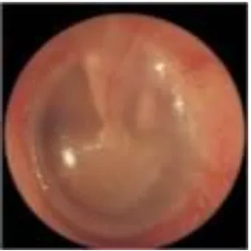
[image:29.595.171.448.112.389.2]Tympanic membrane
FIG 1: NORMAL TYMPANIC MEMBRANE
Tympanic membrane is located at the medial end of the external acoustic meatus.It separate the tympanic cavity from external auditory canal .Lying obliquely forming an angle of 55 degree with floor of meatus. It measures approximately 9-10mm in vertical diameter and 8-9 mm in horizontal diameter. Oval in shape .peripheral part of the tympanic
membrane is thicker and rounded, forms a fibrocartilagenous ring known as annulus or annular ligament, attached at its circumference to the
20
of malleus. The small, triangular area of membrane above these folds is lax and thin named as pars flaccida. The rest of membrane is pars tensa with rich middle fibrous layer.
Tympanic membrane formed from mergence of ectodermal meatal plugs of 1st brachial cleft with endodermal derivative of 1st branchial pouch. It composed of 3 layers. outer cuticular layer or ectodermal layer (keratinized stratified squamous epithelium ) ,middle fibrous layer ,inner endodermal mucosal layer continuous with middle ear mucosa .stratified squamous epithelium of outer layer is again four layer ,stratum basale ,stratum spinosum ,stratum granulosum, stratum corneum.
Stratum basale is the dividing layer and replaced the superficial cell are periodically desquamated .epidermis of the tympanic membrane migrate from umbo outwards mostly towards the posterior superior region. Migration rate is 131 µm/day.
21
Blood supply
Lateral surface of the tympanic membrane supplied through deep auricular branch of internal maxillary artery .medial surface of membrane is supplied through anterior tympanic branch from internal maxillary artery and posterior tympanic branch from stylomastoid artery, inferior tympanic branch from ascending pharyngeal artery. Outer surface drain into external jugular vein, inner surface is drained by venous plexus situated around Eustachian tube.
Innervations:
Anterior half of lateral surface by auriculotemporal nerve and posterior half by tympanic branch of vagus nerve(Arnold nerve) .Inner surface of membrane is supplied by tympanic plexus formed by jacobson’s
nerve(branch of glossopharyngeal nerve).
MIDDLE EAR CLEFT
It consists of
1. Tympanic cavity
2. Mastoid antrum
3. Aditus ad antrum
22 5. Mastoid air cells
The lining epithelium in the middle ear was pseudo stratified ciliated columnar and becomes pavement epithelium near attic and antrum. It is an extension from the nasopharynx .The mucous membrane in middle ear form mucous fold and divides the cavity into various compartments.
MASTOID
The mastoid consists of three parts
1. Aditus ad antrum is a short canal connecting the epitympanum with the mastoid antrum.
2. Mastoid antrum - biggest air cell in the mastoid.
3. Mastoid air cells are variable in number, size and distribution. They communicate with the mastoid antrum.
There are three types of mastoid process: a. Cellular, with large and numerous air cells. b. Diploic, with small and less numerous air cells. c. Sclerotic, with air cells practically absent
23
mainly in petromastoid and squamous parts of the temporal bone. In a well developed mastoid they are grouped as follows
Central group- periantral cells Peripheral group- dural
perisinus sinodural
Tip
Retro facial
Accessory group- squamous
Occipital
Zygomatic
Peritubal
Styloid
EUSTACHIAN TUBE OR AUDITORY TUBE
It connects the cavity of the middle ear and the nasopharynx, it has two parts,
24
It measures 12 mm in length. It runs through squamous and petrous part of temporal bone. Related structure medially carotid canal, inferiorly separated from jugular Fossa, above from tensor tympani
2. Cartilaginous
It measures as 24mm length in adult. Formed partly by cartilage and partly by fibrous connective tissue. The nasopharyngeal end of the tube was wider and mucous membrane covering over this end forms a tubal elevation. The cartilage is fixed to the base of skull in a groove between petrous part and greater wing of sphenoid.
In adult lies at angle of 45 degree and in infants it forms an angle of 10 degree and in horizontal plane. In nasopharynx part, it opened little below the posterior end of inferior turbinate. The tube was lined by ciliated columnar epithelium with more goblet cell and basal cell. In the pharyngeal part, the lumen of the auditory tube surrounded by ostmann’s pad of fat. Which function as closure of the Eustachian tube, Prevented from nasopharyngeal reflux.
25
Blood supply:
Arterial supply
Ascending pharyngeal branch of external carotid and middle meningeal branch from maxillary artery and small branch from pterygoid canal artery.
Venous drainage
Drain into Pterygoid plexus of vein
Functions of Eustachian tube
1. Equilibrate the air pressure in tympanic cavity
2. Product from nasopharyngeal sound pressure and secretion. 3. Drainage of secretions from the middle ear into the nasopharynx
Physiology
Sound transmission
26
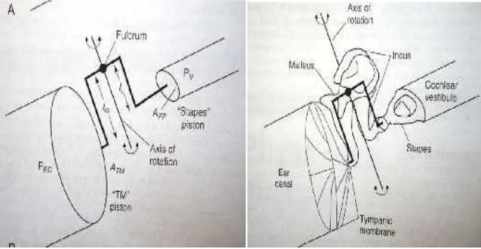
[image:36.595.102.574.307.551.2]Acoustic resistance defined as Sound wave travelling from one medium (air) to another medium like water, 99.9% of sound energy is reflected. This similar impedance mechanism exit in ear (conducted sound from the air to travel to cochlear fluid). This loss of sound energy is compensated by, middle ear convert the sound of lesser force, greater amplitude to greater force and lesser amplitude. It is called impedance matching.
Fig-2; shows ossicle lever ratio
27
process of incus, through the use of this leverage, the force received at footplate of stapes is greater than malleus by 1.3:1 ratio. So middle ear combined transformer ratio is 22:1.
Tympanic membrane Perforation with normal middle ear ossicle and other structure affect the hearing by two different mechanisms. First one due to reduced tympanic membrane surface on which sound pressure exerted leads to ossicular chain vibration reduced .Size of the perforation increases, the surface area loss is also greater, in which sound pressure can act and through the perforation additional sound pressure entering the middle ear act on medial surface of the tympanic membrane against sound pressure on other surface.
The second mechanism result from sound reaching round window directly, This become more with larger perforation .size of perforation increases, hydraulic advantage formed by large surface of tympanic membrane over small oval window disappear , both oval and round window reached by the sound at same time with equal force and Resultant cancellation of vibratory movement.
28
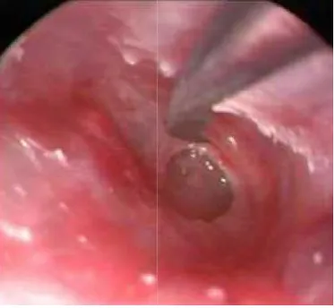
Common causes of tympanic membrane perforation are infection like ASOM, CSOM, viral infection of drum, secondary to otitis externa and trauma.
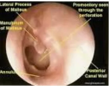
Fig -3; shows Medium sized perforation in antero superior and anteroinferior part of parstensa; margins are regular. Exposed middle ear mucosa appears normal
ASOM(Acute suppurative otitis media ):
[image:38.595.105.461.217.498.2]29
spontaneously. Common organism involved are streptococcus pneumoniae, haemophilus influenza , moraxella cataralis . Persistence of perforation due to recurrent infection or organism is highly virulent.
CSOM(Chronic suppurative otitis media)
Defined as chronic inflammation of mucoperiosteal lining of middle ear cleft with permanent abnormality of the pars tensa or flaccida, likely result of earlier ASOM, negative middle ear pressure or otitis media with effusion.
Classification of CSOM :
1. Inactive mucosal CSOM (Dry perforation)
30
2. Active mucosal CSOM (Perforation with otorrhoea)
Chronic inflammation within the mucoperiosteal lining of middle ear cleft and mastoid produce active mucopurulent discharge via
tympanic membrane perforation.
3. Inactive squamous epithelial CSOM ( Retraction , Atelectasis):
Negative middle ear pressure result in retraction of tympanic membrane. Retraction pocket is defined as invagination of part of tympanic
membrane into middle ear space .it is either fixed to the structure in the middle ear or it’s freely mobile)
4. Active squamous epithelial CSOM:
It is usually associated with cholesteatoma formation Cholesteatoma
It is defined as a sac in the middle ear, which is lined by keratinizing stratified squamous epithelium containing desquamated epithelium as keratin debris. It is also described as skin in the wrong place
31
Other classification:
1. Tubo tympanic type (safe type) 2. Attico - antral type (dangerous type)
TUBO TYMPANIC TYPE
Etiology
1. Predisposing factors
Inadequate / improper treatment of ASOM, infection from
surrounding areas like nose, nasopharynx and oropharynx, some diseases like tuberculosis are chronic from the beginning, pneumatisation of mastoid – sclerotic mastoids are more prone for CSOM.
2. Exciting factors
Gram negative organisms like pseudomonas, proteus, E.coli Streptococcus, Staphylococcus
Symptoms
1. Discharge: Profuse, intermittent, predominantly mucoid
occasionally mucopurulent, non foul smelling, whitish/yellowish and tenacious. It increases with attacks of cold.
32
Active – actively discharging ear at the time of clinical examination
Quiescent – no ear discharge for less than 3 to 6 months period Inactive – no ear discharge for more than 6 months.
Healed – central perforation has healed. 2. Deafness – mild conductive
3. Earache – if associated with otitis externa
Signs
1. Discharge is present in the external auditory canal which is usually mucoidal , tenacious and non-foul smelling.
2. Tympanic membrane –
A central perforation occurs in the pars tensa without any involvement of the margin of the drum. Size of the perforation will range from small to very large perforation in the pars tensa.
Small, pinpoint perforation (less than 25%) is seen in acute suppurative otitis media and also in trauma.
Medium size (25-50%) is frequently round or oval. It could be either dry or moist.
Large perforation (50-75%).
33
Complete or total perforation when there is loss of substance involving the entire tympanic membrane including the annulus found in attico antral disease.
Middle ear can be easily visualized through a large perforation. In case of suppuration, medial wall is covered by thick red granular and sometimes polypoidal mucous membrane.
Other structures that can be identified are as follows Promontory
Oval window
Round window niche Eustachian tube orifice Parts of ossicular chain Stapedius tendon. 3. Tuning fork tests
Rinne – negative
Weber – lateralized to the affected side ABC – normal
INVESTIGATIONS
34 Examination under microscope
To see the margin of the perforation To rule out any ingrowing epithelium To see granulation tissue and polyp To see any evidence of cholesteatoma
For precise collection of swab from middle ear To rule out any hidden pin hole perforation To see status of middle ear mucosa
To see the status of ossicular chain if possible
Pure tone audiogram – mild conductive loss between 20 to 30 db
Patch test – This test is performed as a outpatient procedure. The material used in patch test can be thin paper commonly taken from cigarette foil and which is cut into size of perforation. Before patching, a pure tone audiogram is taken. Then perforation is closed with the same cut thin paper. Patching of perforation is always better if paper is soaked in Vaseline. A repeat audiogram is done after patching.
Interpretations of patch test are
1. Improved hearing means intact ossicular chain.
35
3. No improvement of means in technical fault or improper patching. X-ray of mastoids : to rule out mastoiditis
X-ray of paranasal sinuses : to rule out sinusitis
X-ray soft tissue neck lateral view : to rule out adenoid enlargement. Diagnostic nasal endoscopy
TREATMENT
Medical management
Aural toilet by dry mopping, wet mopping, suction cleaning. Wet mopping is discouraged nowadays. Suction cleaning is the best method but may not be possible in children.
Antibiotic ear drops after culture and sensitivity report.
Surgical management
Removal of septic foci, e.g. Tonsillectomy, adenoidectomy, sinus wash. Myringoplasty if hearing loss is below 40 db, Tympanoplasty if above 40 db
If X-ray mastoid shows mastoiditis, do cortical mastoidectomy
ATTICO-ANTRAL OR DANGEROUS TYPE
36
Cholesteatoma
It is defined as a sac in the middle ear, which is lined by keratinizing stratified squamous epithelium containing desquamated epithelium as keratin debris. It is also described as skin in the wrong place. This structure has a capacity for progressive and independent growth at the expense of underlying bone and has tendency to recur, unless removed completely.
The mechanisms of bony erosion of cholesteatoma are not completely understood. Various factors are mentioned here
1. Earlier it was thought the physical pressure of cholesteatoma causes bony erosion.
2. At the cellular level the chief factor in bony erosion is activation of osteoclasts
3. It is believed that release of inflammatory mediators such as the cytokine, interleukin 1 alpha, from macrophages and epidermal keratinocytes are being important in osteoclast activation. Other humoral factors that have been suggested are prostaglandin, cathepsin D and parathyroid hormone like protein.
37
CLASSIFICATION OF CHOLESTEATOMA
Congenital cholesteatoma
It defined as embryonic epidermal cell rests in the middle ear cleft or temporal bone. It may occur at
Middle ear Petrous apex
Cerebellopontine angle
In middle ear, congenital cholesteatoma usually presents with a whitish mass behind an normal tympanic membrane. Sometime it ruptures through the tympanic membrane spontaneously.
Levenson criteria for diagnosis
A white mass behind intact tympanic membrane Intact pars flaccida and tensa
No history of previous otorrhea
No history of previous ear surgical procedure
Canal atresia and intramembraneous and giant cholesteatoma
Primary acquired cholesteatoma
38
Secondary acquired cholesteatoma
It always occurs in an already diseased ear where there is a pre-existing tympanic membrane perforation
ETIOPATHOLOGY
The exact etiopathology is unknown but various theories have been put forward for the formation of cholesteatoma. Cholesteatoma is a white, pultaceous mass having bone eroding capacity either by expansion or by liberation of some chemical enzyme.
Theories:
39
cell remnants by Wittmack (1933), embryonic epithelial cell rest by Mc Kenzie(1931).
2. Theory of migration – skin of the external auditory meatus will migrate to the middle ear cavity through the tympanic membrane perforation leading to secondary acquired cholesteatoma formation in the middle ear.
3. Metaplasia theory – because of recurrent / chronic infection normal columnar epithelium turns into squamous epithelium by metaplasia 4. Implantation theory – At the time of middle ear surgery squamous
epithelium may get implanted. Other theories of epithelial abnormality
Invasive hyperplasia of basal layer of meatal skin adjoining of the upper margin of tympanic membrane has been postulated in this theory. Papillary invasion with central cornification enters the epitympanum without tympanic membrane perforation. This speaks in favour of primary acquired cholesteatoma
Invasive hyperkeratosis and acanthosis of deep meatal skin has also been postulated.
Typical growth pattern of cholesteatoma
40 Posterior epitympanum
Posterior mesotympanum Anterior epitympanum
It is not unusual for multiple cholesteatoma sac to occur in the same ear involving two or even all three of these typical routes. While great majority of cholesteatoma follow one or more of this common pathway, others assume unusual patterns presumably because of anatomic variation of middle ear fold and the ligaments which channel the cholesteatoma growth.
Pars tensa cholesteatoma
Ossicular chains are involved at a relatively early stage. Long process of incus and stapes suprastructure is the common ossicles involved. This causes moderate to severe conductive deafness. Occasionally the hearing is well preserved by a retraction on to the stapes head or bridging of ossicular chain defect by cholesteatoma (cholesteatoma hearers).
Pars flaccida cholesteatoma
41
chain occurs relatively late in disease process. It is not uncommon to encounter large pars flaccida cholesteatoma with an intact ossicular chain and relatively a minor degree of conductive deafness. Further progression of disease occurs anteriorly in to the anterior epitympanum and posteriorly into the mastoid antrum and its air cell system.
CLINICAL FEATURE
Symptoms:
Ear discharge: it is foul smelling scanty predominantly purulent, occasionally blood stained and has no relation with upper respiratory tract infection.
Deafness: progressive conductive deafness
Itching and pain in the ear: May be caused by otitis externa or may be an early symptoms of complication.
Tinnitus and giddiness: May be early symptoms of complication.
Sign:
Otoscopic examination reveals
42
4. Occasionally granulation tissue or polyp may be seen coming out of perforation.
5. Whitish cholesteatoma flakes can be seen through perforation 6. Mastoid tenderness present
7. Tuning fork shows the rinne test negative, weber test lateralized to the affected side, ABC normal.
Investigations
Examination under microscope
Culture sensitivity from ear discharge
Rigid otoendoscopy to see the facial recess and sinus tympani if possible Audiogram
Imaging
1. X-ray Mastoid (Schuller’s and Law’s view): to see the bony erosion, to see the anatomy of mastoid, occasionally to diagnose cholesteatoma which gives the cotton wool appearance.
2. CT scan : Features seen in high resolution are the following: Blunting of the scutum is the earliest sign
Erosion and destruction of lateral attic wall Widening of aditus
43 Labyrinthine fistula formation Erosion into the facial canal
Dehiscence of the tegmen tympani and sinus plate Destruction of mastoid or natural mastoidectomy
Erosion of posterior and roof of external auditory canal 3. MRI: MRI provides complimentary information by allowing
characterization of soft tissue masses.
MANAGEMENT
Aims and objective
1. Primary objective is to make the ear safe and dry 2. To restore or to improve the hearing
Surgical management
Surgical management (Mastoidectomy) is the main line of treatment in atticoantral disease. Conservative or medical line of management has no role in making the ear safe.
Medical management
44
However ideal medical treatment would be topical agent which has the potential for either eliminating the squamous epithelium or reducing its activity in order to curtail the production of desquamated debris. Studies have shown that 5- flurouracil has some useful activity in this aspect. Initial studies in which 5-flurouracil was applied to the cyst wall of early cholesteatoma, relapsing cholesteatoma and in large discharging cavity showed inhibition of keratin formation and otorrhea.
Traumatic perforation:
Trauma due to blunt or penetrating injuries, thermal or chemical injury, barotraumas, blast injuries. Iatrogenic injury may result from long period of ventilation tube (T tube) or following myringotomy. Barotrauma lead to rapid change of pressure (from 100 to 500 mmhg) causes tympanic membrane rupture. Acoustic trauma due to high sound pressure level (195 db) cause rupture of membrane. Injuries produced by slap lead to column of air trapped in external acoustic meatus, it compressed over the tympanic membrane lead to perforation.
45
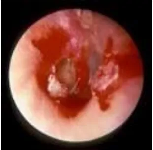
Fig-4; Shows traumatic perforation of the left tympanic membrane with congestion; margins are irregular.
Longitudinal temporal bone fracture commonly involves the tympanic membrane than temporal bone fracture. Penetrating injuries is mostly self inflicted like self cleaning the ear canal with cotton swab or stick .Traumatic injury lead to perforation has more tendencies to heal spontaneously in case of 78-90%
Healing process:
46
perforation heals spontaneously up to 79-90%. Acute otitis media has healed upto 79%. Thermal injury healed up to 38% .Many factor affect the healing process like age > 30,size of perforation ( large) ,location and cause of perforation like posterosuperior ,anterior to handle of malleus, recurrent URI, allergic sensitization of middle ear mucosa , infection ,previous acute episode of necrotizing otitis media or any malnutrition , immunosuppression .
Healing process of tympanic membrane depend on epithelial migration. Two types of migration seen. In centrifugal type, movement from umbo outwards, for removal of keratin wax outward. In centripetal type, mainly responsible for healing process .After injury to the tympanic membrane, mitotic activity increased around the annulus and 2mm away from the wound margin. Healing is regulated by cytokines such as EGF and beta TGF. These factor accelerate the regeneration of epithelium.
Historical aspect:
47
rubber disc attached to silver wire over tympanic membrane perforation with more improvement of hearing .In 1863 yearsly improved hearing by cotton ball placed over the site of perforation.
Blake ( 1879) placed patch of paper over the perforation, it is still used as a test to screen the ossicular chain defect in tympanic membrane perforation. Roosa and okencuff promote healing by chemical cautery using trichloroacetic acid and silver nitrate. With advent of introducing operating microscope detailed examination of ear and use of instrument for manipulating the drum and ossicle was possible. Derlacki introduced repairing the tympanic membrane perforation as office procedure using repeated cautery with success rate of 84%. Berthold 1878 coined the term myringoplastik, used plaster to de-epithelize the drum before placing a full thickness skin graft. Tympanoplasty then popularized by wullstein and zollner, using split thickness graft. Full thickness and split thickness skin graft was originally used , has been replaced by other graft material due to increased reperforation secondary to subsequent infection and desquamation.
48
the graft tended to atrophy after a few months. Heermann introduced temporalis fascia as graft material. It was universally accepted. It become a standard due to ideal handling properties, close resemblance to middle layer of tympanic membrane , easy availability, rarely it undergoes atrophy when middle ear aeration is not proper . Goodhill used perichondrium for grafting. Result more similar to temporalis fascia graft. It was not widely used because limited availability, not easily harvested. Homograft material first used for tympanic membrane perforation by Marquet. The success rate was similar to temporalis fascia, risk of transmission of infection and HIV has limited the use of the material.
Tympanoplasty in children:
Otologic surgery in children has less successful compared to adults. Success rate for pediatric Tympanoplasty are reported from 35-93% due to more incidence of Eustachian tube dysfunction and frequent URI and otitis media in children as the reason for poorer result. The failure rate in children due to following reason
1. Narrow External auditory canal and technical difficulty is more frequent in children than adults.
2. Eustachian tube function is not well developed in children.
49
4. Poorer long term result occur in children than in adult including high reperforation rate.
Techniques:
Preoperative assessment:
It includes complete history taking and otologic examination. History regarding hearing loss, duration of ear discharge , or infection , h/o previous surgery in ear , tinnitus, vertigo , pain in ear and facial paralysis. All previous operative reports must be reviewed, although they may not reflect the current status of middle ear. History of comorbid condition such as diabetes, hypertension, heart, and lung and kidney disease should be documented to determine, if the patient needed preoperative medical attendance.
Examination of pinna and external auditory canal should be done for any evidence of pathology, if present and should be treated. Examination of tympanic membrane using microscopic and otoscopic method to evaluation of the perforation size, location , extent, middle ear mucosa, any TM sclerotic patch , status of annulus and ossicle, presence of cholesteatoma, continuity of lateral attic wall are all evaluated.
50
evaluation is achieved. This means placing the patient on ear drops with or without oral antibiotic for 10-14 days. Strict dry ear precaution should be employed
Patch test using cigarette silver paper was performed. Paper was cut slightly bigger than size of perforation and applied over the drum. Patching of perforation is better if paper soaked in liquid paraffin or Vaseline using microscope. Pure tone audiogram was performed to assess the improvement of hearing. Improved hearing means intact ossicular chain. If decreased hearing means patient have associated ossicular discontinuity or fixity.
51
Myringoplasty- Surgical procedure:
It is considered that fastest and safest way to reconstruct a tympanic membrane perforation. The two accepted techniques for grafting in myringolasty are
1. Overlay technique or lateral grafting 2. Underlay technique or medial grafting.
Overlay myringoplasty and underlay myringoplasty are defined in relation to tympanic membrane remnant .After clearing the squamous epithelium over the tympanic membrane; the temporalis fascia placed over the remnant tympanic membrane with fibrous annulus is termed as overlay myringoplasty. If temporalis fascia graft placed under the remnant tympanic membrane with fibrous annulus either circumferentially (360 degree) or partially, it is termed as underlay myringoplasty.
52
SURGICAL APPROCHES:
1. Transcanal myringoplasty
2. Postaural myringoplasty
3. Endaural myringoplasty
Transcanal myringoplasty
Indication:
1. Through endoscopic method of myringoplasty
2. Small to moderate dry central perforation of tympanic membrane with wide external auditory canal
3. Postoperative residual small perforation in anterior quadrant without any anterior bulge
Surgical Technique:
STEP 1. HARVESTING OF TEMPORALIS FASCIA
53
STEP 2: TRANSCANAL EXPOSURE
Fig-5; Shows central perforation of the tympanic membrane (RT Ear) exposed.
If microscope used self-retaining canal retractor can be used to expose the bony canal and tympanic membrane.
[image:63.595.237.379.116.249.2]STEP 3: FRESHENING THE MARGIN OF PERFORATION
[image:63.595.216.403.466.638.2]54
The margins of perforation are surrounded by squamous epithelium. It prevent graft uptake. So it is freshened using sickle knife and removal of rim using micro scissors.
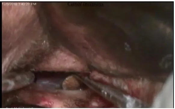
STEP 4: CANAL INCISION AND ELEVATION OF
TYMPANOMEATAL FLAP
[image:64.595.106.563.491.706.2]The canal incision is based on location of perforation like anterior or posterior. For posterior perforation, u shaped incision made in bony canal wall. Superior incision made 11o’clock position in left ear (1 o’ position for right ear), anterior and above to lateral process of malleus, extend up to the bonycartiliginous junction. Inferior incision made at 7 o’ clock position in right ear (5 o’ clock position in left ear), just lateral to annulus and join with the superior incision.
55
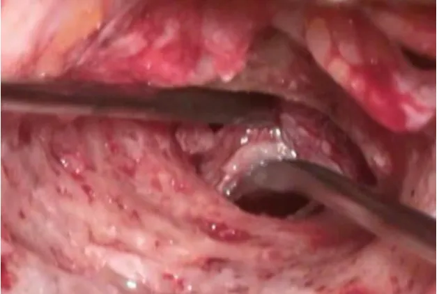
[image:65.595.198.424.308.507.2]Tympanomeatal flap elevated, middle ear entered. If perforation is only in the posterior quadrant, skeletonize the malleus handle is not needed but perforation is extended anteriorly skeletonizing is performed for supporting the temporalis fascia. For anterior perforation superior incision at 12 o’clock position in both ear, inferior incision start at 4 o’ clock position in right ear (8 o’ clock in left ear). It gives easy accessibility to reach anterior perforation
Fig-8; Exposure of middle ear
STEP 5: ASSESSMENT OF OSSICULAR CHAIN
56
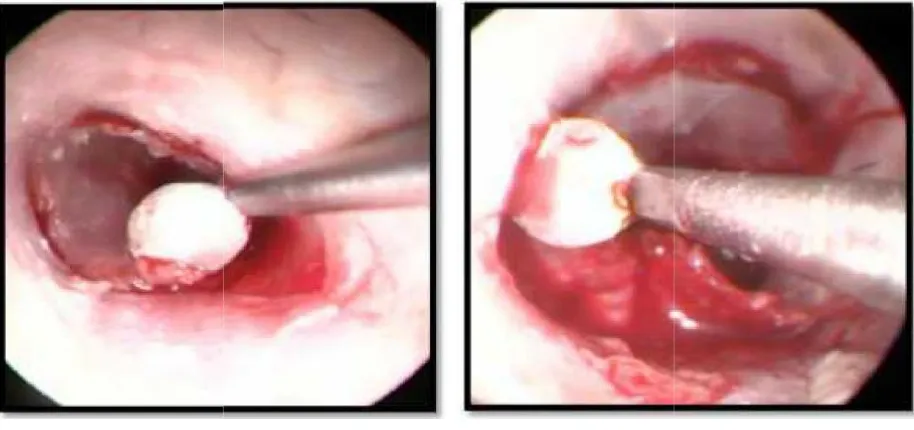
[image:66.595.108.568.113.320.2]STEP 6: GRAFT PLACEMENT:
Fig-9; Placement of graft by underlay technique
Graft bed prepared by filling the middle ear with ointment soaked gel foam. Temporalis fascia graft kept under anterior remnant of tympanic membrane either lateral or medial to malleus handle and over the posterior canal wall. Tympanomeatal flap repositioned, graft stabilized by keeping the gel foam pieces. External auditory canal is filled with medicated aural pack.
POST-AURAL MYRINGOPLASTY:
Indications;
57
Surgical technique:
[image:67.595.102.393.315.497.2]Inferior vertical Canal incision starts at 7 o’ clock position in right ear (5 o’ clock in left ear), 5mm lateral to annulus using circular knife. Similarly superior incision is made at 11 o, clock (1 o’ clock in left ear). Horizontal incision made parallel to annulus joined the superior and inferior incision. Posterior meatal skin flap elevated laterally up to bony cartilaginous junction.
FIG 10.Posterior meatal skin flap elevation
58 FIG11. POSTAURAL INCISION
Middle ear exposed from tympanosquamous suture to the
tympanomastoid suture. Superior and inferior tympanomeatal incision made. Perforation margin freshened and undersurface of tympanic membrane is scraped using plester’s side knife.
If anteriorly narrow or absent remnant tympanic membrane,
59
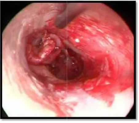
FIG 12:EXPOSURE OF MIDDLE EAR
[image:69.595.100.415.72.283.2]Tympanomeatal flap elevated anteriorly, skeletonizing the malleus handle, assessment of ossicular chain movements in continuity and individual movement and its structure.
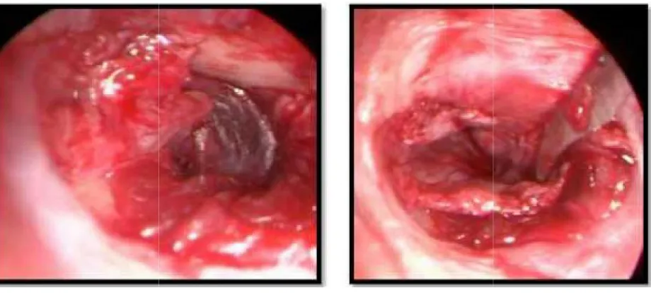
FIG13.GRAFT PLACEMENT
60
underlying bony meatal wall, posterior meatal flap, tympanomeatal flap. Repositioning of tympanomeatal flap and posterior meatal skin flap. Postaural wound closed with 3-0 plain catgut suture.
FIG14: REPOSITIONING OF TYMPANOMEATAL FLAP
In microscopic approach, some condition like narrow external auditory canal , needs canaloplasty. It is defined as circumferential widening of inferior, anterior and posterior bony canal wall to visualization of entire tympanic membrane and annulus to proper placement of fascia graft under direct vision.
ADVANTAGES OF MEDIAL GRAFTING
1. Ideal grafting for Easy visualized and small perforation.
61
3. Avoid complications like tympanic membrane lateralization and blunting.
DISADVANTAGE
1. Anterior meatal recess is less visualized compared to lateral grafting
2. Middle ear space volume is reduced, particularly in cases with severely diseased middle ear mucosa
3. When bed size of graft is limited secondary to large anterior marginal perforation, increased failure rate occurs.
ANTERIOR HITCH TECHNIQUE
In this technique, graft is placed medial to annulus, but lateral to handle of malleus. Has shown to be of use when perforation in anterior to handle.
ADVANTAGE OF LATERAL GRAFTING
1. Anterior meatal recess is well exposed.
2. Middle ear space not gets reduced.
62
DISADVANTAGES OF LATERAL GRAFTING:
1. More complications like lateralization of graft , anterior angle blunting , epithelial pearl formation or cholesteatoma formation.
2. Delayed healing up to 4-8 weeks.
POST-OPERATIVE CARE
Mastoid dressing removed the following day. Alternate day dressing over the incision site and topical antibiotic ear drops twice daily. Removal of suture on 7th POD. Advise to follow dry ear precaution and avoid activity to increase middle ear pressure for initial days. Patient follow up after 3 weeks for examination to ensure that adequate healing has began. Next visit again at six weeks to evaluate the complete healing an audiogram done at this time or third month postoperatively.
RESULTS:
1. CLOSURE OF PERFORATION:
63
reasons are often amenable to revision surgery. If poor Eustachian tube function and recent otorrhoea, require a revision tympanomastoidectomy.
Most authors report a success rate of 90%. Most authors reported less success with anterior perforation closure. Recent studies show that anterior position of tympanic membrane is less vascular. They recommend autologous temporalis fascia graft in this area, because it is less antigenic, low BMR, better able to withstand prolong anoxia.
HEARING:
Anatomical and technical factor responsible for postoperative hearing result. The mucosal status of middle ear was most important predictive factor. The manubrium mallei presence was second most important factor, it allows proper adaption of graft and increase the stability. Tympanic membrane perforation less than 50% of the drum has significantly better than larger one. Halik and symth recommended final air-bone conduction threshold less than 30 db or within 15 db of the other ear to benefit from sound localization and binaural hearing.
COMPLICATION:
64 1. Infection.
2. Failure of graft uptake.
3. Chondritis.
4. Drum cholesteatoma or epithelial pearl formation.
5. Chorda tympani nerve injury.
6. Vertigo.
7. Sensory neural hearing loss
8. Conductive hearing loss.
9. Laterlization of graft.
65
[image:75.595.127.487.116.379.2]Microscopic assisted myringoplasty:
Fig-10; Microscopic and endoscopic view of tympanic membrane
The advent of introducing the operating microscope, result of myringoplasty started showing dramatic improvement. This is attributed to accuracy of surgical technique.
66
[image:76.595.212.406.148.376.2]constantly changing, particularly with transcanal approach, which causes relatively wide microscope movement .
Fig-11; Microscopic view of tympanic membrane through post auricular access
Advantage of microscopic assisted myringoplasty:
1. Depth of perception is more.
2. Binocular vision.
3. Both hands available.
Disadvantage of microscopic assisted myringoplasty:
67
2. In narrow external auditory canal or tortuous external auditory canal , not able to visualize whole tympanic membrane ring and ear canal at same time.
3. Continuous repositioning of patient and surgeon’s head and microscope
4. Hidden structure like sinus tympani, facial recess , attic, hypotymanum not visualized.
[image:77.595.148.405.393.556.2]Endoscopic middle ear surgery :
Fig-12; Wide angle view of endoscope
68
the ear canal . This limitation makes a surgeons to create a parallel port via a postauricular transmastoid approach to gain k access to the middle ear, facial recess, and hypotympanum. Transcanal endoscopic procedure, however bypasses the narrow segment of the ear canal and provides a wide view of middle ear ,so surgeons able to look “around the corner,” even with a zero-degree endoscope is used.
Instrumentation:
Eighteen cm long, 4mm in diameter, wide-angled, zero-degree and 30-degree Hopkins rods are most often used. Recently endoscope of size 3mm has been introduced. Video equipment attached with 3-chip video camera and a monitor. Directly visualizing the monitor, procedures are performed and recorded. Standard microscopic middle ear surgery instruments are used .
69
Poe and colleagues described the use of 1.9 mm rigid endoscope through a myringotomy to aid in the diagnosis of perilympatic fistula. Thomassin and colleagues developed successful rigid endoscopic technique as an adjuvant to conventional cholesteatoma surgery , dramatically reducing the residual rate of disease.
The endoscope lens brings the surgeon view into the depth of the operative field and can provide a wide field of view with perspectives not possible through surgical microscope . Additional angulation of view is accomplished by placing prism on the end of endoscope. Surgical morbidity and operating time can be substantially reduced. Endoscopy within the middle ear may be done through a myringotomy , offering spectacular in viva examination free of artifact of blood , tissue, transduates and injected local anaesthetic agent .
Accordingly, endoscopy may be useful for various diagnostic purposes such as perilympatic fistula exploration . Endoscope also improve the abilty to inspect the entire middle ear after cholesteatoma removal, reducing cholesteatoma residulal rates.
70
instruments. The 4 mm diameter of the scope had not been a limiting factor even in smaller ear canal.
Safety issues with endoscopic ear surgery :
Thermal injury:
While the tip of endoscope heated up very quickly, same time cooled down fastely too. To protect from thermal injury scope tip cooled with savlon (antifog) solution.
Trauma:
Trauma mostly by the tip of the endoscope due to accidental head movement. So movement of head restricted while doing surgery.
Advantages of endoscope :
1. It provides an excellent magnified image with a good resolution
2. With minimal effort it can be used to visualize the nook and corners of middle ear cavity
3. Magnification can be achieved by just getting the endoscope closer to the surgical field
71
5. Middle ear cavity can be visualized easily using an endoscope. Even difficult areas to visualize under microscopy like sinus tympani, facial recess attic, and hypotymanum can easily be examined using an endoscope .
6. Complete view of middle ear , tympanic membrane , and ear canal without the need for continuous repositioning of the surgeon’s head and the microscope .
Major Disadvantages:
1. One hand technique, left hand is used to held the endoscope, so only right hand is used to operate.
2. Steep Learning curve
72
MATERIALS AND METHODS:
The present study was conducted in department of otolaryngology in Madurai medical college from the time period of June 2013-may 2014. A total of 40 patient within the age group of 15-60 years suffering from chronic suppurative otitis media of tubotympanic variety with dry central perforation .
1) DESIGN OF STUDY : Randomized prospective study
2) PERIOD OF STUDY : 1 year
3) SELECTION OF SUBJECT : Age between 15-60 year (Both sexes)
4) INCLUSION CRITERIA:
Subjects with tympanic membrane central perforation due to CSOM or trauma
Subjects with conductive hearing loss due to CSOM or trauma
Subjects with inactive and quiescent CSOM
73
5) EXCLUSION CRITERIA:
1. Patients with active discharge
2. Patients with mastoiditis
3. Patient with sensorineural hearing loss
4. Patients with cholesteatoma.
METHODOLOGY:
All the eligible patients who satisfied the inclusion criteria mentioned below are recruited into the study. Otoscopic examination and tuning fork tests, and pre operative PTA will be done to know the perforation, degree of hearing loss, air bone gap.pre op routine investigations will be done .Patients are selected based on the otoscopy examination of EAC. Patients were selected for particular procedure according to computer generated random table. Post operative outcomes such as % of graft uptake ,improvement in hearing, air bone gap closure, post op hospital stay in two groups are measured and correlation between two will be done.
INVESTIGATION CARRIED OUT IN THE STUDY:
1. Routine pre op blood investigations such as Hb , BT,CT,HIV.
74
Audiometry was consigned to an audiologist who is blinded to the study. Air conduction and bone conduction was performed . Average Air bone gap of each patient was calculated preoperatively and postoperatively at a frequency 500hz, 1000hz, 2000 hz. Audiometric evaluation was also to evaluate the cochlear reserve of patient and also for documentation.
3.CT Scan mastoid
4.Otomicroscopy and otoendoscopy.
Patient were admitted day before surgery , brief history taken , local examination of ear, nose, throat done as per attached proforma. Informed written consent obtained.
ANESTHESIA:
All cases done under the local anesthesia. Xylocaine sensitivity test done for all patient by injecting 0.2 ml of 2% Xylocaine subcutaneously in medial surface of forearm and after ten minutes looking for reaction .
PREOPERATIVE SEDATION :
75
OPERATIVE TECHNIQUE :
The operated ear was painted with povidone, betadine and methylated spirit. After draped ensure complete asepsis. Local infiltration given in external auditory canal at bonycartiliginous junction, and post auricular area using 2% Xylocaine with 1: 10,0000 Adrenaline . Supra auricular incision made above the hairline, temporalis fascia graft harvested. Through post auricular approach, fascia graft harvested through the same incision. It spread on a bowel and dried. In endoscopic method, rigid endoscope of 4mm in diameter, o degree angle of view, 100 mm in length was used. In microscopic assisted method, used microscope with lens distance 250 mm with magnification 10 used. Freshening of perforation done, undersurface of tympanic membrane scraped, canal incision made, tympanomeatal flap elevated, middle ear exposed, ossicle integrity and movement checked. Gel foam bed created, graft placed by underlay technique. Flap repositioned. Canal packed with antibiotic soaked cotton ball. In microscopic assisted method , if patient with tortuous canal , canaloplasty done, if narrow canal , through post auricular approach , myringoplasty done.
POST OPERATIVE PERIOD :
76
discharged on following day. All postauricular approach patient discharged on 7th pod after suture removal.
Patient called for follow up at end of the week for examination and then 1st, 3rd and 6th month. Post operative audiometry taken at 6th month.
Following parameter are checked post operatively:
Primary parameter :
1. Graft uptake at 6th month- categorized as intact tympanic membrane or failure.
2. Post operative hearing at 6th month . post op air – bone gap was considered as objective method to assess the hearing improvement. They grouped as < 10 db , 11-20 db,21-30 db and above 30 db .
Secondary parameter :