Received 20 December 2002/Accepted 17 March 2003
A novel simian immunodeficiency virus (SIV) sequence has been recovered from RNA extracted from the
serum of a mona monkey (Cercopithecus mona) wild born in Nigeria. The sequence was obtained by using novel
generic (degenerate) PCR primers and spans from two-thirds into thegaggene to the 3ⴕ poly(A) tail of the
SIVmonNG1 RNA genome. Analysis of the open reading frames revealed that the SIVmonNG1 genome codes for a Vpu protein, in addition to Gag, Pol, Vif, Vpr, Tat, Rev, Env, and Nef proteins. Previously, only lentiviruses infecting humans (human immunodeficiency virus type 1 [HIV-1]) and chimpanzees (SIVcpz) were known to have
avpugene; more recently, this has also been found in SIVgsn fromCercopithecus nictitans. Overall, SIVmonNG1
most closely resembles SIVgsn: theenvgene sequence groups with HIV-1/SIVcpzenvsequences, whereas thepol
gene sequence clusters closely with thepolsequence of SIVsyk fromCercopithecus albogaris. By bootscanning
and similarity plotting, the first half ofpolresembles SIVsyk, whereas the latter part is closer to SIVcol from
Colobus guereza. The similarities between the complex mosaic genomes of SIVmonNG1 and SIVgsn are con-sistent with a shared or common lineage. These data further highlight the intricate nature of the relationships between the SIVs from different primate species and will be helpful for unraveling these associations.
It is becoming increasingly apparent that many African pri-mates are infected with lentiviruses (33). These simian immu-nodeficiency viruses (SIVs) are of complex evolutionary origin (40, 41). In earlier studies, primate lentiviruses were recovered from chimpanzees (SIVcpz, Pan troglodytes) (13, 21), sooty mangabeys (SIVsm,Cercocebus atys) (20); African green mon-keys (SIVagm, Chlorocebus aethiops) (1, 19, 23, 25), Sykes monkeys (SIVsyk,Cercopithecus albogaris) (11, 18), and man-drills (SIVmnd-1,Mandrillus sphinx) (49). More recently, such viruses have been found and characterized in a wide range of primates, including the red-capped mangabey (SIVrcm, Cerco-cebus torquatus torquatus) (5, 15), guereza colobus monkey (SIVcol, Colobus guereza) (9), L’Hoest’s monkey (SIVlhoest,
Cercopithecus l’hoesti) (17), sun-tailed monkey (SIVsun, Cer-copithecus solatus) (3), talapoin monkey (Myopithecus talapoin) (30), mandrills (SIVmnd-2) (43), and drill monkey (SIVdrl,
Mandrillus leucophaeus) (8). Most recently, a novel SIV has been found in the greater spot-nosed monkey (SIVgsn, Cerco-pithecus nictitans) (10). Some primate lentiviruses are able to cross the species barriers and give rise to new strains: SIVcpz has infected humans more than once to give human immuno-deficiency virus type 1 (HIV-1) (13, 21, 22, 34, 50), and SIVsm has infected humans and macaques to yield HIV-2 and SIVmac, respectively (16, 20, 29). SIVagm has been recovered from baboons (Papiospp.) and a patas monkey (Erythrocebus patas), presumably as a result of natural transmissions in the wild (6, 24, 51). Some SIVs have mosaic or recombinant
ge-nomes, indicating that they have arisen as a result of infections with more than one distinct SIV. Examples are SIVagmSAB from the Sabaeus monkey (23), SIVrcm (5, 15), SIVdrl (8) (J. P. Clewley, unpublished observations), SIVmnd-2 (43), and SIVgsn (10). Evidence of cross-species transmission and mul-tiple infections with SIVs is consistent with the suggestion that the primate lentiviruses originated and evolved in monkeys of theCercopithecusgenus (3, 17, 25).
The SIVs can be classified into distinct lineages based upon their complete genome sequence relationships (4). To date, six such lineages are recognized: SIVcpz/HIV-1, SIVsm/HIV-2/ SIVmac, SIVlhoest/SIVsun/SIVmnd-1, SIVagm, SIVsyk, and SIVcol. New lineages are likely to be defined when the full sequences of several as yet only partially characterized ge-nomes become available (33). The SIVs can also be classified into three groups based on the organization of the accessory genes in their genomes (4). All simian lentiviruses possessvif,
vpr, andnefaccessory genes andtatand revregulatory genes but, until recently, only the SIVcpz and HIV-1 lineage viruses were known to have avpugene (4, 5, 27, 46). SIVgsn from the greater spot-nosed monkey has now also been shown to have a
vpu gene (10). The SIVsm/HIV-2 lineage viruses, including SIVrcm and SIVmnd-2, do not have avpubut do have avpx
gene (5, 43). The Vpu protein and gene have both been ex-tensively studied in the expectation that they might shed light on the unique pathogenesis of HIV-1. For example, although the Vpu protein is not essential for viral replication, removal of the vpu gene leads to a decrease in pathogenicity (44). The origin ofvpuis unclear, but it may have arisen to enhance viral release as a result of increased affinity of the Env protein for the CD4 receptor (45).
In the present study, we have recovered a genome sequence (SIVmonNG1) from lentivirus RNA extracted from the serum * Corresponding author. Mailing address: Sexually Transmitted and
Blood Borne Virus Laboratory, Central Public Health Laboratory, 61 Colindale Ave., London NW9 5HT, United Kingdom. Phone: 44-20-8200-4400, ext. 3245. Fax: 44-20-8200-1569. E-mail: jclewley@phls .org.uk.
6879
on November 8, 2019 by guest
http://jvi.asm.org/
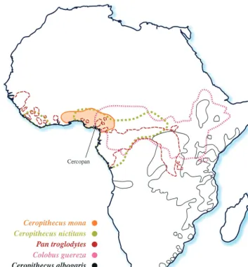
of a mona monkey (Cercopithecus mona). This forest-dwelling guenon has a range from Ghana to Cameroon in western Africa (Fig. 1) (36). There are about 26 species of guenons, of which seven forest species, including the mona, live in southern Nigeria. They are endangered through habitat loss and hunt-ing, and the Cercopan Project, located in southeastern Nigeria, is a nongovernmental organization dedicated to their conser-vation, rescue, and rehabilitation (http://www.cercopan.org/). A partialpolSIV sequence from a mona monkey from Cam-eroon has recently been described (33). Here, we show that the SIVmonNG1 genome, like SIVgsn, has avpugene, and that its
env gene is related to SIVcpz sequences and its pol gene to SIVsyk and SIVcol.
MATERIALS AND METHODS
Specimens and serological testing.Serum samples were obtained from two mona monkeys during quarantine testing at Cercopan. These monkeys were obtained via the bush meat trade and thus were wild born; they were either confiscated by appropriate wildlife officials and subsequently donated to
Cerco-pan or donated by their owners who had purchased them from hunters. In the majority of cases the monkeys at Cercopan become captive at a very young age (6 months or less) and arrived at the sanctuary soon after. The sera were tested at the Central Public Health Laboratory for antibodies cross-reacting with HIV antigens by Innogenetics and Organon Teknika enzyme immunoassays (EIAs). A positive result in these tests was confirmed by using the Genelabs Western blot test.
RNA extraction, PCR amplification, cloning, and sequencing.RNA was ex-tracted from serum by a modified guanidinium isothiocyanate-silica method (7) and converted to cDNA by reverse transcription by using Expand reverse tran-scriptase (RT; Roche Diagnostics, Lewes, United Kingdom). This was PCR amplified by using Expand High Fidelity or Long Template PCR reagents (Roche). Specific primers were used at a concentration of 300 nM, and degen-erate primers were used at a concentration of 1,000 nM. Thermocycling condi-tions were modified appropriately for the target size: typically the first-round amplifications were 94°C for 2 min, followed by 10 cycles of 94°C for 15 s, 50°C for 30 s, and 68°C for 4.5 to 7 min, followed by a further 25 cycles, with an
additional 5-s extension per cycle and a 4°C hold at the end. A total of 1.5l of
[image:2.603.113.466.72.453.2]the primary PCR was added to a secondary PCR mixture and typically cycled at 94°C for 2 min, followed by 4 to 8 cycles of 94°C for 15 s, 42 to 50°C for 30 s, and 68°C (or 72°C for Expand High Fidelity) for 2 to 7 min, followed by a further 27 to 31 cycles, with an additional 5-s extension per cycle and a 4°C hold. On
FIG. 1. Map of Africa showing the natural ranges of the mona monkey,Cercopithecus mona, the greater spot-nosed monkey,Cercopithecus nictitans, the chimpanzee,Pan troglodytes, the Sykes monkey,Cercopithecus albogaris, and the colobus monkey,Colobus guereza. The data were obtained from the study by Rowe (36) and from an online source (http://gorilla.bio.uniroma.it/). The approximate location of the Cercopan Project in Nigeria is arrowed.
6880 BARLOW ET AL. J. VIROL.
on November 8, 2019 by guest
http://jvi.asm.org/
occasion, AccuPrimeTaqpolymerase (Invitrogen, Paisley, United Kingdom) was used for nested-PCR amplifications with previously described touchdown
con-ditions (8). A 3⬘RACE (rapid amplification of cDNA ends) kit (Invitrogen) was
used to recover sequences from the poly(A) tail to thenefgene. PCR products
were cloned with TOPO XL kits (Invitrogen) and plasmids purified with Qiagen kits (Qiagen, Crawley, United Kingdom). Sequences were generated by using a capillary sequencer (CEQ 2000XL DNA analysis system; Beckman Coulter, High Wycombe, United Kingdom) or by commercial providers (J. Bartley, Nat-ural History Museum, London, United Kingdom; MWG Biotech, Milton Keynes, United Kingdom; Cytomix, Cambridge, United Kingdom).
Primers.Initially, previously described primers were used to amplify a small
region of thepolgene (9, 28). PolOR was used for cDNA synthesis and
first-round PCR with Polis4; Polis4/Unipol2 were then used for second-first-round PCR.
New primers were designed in conserved regions of thegag(MN1, MN2, and
MN3),env(MN4, MN5, and MN6) andnef(MN11 and MN14) genes based on
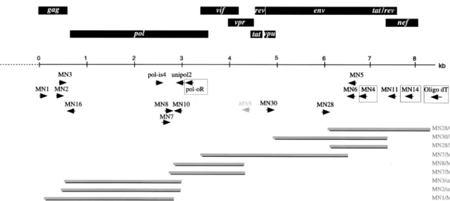
multiple alignments of previously published SIV and HIV sequences. The se-quences of these primers and their locations relative to SIVrcmNG411 (acces-sion no. AF349680) are as follows (Fig. 2): MN1 (GARGAAGSDGCMSANT GGGA; 826 to 847), MN2 (ATNCARAATGCHAAYCCDGAHTG; 1184 to 1206), MN3 (GCHTGYCARGGVGTHGGDGG; 1262 to 1281), MN4 (TKGA GRTTYTTNRYWCCCCA; 7666 to 7685), MN5 (CARVTKYTKCTGYTGCT GCAVTATCCC; 7594 to 7620), MN6 (CCCATTGCAGHDCCHGC; 7531 to 7547), MN11 (CCTTCCAGTCCHCCCTTTBHTTTTA; 8683 to 8709), and MN14 (DBNGRDTYVAABBDCCA; 8954 to 8970).
In addition to the predicted site inenv, MN6 also primed betweenpolandenv.
In subsequent work, MN14, MN4, and PolOR were used for cDNA synthesis and then as first-round PCR primers together with MN1, Polis4, or sequence-specific
primers designed in the smallpolregion between Polis4 and Unipol2.
Second-round PCR primers included MN2, MN3, MN5, MN6, MN11, Unipol2, and sequence-specific primers designed from sequence obtained from the PCR walk. These included the following: MN7 (CATTGAACCAGCCCGAGA), MN8 (T CCTAGCCAAAGAGATAGTCA), MN10 (CGGCTACTAGGATGATACT TC), MN28 (CTACAGCAGCCCAAAGGAGA), and MN30 (GATAATCCGG GAGTCAAGT).
Sequence and phylogenetic analysis.Sequences were analyzed by using pro-grams in the Lasergene suite (DNAStar, Madison, Wis.). CLUSTAL alignments (47) were edited by eye in MacClade 4.0.2 (D. R. Maddison and W. P. Maddison, Sinauer Associates, Sunderland, Mass.), exported into PAUP* 4.0 1 (D. L. Swofford, Sinauer Associates) for gap stripping and first- and second-codon
position alignments. Concatenatedgag,pol,vif,env, andnefnucleotide and
corresponding amino acid alignments were also made. Phylogenetic reconstruc-tions were carried out by using PAUP*, and the most appropriate evolutionary models and parameters were chosen by using Modeltest (35). Tree searches were carried out by using a heuristic search strategy and branch swapping. Neighbor
joining-derived trees were bootstrapped 1,000 times. Protdist (J. Felsenstein, Department of Genetics, University of Washington, Seattle [http://evolution .genetics.washington.edu/phylip/software.html]) was used to estimate the amino acid similarity of aligned predicted protein sequences. Trees were displayed with Treeview (31). RIP (42) and SimPlot (S. Ray, Division of Infectious Diseases, Department of Medicine, Johns Hopkins University School of Medicine [http: //sray.med.som.jhmi.edu/RaySoft/SimPlot/]) were used for similarity and boot-scanning analyses (26). Oligo (MedProbe, Oslo, Norway) was used to design SIVmonNG1 sequence-specific primers.
Nucleotide sequence accession numbers. Nucleotide sequences have been submitted to the EMBL database under accession numbers AJ549283 and AJ549756.
RESULTS
Identification of HIV antibody-positive serum.Serum
sam-ples were obtained from two mona monkeys at Cercopan in Nigeria. One was found to be reactive in both EIA tests, while the other was unreactive. In the Western blot, the EIA-positive sample was strongly reactive with the HIV-1 p66 antigen (the RT) and had weaker reactivities with gp160 (envelope), p31 (the integrase), and p24 (core) antigens of HIV-1, as well as the HIV-2 p36 transmembrane antigen (Fig. 3). This indicated that the mona monkey was infected with a primate lentivirus.
Amplification of SIVmonNG1 RNA. RNA was extracted
from the serum and subjected to nested reverse transcription-PCR with conserved primers in the integrase gene: Polis4/ PolOR and then Polis4/Unipol2 (9, 28). The 598 nucleotides of sequence that were derived from this amplicon were found to be phylogenetically related to other primate lentivirus genome sequences, thus confirming that the mona monkey was infected with a novel SIV. Degenerate primers were then designed based on multiple alignments of eithergag,env, ornefSIV and HIV sequences in order to be able to extend beyond thepol
[image:3.603.62.519.71.275.2]sequences obtained by using Polis4/Unipol2. These primers (MN1, MN2, and MN3 ingag; MN4, MN5, and MN6 in env; MN11 and MN14 innef) were used in RT nested-PCR proto-cols (Fig. 2). For example, MN4 was used as an RT primer and FIG. 2. Genome organization and PCR cloning strategy for SIVmonNG1 RNA. The identified ORFs are indicated by rectangular boxes above the scale bar. The alternative binding site of MN6 is shown in italics. The primers used for PCR are shown below the scale bar; primers used for cDNA synthesis are boxed. The position of the clones is shown by solid bars, and the primers used to obtain them are indicated on the right.
on November 8, 2019 by guest
http://jvi.asm.org/
then MN1 and MN4 or MN5 as first-round primers. Alterna-tively, PolOR or MN14 were used to prime cDNA synthesis. Various combinations of second-round primers were then used, including MN2/Unipol2 and MN1, MN6, and MN11 with specific primers designed on the basis of sequence information derived from amplicons produced with Polis4/Unipol2 (MN7, MN8, and MN10) or with MN7/MN6 (MN28 and MN30). The PCR enzymes and conditions were critical to the successful use of these primers; we mostly used an enzyme mixture with a proofreading polymerase, together with a low annealing tem-perature for the first 10 cycles of the PCR. By these methods we were able to amplify and clone regions of the genome from about two-thirds into thegaggene to the 3⬘poly(A) tail (Fig. 2).
Genomic organization of SIVmonNG1: presence of a vpu
gene. Open reading frames (ORFs) in the sequences were
identified by BLAST searches of the Los Alamos HIV se-quence database (http://hiv-web.lanl.gov) and by nucleotide and derived amino acid phylogenetic comparisons with other SIV sequences. Potential Gag, Pol, Vif, Tat, Vpr, Rev, and Nef proteins were identified. In addition, an ORF in a similar position to thevpugene of HIV-1 and SIVcpz was identified (Fig. 2). Therefore, SIVmonNG1 has a genomic organization more similar to HIV-1, SIVcpz, and SIVgsn than to the HIV-2/SIVsm lineage viruses or to the SIVagm, SIVlhoest, SIVsyk, or SIVcol lineages (5).
Putative Vpu amino acid sequence.The SIVmonNG1
puta-tive Vpu amino acid sequence was not closely related to other Vpu sequences, either HIV-1, SIVcpz, or SIVgsn. The amino acid similarity of SIVmonNG1 Vpu to other Vpu proteins was estimated by using the PHYLIP program Protdist with a man-ually adjusted CLUSTAL W amino acid alignment and ranged from 15.2% for SIVcpzUS to 17.9% for HIV-1 to 23.4% for SIVgsn to 29% for SIVcpzANT (Table 1). Like all Vpu pro-teins, the N-terminal half of the sequence (residues 6 to 33) has many hydrophobic residues and is characteristic of a trans-membrane domain. The C-terminal half of the sequence has many acidic residues but not the EDSGNESXG(E/D) domain that is highly conserved among HIV-1 and SIVcpz strains, nor does it have a similar sequence to SIVgsn in this region (10). It does, however, have the run of glutamic and aspartic acid residues present in all Vpu proteins, and it also has a DNP
motif near the C terminus, in common with other HIV-1 and SIVcpz Vpu sequences.
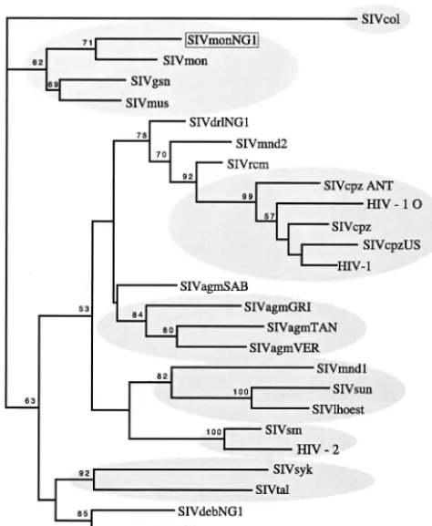
Genomic relatedness of SIVmonNG1 to other SIVs. The
relationship of SIVmonNG1 to other primate lentiviruses was investigated by comparing the sequence obtained to represen-tatives of the six other SIV lineages and to SIVgsn. First, diversity plots were performed by using a multiple alignment of concatenated predicted Gag, Pol, Vif, Env, and Nef gene prod-ucts. Ambiguous sites, as well as sites that contained a gap, were removed from the alignment. The sequence similarity was calculated for a window of 200 amino acids moved along the alignment (Fig. 4). The SIVmonNG1 sequence showed the greatest similarity to SIVgsn across the whole genome, and the most diverse protein was Vif.
[image:4.603.89.487.70.198.2]The second method used for examining the relationship of SIVmonNG1 to other SIV genomes was bootscanning (Fig. 5). For this, as for subsequent phylogenetic analyses, third-codon bases were removed from the alignment since they are satu-rated with nucleotide substitutions and have been shown to be phylogenetically uninformative (10). Bootscanning against sin-gle sequences from the six major SIV lineages showed that the SIVmonNG1 genome is a complex mosaic, with the 5⬘region related to SIVsyk and SIVcol and the 3⬘ region related to SIVcpz and SIVsyk sequences (Fig. 5A). When SIVgsn and SIVcpzANT sequences were included in the bootscanning analysis, the SIVmonNG1 sequence was seen to be most closely related to SIVgsn, apart from a small region in pol
FIG. 3. Antibody profile of serum from the mona monkey revealed on a Genelabs Western blot. At the top is the serum from the mona monkey; below are a negative human serum control, a weak anti-HIV-1-positive human serum control, and a strong anti-HIV-1-positive human serum control.
TABLE 1. Amino acid sequence identity between SIVmonNG1 and other SIV and HIV-1 genomes in Vpu
Sequence Amino acid similarity (%)
gsn71 cpzUS cpzANT cpzGAB M N O
SIVmonNG1 23.4 15.2 29.0 20.9 17.9 18.5 23.9 SIVgsn71 100 20.3 23.9 16.9 15.9 20.0 21.7
SIVcpzUS 100 24.6 32.9 35.6 44.3 30.4
SIVcpzANT 100 24.6 20.0 29.2 28.2
SIVcpzGAB 100 32.9 28.2 31.3
HIV-1 M Z2Z6 100 42.9 28.4
HIV-1 N YBF 100 27.6
HIV-1 O ANT70 100
6882 BARLOW ET AL. J. VIROL.
on November 8, 2019 by guest
http://jvi.asm.org/
[image:4.603.303.542.616.726.2]resembling SIVsyk (Fig. 5B). This substantiates the conclusion that SIVmonNG1 and SIVgsn form a shared lineage.
Comparing the predicted protein sequences of SIVmonNG1 with representatives of other primate lentivirus lineages, SIVmonNG1 was most closely related to SIVgsn (Table 2). For example, for Pol, the similarity score ranged from 34.9% with SIVcpzGAB to 68.3% with SIVgsn. For Env, the similarity ranged from 30.1% with SIVcol to 56.2% for SIVcpzANT to 61.9% for SIVgsn. The SIVmonNG1 Env protein V3 loop is predicted to be 35 amino acids in length and has a QIGAGM TFYS central motif identical to that of SIVgsn; overall, these two V3 loops have 94% identity. The SIVmonNG1 V3 loop is also closely related to SIVcpzANT (71% identity) and similar to other SIVcpz V3 sequences.
The predicted mosaicism of the genome of SIVmonNG1 observed by bootscanning was further analyzed by phyloge-netic analysis of selected genomic regions. Several gene re-gions were investigated, but only the env and pol gave trees with significant bootstrap support for the branching of the SIVmonNG1 and SIVgsn sequences. Analysis of theenvgene by both standard alignments and gap-stripped alignments, and by first- and second-codon positions only, all gave rise to phy-logenetic trees in which SIVmonNG1 env clustered with SIVgsn sequences. For example, Fig. 6 shows a phylogenetic tree derived by maximum likelihood searching, in which the best-fitting model of evolution was evaluated by using Model-test and PAUP* (35). Support for the branching order was found by bootstrapping (1,000 times) a congruent neighbor-joining tree derived with the same model of evolution. Further, the SIVmon and SIVgsn env sequences also formed a clade with HIV-1/SIVcpzenvsequences, supported by a 94 bootstrap proportion, further strengthening the idea that these envelope genes are related.
In contrast to theenvgene, when thepolgene was exam-ined by similar phylogenetic methods the SIVmonNG1 se-quence formed a clade with SIVgsn and SIVsyk, separate from SIVcpz/HIV-1 sequences (Fig. 7). Using bootstrapping with distance methods a high (98 bootstrap proportion) support was found for this grouping. Recently, partialpol sequences (522 nucleotides) have been determined for several previously un-characterized SIVs from African primates in Cameroon (33), including mona monkeys and the mustached monkey ( Cerco-pithecus cephus). Comparing the equivalent region from SIVmonNG1 indicated that it was closest to another mona monkey sequence, SIVmon-99CM-CML1 (Fig. 8). These se-quences formed a separate cluster with theCercopithecus nici-tans (SIVgsn-99CM-CN71) and Cercopithecus cephus
(SIVmus-01CM-S1239) sequences, away from other SIV se-quences, including SIVsyk, which grouped with SIVtal (Fig. 8). Additional sequence data from SIVmus and SIVtal, and any as-yet-uncharacterized SIVs, will enable greater understanding of the relationships between these SIVs.
DISCUSSION
The sequence of more than 8,200 bases of the RNA genome [partial gag to 3⬘ poly(A)] of an SIV from a mona monkey (Cercopithecus mona) was derived by a PCR amplification strategy by using novel degenerate primers located in con-served regions of SIV/HIV genomes. Attempts to recover the 5⬘end by RACE and amplification with a primer based on the presumptive tRNA-binding site were unsuccessful, and insuf-ficient sample remained for further work. Further character-ization of the SIVmonNG1 genome will, therefore, require the collection of more serum. The SIVmonNG1 sequence was obtained from serum collected from a young wild-born mona FIG. 4. Similarity plotting of SIVmonNG1 against the six major SIV lineages (SIVsyk, SIVlhoest, SIVcol, SIVagm, SIVcpz, and SIVsm) and SIVcpzANT and SIVgsn. Alignments of the nonoverlapping regions ofgag,pol,vif,env, andnefwere concatenated together; gaps were removed from the amino acid alignment; a window of 200 residues was used. The relative position of the genome proteins is shown. Sequences for comparison were obtained from the Los Alamos HIV Database/GenBank.
on November 8, 2019 by guest
http://jvi.asm.org/
monkey in the Cercopan Conservation Centre in Nigeria. As with any primate lentivirus that can jump from one species to another, it has to be asked whether the mona monkey is the primary host for this virus or whether it could have come from another monkey. The young age of the monkey (⬍2 months old) is consistent with it being the primary host. The availabil-ity of a smallpolregion sequence independently obtained from a mona monkey from the Cameroon (33) allowed a compari-son with the sequence we obtained (Fig. 8). The two sequences
were clearly related (82% identity over 522 nucleotides), a finding consistent with them being the same SIV from animals at extremes of the species range, which is from Ghana to the Cameroon (Fig. 1) (36). We do not have any evidence that SIVmonNG1 was associated with any specific disease.
The molecular characterization of this genome provides new insights into the phylogeny and evolution of the primate lenti-viruses. Significantly, the genome has a putative vpu gene, which has previously only been found in HIV-1 and SIVcpz FIG. 5. (A) DNA bootscanning comparisons of SIVmonNG1 against single sequences from the major SIV lineages (SIVsyk, SIVlhoest, SIVcol, SIVagmGRI, SIVcpz, and SIVsm). (B) Similar analysis including SIVgsn and SIVcpzANT. Alignments of the nonoverlapping regions ofgag,pol,
vif,env, andnefwere concatenated together, and third-codon positions and gaps were excluded. A sliding window of 450 bases in 40 base steps and 1,000 bootstrap replicates was used for bootscanning with the Kimura 2 parameter model and a transition/transversion ratio of 0.75. The relative positions of the genes are shown at the bottom of the figures.
6884 BARLOW ET AL. J. VIROL.
on November 8, 2019 by guest
http://jvi.asm.org/
genomes and, more recently, in SIVgsn (10). The conceptual translation of this gene yields a sequence that is consistent with it being related to the Vpu proteins of HIV-1, SIVcpz, and SIVgsn although, like these proteins, it is clearly divergent with
an average similarity of 21% (Table 1). HIV-1 Vpu has two domains: an N-terminal transmembrane anchor and a C-ter-minal cytoplasmic domain (38). The transmembrane domain enhances virus release from infected cells, whereas the cyto-plasmic domain is involved in the degradation of CD4 (32, 37, 48). When the derived protein sequence of SIVmonNG1 Vpu was analyzed by molecular modeling (data not shown), the amino terminus (residues 6 to 33) was found, as expected, to be characteristic of a transmembrane domain. The C-terminal (cytoplasmic) half, however, was predicted to have a single alpha-helix, located before two candidate phosphoserine resi-dues. The SIVgsn Vpu was also predicted to have only one alpha-helix. In comparison, by using the same prediction algo-rithms, HIV-1 and SIVcpz Vpu proteins had two alpha-helices, one on either side of the phosphoserines, a finding consistent with the structural data (12, 52, 53). This could be interpreted as indicating a difference in biological activity of the cytoplas-mic domain of the SIVmonNG1 Vpu compared to the equiv-FIG. 6. Phylogenetic comparison ofenvSIV and HIV sequences.
[image:7.603.307.531.72.341.2]An in-frame nucleotide alignment ofenvsequences was derived from the corresponding amino acid alignment, and gaps and codon third-position bases were excluded. The best-fitting model of evolution found by using Modeltest (35) with PAUP* was the general time revers-ible with the following parameters. The rates were as follows: A-C⫽ 1.3738, A-G⫽0.9821, A-T⫽0.3369, C-G⫽1.5349, C-T⫽1.2473, and G-T⫽1.0. The base frequencies were as follows: A⫽0.3135, C⫽0.1323, G⫽0.2273, and T⫽0.3269. The gamma distribution shape parameter (alpha) was 4.6652, and the proportion of invariable sites was 0.0093. The branch lengths of the tree shown were derived by a maximum-likelihood search with the above parameters. The bootstrap values (of 1,000) were derived from a congruent distance/neighbor-joining tree found with the same model of evolution. The scale bar indicates the number of nucleotide substitutions per site. Sequences for comparison were obtained from the Los Alamos HIV Database/GenBank.
FIG. 7. Phylogenetic comparison ofpolSIV and HIV sequences. An in-frame nucleotide alignment ofpolsequences was derived from the corresponding amino acid alignment, and gaps and codon third-position bases were excluded. The best-fitting model of evolution found by using Modeltest (35) with PAUP* was the general time reversible. The rates were as follows: A-C⫽2.9306, A-G⫽3.2906, A-T⫽1.9048, C-G⫽3.1502, C-T⫽5.2621, and G-T⫽1.0. The base frequencies were as follows: A⫽0.3709, C⫽0.2021, G⫽0.2353, and T⫽0.1917. The gamma distribution shape parameter was alpha⫽ 1.2972. The proportion of invariable sites was 0.2594. The branch lengths of the tree shown were derived by a maximum-likelihood search with the above parameters. The bootstrap values (of 1,000) were derived from a congruent distance/neighbor-joining tree found with the same model of evolution. The scale bar indicates the number of nucleotide substitutions per site. Apart from SIVmonNG1, se-quences were obtained from the Los Alamos HIV Database/GenBank.
aNA, not applicable or available. Values in boldface indicate highest sequence
identity scores.
on November 8, 2019 by guest
http://jvi.asm.org/
[image:7.603.42.285.89.194.2] [image:7.603.48.276.300.575.2]alent region of the HIV-1/SIVcpz protein. Of course, this would need to be established experimentally.
Sequence comparisons of the SIVmonNG1 genome further illustrate the complexity of the relationships between the pri-mate lentiviruses. The SIVmonNG1 sequence is most closely related to SIVgsn, as can be seen from the amino acid simi-larity plot (Fig. 4) and, in common with that genome, it can be interpreted as having a complex mosaic structure, as shown by bootscanning (Fig. 5A). Aside from SIVgsn, the 5⬘half of the SIVmonNG1 genome shows similarity to SIVsyk and SIVcol by bootscanning. Again, apart from SIVgsn (Fig. 5B), the 3⬘ half of the genome resembles SIVcpzANT by similarity plot-ting and SIVsyk and SIVcpz by bootscanning. Thevifgene is the most diverse region of the SIVmonNG1 genome (Fig. 4). More definitive analyses of the genetic relationships of SIVmonNG1 were attempted by phylogenetic comparisons of selected genome regions. Trees derived from alignments of
small genome regions suggested by the bootscans have did not high bootstrap support, as was also observed for SIVgsn (10). Trees made from thepolandenvregions were, however, unambiguous. The env region of SIVmonNG1 and SIVgsn formed a clade with SIVcpz/HIV-1 with high bootstrap support (94 BP) (Fig. 6). It is perhaps not surprising thatenvsequences resembling those of the HIV-1/SIVcpz clade are found in an-other SIV that has avpu gene, since the envand vpu genes overlap and are translated from a bicistronic mRNA (39). Thus, theenvgene similarity and the presence of avpugene could be interpreted as indicating that thevpu/envgene regions of SIVmonNG1, SIVgsn, and SIVcpz have a common origin or ancestor. Analysis of thepolregion showed that SIVmonNG1, SIVsyk, and SIVgsn formed a clade with high bootstrap sup-port (98 BP) (Fig. 7), indicating that thepol gene region of SIVmonNG1 has a different origin from theenvgene region. Thus, from the similarity plots, bootscans, and phylogenetic trees, it is clear that SIVmonNG1, like SIVgsn, has a complex mosaic genome, related to the SIVs of otherCercopithecinae
[image:8.603.44.281.72.360.2]monkeys. The present-day ranges of the mona monkey and those of the four other species (chimpanzee, greater spot-nosed monkey, Sykes monkey, and colobus monkey), whose SIVs show the most similarity to SIVmonNG1, are shown in Fig. 1. The ranges of the mona monkey, chimpanzee, greater spot-nosed monkey, and colobus monkey overlap. The Sykes monkey range is adjacent to that of the greater spot-nosed monkey and overlaps with the colobus monkey. This geograph-ical distribution, as well as the similarity of the SIVmonNG1 and SIVgsn genomes from distinct species, is consistent with the idea that the same lineage has infected both species or that SIVgsn passed from the greater spot-nosed to the mona mon-key.
The similarity of SIVmonNG1 and SIVgsn to SIVcpz inenv
raises the question of whether they or the SIVcpz lineage are recombinant. Previously characterized recombinants include SIVagmSAB from the sabaeus African green monkey (23), SIVrcm from red-capped mangabeys (5, 15), SIVmnd-2 from the mandrill (43), and SIVdrl from the drill monkey (8) (Clew-ley, unpublished). As Souquie`re et al. have pointed out, the
Cercopithecinaemonkeys, the mandrills, and drill monkeys are close relatives (43), and the existence of recombinant forms in these species supports the suggestion that the SIVs from them are ancestral and that recombinants play a major role in primate lentivirus evolution (3, 17, 25). The present study provides evidence consistent with an SIVcpz ancestral or pro-genitor virus being present in aCercopithecinaemonkey, sug-gesting that the SIVcpz group is mosaic. As more SIV genomes are characterized, it is likely to become increasingly difficult to recognize pure lineages.
The PCR primers we used to recover sequence information from SIVmonNG1 RNA in serum may help the characteriza-tion of previously unknown SIVs. Many recent SIV studies have used proviral DNA from cultured infected cells as the starting point for the recovery of whole-genome sequence in-formation, either by PCR amplification of unintegrated circu-lar DNA or by screening a genomic library (2, 3, 5, 9, 10, 15, 17–19, 43). These experiments have often relied on degenerate primers to obtain starting sequence information or for inverse PCR of circular DNA (5, 9, 10, 15, 17, 43). Several sets of degenerate primers have been designed and used by different FIG. 8. Maximum-likelihood tree of a 520-bp polsequence, with
significant bootstrap values (of 100) indicated. The sequences repre-sent the amplicon derived by using the Polis4/Unipol2 primers (9, 28). The best-fitting model of evolution found by using Modeltest (35) with PAUP* was the general time reversible. The rates were as follows: A-C ⫽2.4379, A-G⫽5.8145, A-T⫽2.2893, C-G⫽3.5281, C-T⫽12.703, and G-T⫽1.0. The base frequencies were as follows: A⫽0.4341, C ⫽0.1827, G⫽0.2021, and T⫽0.1811. The gamma distribution shape parameter was alpha⫽0.9683. The proportion of invariable sites was 0.2127. Both the branch lengths and the bootstrap proportions (of 100) were derived by maximum-likelihood searching. The scale bar indi-cates the number of nucleotide substitutions per site. SIVmonNG1 (AJ549283), SIVdrlNG1 (AJ310481), and SIVdebNG1 (AJ549756) were sequenced as part of the present study; SIVmon (99CM-CML1), SIVgsn (99CM-CN71), SIVmus (01CM-S1239), SIVmnd2, SIVtal, and SIVdeb are from Peeters et al. (33): these latter sequences and others were obtained from the Los Alamos HIV Database/GenBank.
6886 BARLOW ET AL. J. VIROL.
on November 8, 2019 by guest
http://jvi.asm.org/
ACKNOWLEDGMENTS
We thank John Lewis and Zena Tooze for supplying the serum, without which this work could not have been done, and the Cercopan Project (www.cercopan.org) for their conservation work with the Cer-copithecinae. We also thank Grace McCormack for phylogenetic ad-vice, John Parry and the CPHL HIV-1 Reference Laboratory staff for serological tests, Iain Tatt for performing sequencing reactions, and Philip Mortimer and David Brown for encouragement.
REFERENCES
1. Allan, J. S., M. Short, M. E. Taylor, S. Su, V. M. Hirsch, P. R. Johnson, G. M. Shaw, and B. H. Hahn.1991. Species-specific diversity among simian
immu-nodeficiency viruses from African green monkeys. J. Virol.65:2816–2828.
2. Beer, B. E., E. Bailes, G. Dapolito, B. J. Campbell, R. M. Goeken, M. K. Axthelm, P. D. Markham, J. Bernard, D. Zagury, G. Franchini, P. M. Sharp, and V. M. Hirsch.2000. Patterns of genomic sequence diversity among their
simian immunodeficiency viruses suggest that L’Hoest monkeys (
Cercopithe-cus lhoesti) are a natural lentivirus reservoir. J. Virol.74:3892–3898. 3. Beer, B. E., E. Bailes, R. Goeken, G. Dapolito, C. Coulibaly, S. G. Norley, R.
Kurth, J. P. Gautier, A. Gautier-Hion, D. Vallet, P. M. Sharp, and V. M. Hirsch.1999. Simian immunodeficiency virus (SIV) from sun-tailed monkeys (Cercopithecus solatus): evidence for host-dependent evolution of SIV within theC. lhoestisuperspecies. J. Virol.73:7734–7744.
4. Beer, B. E., E. Bailes, P. M. Sharp, and V. M. Hirsch.1999. Diversity and
evolution of primate lentiviruses, p. 460–474.InC. L. Kuiken, B. Foley, B.
Hahn, P. A. Marx, F. McCutchan, J. W. Mellors, J. I. Mullins, S. Wolinsky, and B. Korber (ed.), Human retroviruses and AIDS. Los Alamos National Laboratory, Los Alamos, N.Mex.
5. Beer, B. E., B. T. Foley, C. L. Kuiken, Z. Tooze, R. M. Goeken, C. R. Brown, J. Hu, M. S. Claire, B. T. Korber, and V. M. Hirsch.2001. Characterization of novel simian immunodeficiency viruses from red-capped mangabeys from
Nigeria (SIVrcmNG409 and -NG411). J. Virol.75:12014–12027.
6. Bibollet-Ruche, F., A. Galat-Luong, G. Cuny, P. Sarni-Manchado, G. Galat, J. P. Durand, X. Pourrut, and F. Veas.1996. Simian immunodeficiency virus
infection in a patas monkey (Erythrocebus patas): evidence for cross-species
transmission from African green monkeys (Cercopithecus aethiops sabaeus)
in the wild. J. Gen. Virol.77:773–781.
7. Boom, R., C. J. A. Sol, M. M. M. Salimans, C. L. Jansen, P. M. E. Wertheim-van Dillen, and J. Wertheim-van der Noorda.1990. Rapid and simple method for
purification of nucleic acids. J. Clin. Microbiol.28:495–503.
8. Clewley, J. P., J. C. M. Lewis, D. W. G. Brown, and E. L. Gadsby.1998. A novel simian immunodeficiency virus (SIVdrl) pol sequence from the drill
monkey,Mandrillus leucophaeus. J. Virol.72:10305–10309.
9. Courgnaud, V., X. Pourrut, F. Bibollet-Ruche, E. Mpoudi-Ngole, A. Bour-geois, E. Delaporte, and M. Peeters.2001. Characterization of a novel simian
immunodeficiency virus from guereza colobus monkeys (Colobus guereza) in
Cameroon: a new lineage in the nonhuman primate lentivirus family. J.
Vi-rol.75:857–866.
10. Courgnaud, V., M. Salemi, X. Pourrut, E. Mpoudi-Ngole, B. Abela, P. Auzel, F. Bibollet-Ruche, B. Hahn, A.-M. Vandamme, E. Delaporte, and M. Peeters.
2002. Characterization of a novel simian immunodeficiency virus with avpu
gene from greater spot-nosed monkeys (Cercopithecus nictitans) provides
new insights into simian/human immunodeficiency virus phylogeny. J. Virol.
76:8298–8309.
11. Emau, P., H. M. McClure, M. Isahakai, J. G. Else, and P. N. Fultz.1991.
Isolation from African Sykes’ monkeys (Cercopithecus mitis) of a lentivirus
related to human and simian immunodeficiency viruses. J. Virol.65:2135–
2140.
12. Federau, T., U. Schubert, J. Flossdorf, P. Henklein, D. Schomburg, and V. Wray.1996. Solution structure of the cytoplasmic domain of the human immunodeficiency virus type 1 encoded virus protein U (Vpu). Int. J. Peptide
Protein Res.47:297–310.
13. Gao, F., E. Bailes, D. L. Robertson, Y. Chen, C. M. Rodenburg, S. F.
novel simian immunodeficiency virus (SIV) from L’Hoest monkeys (
Cerco-pithecus l’hoesti): implications for the origins of SIVmnd and other primate
lentiviruses. J. Virol.73:1036–1045.
18. Hirsch, V. M., G. Dapolito, S. Goldstein, H. M. McClure, P. Emau, P. N. Fultz, M. Isahakai, R. Neroot, G. Myers, and P. R. Johnson.1993. A distinct
African lentivirus from Sykes’ monkeys. J. Virol.67:1517–1528.
19. Hirsch, V. M., C. McGann, G. Dapolito, S. Goldstein, A. Ogen-Odoi, B. Biryawaho, T. Lakwo, and P. R. Johnson.1993. Identification of a new
subgroup of SIVagm in tantalus monkeys. Virology197:426–430.
20. Hirsch, V. M., R. A. Olmsted, M. Murphy-Corb, R. H. Purcell, and P. R. Johnson.1989. An African primate lentivirus (SIVsm) closely related to
HIV-2. Nature339:389–392.
21. Huet, T., R. Cheynier, A. Meyerhans, G. Roelants, and S. Wain-Hobson.
1990. Genetic organization of a chimpanzee lentivirus related to HIV-1.
Nature345:356–359.
22. Janssens, W., K. Fransen, M. Peeters, L. Heyndrickx, J. Motte, L. Bed-jabaga, E. Delaporte, P. Piot, and G. van der Groen.1994. Phylogenetic analysis of a new chimpanzee lentivirus SIVcpz-gab2 from a wild-captured
chimpanzee from Gabon. AIDS Res. Hum. Retrovir.10:1191–1192.
23. Jin, M. J., H. Hui, D. L. Robertson, M. C. Muller, F. Barre-Sinoussi, V. M. Hirsch, J. S. Allan, G. M. Shaw, P. M. Sharp, and B. H. Hahn.1994. Mosaic genome structure of simian immunodeficiency virus from west African green
monkeys. EMBO J.13:2935–2947.
24. Jin, M. J., J. Rogers, J. E. Phillips-Conroy, J. S. Allan, R. C. Desrosiers, G. M. Shaw, P. M. Sharp, and B. H. Hahn.1994. Infection of a yellow baboon with simian immunodeficiency virus from African green monkeys:
evidence for cross-species transmission in the wild. J. Virol.68:8454–8460.
25. Johnson, P. R., A. Fomsgaard, J. Allan, M. Gravell, W. T. London, R. A. Olmsted, and V. M. Hirsch.1990. Simian immunodeficiency viruses from
African green monkeys display unusual genetic diversity. J. Virol.64:1086–
1092.
26. Lole, K. S., R. C. Bollinger, R. S. Paranjape, D. Gadkari, S. S. Kulkami, N. G. Novak, R. Ingersoll, H. W. Sheppard, and S. C. Ray.1999. Full-length human immunodeficiency virus type 1 genomes from subtype C-infected seroconverters in India, with evidence of intersubtype recombination. J.
Vi-rol.73:152–160.
27. McCormick-Davis, C., S. Dalton, D. Singh, and E. Stephens.2000. Compar-ison of Vpu sequences from diverse geographical isolates of HIV type 1 identifies the presence of highly variable domains, additional invariant amino acids, and a signature sequence motif common to subtype C isolates. AIDS
Res. Hum. Retrovir.16:1089–1095.
28. Miura, T., J. Sakuragi, M. Kawamura, M. Fukasawa, E. N. Moriyama, T. Gojobori, K. Ishikawa, J. A. Mingle, V. B. Nettey, H. Akari, M. Enami, H. Tsujimoto, and M. Hayami.1990. Establishment of a phylogenetic survey system for AIDS-related lentiviruses and demonstration of a new HIV-2
subgroup. AIDS4:1257–1261.
29. Novembre, F. J., V. M. Hirsch, H. M. McClure, P. N. Fultz, and P. R. Johnson.1992. SIV from stump-tailed macaques: molecular characterization
of a highly transmissible primate lentivirus. Virology186:783–787.
30. Osterhaus, A. D., N. Pedersen, G. van Amerongen, M. T. Frankenhuis, M. Marthas, E. Reay, T. M. Rose, J. Pamungkas, and M. L. Bosch.1999. Isolation and partial characterization of a lentivirus from talapoin monkey (Myopithecus talapoin). Virology260:116–124.
31. Page, R. D. M.1996. TREEVIEW: an application to display phylogenetic
trees on personal computers. Comput. Appl. Biosci.12:357–358.
32. Paul, M., S. Mazumder, N. Raja, and M. A. Jabbar.1998. Mutational analysis of the human immunodeficiency virus type 1 Vpu transmembrane domain that promotes the enhanced release of virus-like particles from the
plasma membrane of mammalian cells. J. Virol.72:1270–1279.
33. Peeters, M., V. Courgnaud, B. Abela, P. Auzel, X. Pourrut, F. Bibollet-Ruche, S. Loul, F. Liegeois, C. Butel, D. Koulagna, E. Mpoudi-Ngole, G. M. Shaw, B. H. Hahn, and E. Delaporte.2002. Risk to human health from a plethora of simian immunodeficiency viruses in primate bushmeat. Emerg. Infect. Dis.
8:451–457.
34. Peeters, M., C. Honore, T. Huet, L. Bedjabaga, S. Ossari, P. Bussi, R. W. Cooper, and E. Delaporte.1989. Isolation and partial characterization of an
on November 8, 2019 by guest
http://jvi.asm.org/
HIV-related virus occurring naturally in chimpanzees in Gabon. AIDS
3:625–630.
35. Posada, D., and K. A. Crandall.1998. Modeltest: testing the model of DNA
substitution. Bioinformatics14:817–818.
36. Rowe, N.1996. The pictorial guide to the living primates. Pogonias Press, East Hampton, N.Y.
37. Schubert, U., L. C. Anton, J. H. Cox, S. Bour, J. R. Bennink, M. Orlowski, K. Strebel, and J. W. Yewdell.1998. CD4 glycoprotein degradation induced by human immunodeficiency tirus type 1 Vpu protein requires the function
of proteasomes and the ubiquitin-conjugating pathway. J. Virol.72:2280–
2288.
38. Schubert, U., S. Bour, A. Ferrer-Montiel, M. Montal, F. Maldarell, and K. Strebel.1996. The two biological activities of human immunodeficiency virus
type 1 Vpu protein involve two separable structural domains. J. Virol.70:
809–819.
39. Schwartz, S., B. K. Felber, E. M. Fenyo, and G. N. Pavlakis.1990. Env and Vpu proteins of human immunodeficiency virus type 1 are produced from
multiple bicistronic mRNAs. J. Virol.64:5448–5456.
40. Sharp, P. M., E. Bailes, F. Gao, B. E. Beer, V. M. Hirsch, and B. H. Hahn.
2000. Origins and evolution of AIDS viruses: estimating the time scale.
Biochem. Soc. Trans.28:275–282.
41. Sharp, P. M., E. Bailes, D. L. Robertson, F. Gao, and B. H. Hahn.1999.
Origins and evolution of AIDS viruses. Biol. Bull.196:338–342.
42. Siepel, A. C., A. L. Halpern, C. Macken, and B. T. M. Korber.1995. A computer program designed to screen rapidly for HIV type 1 intersubtype
recombinant sequences. AIDS Res. Hum. Retrovir.11:1413–1416.
43. Souquie`re, S., F. Bibollet-Ruche, D. L. Robertson, M. Makuwa, C. Apetrei, R. Onanga, C. Kornfeld, J. C. Plantier, F. Gao, K. Abernethym, L. J. White, W. Karesh, P. Telfer, E. J. Wickings, P. Maucle`re, P. A. Marx, F. Barre-Sinoussi, B. H. Hahn, M. C. Mu¨ller-Trutwin, and F. Simon.2001. Wild
Mandrillus sphinxare carriers of two types of lentivirus. J. Virol.75:7086– 7096.
44. Stephens, E., C. McCormick, E. Pacyniak, D. Griffin, D. Pinson, F. Sun, W. Nothnick, S. Wong, R. Gunderson, N. Berman, and D. Singh.2002. Deletion
of thevpusequences prior to theenvin a simian-human immunodeficiency
virus results in enhanced Env precursor synthesis but is less pathogenic for
pig-tailed macaques. Virology293:252–261.
45. Strebel, K.1996. Structure and function of HIV-1 Vpu, p. III and 19–27.In
G. Myers, B. Korber, B. Foley, K.-T. Jeang, J. W. Mellors, and S. Wain-Hobson (ed.), Human retroviruses and AIDS: a compilation and analysis of nucleic acid and amino acid sequences. Los Alamos National Laboratory, Los Alamos, N.Mex.
46. Strebel, K., T. Klimkait, and M. A. Martin.1988. A novel gene of HIV-1,
vpu, and its 16-kilodalton product. Science241:1221–1223.
47. Thompson, J. D., D. G. Higgins, and T. J. Gibson.1994. CLUSTAL W: improving the sensitivity of progressive multiple sequence alignment through sequence weighting, position specific gap penalties and weight matrix choice.
Nucleic Acids Res.22:4673–4680.
48. Tiganos, E., J. Friborg, B. Allain, N. G. Daniel, X. J. Yao, and E. A. Cohen.
1998. Structural and functional analysis of the membrane-spanning domain
of the human immunodeficiency virus type 1 Vpu protein. Virology251:96–
107.
49. Tsujimoto, H., A. Hasegawa, N. Maki, M. Fukasawa, T. Miura, S. Speidel, R. W. Cooper, E. N. Moriyama, T. Gojobori, and M. Hayami.1989. Sequence of a novel simian immunodeficiency virus from a wild-caught African
man-drill. Nature341:539–541.
50. Vanden Haesevelde, M. M., M. Peeters, G. Janssens, W. Janssens, G. van der Groen, P. M. Sharp, and E. Saman.1996. Sequence analysis of a highly divergent HIV-1-related lentivirus isolated from a wild captured
chimpan-zee. Virology221:346–350.
51. van Rensburg, E. J., S. Engelbrecht, J. Mwenda, J. D. Laten, B. A. Robson, T. Stander, and G. K. Chege.1998. Simian immunodeficiency viruses (SIVs) from eastern and southern Africa: detection of a SIVagm variant from a
chacma baboon. J. Gen. Virol.79:1809–1814.
52. Willbold, D., S. Hoffmann, and P. Rosch.1997. Secondary structure and tertiary fold of the human immunodeficiency virus protein U (Vpu)
cyto-plasmic domain in solution. Eur. J. Biochem.245:581–588.
53. Zheng, S., J. Strzalka, C. Ma, S. J. Opella, B. M. Ocko, and J. K. Blasie.
2001. Structural studies of the HIV-1 accessory protein Vpu in langmuir
monolayers: synchrotron X-ray reflectivity. Biophys. J.80:1837–1850.
6888 BARLOW ET AL. J. VIROL.