ANALYSIS OF FUNCTIONAL OUTCOME FOR UNSTABLE DISTAL RADIUS FRACTURES TREATED WITH CLOSED REDUCTION AND PERCUTANEOUS ‘K’ WIRE FIXATION WITH CASTING AND CLOSED REDUCTION WITH CASTING :
A COMPARATIVE STUDY
Dissertation submitted in partial fulfillment of the regulations for the award of the degree of
BRANCH
Department of Orthopaedic Surgery,
GOVT. STANLEY MEDICAL COLLEGE &HOSPITAL
THE TAMILNADU Dr. M.G.R. MEDICAL
ANALYSIS OF FUNCTIONAL OUTCOME FOR UNSTABLE DISTAL RADIUS FRACTURES TREATED WITH CLOSED REDUCTION AND PERCUTANEOUS ‘K’ WIRE FIXATION WITH CASTING AND CLOSED REDUCTION WITH CASTING :
A COMPARATIVE STUDY
Dissertation submitted in partial fulfillment the regulations for the award of the degree of
MASTER OF SURGERY
BRANCH - II ORTHOPAEDIC SURGERY
Department of Orthopaedic Surgery,
GOVT. STANLEY MEDICAL COLLEGE &HOSPITAL CHENNAI – 600 001
THE TAMILNADU Dr. M.G.R. MEDICAL UNIVERSITY, CHENNAI
APRIL 2014
ANALYSIS OF FUNCTIONAL OUTCOME FOR UNSTABLE DISTAL RADIUS FRACTURES TREATED WITH CLOSED REDUCTION AND PERCUTANEOUS ‘K’ WIRE FIXATION WITH CASTING AND CLOSED REDUCTION WITH CASTING :
the regulations for the award of the degree of
GOVT. STANLEY MEDICAL COLLEGE &HOSPITAL
CERTIFICATE
This is to certify that Dr. SURYAWANSHI VIKRAM VILAS, post graduate student (2011 - 2014) in the Department of Orthopaedics and Traumatology, Govt. Stanley Medical College, Chennai has done this dissertation on ‘ ANALYSIS OF FUNCTIONAL OUTCOME FOR UNSTABLE DISTAL RADIUS FRACTURES TREATED WITH CLOSED REDUCTION AND PERCUTANEOUS ‘K’ WIRE FIXATION WITH CASTING AND CLOSED REDUCTION WITH CASTING : A COMPARATIVE STUDY ’ under my guidance and supervision in partial fulfillment of the regulation laid down by the Tamil Nadu Dr. M.G.R Medical University, Chennai for MS (Orthopaedics) degree examination to be held on April 2014.
PROF. DR. R. ARUNMOZHIMARAN VIJAYABABU M.S. Ortho ; D.Ortho Professor and Head of the Department,
Department of Orthopaedics & Traumatology Govt. Stanley Medical College & Hospital Chennai - 01.
PROF. S. GEETHALAKSHMI M.D. Ph.D DEAN
ACKNOWLEDGEMENT
I express my thanks and gratitude to our respected Dean Prof. Dr. GEETHALAKSHMI M.D., Ph.D, Stanley Medical College & Hospital, Chennai for having given permission to me for conducting this study and utilise the clinical resources and materials of this hospital.
I have great pleasure in placing on record my deep sense of gratitude and thanks to my teacher
Prof. Dr. ARUNMOZHIMARAN VIJAYABABU M.S.Ortho., D.Ortho Professor and Head of the Department of Orthopaedics and Traumatology, Govt. Stanley Medical College and Hospital, Chennai for his constant guidance, encouragement and untiring help throughout the preparation of this dissertation.
I owe much to Prof. Dr. R. SELVARAJ M.S.Ortho., D.Ortho., MNAMS who stood by me in every step of this study and without whose guidance, this study would not have been completed.
I express my sincere gratitude to my beloved teacher Prof. Dr. ANTONY VIMALRAJ M.S Ortho who have been my inspiration in Orthopaedics, for his help, guidance and advice given to me in the preparation of this study.
I sincerely thank to my teachers,
Dr.RAJGANESH.R, Dr.A.N.SARATHBABU, Dr.SURESHBABU.,
Dr.P.BALAKRISHNAN, Dr.GANESHANANDKUMAR.T,
Dr.R.KARUSHANMUGA KARTHIKEYAN and my colleagues for their help and motivation in doing this study.
I thank Prof. Dr. SINIVASAN M.D. (S.P.M) for his valuable help in statistical analysis.
I wish to express my thanks to anaesthesiologists, staff members, and theatre staff for the help they have rendered.
I thank all my PATIENTS who gave full cooperation for this study without whom this study wouldn’t been possible.
Above all it is the blessings of The Almighty that made this study a successful one and to Him I offer my sincere prayers.
I dedicate this work to my loving Mother & Father who have been eternal inspiration for my achievements.
DECLARATION
I, DR. SURYAWANSHI VIKRAM VILAS, solemnly declare that this dissertation entitled
‘ANALYSIS OF FUNCTIONAL OUTCOME FOR UNSTABLE DISTAL RADIUS FRACTURES TREATED WITH CLOSED REDUCTION AND PERCUTANEOUS ‘K’ WIRE FIXATION WITH CASTING AND CLOSED REDUCTION WITH CASTING : A COMPARATIVE STUDY ’
Is a bonafide work done by me at Government Stanley Medical College, Chennai between 2012-2014 under the guidance and supervision of our respected Head of The Department
Prof. Dr. ARUNMOZHIMARAN VIJAYABABU M.S.Ortho., D.Ortho. This dissertation is submitted to THE TAMIL NADU DR.M.G.R MEDIAL UNIVERSITY, CHENNAI, towards partial fulfillment of regulations for the award of M.S Degree Branch II in Orthopaedic surgery.
CONTENTS PAGE No.
1. INTRODUCTION 1
2. AIM 4
3. REVIEW OF LITERATURE 5
4. MATERIALS AND METHODS 35
5. OBSERVATION AND FOLLOW-UP RESULTS 44
6. ILLUSTRATIVE CASES 59
7. DISCUSSION 71
8. CONCLUSION 76
9. BIBLIOGRAPHY 77
10.APPENDICES I. DISABILITY OF ARM , SHOULDER AND HAND (DASH) SCORING 80
II. LINDSTOM ANATOMICAL-RADIOLOGICAL GRADING 82
III. CLINICAL PROFORMA 83
IV. CONSENT FORM 86
ANALYSIS OF FUNCTIONAL OUTCOME FOR UNSTABLE DISTAL RADIUS FRACTURES TREATED WITH CLOSED REDUCTION AND PERCUTANEOUS ‘K’ WIRE FIXATION WITH CASTING AND CLOSED REDUCTION WITH CASTING :
A COMPARATIVE STUDY
ABSTRACT
Background : Fractures of the distal radius are among the most common fractures and closed in most of the cases, has long been treated by closed reduction with casting . Although cast does provide support, it may fail to maintain the reduction. Percutaneous ‘K’ Wire fixation is a simple way of providing additional stability to immobilization in cast in an unstable fracture of distal radius in which anatomical reduction is obtainable.
Objective : The purpose of present study was to compare two treatment methodologies (1) Closed reduction along with percutaneous K wire fixation with casting (2) Closed reduction and casting alone for the treatment of displaced unstable extra-articular and simple intra-articular fractures of distal part of radius, with specific emphasis on functional and radiological outcome.
Results : On functional analysis based upon Disability of Arm, Shoulder and Hand (DASH) scoring system the K wire fixation and casting group had lower mean scores compared with closed reduction and casting group both at nine (16.59 compared with 18.41 with p value 0.593) and at twelve weeks (17.24 compared with 17.76 with p value 0.877) indicating no significant difference in terms of functional outcome; though lower scores indicates better outcome considered on individual basis. At end of twelfth week post-intervention thirteen patients (76.87%) of K wire and casting group resumed to their regular work as compared to ten (58.82%) patients of closed reduction and casting group. On radiological assessment both group of patients had significant improvement in radiological criteria viz; Radial Length, Volar tilt, Radial Inclination and Ulnar variance post-intervention (with p values for each of parameters <0.005). Also there was no significant changes in all these
parameters between ninth and twelfth weeks post-intervention in both of these groups; although changes in K wire and casting group were lower as compared with closed reduction and casting group. In K wire and casting group two patients (10%) developed pin site infection. Finger stiffness was major problem in either group. In K wire and casting group seven patients (35%) had finger stiffness compared with nine (45%) patients in closed reduction and cast immobilisation group.
Conclusions : In unstable distal radius fractures both the techniques of managements K wire fixation and casting and closed reduction and casting gives near equal results in terms of functional outcome. Better anatomical reduction and maintenance of reduction can be expected with K wire fixation and casting group.
1
INTRODUCTION
Fractures of the distal radius are among the most common fractures encountered in orthopaedic emergency practice. Almost two centuries before Sir Abraham Colles described a fracture distal radius in 1814. Still there is no consensus regarding the description , assessment and management of the outcomes of fracture distal radius.
Distal radius fracture being the most common fracture and closed in most of the cases, has long been treated by closed reduction with casting . Although cast does provide support, it may fail to maintain the reduction. Hence, in a majority of cases, with cast in situ satisfactory reduction will redisplace or reangle resulting in a poor anatomical and functional outcome.
Several factors have been associated with redisplacement after closed manipulation of a distal radius fracture
.
The initial displacement of the fracture. The greater the degree of the
2
imparted to the fracture, resulting in a higher likelihood that closed treatment will be unsuccessful.
The age of the patient. Fractures in elderly patients with osteopenic bones
tend to displace particularly late.
The extent of metaphyseal comminution (the metaphyseal defect)
Finally, displacement after closed treatment is a predictor of instability,
and repeat manipulation is unlikely to result in a successful radiographic outcome.
Distal radius fractures with displacement are considered unstable when alignment cannot be maintained after closed reduction in a forearm plaster. Prior attempts have been made by different studies to identify the risk factors for the instability from which we can predict instability at initial presentation. To prevent or minimize the loss of reduction of unstable distal radius fractures various methods has been devised and includes
1) Percutaneous ‘K’ Wire fixation 2) External fixator
3) External fixator with augmentation with pins
4) Open reduction with internal fixation with or without bone grafting
3
4
AIM
5
REVIEW OF LITERATURE
HISTORY
Sir Abraham Colles, in the year 1814, a surgeon from Ireland described the most common fracture pattern affecting the distal radius and that even before the invention of X rays. Ponteau, a French surgeon is said to have described the same fracture earlier in 1783.
Hutchinson described radial styloid fracture, after the introduction of radiography and named it as Chauffeur’s fracture. In the nineteenth century two more surgeons notably Barton and Smith also described fractures of distal radius.
These eponyms were important at one time; however, current diagnosis and treatment involve understanding the physiology and biomechanics of the fracture.
6
In 1944, Anderson and O’Neil described external fixator for management of distal radius fractures.
Lindstrom in 1959, his study on the end results of fractures of distal distal radius, published in Journal Of Acta Orthopaedica Scandinavia.
Frykman, introduced his classification in 1967.
Skeletal transfixation with casting as an alternative method described by Cole and Obletz.
Volar buttress plate, introduced by Ellis in 1965.
Diego L. Fernandez a Swiss orthopaedician in 1985 , introduced mechanism based classification for distal radius fractures.
In 1996 Hudak et al published their approachto evaluation of disability of upper limb disorders : the DASH (Disability of Arm, Shoulder and Hand) score.
7
EPIDEMIOLOGY
Incidence
Distal radius fractures are among the most common fractures of upper extremity. Fracture of distal radius represent approximately one-sixth of all fractures treated in emergency department.
Age
The incidence of this injury appears to be both gender and age specific. There are three main peaks of fracture distribution: the first peak is in children ages 5 to 14, the second is in males under age 50, and the third peak is in females over the age of 40 years.
Sex
patients resulting from trivial trauma; associated with all of the risk factors for osteoporosis, and the other is a traumatic injury in younger males may be high energy trauma.
Risk Factors
Osteoporosis, female gender, early menopause are the major risk factors.
ANATOMY
The distal radius consists of the (a) metaphysis, (b) scaphoid fossa, (c) lunate fossa, and (d) sigmoid notch. Th
antero-posterior and the lateral planes with thinner cortical bone lying dorsally and radially . Scaphoid fossa, lunate fossa and sigmoid notch articulates with scaphoid, lunate and ulnar head respectively
Distal Radius And Articulations
8
patients resulting from trivial trauma; associated with all of the risk factors for osteoporosis, and the other is a traumatic injury in younger males may be high energy trauma.
Osteoporosis, female gender, early menopause are the major risk factors.
The distal radius consists of the (a) metaphysis, (b) scaphoid fossa, (c) lunate fossa, and (d) sigmoid notch. The metaphysis is flared distally in both the posterior and the lateral planes with thinner cortical bone lying dorsally and radially . Scaphoid fossa, lunate fossa and sigmoid notch articulates with
ate and ulnar head respectively.
Distal Radius And Articulations
patients resulting from trivial trauma; associated with all of the risk factors for osteoporosis, and the other is a traumatic injury in younger males may
Osteoporosis, female gender, early menopause are the major risk factors.
The distal radius and hand as a single unit articulate and rotate about head of ulna via sigmoid notch of the radius.
Rikkli et al interpreted the wrist as consisting of three distinct columns, each of which is subjected to different forces and must be addressed as discrete elements.
The radial column consists of the scaphoid fossa and the radial styloid. Impaction of the scaphoid on the articular surface results in a shear moment on the radial styloid causing failure laterally at the
The intermediate column consists of the lunate fossa and the sigmoid notch of the radius. The intermediate column may be considered the
9
The distal radius and hand as a single unit articulate and rotate about head of ulna via sigmoid notch of the radius.
et al interpreted the wrist as consisting of three distinct columns, each of which is subjected to different forces and must be addressed
The radial column consists of the scaphoid fossa and the radial styloid. Impaction of the scaphoid on the articular surface results in a shear moment on the radial styloid causing failure laterally at the radial cortex.
The intermediate column consists of the lunate fossa and the sigmoid notch of the radius. The intermediate column may be considered the The distal radius and hand as a single unit articulate and rotate about head of
et al interpreted the wrist as consisting of three distinct columns, each of which is subjected to different forces and must be addressed
The radial column consists of the scaphoid fossa and the radial styloid. Impaction of the scaphoid on the articular surface results in a shear moment on
10
cornerstone of the radius because it is critical for both articular congruity and distal radioulnar function. Failure of the intermediate column occurs as a result of impaction of the lunate on the articular surface with dorsal comminution. A direct buttress of the dorsal ulnar aspect of the radius stabilizes the column. Collapse of the lunate facet results in radiocarpal incongruity, and collapse of the radial metaphysis results in radioulnar incongruity
.
The ulnar column consists of the ulna styloid, but also should include the TFCC ( Triangular Fibro Cartilage Complex ) and the ulnocarpal ligaments.
11
LIGAMENTS
The distal radius articulation with carpal bones and ulnar head is augmented by number of ligaments which play vital role in stability, load transfer and wrist kinematics.
The extrinsic ligaments of the wrist play a major role in the use of indirect reduction techniques. The palmar extrinsic ligaments are attached to the distal radius, and these ligaments are relied on to reduce the components of a fracture using closed methods. There are two factors about these ligaments that make them significant for reduction. First, the orientation of the extrinsic ligaments from the radial styloid is oblique relative to the more vertical orientation of the ligaments attached to the lunate facet.
12
for this reason that it is difficult to achieve reduction of the normal degrees of palmar tilt using distraction alone.
13
EXTRINSIC LIGAMENTS
They connect forearm bones and carpal bones Palmar Radio Carpal ligaments: 1)Radio Scapho Capitate ligament
- radial component of arcuate complex 2) Long Radio Lunate ligament
14
2) Radio Triquetral ligament 3) Dorsal Intercarpal ligament Ulno Carpal ligaments: 1) Ulno Capitate ligament- ulnar component of cruciate ligament 2) Ulno Triquetral ligament
3) Ulno Lunate ligament
Distal Radio Ulnar ligaments:
1) Triangular Fibro Cartilage Complex
It is the most important stabilizer of Distal Radio Ulnar Joint. It arises along the entire ulnar aspect of the distal articular surface of the radius, at the distal margin of the sigmoid notch. It is inserted into base of ulnar styloid, lunate, triquetrum, hamate and finally at the base of fifth metacarpal.
The central 80% of Triangular Fibro Cartilage Complex is avascular.
15
INTRINSIC LIGAMENTSThey interconnect carpal bones. Important are Scapho Lunate interosseous ligament and Luno Triquetral interosseous ligament.
KINEMATICS
The muscles of the wrist are attached to the metacarpals. Capitate act as the centre of rotation for wrist joint.
Wrist flexion and extension occur equally through radio carpal and midcarpal joints.
Radial and ulnar deviations occur 60% through Mid-carpal joint and remaining through radio carpal joint.
Normal range of movements:
16
iii. Supination 0 to 70-90º iv. Pronation 0 to 70-90º17
RADIOGRAPHIC PARAMETERS
Radial Height or Length
18
Radial InclinationAngle between longitudinal radial axis and a line touching tip of radial styloid and radial articular surface, measured in postero-anterior view. Normal angle is 22-23˚.
Palmar Tilt
Measured in laterl view it’s angle between plane perpendicular to longitudinal radial axis and plane of distal articular surface. Normal angle is 11-12˚.
Ulnar Variance
It’s difference between radial and ulnar articular sufaces with carpals. It’s measured in postero-anterior view. It may be positive, negative or neutral. Positive value indicates loss of radial height. Normal value is 0.9 to 1mm.
In a suspected case of distal radius fracture standard postero-anterior and lateral views of x-ray are taken.
19
In the postero-anterior view x-ray following parameters are given importance
Loss of radial height
Radial inclination
Ulnar variance
Intra-articular step-off
Fracture comminution
Associated ulnar styloid fracture
Distal radio-ulnar joint injury
Following parameters attended in lateral view of x-ray
Palmar tilt
Metaphyseal comminution
Volar cortex displacement
An oblique view may be obtained to assess the extent of comminution. Contra lateral wrist postero-anterior and lateral view x-ray are taken to assess the patients normal radiological parameters.
20
and remaining by distal ulna. 2.5 mm loss in radial length results in 42% load on ulna and with 20 degree of dorsal angulation, ulna bears 50% of load.
Restoration of radial length is the most important factor for preservation of function. Loss of radial length can lead to ulnar impaction or dysfunction of DRUJ, with limited range of motion in pronation and supination, depending on the volar or dorsal subluxation of the ulnar head within the sigmoid notch.
Residual dorsal angulation can precipitate ulnar impaction, midcarpal instability and altered stress concentration which may lead to early arthritis. Porter, in his study, felt that loss of function did not occur until at least 20 degrees of palmar tilt was lost.
21
MECHANISM OF INJURY
A fall on the outstretched hand is the most common mechanism leading to distal radius fracture. The fracture pattern determined by the following variables
1) Velocity
2) Position of hand and wrist at impact 3) Degree of rotation of forearm
4) The individual’s bone quality and density
In a forward fall in which the forearm is pronated and the hand and wrist extended, the body weight of the patient is transmitted along the axis of radius resulting in bending forces at the level of metaphyseal bone. The volar cortex fails under tensile stress and the dorsal cortex fails from compressive forces at impact.
Impaction and collapse of the cancellous bone of the metaphysis also occur due to penetration of the harder and stiffer cortical bone at the proximal diaphyseal section. With dorsally displaced fractures, the distal fragment supinates with respect to the radial diaphysis.
22
Ulnar styloid fractures have been identified in approximately half of distal radius fractures. The Triangular Fibro Cartilage can be injured with or without an associated fracture of ulnar styloid.
CLASSIFICATION
The classification systems available the for distal radius fractures are I. Fernandez and Geissler classification
II. A.O. classification III. Frykman classification
IV. Gartland and Werely classification V. Melone classification
VI. Rayhack universal classification VII. Mayo clinic classification
VIII. Lindstrom classification IX. Thomas classification
23
Fernandez and Geissler ClassificationFernandez proposed a mechanism-based classification system that would address the potential for ligamentous injury and thereby assist in treatment recommendations :
Type I: Metaphyseal bending fractures with the inherent problems of
loss of palmar tilt and radial shortening relative to the ulna (DRUJ injuries).
Type II: Shearing fractures requiring reduction and often buttressing of
the articular segment.
Type III: Compression of the articular surface without the
characteristic fragmentation; also includes the potential for significant interosseous ligament injury.
Type IV: Avulsion fractures or radio-carpal fracture dislocations.
Type V: Combined injuries with significant soft tissue involvement due
24
25
This systems also addresses DRUJ instabiltyType I : Stable following reduction and DRUJ is congruent and stable
A) Avulsion fracture of styloid tip B) Stable ulnar neck fracture
Type II : Unstable , following reduction subluxation or dislocation of ulnar head occurs
A)Substance tear of TFCC and/or palmar and dorsal
capsular ligaments
B) Avulsion fracture base of ulnar styloid
Type III : Potentially unstable, subluxation is possible
A) Intraarticular # of sigmoid notch
26
A.O. (Arbeitsgemeinschaft für Osteosynthesefragen) Classification The A.O. classification system emphasizes over
i. The increasing severity of the bony injury ii. The displacement of the distal fragment iii. Extent of articular involvement
27
This classification system have three main categories . Each of them further devided in subclasses
A. Extra-articular B. Partial articular C. Intra-articular
Type A – Extra articular fracture.
A1 – Extra articular ulnar fracture A1.1 – Styloid process fracture
A1.2 – Simple fracture of metaphysis
A1.3 – Multifragmentary metaphyseal fracture A2 – Simple or impacted extra articular radius fracture. A2.1 – Undisplaced
A2.2 – With dorsal tilting A2.3 – With anterior tilting
A3 – Simple or impacted multi fragment extra articular fracture. A3.1 – With axial impaction and shortening
28
Type B – Partially articular fracture.B1- Sagittal rim fracture B1.1 – Simple lateral
B1.2 – Multifragmentary lateral B1.3 – Medial
B2 – Dorsal rim fracture. B2.1 – Simple
B2.2 – With an additional lateral sagittal fracture. B2.3 – With dorsal dislocation of the carpus. B3 – Volar rim fracture.
B3.1 – Simple with a small fragment B3.2 – Simple with a large fragment
B3.3 – Multi fragmentary
Type C – Intra articular fracture.
29
C1.3 - Articular fracture line in frontal plane.
C2 – Simple articular, multi fragment metaphyseal fracture. C2.1 - Articular fracture line in sagittal plane.
C2.2 - Articular fracture line in frontal plane.
C2.3 – Metaphyseal fracture extends into the diaphysis
C3 – Complete articular multi fragment metaphyseal fractures. C3.1- Metaphyseal simple
C3.2 – Metaphyseal fracture also multi fragmentary C3.3 – Multi fragmentary metaphyseal fracture extending
into the diaphysis.
Frykman classification
A classification that incorporated individual involvement of the radio carpal and radio-ulnar joints along distal ulna fracture
30
classification
A classification that incorporated individual involvement of the radio ulnar joints along distal ulna fracture
Despite the plethora of classif
perceived deficits in our ability to consistently and accurately classify these fractures in a manner that provides both prognosis and treatment guidance. It has also become increasingly apparent that outcome after th
also depend on soft tissue injury, including inter DRUJ instability.
31
32
For an extra articular fracture, either one of the following features on initial presentation indicates instability
1) Dorsal angulation of more than 20 degrees 2) Dorsal communication more than 50% of width 3) Radial shortening of more than 5mm
4) Volar Comminution
5) Translation more than 1 cm 6) Severe osteoporosis.
COMPLICATIONS
Complications of distal radius fractures range from 20 to 30% and are
consequence of injury or of treatment. Complications may involve soft tissue (tendon, nerve, arterial or fascial complication, chronic regional pain syndrome) or bone and joint (malunion, nonunion, osteoarthritis). They may be categorised as
Immediate complications: 1) Compartment syndrome.
33
4) Acute Carpal Tunnel Syndrome. 5) Open fractures6) Missed associated injuries.
Early complications ( less than 6 weeks ):
1) Loss of reduction
2) Plaster related complications 3) Deep infections
4) Carpal Tunnel Syndrome. 5) Tendon rupture.
Late complications ( more than 6 weeks ): 1) Carpal Tunnel Syndrome.
2) Reflex Sympathetic Dystrophy 3) Malunion
4) Delayed union
5) Post traumatic arthritis
34
Complications related to ‘K’ wire :1) Pin site infection 2) Pin loosening
35
MATERIALS AND METHODS
This is a prospective randomised study conducted at Government Stanley Medical College and Hospital, Chennai from April 2012 to October 2013.
Patients between age group of 18 to 70 years with unstable distal radius fracture of A.O. type A2, A3, C1 were enrolled in this study.
Patient Inclusion Crieteria : 1. Age ≥18 years
2. Displaced extra-articular distal radius fractures 3. Simple intra-articular distal radius fractures 4. Closed distal radius fractures
Patient Exclusion Crieteria :
1. Patients who rely on others for basic activities
2. Complex articular fractures with more than one sagittal split
36
4. Fractures with neurovascular injuries
5. Associated with musculoskeletal injuries to ipsilateral upper limb
On first presentation of patient following things noted 1. Associated deformity
2. Skin condition
3. Neurovascular deficit
4. Movements of shoulder, elbow and fingers 5. Forearm rotatory movements
Preoperative Radiological Assessment
Standard postero-anterior and lateral view x-rays taken for affected and unaffected distal radii. Following parameters evaluated
37
4) Ulnar variance5) Metaphyseal comminution 6) Intra-articular step-off
Patients were divided in either group randomly. Operative procedures were carried out under brachial plexus block or general anaesthesia. Closed reduction procedures were carried out under hematoma block with subsequent check x-rays. After procedures all patients were applied below elbow plaster slab on dorsal forearm along with three point fixation.
Acceptable reduction criteria for distal radius fracture is 1) Radial shortening <5 mm at distal radio-ulnar joint 2) Radial inclination on postero-anterior radiographs >15˚
3) Sagittal tilt on lateral projection between 15˚ dorsal tilt and 20˚ volar tilt
38
CLOSED REDUCTION WITH CAST IMMOBILSATION GROUP
Displaced fractures reduced by longitudinal traction and gentle
manipulation. Traction is applied for disimpaction of the bone surfaces; holding the thumb, index finger, and middle finger. Counter-traction applied at arm with flexed elbow of patient by an assistant. Translation reduction manoeuvres are used. With maintained traction at the fracture site, flexion and ulnar deviation manoeuvres were applied to reduce the distal fragment. Finally the fracture was locked in reduced position by applying slight pronation, flexion andulnar deviation forces.
With this maintained reduction below elbow plaster slab applied over dorso-radial aspect of forearm. Moulding of the splint using the three-point pressure technique is used to lessen the chances of re-displacement. Limb elevation given with cuff and collar as a sling.
39
40
Patients were reviewed at 1st week, 3rd week, 6th week. At this stage fracture union was confirmed radiologicaly and cast removal done. Elasto-crepe bandage applied for couple of weeks hereafter.
Patients were encouraged for wrist movements. patients reviewed hereafter at a span of 3 weeks at 9th, 12th week respectively. At the end of 9th, 12th week functional and radiological outcome assessed.
PERCUTANEOUS K-WIRE FIXATION WITH CASTING GROUP
In K-wire fixation group, the affected limb painted and draped. The fracture reduction done with same manoeuvre as closed reduction group. Fracture reduction and fragments alignment were checked under C-arm. After satisfactory reduction percutaneous K-wire fixation done.
41
made over the tip of the radial styloid. The radial styloid is exposed by blunt dissection and great care is taken not to injure the sensory branch of the radial nerve or the tendons of the first and third extensor compartments.
The K wire tip is introduced between the soft tissues. After checking reduction and anticipated direction of the K-wire using image intensification, the wire is introduced carefully with a power drill. The K-wire just penetrated the medial cortex of the radial shaft. A second K-K-wire is introduced through the radial styloid in the same manner, but in a divergent direction.
Third K-wire: Insertion from the dorsoulnar aspect. A second incision is made between the fourth and fifth extensor compartments. Blunt dissection to the bone is carried out. Under image intensifier control, the third K-wire is introduced from the dorsoulnar rim of the radius into the anterior cortex of the radial shaft.
42
forearm was splinted with dorsal below-elbow plaster slab, arm cuff and collar sling provided for forearm support.
Postoperatively limb elevation was given to prevent oedema development. Intravenous antibiotics were given for two days followed by oral antibiotics for three more days. Patients were encouraged for active finger movements with special emphasis on ‘Six-Pack’ exercise regime. Elbow and shoulder movements also taught.
Patients were discharged at 3-4 days post-op and reviewed after 1 week. At 1st visit any sign of pin site infection was noted. Plaster slab was replaced with circumferential cast. Patients were reviewed after this at 3rd week of postop.
At 3rd week any cast loosening noted, standard postero-anterior and lateral x-rays taken to assure K wire position and maintenance of fracture reduction. At 6th post-op week cast removal and K wire removal done after fracture union was confirmed radiologicaly . At the end of 6 weeks elasto-crepe bandage was applied for further 1 week.
43
44
OBSERVATIONS AND RESULTS
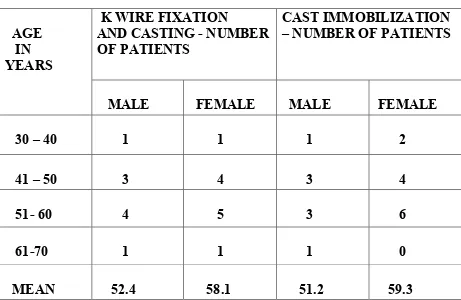
[image:53.595.73.530.496.670.2]Forty patients were enrolled in this study. Twenty patients treated with closed reduction and casting and twenty patients were treated with K wire fixation and casting. Among them seventeen were male and twenty three were female. The mean age for males was 51.8 years and 58.7 years for females. The dominant side was affected in 40% of K wire and casting group and 45% in cast immobilization group. Metaphyseal comminution was present in 57.5% patients.
Table 1. NUMBER OF PATIENTS
K WIRE FIXATION AND CASTING
CAST
IMMOBILZATION
MALE 9 8
FEMALE 11 12
SUM 20 20
45
Table 2. AGE OF PATIENTS
AGE IN YEARS
K WIRE FIXATION AND CASTING - NUMBER OF PATIENTS
CAST IMMOBILIZATION – NUMBER OF PATIENTS
MALE FEMALE MALE FEMALE 30 – 40 1 1 1 2 41 – 50 3 4 3 4 51- 60 4 5 3 6 61-70 1 1 1 0 MEAN 52.4 58.1 51.2 59.3
Table 3. SIDE OF INJURY
SIDE OF INJURY K WIRE FIXATION AND CASTING
(NO. OF PATIENTS )
CAST
IMMOBILIZATION
(NO. OF PATIENTS )
RIGHT 8 9
LEFT 12 11
%DOMINANT SIDE INJURY
[image:54.595.68.546.510.670.2]
46
Table 4. FRACTURE TYPE BY AO CLASSIFICATION
AO TYPE K WIRE
AND CASTING
(NO. OF PATIENTS )
CAST IMMOBILIZATION
(NO. OF PATIENTS AT)
A 2 6 8 A 3 12 11 C 1 2 1
The mechanism of injury by fall on outstretched hand was present in thirty five patients. Five patients sustained distal radius fracture in road traffic accidents.
All except two patients were treated on the same day of injury; two patient of K wire and casting group were treated on next day of injury.
47
developed tendonopathy that resolved with physiotherapy on follow-up at ninth week.
Seven patients (35%) of this group had finger stiffness at six week follow-up that gradually resolved by twelve weeks in five patients and at end twelve weeks only two patients had finger stiffness of mild degree. Two patients (10 %) had shoulder stiffness unrelated to operative procedure that gradually resolved by nine weeks with shoulder mobilization exercises. No patient of this group developed median nerve neuropathy or other complications viz; compartment syndrome, carpel tunnel syndrome.
In closed reduction and casting group major complications were finger stiffness in nine patients (45%) and tendonopathy in four patients (20%). At end of twelve weeks finger stiffness resolved in six patients out of nine patients and rest of three patients had finger stiffness of mild to moderate degree at end of twelve weeks. Among four patients of tendonopathy two patients got complete relief by ninth week and rest of two patients had no complaints by twelfth week.
48
Loss of follow-up in K wire and casting group was three patients and remaining seventeen patients were followed up to twelve weeks. In closed reduction and casting group loss of follow-up was three patients and rest of seventeen patients were followed up to twelve weeks.
49
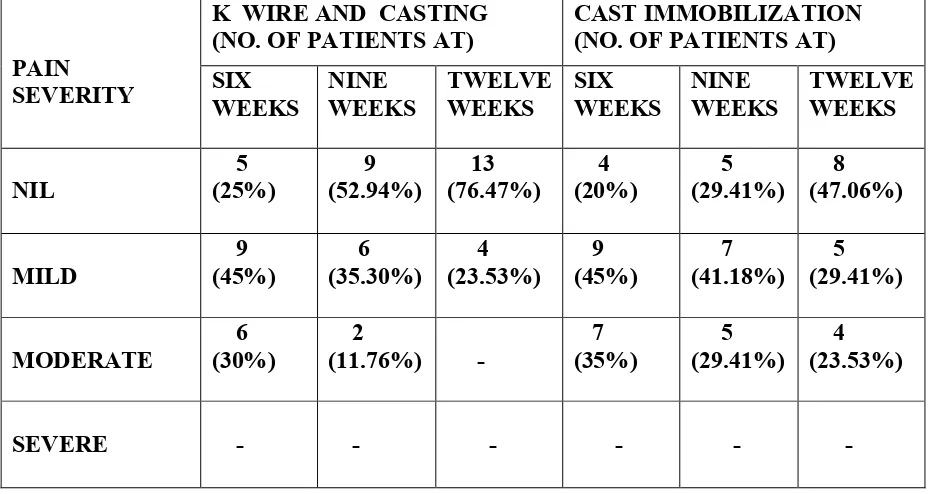
[image:58.595.66.532.276.523.2]At end of each of sixth, ninth and twelfth weeks pain over the affected side evaluated and categorised as nil, mild, moderate and severe according to patient’s response.
Table 5. PAIN
PAIN SEVERITY
K WIRE AND CASTING (NO. OF PATIENTS AT)
CAST IMMOBILIZATION (NO. OF PATIENTS AT) SIX WEEKS NINE WEEKS TWELVE WEEKS SIX WEEKS NINE WEEKS TWELVE WEEKS NIL 5 (25%) 9 (52.94%) 13 (76.47%) 4 (20%) 5 (29.41%) 8 (47.06%) MILD 9 (45%) 6 (35.30%) 4 (23.53%) 9 (45%) 7 (41.18%) 5 (29.41%) MODERATE 6 (30%) 2
(11.76%) -
50
Table 6. GRIP STRENGTH
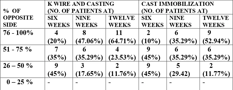
% OF OPPOSITE SIDE
K WIRE AND CASTING (NO. OF PATIENTS AT)
CAST IMMOBILIZATION (NO. OF PATIENTS AT) SIX WEEKS NINE WEEKS TWELVE WEEKS SIX WEEKS NINE WEEKS TWELVE WEEKS
76 - 100% 4 (20%) 8 (47.06%) 11 (64.71%) 2 (10%) 6 (35.29%) 9 (52.94%) 51 - 75 % 7
(35%) 6 (35.29%) 4 (23.53%) 9 (45%) 6 (35.29%) 6 (35.29%) 26 – 50 % 9
(45%) 3 (17.65%) 2 (11.76%) 9 (45%) 5 (29.42) 2 (11.77%)
0 – 25 % - - - -
Table 7. STIFFNESS
K WIRE FIAXTION AND CASTING
CAST
IMMOBILIZATION 6 WEEKS 7 (41.17%) 9 (52.94%)
12 WEEKS 2 (11.76%) 3 (17.64% )
51
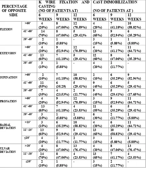
[image:60.595.72.548.206.761.2]The range of movements of palmar flxion, dorsi flexion, radial and ulnar deviation, supination and pronation were measured at end of 6th, 9th and 12th week and compared with opposite side.
Table 8. RANGE OF MOVEMENTS
PERCENTAGE OF OPPOSITE SIDE
K WIRE FIXATION AND CASTING
(NO OF PATIENTS AT)
CAST IMMOBILIZATION (NO OF PATIENTS AT ) 6 WEEKS 9 WEEKS 12 WEEKS 6 WEEKS 9 WEEKS 12 WEEKS FLEXION
˃60˚ 4 (20%) 8 (47.06%) 12 (70.59%) 4 (20%) 7 (41.18%) 10 (58.82%)
41˚-60˚ 14 (70%) 8 (47.06%) 5 (29.41)% 13 (65%) 9 (52.94%) 6 (35.29%)
20˚-40˚ 2
(10%) 1 (5.88%)
- 3
(15%) 1 (5.88%) 1 (5.88%) EXTENSION
˃60˚ 4
(20%) 9 (52.94%) 12 (70.59%) 4 (20%) 7 (41.17%) 11 (64.71%)
41˚-60˚ 13 (65%) 7 (41.18%) 5 (29.41%) 12 (60%) 8 (47.06%) 6 (35.29%)
20˚-40˚ 3 (15%)
1 (5.88%)
- 4
(20%) 2
(11.77%) -
SUPINATION
˃60˚ 2 (10%) 7 (41.18%) 10 (58.82%) 2 (10%) 6 (35.29%) 9 (52.94%)
41˚-60˚ 11 (55%) 6 (35.29) 5 (29.41%) 9 (45%) 6 (35.29%) 5 (29.41%)
20˚-40˚ 7
(35%) 4 (23.53%) 2 (11.77%) 9 (45%) 5 (29.41%) 3 (17.65%) PRONATION
˃60˚ 4 (20%) 9 (52.94%) 12 (70.59%) 3 (15%) 9 (52.94%) 11 (64.71%)
41˚-60˚ 13 (65%) 7 (41.18%) 4 (23.53%) 11 (55%) 6 (35.29%) 5 (29.41%)
20˚-40˚ 3 (15%) 1 (5.88%) 1 (5.88%) 6 (30%) 2 (11.77%) 1 (5.88%) RADIAL DEVIATION
˃15˚ 3
(15%) 6 (35.29%) 10 (58.82%) 4 (20%) 6 (35.29%) 11 (64.71%)
11˚-15˚ 13 (65%) 9 (52.94%) 5 (29.41%) 13 (65%) 10 (58.82%) 5 (29.41%)
≤10˚ 4 (20%) 2 (11.77%) 2 (11.77%) 3 (15%) 1 (5.88%) 1 (5.88%) ULNAR DEVIATION
˃20˚ 4
(20%) 8 (47.06%) 13 (76.47%) 4 (20%) 8 (47.06%) 13 (76.47%)
11˚-20˚ 14 (70%) 8 (47.06%) 4 (23.53%) 13 (65%) 7 (41.17%) 4 (23.53%)
≤10˚ 2 (10%)
1 (5.88%)
- 3
(15%) 2
52
Table 9. FUNCTIONAL STATUS AT 12 WEEKS
Functional status was evaluated whether patient is able to adapt to his or her activities both household and occupational.
K WIRE AND CASTING
CAST IMMOBILIZATION
REGULAR WORK
13 (76.47%) 10 (58.82%)
RESTRICTED WORK
3 (17.65%)
5 (29.41%)
UNABLE TO
WORK
1 (5.88 %) 2 (11.77%)
53
[image:62.595.70.529.232.434.2]In both groups both forearm with wrist x-rays taken and compared.
Table 10. K WIRE AND CASTING GROUP – RADIOLOGICAL
ASSESMENT
Average measurements Pre-operative (20 patients) Post-operative (20 patients) Nine weeks (17 patients) Twelve weeks (17 patients) RADIALLENGTH (mm) 4.1 10.7 10.2 9.8
VOLAR
TILT (˚) -23.3 +8.85 +6.9 +5.78
RADIAL
ANGULATION(˚) 11 19.15 18.02 17.44 ULNAR
VARIANCE(mm) +1.05 +0.4 +0.8 +1.02
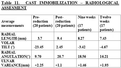
Table 11. CAST IMMOBILIZATION – RADIOLOGICAL
ASSESMENT
Average measurements Pre-reduction (20 patients) Post- reduction (20 patients) Nine weeks (17 patients) Twelve weeks (17 patients) RADIALLENGTH (mm) 3.7 9.4 8.27 7.43 VOLAR
TILT (˚) -23.45 2.45 -3.42 -4.67
RADIAL
ANGULATION(˚) 9.70 20.7 18.56 16.21 ULNAR
[image:62.595.67.530.487.746.2]54
[image:63.595.70.546.389.647.2]Wilcoxon Signed Ranks Test applied to assess radiological outcome. Both the treatment methods showed significant outcome at the time of post-intervention. While comparing the radiological parameters at 9th and 12th weeks both groups have non-significant changes in radiological parameters.
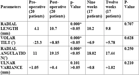
Table 12. K WIRE AND CASTING GROUP – RADIOLOGICAL
ASSESMENT
Parameters Pre-operative (20 patients) Post-operative (20 patients) p-Value Nine weeks (17 patients) Twelve weeks (17 patients) P-Value RADIAL LENGTH (mm)4.1 10.7
0.000*
<0.05 10.2 9.8
0.707
VOLAR
TILT (˚) -23.3 +8.85
0.000*
<0.05 +6.9 +5.78
0.628 RADIAL
ANGULATIO N(˚)
11 19.15
0.000*
<0.05 18.02 17.44
0.250
ULNAR VARIANCE (mm)
+1.05 +0.4
0.101
>0.05 +0.8 +1.02
0.210
55
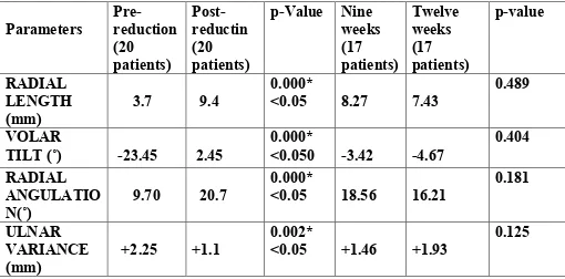
Table 13. CAST IMMOBILIZATION – RADIOLOGICAL
ASSESMENT
Parameters Pre-reduction (20 patients) Post- reductin (20 patients)p-Value Nine weeks (17 patients) Twelve weeks (17 patients) p-value RADIAL LENGTH (mm)
3.7 9.4
0.000*
<0.05 8.27 7.43
0.489
VOLAR
TILT (˚) -23.45 2.45
0.000*
<0.050 -3.42 -4.67
0.404 RADIAL
ANGULATIO N(˚)
9.70 20.7
0.000*
<0.05 18.56 16.21
0.181
ULNAR VARIANCE (mm)
+2.25 +1.1
0.002*
<0.05 +1.46 +1.93
0.125
[image:64.595.67.577.163.415.2]56
[image:65.595.67.528.501.732.2]Anatomical assessment was done according to Lindstrom and Frykman criteria. In K wire group five (29.41%) patients had grade I i.e. no deformity as compared to two (11.76%) patients in cast immobilization group. In K wire group majority patients i.e. ten (58.83%) had grade II outcome i.e. mild deformity. In cast immobilization group grade II (mild deformity) and grade III (moderate deformity) outcome was observed in equal number of patients i.e. six (35.29%). Grade IV outcome i.e. severe deformity was present in three (17.66%) patients of cast immobilization as compared to none of patient had severe deformity in K wire fixation group.
Table 14. ANATOMICAL OUTCOME (AT 12
thWEEK)
LINDSTROM AND FRYKMAN GRADING
K WIRE FIXATION AND CASTING
CAST IMMOBILIZATION
Number Of
Patients Percentage
Number Of
Patients Percentage
GRADE I 5 29.41 % 2 11.76 % GRADE II 10 58.83 % 6 35.29 % GRADE III 2 11.76 %
57
[image:66.595.60.530.489.654.2]The functional outcome was evaluated at end of nine and twelve weeks with Disability of Arm Shoulder and Hand (DASH) scoring. This system consists of thirty set of questionnaire for subjective evaluation including activities of daily living. By this system average scores were calculated and were 45.31 for K wire and casting group at nine weeks as compared to 46.67 for cast immobilization group. At end of twelve weeks scores were 22.74 and 23.40 respectively for K wire and casting and cast immobilization groups.
Table 15 . FUNCTIONAL ASSESSMENT
DISABILITY OF ARM, SHOULDER AND HAND (DASH) SCORING
AVERAGE DASH
SCORE AT END OF
K WIRE FIXATION AND CASTING
(17 patients )
CAST
IMMOBILIZATION (17 patients )
9 WEEKS 45.31 46.67
12 WEEKS 22.74 23.40
*
Higher scores indicates poorer outcome58
[image:67.595.66.532.472.713.2]
Mann-Whitney test was used for statistical evaluation of functional outcome. With this test mean DASH scores were compared in two groups at the end of nine and twelve weeks. At end of nine weeks p value was 0.593 (˃0.005) and at end of twelve weeks p value was 0.877 (˃0.005) indicating no significant difference between two groups in terms of functional outcome.
Table 16 . FUNCTIONAL ASSESSMENT
DISABILITY OF ARM, SHOULDER AND HAND (DASH) SCORING (Mann-Whitney Test applied)
MEAN DASH SCORE AT END OF
K WIRE
FIXATION AND CASTING
(Mean Rank) N=17
CAST
IMMOBILIZATION (Mean Rank) N=17
p- Value
9 WEEKS 16.59 18.41 0.593
CASE ILLUSTRATIONS
CASE 1 – K WIRE WITH CASTING
59
CASE ILLUSTRATIONS
K WIRE WITH CASTING
PREOPERATIVE
POSTOPERATIVE
9 WEEKS POST-OP
12 WEEKS POST
60
12 WEEKS POST-OP (Comparison with normal side)
RANGE OF MOVEMENTS
CASE 2 - K WIRE AND CASTING
61
K WIRE AND CASTING
PREOPERATIVE
POSTOPERATIVE
9 WEEKS POSTOP
12 WEEKS POSTOP (Comparison with normal side )
RANGE OF MOVEMENTS
62
12 WEEKS POSTOP (Comparison with normal side )
RANGE OF MOVEMENTS
CASE 3 – K WIRE FIXATION AND CASTING
PREOPERATIVE
POSTOPERATIVE
9 WEEKS POST OP
63
WIRE FIXATION AND CASTING
PREOPERATIVE
POSTOPERATIVE
9 WEEKS POST OP
12 WEEKS POSTOP (Comparison with normal side )
RANGE OF MOVEMENTS
64
12 WEEKS POSTOP (Comparison with normal side )
RANGE OF MOVEMENTS
CASE 1. CLOSED REDUCTION AND CASTING
PREREDUCTION
POSTREDUCTION
9 WEEKS POSTREDUCTION
65
CASE 1. CLOSED REDUCTION AND CASTING
PREREDUCTION
POSTREDUCTION
9 WEEKS POSTREDUCTION
PREREDUCTION
12 WEEKS POSTREDUCTION (comparison with normal side )
RANGE OF MOVEMENTS
66
12 WEEKS POSTREDUCTION (comparison with normal side )
RANGE OF MOVEMENTS
CASE 2 –CLOSED REDUCTION AND CASTING
9 WEEKS POSTREDUCTION
67
CLOSED REDUCTION AND CASTING
PREREDUCTIONPOSTREDUCITON
9 WEEKS POSTREDUCTION
12 WEEKS POSTREDUCTION (comparison wit
RANGE OF MOVEMENTS
68
12 WEEKS POSTREDUCTION (comparison wit
RANGE OF MOVEMENTS
CASE 3 – CLOSED REDUCTION WITH CASTIN
PREREDUCTION
POSTREDUCTION
9 WEEKS POSTREDUCTION
69
CLOSED REDUCTION WITH CASTING
PREREDUCTION
POSTREDUCTION
9 WEEKS POSTREDUCTION
70
12 WEEKS POSTREDUCTION
RANGE OF MOVEMENTS
71
DISCUSSION
In this study, forty patients with unstable fractures of distal radius treated with K wire fixation and casting and closed reduction and casting were analysed in terms of functional and radiological outcome.
On functional analysis based upon Disability of Arm, Shoulder and Hand (DASH) scoring system the K wire fixation and casting group had lower mean scores compared with closed reduction and casting group both at nine (16.59 compared with 18.41 with p value 0.593) and at twelve weeks (17.24 compared with 17.76 with p value 0.877) indicating no significant difference in terms of functional outcome; though lower scores indicates better outcome considered on individual basis. At end of twelfth week post-intervention thirteen patients (76.87%) of K wire and casting group resumed to their regular work as compared to ten (58.82%) patients of closed reduction and casting group.
72
group grade II (mild deformity) and grade III (moderate deformity) outcome was observed in equal number of patients i.e. six (35.29%). Grade IV outcome i.e. severe deformity was present in three (17.66%) patients of cast immobilization as compared to none of patient had severe deformity in K wire fixation group.
On radiological assessment both group of patients had significant improvement in radiological criteria viz; Radial Length, Volar tilt, Radial Inclination and Ulnar variance post-intervention (with p values for each of parameters <0.005). Also there was no significant changes in all these parameters between ninth and twelfth weeks post-intervention in both of these groups; although changes in K wire and casting group were lower as compared with closed reduction and casting group
In K wire and casting group two patients (10%) developed pin site infection. In one patient infection resolved with antibiotics without further sequelae. In other patient infection got deeper necessitating K wire removal at third week.
73
Thus both types of intervention produced statistically similar results in terms of functional outcome according to DASH scoring system. Also the changes in radiological parameters were non-significant between ninth and twelfth weeks post-intervention in both groups of patients.
T. Azzopardi and et al in their study observed that functional outcomes were not significant in K wire as compared with closed reduction and casting group. Anatomically supplementary fixation by K wire was only marginally superior to cast immobilization alone in reducing displacement of the fracture after closed manipulation.
Restoration of normal anatomy is crucial for restoration of function. Normally 82% of the compressive load at wrist joint is borne by distal radius and remaining by distal ulna. 2.5 mm loss in radial length results in 42% load on ulna and with 20 degree of dorsal angulation, ulna bears 50% of load.
74
Radiocarpal articular congruity remains the most clinically significant radiographic parameter regarding both functional outcome and future degenerative changes
.
If 2 mm of incongruity were present, there was a 100% incidence of degenerative changes on plain x-rays.
Residual dorsal angulation can precipitate ulnar impaction, midcarpal instability and altered stress concentration which may lead to early arthritis. Gartland and Werley concluded that residual dorsal tilt has a more direct effect on outcome than residual radial deviation, radial shortening, or loss of integrity of the radioulnar joint. Porter, in his study, felt that loss of function did not occur until at least 20 degrees of palmar tilt was lost.
In both treatment methods radial length, ulnar varience, radial angulation are restored to near normal but correction of dorsal tilt is not complete. This is because the fact that volar ligaments are stronger on distraction before relatively ‘Z’ oriented dorsal ligaments. So, on distraction volar cortex is brought out to length before dorsal cortex preventing full correction of dorsal tilt.
75
76
CONCLUSION
Fractures of distal radius are common and appear simple, affect the function of wrist and hand considerably. It is most common fracture encountered in outpatient department. Majority of these fractures are unstable resulting in loss of reduction and hence malunion, poor range of motion and early arthritis, altered wrist kinematics and early arthritis. For better outcome of management the goals of treatment are
1. To achieve perfect anatomical reduction and maintain the reduction till union
2. Early mobilisation to achieve good range of movements and to prevent stiffness.
3. To prevent early and late complication
77
BIBLIOGRAPHY
1) Clancey G. Percutaneous Kirschner-wire fixation of Colles fractures: a prospectivestudy of thirty cases.J Bone Joint Surg [Am]1984;66-A:1008-14.
2) Mah ET, Atkinson RN.Percutaneous Kirschner wire stabilisation following closed reduction of Colles’ fractures.J Hand Surg [Am] 3) Rayhack JM. The history and evolution of percutaneous pinning of
displaced distal radius fractures. Orthop Clin North Am 1993;24:287-300 4) Green DP. Pins and plaster treatment of comminuted fractures of the
distal end of the radius. J Bone Joint Surg [Am] 1975;57-A:304-10
5) Weber SC, Szabo RM. Severely comminuted distal radial fracture as an unsolved problem: complications associated with external fixation and pins and plaster techniques. J Hand Surg [Am] 1986;11:157-65.
6) Frykman G. Fractures of the distal radius including sequelae-
shoulder-hand-finger syndrome, disturbance in the distal radio-ulnar joint and impairment of nerve function: a clinical and experimental study. Acta Orthop Scand 1967;Suppl 108:3.
78
8) Rodriguez-Merchan EC. Plaster cast versus percutaneous pin fixation for comminuted fractures of the distal radius in patients between 46 and 65 years of age.J Orthop Trauma 1997;11:212-17.
9) Naidu SH, Capo JT, Moulton M, Ciccone W 2nd, Radin A. Percutaneous pinning of distal radius fractures: a biomechanical study. J Hand Surg [Am] 1997;22:252-7.
10) Jesse B, Jupiter MD. Complex Articular Fractures of the Distal Radius: Classification and Management. J Am Acad Orthop
Surg 1997; 5: 119-29.
11) Knirk JL, Jupiter JB. Intra-articular fractures of the distal end of the radius in young adults. J Bone Joint Surg 1986; 68: 647-59.
12) Wong TC, Chiu Y. Casting Versus Percutaneous Pinning for Extra-ArticularFractures of the Distal Radius in an Elderly Chinese Population: A Prospective Randomized Controlled Trial. J Hand Surg 35E:202-208, 2010.
13) Hudak PL, Amadio PC, Bombardier C. Development of an upper extremity outcome measure: the DASH (disabilities of the arm, shoulder and hand) [corrected]. The Upper Extremity Collaborative Group(UECG) Am J Ind Med. 1996 Jun;29(6):602-8. Erratum in: Am J Ind Med 1996 Sep0(3):372.
79
Bone and Joint Surgery (Br); 1988; 70-B, 649- 651.
15) Diego L.Fernandez and Andrew K. Palmer: Fractures of the distal radius – Green’s Operative hand surgery. 929-989. 16) Mark S. Cohen, Robert Y.McMurtry and Jesse B.Jupiter:
Fractures of the distal radius – Skeletal Trauma. 1315-1361. 17) A.Sarmiento, G.W Pratt, N.C Berry and W.F Sinclair: Colles’ fracture, functional bracing in supination - Journal of
Bone and Joint Surgery (Am); 1975; 57, 311-317.
18) W.Van der Linden and R. Ericsson: Colles fracture. How should its displacement be measured and how should it be
immobilized?- Journal of Bone and Joint Surgery (Am) 1981;63, 1285-1288.
19) J.B.Jupiter: Current concepts review – Fractures of distal end of radius. Journal of Bone and Joint Surgery (Am) 1991; 73,
461-469.
20) T. Azzopardi, S. Ehrendorfer, T. Coulton, M. Abela Unstable extra-articular fractures of the distal radius a prospective, randomised study ofimmobilisation in a cast versus supplementary percutaneous pinning. J Bone Joint Surg Br June 2005 vol. 87-B no. 6 837-840
APPENDIX I
DISABILITY OF ARM, SHOULD
SCORING TO EVALAUATE FUNCTIONAL OUTCOME
80
APPENDIX I
DISABILITY OF ARM, SHOULDER AND HAND (DASH) SCORING TO EVALAUATE FUNCTIONAL OUTCOME
82
APPENDIX II
LINDSTROM AND FRYKMAN GRADING FOR ANANTOMICAL OUTCOME
DEFORMITY GRADE
DORSAL
ANGULATION
RADIAL
SHORTENING
GRADE I (No significant deformity)
Not exceeding
neutral < 3 mm
GRADE II (Mild deformity)
1-10˚ 3 - 6 mm
GRADE III (Moderate deformity)
11-14˚ 7 - 11 mm
GRADE IV (Severe deformity)
83
APPENDIX III
CONSENT FORM ÑV Jl×Rp T¥Ym
U¦dLhÓ GÛm× Ø±®tÏ Lm© ùTôÚj§ AßûY £¡fûN ùNnRYu TXuLs Utßm ®û[ÜLs
BWônf£ ¨ûXVm : AWÑ vPôu UÚjÕYUû]
ùNuû] - 600 001.
TeÏ ùTßm úSôVô°«u ùTVo :
YVÕ :
Bi/ùTi :
TeÏ ùTßTY¬u Gi :
¸rLiP Yt±p TeÏ ùTßTYo ()ϱdLÜm
1. úUúX ϱl©PlThÓs[ UÚjÕY Bn®u ®YWeLs
G]dÏ GuàûPV RônùUô¯«p ®[dLlThPÕ.
GuàûPV NkúRLeLû[ úLhLÜm, ARtLô] RÏkR ®[dLeLû[ ùT\Üm YônlT°dLlThPÕ.
2. Sôu CqYôn®p Ru²fûNVôL TeúLt¡ú\u. GkR LôWQj§]ôÛm GkRd LhPj§Ûm Gq®Rf NhP £dLÛdÏm EhTPôUp Sôu CqYôn®p CÚkÕ ®X¡d ùLôs[Xôm GuTûR A±kÕ ùLôiúPu.
84
4. CkR Bn®p Sôu TeÏùT\ Jl×d ùLôs¡ú\u. G]dÏ ùLôÓdLlThP A±ÜûWL°uT¥ SPkÕ ùLôsYÕPu CkR BnûY úUtùLôsÞm UÚjÕY A¦dÏ EiûUÙPu CÚlúTu Gußm Eߧ A°d¡ú\u.
5. GuàûPV EPp ¨ûX Tô§dLlThPôúXô ApXÕ G§oTôWôR YZdLj§tÏ Uô\ô] úSônϱ ùRuThPôúXô EPú] ARû] UÚjÕY A¦dÏ ùR¬®lúTu G] Eߧ A°d¡ú\u.
6. AßûY£¡fûNdÏ Øu]Úm, AßûY £¡fûN«u
úTôÕm, ARtÏ ©u]Úm CWjRl T¬úNôRû]Ls,
FÓL§oLs (X-Ray) GÓdLÜm, UVdLUÚkÕ ùLôÓjÕ
AßûY£¡fûN êXm U¦dLhÓ GÛm× Ø±®tÏ Lm© ùTôÚjRÜm NmURm A°d¡ú\u.
7. Gu SXu LÚ§úV CkR BnÜ úUtùLôs[lTÓ¡\Õ Guß ùR¬kÕ CkR Bn®tÏ Jl×Rp A°d¡ú\u.
Sôs :
CPm :
TeúLtTY¬u ûLùVôlTm ApXÕ
CPÕ ûL ùTÚ®Wp úWûL (CkR T¥Ym T¥jÕ LôhPlThÓ ×¬kÕ
ûLúWûL A°d¡uú\u)
TeúLtTY¬u ùTVo Utßm ®XôNm
---
85
úSôVô° RLYp T¥Ym
U¦dLhÓ GÛm× Ø±®tÏ Lm© ùTôÚj§ AßûY £¡fûN ùNnRYu TXuLs Utßm ®û[ÜLs
úSôVô°dLô] RLYpLs
BWônf£«u úSôdLØm BRôWeLÞm
CkR Bn®u êXm U¦dLhÓ GÛm× Ø±®tÏ Lm© ùTôÚj§ AßûY £¡fûN ùNnRYu TXuLs Utßm ®û[ÜLs LiP±VlTÓm.
A[ÅÓLs
FÓL§o (X-Ray) TPj§p AßûY £¡fûNdÏl©u EûPkR GÛm×
Ju\ôL úNÚRp.
AßûY £¡fûNdÏl©u Gq®R ETLWQj§u ER®Ùªu± Ïß¡V LôXj§p ûL ØÝûUVôL ÏQUûPRp.
BnÜ Øû\Ls
AßûY £¡fûN êXm Lm© êXm GÛm× Ø±ûY N¬ ùN