Variation in Adenovirus Transgene Expression between BALB/c and
C57BL/6 Mice Is Associated with Differences in Interleukin-12
and Gamma Interferon Production and NK Cell Activation
YUFENG PENG,1ERIK FALCK-PEDERSEN,2ANDKEITH B. ELKON1*
Departments of Medicine1and Microbiology and Immunology,2Weill Medical College
of Cornell University, New York, New York 10021
Received 30 October 2000/Accepted 13 February 2001
The innate immune response against replication-defective adenoviruses (Ad) is poorly defined. We and others have previously observed striking differences in the rate at which the Ad vector itself or the virus encoding a variety of transgenes is eliminated in different mouse strains. Here, we report that Ad infection of BALB/ mice is associated with sixfold-higher levels of serum alanine aminotransferase and that Ad transgenes induce two- to threefold-higher levels of intrahepatic NK cells and NK activity compared to C57BL/6 mice. The
increase in NK activation in BALB/c mice was associated with⬃4-fold higher level of mRNA expression of a
newly described NKG2 receptor activator, H-60, as well as increased expression of interleukin-12 and gamma interferon mRNAs in BALB/c mice compared to C57BL/6 mice. NK depletion in BALB/c mice or defective NK function in C3H beige mice extended transgene expression compared to their appropriate controls, and attenuation of NK together with CD8 T-cell function had a synergistic effect. These findings indicate that there are intrinsic differences in the innate immune responses of different mouse strains to Ad and Ad transgenes and that NK cells, in cooperation with CD8 T cells, play a pivotal role in the early extinction of transgene expression in BALB/c mice.
It is now well established that the major obstacle to adeno-virus (Ad)-mediated gene therapy is the induction of a robust immune response to, and elimination of, virus-infected cells (reviewed in reference 34). Whereas the role of conventional lymphocyte subsets has been fairly extensively evaluated (36), little attention has been paid to the innate immune response to the replication-defective virus. Early responses are particularly important to define in view of the potential for high-dose Ad gene therapy to cause mortality (14).
Based on the evidence that CD8⫹T cells play a dominant
role in elimination of replication-defective Ad (36), we re-cently demonstrated that an Ad-encoded soluble CD8 (sCD8) protein could prolong transgene expression in C57BL/6 (B6) and C3H mice (20). Unlike these two strains of mice however, BALB/c mice rapidly eliminated the Ad vector expressing sCD8. Paradoxically, in vitro studies revealed that splenic T cells obtained from BALB/c mice were less activated and ex-hibited less antigen-specific cytotoxicity after restimulation compared to B6 mice (20). This finding suggested that the rapid extinction of transgene expression was most likely facil-itated by non-CD8 effector cell(s).
Most virus infections of mammalian cells activate natural killer (NK) cells (2, 16) through modulation of major histo-compatibility complex class I on the surface of infected cells and/or by the release of cytokines such as interleukin-12 (IL-12), IL-18, and gamma interferon (IFN-␥) (2). Very recently, ligands responsible for activating NK cells through the lectin-like NKG2 receptor have been identified, but their roles in
virus infections have not been delineated (3). Once triggered, NK cells secrete large amounts of IFN-␥, a cytokine that not only amplifies the innate immune response of NK cells and macrophages but also activates CD8⫹T cells (2). To determine
whether NK cells contributed to the innate immune response to Ad vectors and could explain the rapidity of transgene silencing in BALB/c mice, we evaluated the early cytokine response and the role of NK cells in Ad-infected mice. The results of these studies indicate that BALB/c mice have per-sistent IL-12 expression associated with enhanced NK activity.
MATERIALS AND METHODS
Mice.C57BL/6 (H2b), BALB/c/J (H2d), C3H/HeJ (H2k), and beige C3H/HeJ (H2k) mice and IL-12 p35-deficient mice in the BALB/c/J (H2d) background were purchased from Jackson Laboratory, Bar Harbor, Maine. BALB/c.SCID mice were bred and maintained at the Hospital for Special Surgery in a patho-gen-free environment.
Ad-encoded transgenes.All viruses used in this study, including Ad null, lacked E1 and E3 (Ad type 5) and were replication defective (20). Ad type 5 expressing the ectodomain of murine CD8-␣(Ly2.1) or chloramphenicol acetyl-transferase (CAT) (AdsCD8 or AdCAT) was produced as described elsewhere (20). Briefly, the pAd vector containing the transgene was cotransfected with PJM17 into 293 cells. The cell lysate was used to infect 293 cells. Viral DNA was extracted by a modified Hirt assay, and recombination was verified by restriction enzyme digestion, PCR, and protein production (see below). The viruses were further plaque purified. Each clone was rescreened as above. Finally, large-scale virus was purified by two-step CsCl concentration and stored in glycerol at⫺20°C or sucrose at⫺70°C. Quantitation of viral particles was measured at optical density at (OD) 260 nm.
Injection of mice with Ad vectors and evaluation of virus expression.Mice were injected with 2.0⫻1010particles of virus by the intravenous route unless
indicated otherwise and bled at 7- or 14-day intervals. Persistence of transgene expression was determined by detection of the transgene product secreted into the serum (sCD8) or by protein expression in the liver (CAT) as described previously (21). In brief, CAT activity in liver homogenates was quantified by thin-layer chromatography of14C-labeled substrate (21), and secreted transgene
products by were quantitated by enzyme-linked immunosorbent assay (ELISA)
* Corresponding author. Mailing address: Research Division, Hos-pital for Special Surgery-Weill Medical College of Cornell University, 535 E. 70th St., New York, NY 10021. Phone: (212) 606-1409. Fax: (212) 774-2337. E-mail: elkonk@hss.edu.
4540
on November 9, 2019 by guest
http://jvi.asm.org/
as described below. To verify active transcription of transgenes, we isolated total RNA from liver homogenates and performed PCR amplification of the trans-gene with primers (5⬘to 3⬘) that were specific for Ad sequences immediately flanking the transgenes (CCCAGGTCCAACTGCAGCCC and GGTACTTGT GAGCCAAGGCAG). The amplification conditions were 35 cycles of 95°C for 30 s, 55°C for 30 s, and 72°C for 50 s. PCR of-actin with the primers CCCT GGCTGCCTCAACACC and GAGCAAACATCCCCCAAAGT was used as a control.
Antibodies and flow cytometry analysis.Flow cytometry analysis was per-formed using a FACScan with CellQuest software (Becton Dickinson, Mountain View, Calif.) as described previously (21). Monoclonal antibodies against the following antigens were used for staining: CD4 (PharMingen, San Diego, Calif.); CD8 (PharMingen); and DX5 (PharMingen), a pan-NK cell marker.
Isolation of IHL.Intrahepatic lymphocytes (IHL) were isolated as described by Watanabe et al. (33). Briefly, livers from day 10-infected mice were perfused with 10 ml of Hanks buffered salt solution and cut into small pieces. These small pieces were forced through a metal mesh. The liver slurry was digested with liver digestion medium (Gibco) at 37°C for 40 min followed by washing. The cell pellet was resuspended in 35 ml (two livers) of medium (RPMI 1640 supplemented with 10% fetal calf serum) and centrifuged through 15 ml of Ficoll (Amersham Pharmacia Ficoll-Hypaque). IHL were collected at the interface, washed, and used for phenotypic or functional assays.
In vivo and in vitro cytotoxicity assays.The serum levels of glutamic pyruvic transaminase/alanine aminotransferase (ALT), a marker of hepatocyte injury in vivo, was quantified using a commercial diagnostic kit (Sigma) (26). Ex vivo CD8 T-cell cytotoxicity was performed using the appropriate major histocompatibility complex-matched cells (C57SV,H2bfor B6; SVBALB,H2dfor BALB/c [28]) as targets. The target cells were labeled with [3H]thymidine (5Ci/ml) (15) and
infected with AdCAT (multiplicity of infection of 100). The next day, the labeled infected cells were used as targets for cytotoxic T lymphocytes (CTI) at different effector/target (E/T) ratios. Lysis was allowed to proceed for 5 h, after which the plates were extensively washed and the remaining cells were detached with trypsin-EDTA (Gibco). The cells were harvested and counted in a Microbeta Trilux scintillation counter (Wallac, Gaithersburg, Md.). The percentage of spe-cific lysis was calculated from the formula 100⫻[(S⫺E)/S], whereSandEare the spontaneous and experimental counts per minute, respectively. Standard deviations (SD) were derived from triplicates within experiments.
NK cell cytotoxicity was evaluated by a Cr release assay. Spleen cells or IHL were incubated with51Cr-labeled YAC-1 cells at various E/T ratios, and release
of Cr was quantified on a␥counter. The percentage of lysis was calculated according to the formula [(cpm sample⫺cpm spontaneous)/(cpm maximum⫺ cpm spontaneous)]⫻100. As a control for specificity, infected mice were pre-treated with anti-asialo GM1 antibody (anti-GM1) to deplete NK cell activity (see below).
ELISAs.CD8 levels in mouse serum were quantified by sandwich ELISAs using two different monoclonal antibodies to the mouse CD8␣chain, TIB 105 hybridoma (American Type Culture Collection) and biotinylated mouse YST 169 (CALTAG Laboratories, Burlingame, Calif.). ELISA plates were coated overnight with TIB 105 (5g/ml) at 4°C. The plates were blocked with phos-phate-buffered saline–3% bovine serum albumin for 1 h at room temperature and then incubated with the 1/3-diluted serum sample for 4 to 5 h in room temperature. The plates were washed and sequentially incubated with biotinyl-ated secondary antibody, avidin-alkaline phosphatase, and substrate. The OD was read at 405 nm. The level of expression of each protein in the serum was normalized to the peak level of the corresponding protein in SCID mouse serum as calculated by relative expression (OD of test sample/OD of SCID serum). IFN-␥production by day 7 AdCAT-infected mice was quantified following in-cubation of IHL with YAC-1 cells at a ratio of 1:10 at 37°C for 24 h. In some cases, IL-12 (10 ng/ml) was added. IFN-␥in the supernatant was quantified using a sandwich ELISA (PharMingen) with R4-6A2 as capture antibody, biotinylated XMG1.2 as detecting antibody, and purified recombinant IFN-␥(Gibco BRL, Grand Island, N.Y.) as a standard.
Cytokine mRNA expression.To compare the cytokine profiles induced by Ad vector infection, spleen and liver RNA samples were collected at days 0, 3, and 7 postinfection. Liver (100g) or spleen (30g) RNA was hybridized with in vitro-transcribed MCK-2b template (PharMingen). The RNase protection assay was performed according to the manufacturer’s instructions. Semiquantitative assessment of IFN-␥, H-60, and IL-12 p35 and p40 subunit expression was performed using the following primer pairs: IFN-␥(5⬘33⬘), CATGGCTGTTT CTGGCTGTTACTG and TTGGCGCTGGACCTGTGG (94°C for 30 s, 65°C for 45 s, and 72°C for 1 min; 30 cycles); H-60 (5⬘33⬘), TGTGCTGATTTGTC CCAAAA and CCGGCACCTTTAATGTTGAT (94°C for 30 s, 55°C for 45 s, and 72°C for 1 min; 30 cycles); p35 (5⬘33⬘), TGCCAGGTGTCTTAGCCAGG
TCC and CGCAGAGTCTCGCCATTAT (94°C for 30 s, 62°C for 45 s, and 72°C for 1 min; 35 cycles); p40 ACCTGTGACACGCCTGAAGA and TGATGATG TCCCTGATGAGAAGC (94°C for 30 s, 62°C for 45 s, and 72°C for 1 min; 32 cycles). mRNA expression was quantified by densitometry.
Depletion and repletion of NK cells.Depletion of NK cells was performed as instructed by the manufacturer. Briefly, 25l of anti-GM1 (Wako Chemical USA, Richmond, Va.) was injected every 4 to 5 days intravenously, starting at day⫺1 for 2 weeks. In vitro depletion of NK cells from SCID splenocytes was achieved by incubating anti-GM1 with Low-Tox-M rabbit complement (Cedar-lane). Rabbit immunoglobulin G (IgG) was used in the control group.
Statistical analysis.Test samples were analyzed for normal distribution and then compared by either Student’sttest (normal distribution) or the Mann-Whitney rank sum test (nonparametric data).
RESULTS
BALB/c mice mount a greater NK cell response to Ad
trans-genes compared to B6 mice.To determine whether differences
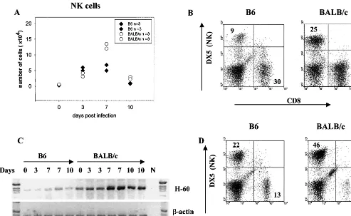
in NK activity could explain the variable strain responses, we injected B6 and BALB/c mice with AdCAT and harvested IHL at day 0 and at days 3, 7, and 10 postinfection. Whereas the percentage and absolute number of NK cells were similar in B6 and BALB/c mice at days 0 and 3, BALB/c mice had a signif-icantly higher number (Fig. 1A) and proportion (Fig. 1B) of NK cells at day 7 postinfection. Very recently, the ligands for the murine NKG2 receptor have been identified as the minor histocompatibility antigens H-60 and Rael (3). To determine whether the NK-activating ligand H-60 was increased in the liver following AdCAT infection, we performed reverse tran-scription-PCR (RT-PCR) analysis of liver lysates. As shown in Fig. 1C, the peak expression of NK infiltration corresponded with an⬃4-fold increase in the mRNA expression of H-60 in BALB/c but not B6 mice. To exclude the possibility that the NK response was transgene specific, B6 and BALB/c mice were injected with the Ad null vector and NK cells were eval-uated at days 3 and 7. As shown in Fig. 1D, the proportion of NK cells was twofold higher in BALB/c mice but peaked at day 3 rather than day 7. The reason for the difference in the kinetics of NK responses is uncertain but presumably reflects modulation of NK activators by the expression of the trans-gene.
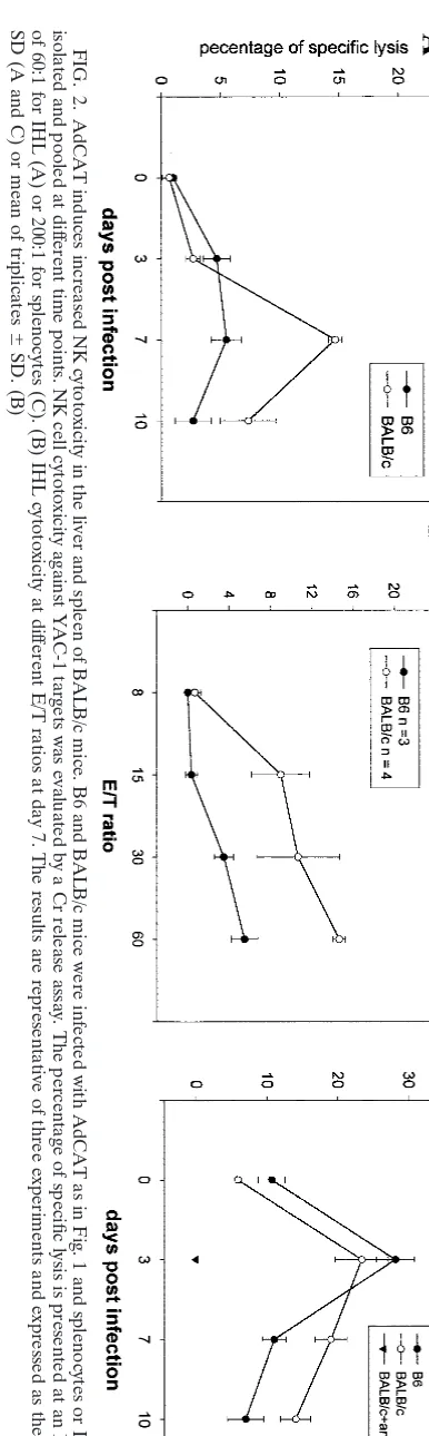
To determine whether the higher number of intrahepatic NK cells in BALB/c mice was associated with increased NK activity, we compared NK-mediated cytotoxicity against the target, YAC-1, at various time points after infection. As shown in Fig. 2A, AdCAT infection induced comparable NK cell cytotoxicity at day 3, whereas the activity at day 7 was signifi-cantly higher in BALB/c-derived than in B6-derived IHL. A representative example of IHL cytotoxicity at day 7 is shown in Fig. 2B. A similar increase in NK activity in BALB/c compared to B6 mice was observed in splenic lymphocytes, whereas little to no cytotoxic activity against this target was observed in BALB/c mice treated with anti-GM1 (Fig. 2C). Since Liu et al. (11) reported that hepatic enzymes such as ALT reflect NK-induced hepatocyte injury, we quantified serum ALT in Ad null-infected mice. Whereas ALT was close to baseline in both B6 and BALB/c mice at day 3, BALB/c mice had ⬃6-fold higher serum ALT at day 7 compared to B6 mice (390⫾107 versus 59 ⫾ 3 U/ml [n ⫽ 6]). These findings indicate that BALB/c mount a more intense NK response to Ad and Ad-CAT compared to B6 mice.
on November 9, 2019 by guest
http://jvi.asm.org/
NK depletion or an intrinsic deficiency in NK cell function
is associated with extended transgene expression.To directly
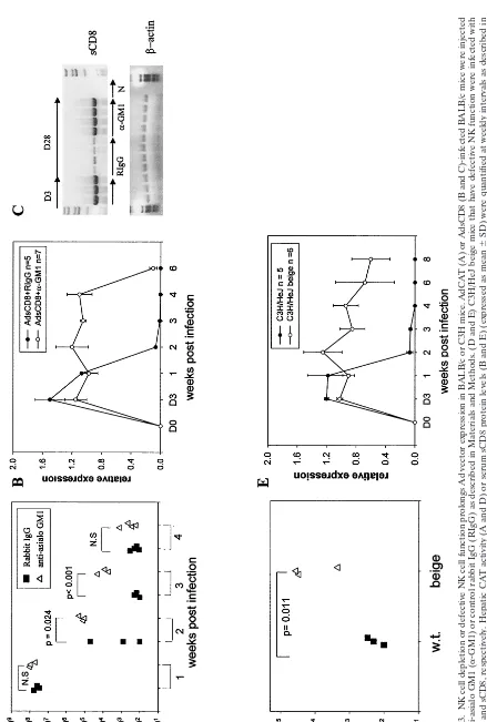
address the role of NK cell activation in the response to Ad vectors, BALB/c mice were depleted of NK cells with anti-GM1 and infected with AdCAT or AdsCD8, and expression of the appropriate transgene product was quantified at weekly intervals. As shown in Fig. 3A to C, CAT activity and serum sCD8 protein and mRNA levels were significantly higher in NK-depleted compared to control mice injected with normal rabbit IgG at the times indicated. However, transgene expres-sion was extinguished between weeks 4 to 6 postinfection, the time at which the rabbit antibody could no longer be detected in the circulation (not shown).
C3H/HeJ beige (beige) mice have a spontaneous mutation in theLystgene leading to defective NK cell cytotoxicity (24). To determine whether NK cells also participate in the innate immune response to Ad vectors in C3H mice, we infected beige mice with AdCAT or AdsCD8 and quantified transgene expression as in BALB/c mice. CAT expression was 10- to 100-fold higher in beige mice than in strain-matched controls at 4 weeks postinfection (Fig. 3D), and sCD8 levels were sig-nificantly higher than in wild-type controls up to 8 weeks
postinfection (Fig. 3E). These findings indicate that NK cells contribute to the attenuation of transgene expression in Ad vector-infected cells in both BALB/c and C3H mice and that sCD8 was expressed longer than AdCAT.
Synergistic effect of CD8ⴙand NK cell inhibition in BALB/c
and C3H mice. The longer duration of expression of sCD8
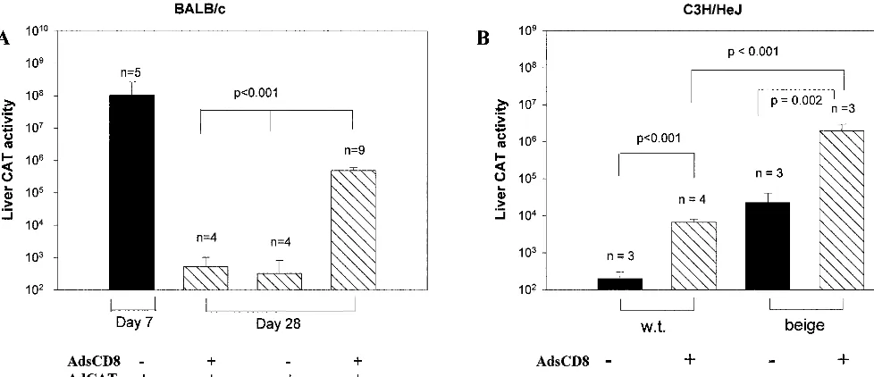
compared to CAT (Fig. 3) could be explained by a more potent immune response to the foreign transgene, CAT, or the com-bined effect of NK depletion and CD8 neutralization. To test whether combined NK cell depletion and sCD8 had a more potent effect than either alone, BALB/c mice were depleted of NK cells and coinjected with AdsCD8 and AdCAT. As shown in Fig. 4A, BALB/c mice that received both transgenes had significantly higher CAT activity at day 28 postinjection than mice treated with either manipulation alone.
[image:3.612.57.550.74.377.2]To exclude the possibility that the results observed were due to unwanted effects of the antibody depletion, we tested whether a similar effect could be observed in C3H beige mice. As shown in Fig. 4B, sCD8 conferred significant protection to AdCAT in both wild-type and beige mice, but CAT expression was almost 1,000-fold higher in beige than in to wild-type mice. These findings indicate that loss of CD8 and NK function has FIG. 1. AdCAT induces increased intrahepatic NK cells in BALB/c mice. (A and B) B6 and BALB/c mice were infected with 2⫻1010particle
of AdCAT and sacrified either before (day 0) or at different time points after infection. IHL from three mice were isolated and pooled, and the numbers and percentages of DX5⫹(FL2) and CD8⫹(FL1) cells per liver were determined by flow cytometry. The numbers in panel A were derived
from two separate experiments. A representative flow cytometry analysis of IHL from day 7-infected mice is shown. (C) Total liver RNA was collected and pooled from day 0, 3, 7, and 10 AdCAT-infected B6 and BALB/c mice (n⫽3 per group). RT-PCR was performed to amplify the H-60 and-actin mRNA transcripts as described in Materials and Methods. Each lane includes pooled RNA from three mice and represents an independent experiment. N, negative control. (D) B6 and BALB/c mice were infected with 2⫻1010particles of Ad null and sacrificed at different
time points after infection as in panel A. IHL from each group were isolated and pooled, and the percentages of DX5⫹(FL2) and CD8⫹(FL1)
cells per liver were determined by flow cytometry. Flow cytometry analysis of IHL from day 3 infected mice is shown and is representative of three experiments performed.
on November 9, 2019 by guest
http://jvi.asm.org/
a synergistic negative effect on the immune response to Ad transgenes in BALB/c and C3H mice.
Effect of NK cell depletion or repletion on intrahepatic CD8
T-cell infiltration.The temporal association between NK and
CD8 T-cell infiltration in the liver of BALB/c mice in response to Ad null and AdCAT (Fig. 1), as well as the synergistic effect of NK and CD8 T-cell function on transgene expression, sug-gested the possibility of a cooperative interaction between NK and CD8⫹T cells. To examine whether NK cells could directly
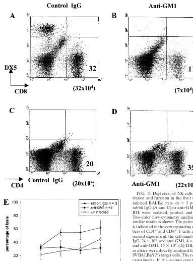
affect CD8 T-cell activation, we first removed NK cells from AdCAT-infected BALB/c mice and analyzed IHL phenotype and function at day 7 postinfection. As shown in Fig. 5A to D, intrahepatic CD8 T cells were reduced 4- to 5-fold, whereas no reduction of the number of CD4 T cells was observed. IHL CD8 T-cell cytotoxicity against virus-infected fibroblast targets was significantly lower in NK-depleted mice (Fig. 5E). NK cell depletion did not affect the proliferation and cytotoxicity of splenic T cells (data not shown), indicating that the NK-CD8 T-cell interaction was compartment specific.
Since antibody depletion with anti-GM1 may not be com-pletely specific for NK cells (27, 31), we assessed whether local CD8 T-cell function could be enhanced by increasing NK cell numbers. Since⬃70% spleen cells in SCID mice are NK cells (DX5⫹[data not shown]), we transferred either total or
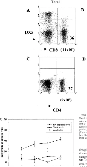
[image:4.612.76.269.72.717.2]NK-depleted SCID splenocytes to day 1 AdCAT-infected BALB/c mice. The increased percentages of NK cell could readily be detected in the peripheral blood of the NK-depleted recipients compared to the control group at day 1 (7.65 ⫾ 0.47 versus 5.9 ⫾ 0.26 [n ⫽ 6], P ⫽ 0.01). At day 3 posttransfer, we analyzed the composition and function of IHLs. As shown in Fig. 6A to D, the number of intrahepatic CD8 but not CD4 cells was higher in mice that received total SCID splenocytes than in mice that received NK-depleted SCID splenocytes. Furthermore, CD8 CTL activity was substantially enhanced after NK cell transfer (Fig. 6E). Similar results were observed at day 7 postinfection (CD8⫹T-cell number, 21 versus 6.4⫻
104; CTL activity, 45⫾2% versus 20⫾0.5% at E/T ratio of 60:1 in mice receiving total [n⫽3] versus NK-depleted ([n⫽
3] splenocytes). These results strongly support the idea that NK cells can directly affect the recruitment and function of antigen-specific CD8 T cells.
The IL-12 p35 subunit is differentially expressed in BALB/c
and B6 mice after infection.The cytokines IL-12 and IL-18 are
potent NK cell activators in vivo and in vitro (32). To investi-gate whether differential cytokine expression was responsible for the greater NK activation observed in BALB/c than in B6 mice, we quantified cytokine mRNA expression in the spleen and liver of infected mice by an RNase protection assay. Spleen cells from B6 and BALB/c mice produced comparable levels of IL-12 p35 before viral infection (not shown), but at days 3 and 7 postinfection, the mRNA level of p35 was two- to threefold higher in BALB/c than in B6 mice (Fig. 7A). In the liver, neither p35 nor p40 was detected in total liver RNA derived from B6 or BALB/c mice at day 3 postinfection (not shown). However, at day 7, the p40 subunit was detected in both strains whereas the p35 subunit was only weakly detected in BALB/c mice (Fig. 7B).
To confirm these findings, RT-PCR was used to amplify the p35 and p40 IL-12 transcripts from liver RNA obtained at days 7 and 10 postinfection. Consistent with the results obtained in
FIG. 2. AdCAT induces increased NK cytotoxicity in the liver and spleen of BALB/c mice. B6 and BALB/c mice were infected with AdCAT as in Fig. 1 and spleno cytes or IHL were isolated and pooled at dif ferent time points. NK cell cytotoxicity against YAC-1 targets was evaluated by a Cr release assay. The percentage of specifi c lysis is presented at an E/T ratio of 60:1 for IHL (A) or 200:1 for splenocytes (C). (B) IHL cytotoxicity at dif ferent E/T ratios at day 7. The results are representative of three experime nts and expressed as the mean ⫾ SD (A and C) or mean of triplicates ⫾ SD. (B)
on November 9, 2019 by guest
http://jvi.asm.org/
FIG.
3.
NK
cell
depletion
or
defective
NK
cell
function
prolongs
Ad
vector
expression
in
BALB/c
or
C3H
mice.
AdCAT
(A)
or
AdsCD8
(B
and
C)-infected
BALB/
c
mice
were
injected
with
anti-asialo
GM1
(
␣
-GM1)
or
control
rabbit
IgG
(RIgG)
as
described
in
Materials
and
Methods.
(D
and
E)
C3H/HeJ
beige
mice
that
have
defective
NK
function
were
infected
wit
h
AdCAT
and
sCD8,
respectively.
Hepatic
CAT
activity
(A
and
D)
or
serum
sCD8
protein
levels
(B
and
E)
(expressed
as
mean
⫾
SD)
were
quantified
at
weekly
intervals
as
described
in
Materials
and
Methods.
(C)
AdsCD8mRNA
expression
assessed
by
RT-PCR
using
primers
common
to
Ad
vector
sequences
flanking
the
transgene
(Ad5
⬘
and
Ad3
⬘
).

-Actin
was
used
as
a
control.
N,
negative
control
from
uninfected
mice;
NS,
not
significant.
on November 9, 2019 by guest
http://jvi.asm.org/
[image:5.612.85.524.69.720.2]the RNase protection assay, hepatic p35 transcripts were higher in BALB/c than in B6 mice at day 7 postinfection, whereas the levels of p40 transcripts were comparable (Fig. 7C). This difference persisted until day 10 postinfection, al-though fewer p40 transcripts were present in the liver of B6 compared to BALB/c mice at this time point. The different induction pattern of the p35 subunit in both the spleen and liver of B6 compared to BALB/c mice indicates an intrinsic difference between these two strains in their responses to Ad vector infection.
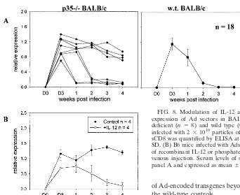
Modulation of bioactive IL-12 levels influences Ad vector
expression in BALB/c mice.If IL-12 plays a nonredundant role
in promoting the NK response to Ad vectors in BALB/c mice, then mice with a selective deficiency of the p35 subunit of IL-12 should demonstrate extended expression of Ad trans-genes. As shown in Fig. 8A, four of eight p35-deficient BALB/c mice infected with AdsCD8 expressed sCD8 for at least 4 weeks postinfection, whereas all 18 wild-type BALB/c mice eliminated AdsCD8 within⬃2 weeks (20).
Since B6 mice had a lower IL-12 response to Ad vectors associated with a slower attenuation of expression of Ad vec-tors such as AdsCD8 (20), a second prediction from these studies is that addition of IL-12 should enhance the immune response to AdCD8 in B6 mice. As shown in Fig. 8B, the expression level of sCD8 (and CAT [data not shown]) were significantly lower in B6 mice that had received IL-12 than in untreated B6 mice.
Increased IL-12 expression is associated with enhanced
IFN-␥production by BALB/c mice.The p40-p35 heterodimer
forms bioactive IL-12, which stimulates NK cells to produce IFN-␥, whereas the p40-p40 homodimer inhibits IL-12 func-tion. Because p35 and p40 subunits are differentially regulated in the liver of infected BALB/c and B6 mice and IL-12 pro-motes IFN-␥expression (32), we compared IFN-␥production by IHL between these two strains. As shown in Fig. 9A, the
much higher increase in IFN-␥mRNA expression at day 7 in BALB/c than in B6 mice is consistent with a role for IL-12 in augmenting IFN-␥production in BALB/c mice. Restimulation of NK cells with the YAC-1 target induced a substantially higher level of IFN-␥from day 7-infected BALB/c IHL com-pared to B6 IHL. Even when exogenous IL-12 was added to the cultures, BALB/c mice produced⬃3-fold more IFN-␥than did B6 mice, in keeping with the higher number of NK cells in BALB/c livers. Taken together, these findings indicate that bioactive IL-12 promotes immune responses to Ad vectors by enhancing NK function, at least in part, through increased production of IFN-␥.
DISCUSSION
In this and previous (20, 21) reports, BALB/c but not B6 mice rapidly eliminated Ad vectors expressing CAT, sCD8, Fas, and tumor necrosis factor (TNF) receptor. Another recent study that methodically evaluated strain variation of the ex-pression of the transgene hAAT also observed more rapid clearance of the Ad virus genome in BALB/c than in B6 mice (26), although strain differences to Ad-encoded transgenes have not uniformly been found (17, 28). The rapid elimination of Ad transgene expression in BALB/c mice could not simply be explained by increased CD8 T-cell cytotoxicity since neu-tralization of CD8 T-cell function or antibody-mediated CD8 depletion only had a modest effect on transgene expression (20). These strain differences suggested that another early component of the host immune response was required for rapid elimination or silencing of many Ad vectors in BALB/c mice.
[image:6.612.61.550.75.286.2]NK cells play a pivotal role in the early host defense against many viruses (reviewed in reference 2). In some cases, strain variation in resistance to the virus correlates with NK activity. For example, in 10 of 11 mouse strains infected with murine FIG. 4. Attenuation of NK and CD8 function promotes synergistic CAT expression in BALB/c and C3H mice. (A) BALB/c mice (n⫽4 to 9) were infected with AdCAT and/or AdCD8 and depleted of NK cells as shown. Hepatic CAT expression was quantified at day 7 or 28 postinfection. Error bars show SD. (B) C3H/HeJ beige or C3H/HeJ wild-type (w.t.) mice (n⫽3 per group) were infected with AdCAT alone and AdsCD8 as shown. Hepatic CAT expression was quantified at day 28 postinfection. Error bars show SD.
on November 9, 2019 by guest
http://jvi.asm.org/
cytomegalovirus, resistance correlated with the extent of NK activation (1). In the present report, we observed increased splenic and hepatic NK cell infiltration as well as increased NK cytotoxicity by IHL in BALB/c compared to B6 mice following AdCAT infection. Antibody-mediated depletion of NK cells in BALB/c mice, or a spontaneous mutation leading to defective NK function in C3H beige mice, increased expression of Ad-CAT compared to their wild-type controls. Taken together, these findings strongly support an important role for NK cells
[image:7.612.62.445.71.588.2]in the immune response against AdCAT and demonstrate a heightened role for these cells in BALB/c and C3H compared to B6 mice. Of considerable interest, a fourfold increase in mRNA expression of the minor histocompatibility antigen H-60, recently shown to be a ligand for the NKG2 activation receptor (3), was observed to increase around day 7 postinfec-tion. Since H-60 is expressed in a strain-specific fashion (BALB.B but not B6 [13]), it is tempting to speculate that H-60 may be involved not only in NK cell activation but also in macrophage production of TNF-␣(5, 10, 21) and nitric oxide. FIG. 5. Depletion of NK cells selectively reduces CD8 T-cell infil-tration and function in the liver of BALB/c mice. (A to D) AdCAT-infected BALB/c mice (n⫽3 per group) were treated with control rabbit IgG (A and C) or anti-GM1 (B and D). At day 7 postinfection, IHL were isolated, pooled, and stained for CD8, DX5, and CD4. Two-color flow cytometry analysis from one of two experiments with similar results is shown. The percentage of each cell type (where⬎1%) is indicated in the corresponding quadrant, whereas the absolute num-bers of CD4⫹and CD8⫹T cells are shown below the quadrant. In the
second experiment, the cell numbers were as follows: for CD8⫹, rabbit
IgG, 24⫻104, and anti-GM1, 4⫻104; for CD4⫹, rabbit IgG, 10⫻104,
and anti-GM1, 15⫻104. (E) IHL isolated from BABL/c mice treated
as above were directly analyzed for the ability to lyse AdCAT-infected SVBALB(H2d) target cells. The result is representative of two separate experiments. In the second experiment at E/T⫽50:1, percentages of cell lysis were as follows: rabbit IgG, 50⫾7; anti-GM1, 25⫾47; and uninfected, 27 ⫾ 5. “Uninfected” refers to lysis observed in cells obtained from uninfected mice.
on November 9, 2019 by guest
http://jvi.asm.org/
While these studies were in progress, Liu et al. (11) reported that NK cells induced apoptotic liver injury associated with the release of liver enzymes (ALT) at days 6 to 9 postinfection in Ad-gal-infected B6 mice. Since ALT release was also ob-served in SCID mice and could be attenuated by anti-GM1, apoptotic liver injury was attributed to NK⫹CD3⫺cells.
Al-though comparisons were not made between different normal strains, the authors noted that in T-cell-deficient mice on a B6 background, poly(I-C) was required for efficient activation of NK cells, whereas in Ad-infected BALB/c SCID mice, NK cells were fully activated (11). We observed that BALB/c mice had sixfold-higher serum ALT at day 7 postinfection compared to the B6 strain in response to the Ad null vector. These results are consistent with the⬃8-fold increase observed in the study by Christ et al. (4) and the very high ALT response in BALB/c mice infected with AdhAAT (26). The study by Liu et al. (11) supports an important role for NK cells in the murine innate response to replication-defective Ad vectors, and evaluation of hepatic enzymes is consistent with our observations that BALB/c mice have an increased NK response to infection with Ad.
[image:8.612.60.352.76.635.2]Despite the evidence for heightened NK involvement in the FIG. 6. Adoptive transfer of NK cells selectively enhances CD8 T-cell infiltration and function in the liver of BALB/c mice. BALB/c mice were infected with AdCAT and at day 1 postinfection injected with 5⫻106SCID spleen cells (Total) or an equal number of
NK-depleted SCID spleen cells. At day 3 postinfection, IHL were isolated, pooled, and evaluated for phenotype (A to D) and CD8 cytotoxicity (E) as in Fig. 5. Similar results were obtained at day 7 postinfection (see Results). “Uninfected” refers to lysis observed in cells obtained from uninfected cells.
on November 9, 2019 by guest
http://jvi.asm.org/
immune response to Ad vectors in BALB/c and C3H mice, Ad-encoded transgenes persist for months in BALB/c SCID mice (5), indicating that NK cells acting alone cannot eliminate Ad vectors. This observation is not surprising since the primary role of NK cells in response to viruses and, probably most pathogens, is to prime other cells and pathways for effector function (2). In this regard, we noted that increased NK infil-tration in the liver of BALB/c mice was followed by increased numbers of intrahepatic CD8⫹T cells, similar to the sequence
of events during murine cytomegalovirus (16) and Rous sar-coma virus (8) infections. Specific manipulations to either de-plete (by anti-GM) or enrich (by adoptive transfer of) NK cells resulted in the selective reduction or increase in both numbers and activity of intrahepatic CD8⫹ T cells. Together, these
findings indicate that the results are not an artifact of coex-pression of asialo-GM1 on CD4⫹or CD8⫹T cells (27), but
rather that the expansion and functional activity of CD8⫹T
cells is dependent in part on NK cells in BALB/c mice. Despite the sequence of events described above, attenuation of CD8 function in BALB/c mice had a modest effect on transgene expression (20), whereas the combined blockade of NK and CD8 T-cell function had a striking effect on transgene expression, increasing CAT activity by several orders of mag-nitude in both BALB/c and C3H mice. Although these findings could be explained by incomplete neutralization or depletion of CD8 cells, we believe that the cumulative results are most consistent with the idea that NK cells promote CD8 T-cell
recruitment and priming, but that the CD8 T cells produce chemokines, cytokines, or ligands that feed back on NK cells in a positive amplification loop. This hypothesis does not exclude additional roles for NK1.1 (37) or other cells of the innate or adaptive immune systems.
IL-12, a cytokine produced by macrophages or dendritic cells in response to microbial infection (32), was originally identified as a strong NK cell stimulator that promotes Th1 development, in part through activation of IFN-␥ (9). Al-though IL-12 can augment CD8 CTL activity directly, this is seen with pharmacologic rather than physiologic concentra-tions of IL-12 (12) Biologically active IL-12 is composed of two subunits, p35 and p40, that are encoded by separate genes and differentially regulated. Both subunits have to be synthesized in the same cell to form bioactive IL-12, whereas secretion of p40 alone results in a potent competitive inhibitor of IL-12. BALB/c mice produced a higher level of expression of both p35 and p40 mRNA in the liver from days 7 to 10 postinfection compared to B6 mice. Given that most Ad particles are cleared by Kupffer cells during the first 24 h of infection (35), the delayed expression of IL-12 suggests that the source of this cytokine was infiltrating cells such as monocytes (36, 38) rather than Kupffer cells. The kinetics of IFN-␥mRNA expression in the liver paralleled that of IL-12 and IHL obtained from BALB/c mice showed a striking increase in IFN-␥following exposure to a NK target in vitro. Together, these findings strongly suggest that IL-12 is instrumental in augmenting IFN-FIG. 7. BALB/c mice demonstrate high splenic and hepatic IL-12 p35 mRNA expression following Ad infection. B6 and BALB/c mice were injected with 2⫻1010particles of AdCAT or AdsCD8. RNA from the whole spleen (A) and liver (B) was harvested at days 3 and 7 postinfection.
mRNA expression was quantified by an RNase protection assay according to the manufacturer’s instructions, using 30g (A) or 100g (B) of total RNA. A housekeeping gene (L32; short exposure time) is shown as a control. Total liver RNA was collected from day 7(C) and day 10 (D) Ad-infected B6 and BALB/c mice. RT-PCR was performed to amplify the p35 and p40 IL-12 mRNA transcripts as well as-actin as described in Materials and Methods. P, positive control; M, marker.
on November 9, 2019 by guest
http://jvi.asm.org/
␥production and that these cytokines play a pivotal role in the rapid extinction of Ad transgene expression in BALB/c mice. The role of IL-12 in strain-specific regulation of the innate immune response to Ad vectors was confirmed by manipula-tion of IL-12 activity in BALB/c and B6 mice. Thus, four of eight p35-deficient BALB/c mice demonstrated prolonged ex-pression of sCD8, whereas administration of low dose recom-binant IL-12 reduced expression of AdsCD8 and AdCAT in B6 mice. It is not clear whether the variability in the response p35⫺/⫺BALB/c mice is explained by a compensatory effect of
other cytokines such as IL-18 (30) which, in individual mice, may be stochastic (23). However, the extended expression of transgenes in p35⫺/⫺mice is highly significant since expression
of Ad-encoded transgenes beyond 2 weeks was not observed in the wild-type controls.
The rapid extinction of Ad transgene expression in BALB/c mice associated with enhanced IL-12 and IFN-␥production and NK and CD8 T-cell activation reported here contrasts with the delayed clearance and Th2 cytokine profile characteristic of the BALB/c immune response to Leishmania major(19). However, the time course and cells implicated in the key ef-fector phase are different in these two infections. Indeed, BALB/c mice also produce significantly more IL-12 compared to B6 mice in response toL. majorinfection, but this response is quenched by increased production of macrophage-deactivat-ing cytokines such as IL-10 and transformmacrophage-deactivat-ing growth factor
[image:10.612.109.451.69.348.2](25) and/or by a more rapid and sustained down-regulation of the IL-12 receptor2 chain (29). Unlike the response to Ad, efficient clearance of the parasite is critically dependent on cytokine polarized CD4 T cells, whereas CD8 T cells play a minor role (7). Since cytokines such as IFN-␥and TNF-␣may FIG. 8. Modulation of IL-12 activity influences the duration of expression of Ad vectors in BALB/c and B6 mice. (A) IL-12p35-deficient (n⫽ 8) and wild type (w.t) (n⫽18) BALB/c mice were infected with 2⫻1010particles of AdsCD8, and the serum level of
sCD8 was quantified by ELISA at weekly intervals. Error bars show SD. (B) B6 mice infected with AdsCD8 as in panel A, received 10 ng of recombinant IL-12 or phosphate-buffered saline (control) by intra-venous injection. Serum levels of soluble CD8 were quantified as in panel A and expressed as mean⫾SD.
FIG. 9. Increased intrahepatic production of IFN-␥by BALB/c mice. (A) BALB/c and B6 mice were infected with AdCAT, and the total liver cDNA used for Fig. 1C was used to amplify IFN-␥and the-actin control by PCR. Each lane contains pooled RNA from three mice and represents an independent experiment. (B) B6 and BALB/c mice were infected with AdCAT as in Fig. 1. IHL were harvested on day 7, pooled, and incubated with YAC-1 cells at a ratio of 1:10, with or without IL-12 (10 ng/ml) in the culture medium. Culture supernatants were collected 24 h later and assayed for IFN-␥concentrations by ELISA. When IHL were incubated without YAC-1 cells,⬍30 U of IFN-␥was detected. The results are representative of three experiments.
on November 9, 2019 by guest
http://jvi.asm.org/
[image:10.612.66.540.535.669.2]attenuate transgene expression at the level of transcription (6, 22) as well as by promoting cytotoxic effector function, we cannot be certain whether reduced expression of transgenes reported here was also associated with virus genome clearance. However, in at least one other study where both transgene and virus genome were quantified in BALB/c mice, loss of trans-gene expression was associated with elimination of the Ad genome (26).
In summary, the findings in this study provide a mechanistic explanation for the different strain responses to Ad vectors in BALB/c and B6 mice. Although the exaggerated responses of BALB/c mice to Ad null reported here and elsewhere (4) strongly suggest that there is an intrinsic strain difference in response to Ad, the nature and level of expression of the transgene can almost certainly modulate the quality of the immune response (17, 18, 26). In Rous sarcoma virus infec-tions, preexposure of mice to different virus-encoded proteins can also profoundly alter the nature of the immune response (8). Further attention to the role of innate immune responses (5, 10) to both the Ad and transgene product may be required to optimize Ad-based gene delivery systems in the future.
ACKNOWLEDGMENTS
We thank Xiaojing Ma, Weill Medical College, for helpful discus-sions.
This work was supported in part by grant HL-9308-L (K.B.E. and E.F.-P.) from the National Institutes of Health.
REFERENCES
1.Bancroft, G. J., G. R. Shellam, and J. E. Chalmer.1981. Genetic influences on the augmentation of natural killer (NK) cells during murine cytomega-lovirus infection: correlation with patterns of resistance. J. Immunol.126:
988–994.
2.Biron, C. A., K. B. Nguyen, G. C. Pien, L. P. Cousens, and T. P. Salazar-Mather.1999. Natural killer cells in antiviral defense: function and regula-tion by innate cytokines. Annu. Rev. Immunol.17:189–220.
3.Cerwenka, A., A. B. Bakker, T. McClanahan, J. Wagner, J. Wu, J. H. Phillips, and L. L. Lanier.2000. Retinoic acid early inducible genes define a ligand family for the activating NKG2D receptor in mice. Immunity12:721– 727.
4.Christ, M., B. Louis, F. Stoeckel, A. Dieterle, L. Grave, D. Dreyer, J. Kintz, D. Ali Hadji, M. Lusky, and M. Mehtali.2000. Modulation of the inflam-matory properties and hepatotoxicity of recombinant adenovirus vectors by the viral E4 gene products. Hum. Gene Ther.11:415–427.
5.Elkon, K. B., C. C. Liu, J. G. Gall, J. Trevejo, M. W. Marino, K. A. Abra-hamsen, X. Song, J. L. Zhou, L. J. Old, R. G. Crystal, and E. Falck-Pedersen.
1997. Tumor necrosis factor alpha plays a central role in immune-mediated clearance of adenoviral vectors. Proc. Natl. Acad. Sci. USA94:9814–9819. 6.Harms, J. S., and G. A. Splitter.1995. Interferon-gamma inhibits transgene
expression driven by SV40 or CMV promoters but augments expression driven by the mammalian MHC I promoter. Hum. Gene Ther.6:1291–1297. 7.Huber, M., E. Timms, T. W. Mak, M. Rollinghoff, and M. Lohoff.1998. Effective and long-lasting immunity against the parasiteLeishmania majorin CD8-deficient mice. Infect. Immun.66:3968–3970.
8.Hussell, T., and P. J. Openshaw.1998. Intracellular IFN-gamma expression in natural killer cells preceeds lung CD8⫹T cell recruitment during
respi-ratory syncytial virus infection. J. Gen. Virol.79:2593–2601.
9.Kobayashi, M., L. Fitz, M. Ryan, R. M. Hewick, S. C. Clark, S. Chan, R. Loudon, F. Sherman, B. Perussia, and G. Trinchieri.1989. Identification and purification of natural killer cell stimulatory factor (NKSF), a cytokine with multiple biologic effects on human lymphocytes. J. Exp. Med.170:827–845. 10. Lieber, A., C. Y. He, L. Meuse, D. Schowalter, I. Kirillova, B. Winther, and M. A. Kay.1997. The role of Kupffer cell activation and viral gene expression in early liver toxicity after infusion of recombinant adenovirus vectors. J. Vi-rol.71:8798–8807.
11. Liu, Z. X., S. Govindarajan, S. Okamoto, and G. Dennert.2000. NK cells cause liver injury and facilitate the induction of T cell-mediated immunity to a viral liver infection. J. Immunol.164:6480–6486.
12. Magram, J., S. E. Connaughton, R. R. Warrier, D. M. Carvajal, C. Y. Wu, J. Ferrante, C. Stewart, U. Sarmiento, D. A. Faherty, and M. K. Gately.1996. IL-12-deficient mice are defective in IFN gamma production and type 1 cytokine responses. Immunity4:471–481.
13. Malarkannan, S., P. P. Shih, P. A. Eden, T. Horng, A. R. Zuberi, G. Chris-tianson, D. Roopenian, and N. Shastri.1998. The molecular and functional characterization of a dominant minor H antigen, H60. J. Immunol.161:
3501–3509.
14. Marshall, E.1999. Gene therapy death prompts review of adenovirus vector. Science286:2244–2245.
15. Matzinger, P.1991. The JAM test. A simple assay for DNA fragmentation and cell death. J. Immunol. Methods145:185–192.
16. McIntyre, K. W., and R. M. Welsh.1986. Accumulation of natural killer and cytotoxic T large granular lymphocytes in the liver during virus infection. J. Exp. Med.164:1667–1681.
17. Michou, A. I., L. Santoro, M. Christ, V. Julliard, A. Pavirani, and M. Mehtali.1997. Adenovirus-mediated gene transfer: influence of transgene, mouse strain and type of immune response on persistence of transgene expression. Gene Ther.4:473–482.
18. Morral, N., W. O’Neal, H. Zhou, C. Langston, and A. Beaudet.1997. Im-mune responses to reporter proteins and high viral dose limit duration of expression with adenoviral vectors: comparison of E2a wild type and E2a deleted vectors. Hum. Gene Ther.8:1275–1286.
19. Mosmann, T. R., and R. L. Coffman.1989. TH1 and TH2 cells: different patterns of lymphokine secretion lead to different functional properties. Annu. Rev. Immunol.7:145–173.
20. Peng, Y., E. Falck-Pedersen, and K. B. Elkon.2000. Soluble CD8 attenuates cytotoxic T cell responses against replication-defective adenovirus affording transprotection of transgenes in vivo. J. Immunol.165:1470–1478. 21. Peng, Y., J. Trevejo, J. Zhou, M. W. Marino, R. G. Crystal, E.
Falck-Pedersen, and K. B. Elkon.1999. Inhibition of tumor necrosis factor alpha by an adenovirus-encoded soluble fusion protein extends transgene expression in the liver and lung. J. Virol.73:5098–5109.
22. Qin, L., Y. Ding, D. R. Pahud, E. Chang, M. J. Imperiale, and J. S. Brom-berg.1997. Promoter attenuation in gene therapy: interferon-gamma and tumor necrosis factor-alpha inhibit transgene expression. Hum. Gene Ther.
8:2019–2029.
23. Reiner, S. L., and R. A. Seder.1999. Dealing from the evolutionary pawn-shop: how lymphocytes make decisions. Immunity11:1–10.
24. Roder, J., and A. Duwe.1979. The beige mutation in the mouse selectively impairs natural killer cell function. Nature.278:451–453.
25. Scharton-Kersten, T., L. C. Afonso, M. Wysocka, G. Trinchieri, and P. Scott.
1995. IL-12 is required for natural killer cell activation and subsequent T helper 1 cell development in experimental leishmaniasis. J. Immunol.154:
5320–5330.
26. Schowalter, D. B., C. L. Himeda, B. L. Winther, C. B. Wilson, and M. A. Kay.
1999. Implication of interfering antibody formation and apoptosis as two different mechanisms leading to variable duration of adenovirus-mediated transgene expression in immune-competent mice. J. Virol.73:4755–4766. 27. Slifka, M. K., R. R. Pagarigan, and J. L. Whitton.2000. NK markers are
expressed on a high percentage of virus-specific CD8⫹and CD4⫹T cells. J. Immunol.164:2009–2015.
28. Suzuki, M., R. Singh, M. A. Moore, W. R. Song, and R. G. Crystal.1998. Similarity of strain- and route-dependent murine responses to an adenovirus vector using the homologous thrombopoietin cDNA as the reporter genes. Hum. Gene Ther.9:1223–1231.
29. Szabo, S. J., A. S. Dighe, U. Gubler, and K. M. Murphy.1997. Regulation of the interleukin (IL)-12R beta 2 subunit expression in developing T helper 1 (Th1) and Th2 cells. J. Exp. Med.185:817–824.
30. Takeda, K., H. Tsutsui, T. Yoshimoto, O. Adachi, N. Yoshida, T. Kishimoto, H. Okamura, K. Nakanishi, and S. Akira.1998. Defective NK cell activity and Th1 response in IL-18-deficient mice. Immunity8:383–390.
31. Trambley, J., A. W. Bingaman, A. Lin, E. T. Elwood, S. Y. Waitze, J. Ha, M. M. Durham, M. Corbascio, S. R. Cowan, T. C. Pearson, and C. P. Larsen.
1999. Asialo GM1(⫹) CD8(⫹) T cells play a critical role in costimulation blockade-resistant allograft rejection. J. Clin. Investig.104:1715–1722. 32. Trinchieri, G.1998. Interleukin-12: a cytokine at the interface of
inflamma-tion and immunity. Adv. Immunol.70:83–243.
33. Watanabe, H., K. Ohtsuka, M. Kimura, Y. Ikarashi, K. Ohmori, A. Kusumi, T. Ohteki, S. Seki, and T. Abo.1992. Details of an isolation method for hepatic lymphocytes in mice. J. Immunol. Methods146:145–154. 34. Wilson, J. M.1996. Adenoviruses as gene-delivery vehicles. N. Engl. J. Med.
334:1185–1187.
35. Worgall, S., G. Wolff, E. Falck-Pedersen, and R. G. Crystal.1997. Innate immune mechanisms dominate elimination of adenoviral vectors following in vivo administration. Hum. Gene Ther.8:37–44.
36. Yang, Y., H. C. Ertl, and J. M. Wilson.1994. MHC class I-restricted cytotoxic T lymphocytes to viral antigens destroy hepatocytes in mice infected with E1-deleted recombinant adenoviruses. Immunity1:433–442.
37. Yang, Y., and J. M. Wilson.1995. Clearance of adenovirus-infected hepato-cytes by MHC class I-restricted CD4⫹CTLs in vivo. J. Immunol.155:2564– 2570.
38. Zhang, H. G., T. Zhou, P. Yang, C. K. Edwards III, D. T. Curiel, and J. D. Mountz.1998. Inhibition of tumor necrosis factor alpha decreases inflam-mation and prolongs adenovirus gene expression in lung and liver. Hum. Gene Ther.9:1875–1884.