Dissertation on
A MORPHOLOGICAL STUDY AND
MORPHOMETRIC ANALYSIS OF THE
JUGULAR FORAMEN
Submitted in partial fulfillment for
M.D. DEGREE EXAMINATION BRANCH- XXIII, ANATOMY
Upgraded Institute of Anatomy
Madras Medical College & Rajiv Gandhi Government General Hospital,
Chennai - 600 003
THE TAMILNADU Dr. M.G.R. MEDICAL UNIVERSITY
CHENNAI – 600 032
TAMILNADU
CERTIFICATE
This is to certify that this dissertation entitled
“A MORPHOLOGICAL STUDY AND
MORPHOMETRIC
ANALYSIS OF THE JUGULAR FORAMEN”
is a bonafide record of the research work done by Dr. PREFULLA.P.R, Post graduate student in the Institute of Anatomy, Madras Medical College and Rajiv Gandhi Government General Hospital, Chennai-03, in partial fulfillment of the regulations laid down by The Tamil Nadu Dr. M.G.R. Medical University for the award of M.D. Degree Branch XXIII-Anatomy, under my guidance and supervision during the academic year from 2013-2016.Dr. Sudha Seshayyan, M.B.B.S., M.S.,
Director & Professor, Institute of Anatomy, Madras Medical College, Chennai– 600 003.
Dr. B. Chezhian, M.B.B.S., M.S.,
Associate Professor, Institute of Anatomy, Madras Medical College, Chennai– 600 003.
Dean
Madras Medical College and
ACKNOWLEDGEMENT
I wish to express exquisite thankfulness and gratitude to my most respected teachers, guide Dr. B. Chezhian, Associate Professor and co-guide Dr. Sudha Seshayyan, M.S., Director and Professor, Institute of Anatomy, Madras Medical College, Chennai – 3, for their invaluable guidance, persistent support and quest for perfection which has made this dissertation take its present shape.
I am thankful to Dr.R.Vimala, M.D., Dean, Madras Medical College, Chennai-3 for permitting me to avail the facilities in this college for performing this study.
My heartfelt thanks to Dr. V.Lokanayaki and Dr.B.Santhi,
Associate Professors, Dr.V.Lakshmi, Dr.T.Anitha, Dr. P. Kanagavalli,
Dr. J. Sreevidya, Dr.Elamathi Bose, Dr.S.Arrchana, Assistant
Professors, Institute of Anatomy, MadrasMedical College, Chennai-3 for their valuable suggestions and encouragement throughout the study.
My gratefulness to Dr.Vanitha, M.D., Director, Barnard Institute of Radiology, Rajiv Gandhi Govt.General Hospital,Chennai – 3 and
Dr.Babu Peter, M.D., Associate Professor and Dr.Ashraf, post graduate
I earnestly thank my senior post graduate students
Dr.V.Anuradha, Dr.B.J.Bhuvaneswari, Dr.Elizabeth Priyadarisini
and Dr.E.Srividhya who have been supportive and encouraging
throughout the study.
I extend my heartfelt thanks to my colleagues Dr.S.Keerthi,
Dr.N.V.Ganga and junior post graduate students for their constant
encouragement and unstinted co-operation.
I am especially thankful to Mr. Mathews and Mr. Senthil kumar, technicians, who extended great support for this study and all other staff members including Mr. Pugazhendi, Mr.Jagadeesan, Mr.Maneesh
and Mr. Devaraj for helping me to carry out the study.
I thank my parents, Mr.P.V.Premachandran &
Mrs.P.R.Preetha, my parents in law, Dr.C.K.Sreedharan &
Mrs.K.B.Nirmala who have showered their choicest blessings on me and
supported me in my every step.
I am grateful beyond words to my husband Mr. Rajiv.N.S and my daughter Adhvika.P.R who in all possible ways supported me in making this study a reality.
LEGEND
APD - Anteroposterior Diameter FJD - Depth of Jugular Fossa FJW - Width of Jugular Fossa FM - Foramen Magnum IJV - Internal Jugular Vein
JB - Jugular Bulb/ Superior bulb of internal jugular vein JF - Jugular Foramen
Lt AJF - Area of left Jugular Foramen MLD - Mediolateral Diameter PCC - Posterior Condylar Canal
‘p’ value - Probability of observing the difference by chance Rt AJF - Area of right Jugular Foramen
CONTENTS
SL.NO. TITLE PAGE NO.
1. INTRODUCTION 1
2. AIM OF THE STUDY 4
3. REVIEW OF LITERATURE 8
4. EMBRYOLOGY 35
5. MATERIALS AND METHODS 37
6. OBSERVATION 41
7. DISCUSSION 65
8. CONCLUSION 94
A MORPHOLOGICAL STUDY AND MORPHOMETRIC
ANALYSIS OF THE JUGULAR FORAMEN
ABSTRACT
The Jugular Foramen is a complex crossroad of neurovascular structures at the skull base. Anomalies of the jugular bulb, glomus jugulare tumours, schwannomas, metastatic lesions and infiltrating inflammatory processes attract the clinicians attention to this region.. Knowledge of the anatomy of Jugular Foramen is vital for a favourable surgical outcome in the technically challenging operations of this region.
This study was aimed at analysing the Jugular Foramen morphologically and morphometrically. 100 adult human dry skulls at the Institute of Anatomy, Madras Medical College were used for the study. The features of the Jugular Foramen and Jugular Fossa were studied sincerely and the numerical data obtained were analysed statistically.
The data obtained will be useful for neurosurgeons and otorhinolaryngologists for achieving a less morbid and more favourable outcome in surgeries of the Jugular Foramen region. The findings are also enlightening to anthropologists, radiologists and anatomists.
Jugular Foramen
1
INTRODUCTION
Cranial foramina are the only portals to the otherwise closed cranium. Knowledge about the foramina in the base of skull is of utmost importance considering the delicate neurovascular structures that traverse through their narrow confines. The Jugular Foramen is a complex crossroad of neurovascular structures in the skull base.
The Jugular Foramen is a large irregular hiatus in the base of skull, between the jugular notch of the anterior border of the jugular process of the occipital bone 25,52,53,54 and the jugular fossa of the petrous part of the temporal bone. It is located at the posterior end of the petro-occipital suture. Anteriorly it is related to the lower opening of the carotid canal which is separated from it by a raised ridge of bone. On the lateral side of the Jugular Foramen lies the medial aspect of the sheath of the styloid process. Medially it is separated from the anterior condylar canal by a thin osseous bar (Fig.1)
Fig.2: Right Jugular Foramen showing the Jugular Fossa.
2
posterior larger venous (sigmoid) part transmits the internal jugular vein.
26,52
The jugular foramen contains the transition of sigmoid sinus to internal jugular vein and the termination of inferior petrosal sinus 14, with 60% of the area of all venous foramina of the skull occupied by the jugular foramina.24
Where the sigmoid sinus continues as the internal jugular vein, the jugular fossa of the petrous part of temporal bone (Fig. 2) is recessed out upwards and laterally to accommodate the superior bulb of the internal jugular vein, and it separates the same from the tympanic cavity.13,14,25 Dome of the jugular bulb is generally covered by a bone, which is called the dome of the Jugular Fossa. The lateral wall of the fossa is pierced by mastoid canaliculus, which transmits the auricular branch of vagus nerve (Arnold’s nerve). On or near the ridge between the Jugular Foramen and lower opening of the carotid canal lies the canaliculus for the tympanic nerve which transmits the tympanic branch of glossopharyngeal nerve (Jacobson’s nerve).38,52 Other structures that the Jugular Foramen transmits are the meningeal branches of the ascending pharyngeal artery in the anterior compartment and meningeal branch of occipital artery in the posterior compartment.25
3
4
AIM OF THE STUDY
A plethora of pathological conditions may occur in the Jugular Foramen, arising from structures within it or from contiguous structures. The neoplastic pathologies tend to alter the normal anatomy of Jugular Foramen by invasion, erosion or expansion. These tumours pose significant diagnostic as well as surgical challenges since they demand a microsurgical approach to this region. The Jugular Foramen varies considerably in size and shape, along with the internal jugular vein.57The foramen’s complex shape, its formation by two bones, and the delicate neurovascular structures that pass through it further compound its anatomy.
The most common intrinsic lesion in the Jugular Foramen (JF) is hypervascular paraganglioma (glomus jugulare) located in the jugular fossa,39,60 followed by neurogenic tumours like schwannoma and neurofibroma. Less common lesions include meningioma, haemangiopericytoma, chondrosarcoma, and plasmacytoma.
5
oral cavity may give rise to JF metastases. JF fracture may result from trauma to skull base. Endolymphatic sac tumours arising at the posteromedial aspect of the petrous bone can frequently extend to the Jugular Foramen. 55,58,60
In Computerised Tomography images most tumours of the Jugular Foramen manifest as areas of infiltrative bone destruction, although schwannoma and meningioma cause smooth enlargement of the foramen.8 Anatomical variants like asymmetrically enlarged Jugular Foramen, high jugular bulb, high dehiscent jugular bulb, or a jugular bulb diverticulum may be misdiagnosed as pseudomasses in the JF.22,55 In such aragangliomas can be ruled out by the presence cases the hypervascular p
of preservation of normal bony margins and intact compartmentation by intrajugular process in the asymmetrically but proportionately enlarged JF.16
Typically JF tumours produce the Jugular Foramen syndrome (Vernet’s syndrome) characterised by IX, X and XI cranial nerve palsy and may produce other related syndromes depending on the extension of the tumour. 49
6
accurate knowledge of the normal anatomy and variations of the region, success can be achieved with reasonable morbidity and mortality rates.39Thus the knowledge of morphological details and dimensions of the Jugular Foramen as well as its variations would be of great value to neurologists, radiologists, otorhinolaryngologists and neurosurgeons.
Aim of the present study is to analyse the Jugular Foramen morphologically and morphometrically. Hopefully the data will be useful to radiologists, ENT surgeons and neurosurgeons for preoperative planning and management of Jugular Foramen surgeries.
The parameters studied are:
1. Maximum mediolateral diameter of right and left Jugular Foramina 2. Maximum anteroposterior diameter of right and left Jugular
Foramina
3. Area of right and left Jugular Foramina 4. Width of right and left Jugular Fossae 5. Depth of right and left Jugular Fossae
6. Presence of domed right and left Jugular Fossae
7. Number of septa in the right and left Jugular Foramina
7
9. Presence of accessory opening in the walls of right and left Jugular Foramina
8
REVIEW OF LITERATURE
MEDIOLATERAL DIAMETER OF THE JUGULAR FORAMEN
(MLD)
Aydinlioglu A et al6 (2001) studied Eastern Anatolian skulls and
reported that MLD on the right and left sides were 13.7mm 12.3mm respectively.
OE Idowu34 (2004) studied 40 JF of 20 Nigerian skulls and stated
that mean MLD on the right and left sides were 13.90 mm and 14.11 mm respectively.
Ekinci et al15 (2009) conducted a study on 70 skulls of Turkish
population and reported that MLD on the right side (Rt MLD) was 16.0mm and MLD on the left side (Lt MLD) was 15.5 mm.
Namita A Sharma et al33 (2011) in their study of the foramina of
skull base in 50 dry skulls said that the MLD of JF were 15.59+/- 2.64mm and 13.83+/- 4.94mm on the right and left sides respectively.
Ketu Chauhan et al28 (2011) in their study of 50 dry skulls
9
Anjali Singla et al4 (2012) studied 50 adult dry skulls and
observed that the MLD of the JF were 15.67mm and 14.85mm on the right and left sides respectively.
Osunwoke EA et al35 (2012) in their study of 120 dry skulls
reported that the MLD of the JF were 15.76±0.22mm and 13.39±0.23mm respectively on the right and left sides. They stated that the mean length of the right JF was larger than that of the left JF.
Anitha MR et al3 (2013) in their study of the JF of 100 adult dry
skulls reported that Rt MLD and Lt MLD were 15.21mm and 13.39mm respectively. In 88% of skulls the length of the JF on the right side was more than that of the left side and in 12% of skulls length of the left side JF was more.
Rahul Rai et al41 (2013) studied 100 dry skulls and observed that
MLD of right JF was 12.90mm and the same of left JF was 13.01mm.
Shifan Khanday et al48 (2013) analysed 648 Jugular Foramina of
324 skulls and found out that Rt MLD and Lt MLD were 14.6 mm and 13.9 mm respectively.
Vijisha P et al57 (2013) in their study of 30 adult dry skulls
10
Avanish Kumar et al5 (2014) in their study of JF of 68 skulls,
stated that the MLD was 13.6mm on right side and 13.9 mm on left side.
Chandni Gupta et al9 (2014) studied 50 adult dry skulls and
reported that Rt MLD and Lt MLD were 16.52 mm and 16.02 mm respectively.
Roma Patel et al44 (2014) in their study of 100 dry skulls stated
that MLD of right and left JF were 12.17mm and 11mm respectively.
N. Himabindu et al32 (2015) analysed the JF of 110 adult dry
skulls and reported that Rt MLD was 14.6mm and Lt MLD was 12.69mm.
ANTEROPOSTERIOR DIAMETER OF THE JUGULAR
FORAMEN (APD)
Aydinlioglu A et al6 (2001) studied Eastern Anatolian skulls and
reported that right APD (Rt APD) and left APD (Lt APD) were 12.2mm and 10.9mm respectively.
OE Idowu34 (2004) studied 40 JF of 20 Nigerian skulls and stated
11
Ekinci et al15 (2009) conducted a study on 70 skulls of Turkish
population and reported that APD on right side was 8.4mm and the same on the left side was 7.6 mm.
Ketu Chauhan et al28 (2011) in their study of 50 dry skulls
reported that APD were 9.9mm and 7.9mm on the right and left sides respectively.
Namita A Sharma et al33 (2011) in their study of the foramina of
skull base in 50 dry skulls said that Rt APD and Lt APD were 9.02+/-1.79mm and 7.73+/- 9.02+/-1.79mm respectively.
Anjali Singla et al4 (2012) studied 50 adult dry skulls and
observed that APD of the right JF was 9.32 mm and that of the left JF was 7.34mm. They also reported that the width of the JF was more on right side.
Osunwoke EA et al35 (2012) in their study of 120 dry skulls
reported that APD of JF were 9.34±0.18mm and 7.54±0.20mm respectively on right and left sides. They stated that mean width of right JF was larger than that of left JF.
Anitha MR et al3 (2013) in their study of the JF of 100 adult dry
12
respectively. In 74% of the skulls width of the right side was more than that of the left side whereas in 16% of skulls width of the left JF was more.
Rahul Rai et al41 (2013) studied 100 dry skulls and observed that
Rt APD was 8.90mm and Lt APD was 6.91mm.
Shifan Khanday et al48 (2013) analysed 648 JF of 324 skulls and
found out that APD of JF on the right side was 10.06mm and that on the left side was 8.9mm.
Vijisha P et al57 (2013) in their study of 30 adult dry skulls
reported that APD of right JF was 12.13mm and APD of left JF was 9.27mm.
Avanish Kumar et al5 (2014) in their study of JF of 68 skulls,
stated that mean Rt APD was 10.6mm and mean Lt APD was 9.2 mm.
Chandni Gupta et al9 (2014) studied 50 adult dry skulls and
reported that APD were 11.22 mm and 9.52 mm on right and left sides respectively.
Roma Patel et al44 (2014) in their study of 100 dry skulls stated
13
N. Himabindu et al32 (2015) analysed the JF of 110 adult dry
skulls and reported that the APD on the right side was 9.61mm and the same on the left side was 8.24mm.
AREA OF JUGULAR FORAMEN (AJF)
OE Idowu34 (2004) studied 40 JF of 20 Nigerian skulls and stated
that mean JF area on the right was 437.49 mm2 and that on the left was 419.48 mm2.
Shifan Khanday et al48 (2013) analysed 648 JF of 324 skulls and
found out that the mean area of JF on right side was 118 mm2 and on left side was 90 mm2.
Vijisha P et al57 (2013) in their study of 30 adult dry skulls
reported that area of right JF was 210.87mm2 and that of the left JF was 141.93 mm2.
Chandni Gupta et al9 (2014) studied 50 adult dry skulls and
14
SIDE DOMINANCE OF THE JUGULAR FORAMEN
Peter.L.Williams and Roger Warwick38 (1980) in ‘Gray’s
Anatomy’ stated that the Jugular Foramen is usually larger on the right side of the skull.
R R Sturrock40 (1988) in his study of 156 adult dry skulls
observed that the right JF was larger than the left JF in 68.6% whereas the left was larger in 23.1%, no difference in size with side was observed in 8.3%.
MT Hatiboglu et al30 (1991) did a study on 300 Anatolian skulls
of 17th and 18th centuries and reported that in 61.6% the right JF was larger whereas in 26% the left was larger and rest of the specimens had both JF of equal size.
SA Ayeni et al46 (1995) studied the microsurgical anatomy of JF in
10 cadavers, and reported that the right foramen was larger than the left in 70%, equal in 20% and smaller in 10%.
H.L. Guido et al19 (1997) studied 100 Brazilian skulls and stated
15
OE Idowu34 (2004) studied 40 JF of 20 Nigerian skulls and stated
that side predominance of one of the JF appeared in 80% of cases. When present, the predominance of right JF was in 55% and that of left JF was in 25%.
Wysocki et al24 (2006) in their study of 100 adult human dry skulls
observed that in 54% of the skulls there was right sided dominance of the JF compared to 27% of the skulls with a left sided dominance. In the remaining 19% of skulls there was symmetry with no side dominance.
MM Patel et al37 (2007) in their study of 91 skulls stated that
60.4% of cases showed that right foramen was larger than the left foramen whereas 24.2% of cases showed that right foramen was equal to the left and in 15.4% of cases the left was larger than the right.
Keles B et al27 (2009) studied the JF anatomy by microscopic
dissection of 22 cadaveric head specimens and documented that right JF was larger than the left in 72.7%.
Ekinci et al15 (2009) conducted a study on 70 skulls of Turkish
16
Hussain Saheb et al20 (2010) studied 125 adult dry skulls and
observed that right JF was larger than the left in 64.8% and equal in size to the left in 10.4%. The remaining skulls had a larger JF on the left side.
Ruchira Sethi et al45 (2011) analysed 56 adult dry skulls and
reported that the right JF was larger in 53.5% and left JF was larger in 7.1% skulls. In the remaining 39.4% skulls the JF were equal in size on both sides.
Osunwoke EA et al35 (2012) studied 120 dry skulls and stated that
right JF was larger than the left JF.
SM Akram Hossain et al50 (2012) studied 55 adult dry skulls and
observed that in 58 % right JF was larger than the left, in 22 % smaller and in 20 % right JF was equal in size to the left.
Rahul Rai et al41 (2013) studied 100 dry skulls and observed that
the right JF was larger than the left in 74%, smaller in 19% and both were equal in size in 7%.
Avanish Kumar et al5 (2014) in their study of JF of 68 skulls,
17
Roma Patel et al44 (2014) in their study of 100 dry skulls stated
that size of the JF varied on the two sides. The right JF larger than the left were 75%, right smaller than the left were 23% and both were of equal size in 2%.
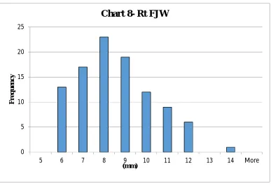
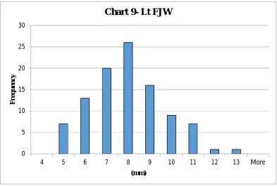
WIDTH OF THE JUGULAR FOSSA (FJW)
Anjali Singla et al4 (2012) in their study of 50 adult dry skulls
observed that the mean FJW were 8.99mm on the right side and 7.54mm on the left side. They also reported that width of the Jugular Fossa was larger on the right side.
Chandni Gupta et al9 (2014) studied 50 adult dry skulls and
reported that the mean FJW were 6.83 mm and 5.69mm on the right and left sides respectively.
DEPTH OF THE JUGULAR FOSSA (FJD)
Aydinlioglu A et al6 (2001) studied Eastern Anatolian skulls and
reported that mean FJD were 14mm on right side(Rt FJD) and 13.7 mm on left side(Lt FJD).
Ketu Chauhan et al28 (2011) in their study of 50 dry skulls
18
Anjali Singla et al4 (2012) studied 50 adult dry skulls and reported
that Rt FJD was 11.11mm and Lt FJD was 11.04mm.
Shifan Khanday et al48 (2013) analysed 648 JF of 324 skulls and
documented that Rt FJD was 10.1mm and Lt FJD was 9.0 mm.
Chandni Gupta et al9 (2014) studied 50 adult dry skulls and
reported that Rt FJD and Lt FJD were 11.58mm and 11.13mm respectively.
Ivan Jovanovic et al22 (2014) in their study of 37 sagittal sections
(17 left and 20 right) of dried adult human skulls reported that Rt FJD was 9.43mm and Lt FJD was 8.10mm.
PRESENCE OF DOMED BONY ROOF OF JUGULAR FOSSA
A.S. Breathnack1 (1948) in ‘Frazer’s anatomy of the human
skeleton’ stated that the Jugular Fossa is frequently very deep, lodging the bulb of IJV.
R R Sturrock40(1988) in his study of 156 adult dry skulls found
19
MT Hatiboglu et al30 (1991) did a study on 300 Anatolian skulls
of 17th & 18th centuries and reported that dome was present bilaterally in 49%, unilaterally on the right side in 36.6% and on the left in 4.6%. A domed roof was not found in the remaining 10.3% of skulls.
H.L. Guido et al19 (1997) studied 100 Brazilian skulls and stated
that dome was present bilaterally in 86%, unilaterally on right side in 10% and left side in 4%.
MM Patel et al37 (2007) in their study of 91 skulls stated that
dome was found bilaterally in 21% and was absent bilaterally in 25.3% of cases.
Hussain Saheb et al20 (2010) studied 125 adult dry skulls and
observed the dome bilaterally in 49.6%, unilaterally on right side in 27.2% and left side in 8.8%. It was absent in 14.4%.
SA Athavale51 (2010) studied 116 dry skulls and reported that
dome was present on right side in 78% and on left side in 69.4%.
P ereira et a l1 7 (20 10 ) studied 111 dry skulls and observed a
20
Namita A Sharma et al33 (2011) in their study of 50 dry skulls
said that the dome was present bilaterally in 58%, unilaterally on right side in 28% and on left side in 8%.
Anjali Singla et al4 (2012) in their study of 50 adult dry skulls
reported that dome was present bilaterally in 66%, unilaterally in 6% on right side and in 16% on left side and was absent bilaterally in 12%.
SM Akram Hossain et al50 (2012) studied 55 adult dry skulls and
observed that the dome was present bilaterally, in all the specimens.
Vijisha P et al57 (2013) in their study of 30 adult dry skulls
reported that dome was observed only on right side in 26.6% and only on left side in 3.33 % and bilaterally in 70%.
Shifan Khanday et al48 (2013) studied 648 JF of 324 skulls and
found out that in 20% skulls dome was present bilaterally. It was present unilaterally on the right side in 40% and on the left side in 29%.The dome was absent in 11%.
Rahul Rai et al41 (2013) studied 100 dry skulls and observed that
21
Avanish Kumar et al5 (2014) in their study of JF of 68 skulls,
stated that the dome was observed bilaterally in 57.35%, only on the right side in 29.4%, only on the left side in 8.82% and was absent bilaterally in 4.41%.
Chandni Gupta et al9 (2014) studied 50 adult dry skulls and
reported that dome was present in 74 % on right side and 58% on left side.
Peiris HRD et al18 (2014) analysed 75 dry skulls and reported that
the dome indicating the presence of a jugular bulb was present on right side in 34.9% and left side in 47.6% of skulls.
Roma Patel et al44 (2014) in their study of 100 dry skulls stated
that the jugular bulb dome was present bilaterally in 23%, unilaterally on right side in 30%, left side in 11% and absent bilaterally in 36%.
INCIDENCE OF SEPTATE JUGULAR FORAMEN
Peter.L.Williams and Roger Warwick38 (1980) in ‘ Gray’s
22
A.S. Breathnack1 (1948) in ‘Frazer’s anatomy of the human
skeleton’ stated that JF has a larger part laterally for the vein and a smaller part medially for the nerves, these two parts are separated in part or whole by a small bony projection from the occipital bone above the anterior condylar canal, which is called the intrajugular process.
R R Sturrock40 (1988) in his study of 156 adult dry skulls
observed septate right JF in 4.5% and septate left JF in 14.1% specimens.
MT Hatiboglu et al30 (1991) did a study on 300 Anatolian skulls
of 17th & 18th centuries and reported that in 8.2% right JF was septate whereas in 23.9% left was septate.
H.L. Guido et al19 (1997) studied 100 Brazilian skulls and stated
that one septum was present bilaterally in 5%, unilaterally only on the right JF in 7% and only on the left JF in 3% .Tripartite division was present in 6% of right JF and 5% of left JF.
I Tekdemir et al21 (2001) studied 80 JF of 40 dry skulls and
observed 13 (16.25%) bony septa (5 bilateral, 3 unilateral).
MM Patel et al37 (2007) in their study of 91 skulls stated that JF
23
DR Sawyer et al10 (2009) studied bridging of JF in 234 skulls and
reported septate JF in 8.1%, with the right showing a significantly higher incidence than the left.
Ekinci et al15 (2009) conducted a study on 70 skulls of Turkish
population and reported that bony bridging was observed in 20% of cases and was not in 80%. In addition one tripartite JF was observed.
Hussain Saheb et al20 (2010) observed that right JF was septate in
66.4% and left JF was septate in 75.2% of 125 adult dry skulls studied.
Pereira et al 17(2010) studied the Jugular Foramina of 111 dry
skulls and reported that 83.8% of skulls lacked the septum bilaterally.
Ruchira Sethi et al45 (2011) analysed 56 adult dry skulls and
reported the presence of one septum unilaterally in 17.8% skulls on right side and in 14.3% skulls on left side. They also described a tripartite JF in 10.7% skulls bilaterally.
Namita A Sharma et al33 (2011) in their study of JF in 50 dry
skulls said that complete septation was present in 10% of the skulls and incomplete in 36% skulls.
Anjali Singla et al4 (2012) studied 50 adult dry skulls and reported
24
SM Akram Hossain et al50 (2012) studied 55 adult dry skulls and
observed that JF was septate in 100% of the specimens.
Vijisha P et al57 (2013) in their study of 30 adult dry skulls
reported that JF was septate in 83.3% on right side and 86.6% on left side.
Anitha MR et al3 (2013) in their study of the Jugular Foramina of
100 adult dry skulls reported that JF was septate in 8% on right side and 11% on left side.
Shifan Khanday et al48 (2013) analysed 648 JF of 324 skulls and
found out that the right JF was septate in 36.3% and left JF in 24.1%.
Rahul Rai et al41 (2013) studied 100 dry skulls and observed that
the JF were septate in all specimens.
Avanish Kumar et al5 (2014) in their study of JF of 68 skulls,
stated that a single septum was present in all the JF.
Chandni Gupta et al9 (2014) studied 50 adult dry skulls and
25
Peiris HRD et al18 (2014) analysed 75 dry skulls and reported that
84.7% of the left JF and 80.6% of the right JF were septate.
Roma Patel et al44 (2014) in their study of 100 dry skulls stated
that 45% of the JF on right side and 39% on left side were septate.
N. Himabindu et al32 (2015) analysed the JF of 110 adult dry
skulls and reported that absence of septation on right was in 16.70% and left in 18.70% of the specimens.
TRIPARTITE JUGULAR FORAMEN
Peter.L.Williams and Roger Warwick38 (1980) in their book
stated that sometimes margins of the notch in which the inferior ganglion of IX cranial nerve is lodged, extend to divide the JF into two or three compartments.
in ‘Quain's Elements of Anatomy’ described
T.H Bryce54 (1915)
26
MT Hatiboglu et al30 (1991) did a study on 300 Anatolian skulls
of 17th and 18th centuries and reported that the inferior petrosal sinus occasionally passed through a small separate opening in JF. The occurrence of this small separate opening was seen on the right in 5.6% and on the left in 4.6%.
H.L. Guido et al19 (1997) studied 100 Brazilian skulls and stated
that JF was tripartite on the right side in 6% and left side in 5%.
I Tekdemir et al21 (2001) studied 80 JF of 40 dry skulls and
reported that in one specimen (1.25%) the JF was divided anatomically into three parts.
Ekinci et al15 (2009) conducted a study on 140 JF in 70 skulls of
Turkish population and reported the presence of one tripartite JF (0.71%).
Ruchira Sethi et al45 (2011) analysed 56 adult dry skulls and
reported the presence of tripartite JF in 10.7% skulls bilaterally.
Anjali Singla et al4 (2012) studied 50 adult dry skulls and reported
6% of specimens to have tripartite JF bilaterally.
Shifan Khanday et al48 (2013) analysed 324 skulls and found out
27
TYPE OF SEPTUM IN JUGULAR FORAMEN
.C Brash and E.B.Jamieson25 (1937) in ‘Cunningham’s textbook
J
of anatomy’ stated that in some skulls a curved process, called intrajugular process projects from the floor of jugular notch of the occipital bone and partially or completely subdivides the JF. Sometimes spicules of bone project across the foramen from its petrosal and occipital margins, and may divide the same into compartments.
A.S. Breathnack1 (1948) in ‘Frazer’s anatomy of the human
skeleton’ wrote that JF is divided by an intrajugular process of bone from either or both the occipital and temporal sides.
in ‘Quain's Elements of Anatomy’ stated that
T.H Bryce54 (1915)
at the forepart of jugular notch of the jugular process of occipital bone, a small projection is frequently present, called the intrajugular process. Occasionally it is prolonged into a spicule of bone, which meets the petrosal part of temporal bone.
Y. Dodo62 (1986) studied anatomical nature and pattern of
28
intrajugular processes of JF. The observations were: (1) Intrajugular process of the temporal bone is situated posterior to the triangular depression of petrous part of the bone. (2) Bony bridging of JF is established by the contact of intrajugular process of temporal bone with bony process of occipital bone projecting either from just above the anterior condylar canal (Type I) or from posterior to the anterior condylar canal (Type II). (3) If both the processes of occipital bone reach the intrajugular process of temporal bone simultaneously, JF is divided into 3 compartments. (4) In the case of Type I bridging, the anteromedial compartment transmits the IX cranial nerve, while the posterolateral compartment gives passage to X nerve, XI nerve and IJV. (5) In the case of Type II bridging, the anteromedial compartment contains the glossopharyngeal, vagus and accessory nerves, and the posterolateral compartment transmits IJV. (6) When tripartite division of JF occurs, the anteromedial compartment transmits the IX nerve, the middle compartment contains the X and XI nerves, and the posterolateral compartment transmits IJV.
R R Sturrock40 (1988) studied 156 adult dry skulls and observed
29
one incomplete septum in 10.9% specimens. He also stated that the incomplete septa in life were probably completed by cartilage.
MT Hatiboglu et al30 (1991) studied 300 Anatolian skulls of 17th
and 18th centuries and reported that complete septum (intrajugular process) was present in 5.6% of right JF and 4.3% of left JF. Incomplete or partial septum was observed in 2.6% of right and 19.6% of left JF. The JF had two septa in 5.6% on right and 4.6% on left side.
A.L.Rhoton Jr et al2 (2000) describedthat the junction of petrosal
and sigmoid parts of JF is the site of bony prominences on the opposing surfaces of temporal and occipital bones. These are called the intrajugular processes, which are joined by a fibrous or less frequently an osseous bridge, the intrajugular septum. This separates the sigmoid and petrosal parts of JF. The IX, X and IX cranial nerves are related to the medial margin of the intrajugular process of temporal bone.
OE Idowu34 (2004) studied 40 JF of 20 Nigerian skulls and stated
that a bony bridge completely partitioned the JF in 3 (7.5%) of the JF.
MM Patel et al37 (2007) in their study of 91 skulls stated that
30
J. Linn et al23 (2009) in their MRI study stated that intraforaminal
compartments could be identified by depicting the temporal and occipital intrajugular processes as well as the dural septum separating petrosal from sigmoid portion.
Keles B et al27 (2009) studied JF anatomy by microscopic
dissection of 22 cadaveric head specimens and observed the presence of a complete bony septum bilaterally in 13.6% specimens.
Hussain Saheb et al20 (2010) studied 125 adult dry skulls and
observed that one complete septum was present in 20.8% on right side and 16.8% on left side. One incomplete septum was present in 45.6% on right side and 58.4% on left side.
P ereira et a l1 7 (2010 ) studied 111 dry skulls and reported that
0.9% showed a complete septum bilaterally and 0.9% showed incomplete septum bilaterally, 14.4% had unilateral septate JF.
Ketu Chauhan et al28 (2011) in their study of 50 dry skulls
reported that JF was partitioned by a complete septum in 6% and 8% of specimens on right and left sides respectively.
Ruchira Sethi et al45 (2011) analysed 56 adult dry skulls and
31
of the 14.3% septate left JF, 4.3% had partial septum. They also described a tripartite JF in 10.7% skulls bilaterally. They reported that incomplete or partial septa were never observed bilaterally.
Namita A Sharma et al33 (2011) in their study of 50 dry skulls
said that complete septation was present in 10% skulls, in which 6% was bilateral. It was present unilaterally on right side in 2% and 2% on left side. Partial septation was present totally in 36% with incidence bilaterally in 18%, unilaterally on right side in 4% and left side in 14 %.
Anjali Singla et al4 (2012) studied 50 adult dry skulls and reported
that bilaterally one complete septum was present in 4% and two complete septa in 6%. Unilaterally 4% had one complete septum on right side. The remaining specimens had a partial septum.
SM Akram Hossain et al50 (2012) studied 55 adult dry skulls and
observed that septum was complete on right side in 76.36% and left side in 90.91% while incomplete on right side in 23.64% and left side in 9.09%.
Vijisha P et al57 (2013) in their study of 30 adult dry skulls
32
Rahul Rai et al41 (2013) studied 100 dry skulls and observed that
septum was complete on right side in 68% and on left side in 21% whereas incomplete on right side in 32% and on left side in 79%.
Shifan Khanday et al48 (2013) studied 648 JF of 324 skulls and
found out that 12.6% of right JF and 12.8% of left JF had one complete septum. One partial septum was noticed in 23.7% of right JF and 11.3% of left JF.
Avanish Kumar et al5 (2014) in their study of JF of 68 skulls,
stated that complete septum was present in 16.18% and 8.80%, and incomplete septum in 83.82% and 91.2% on right and left sides respectively.
Chandni Gupta et al9 (2014) studied 50 adult dry skulls and
reported that one complete septum was present in 44% on right side and 42% on left side whereas the rest of the specimens had one incomplete or partial septum.
Ivan Jovanovic et al22 (2014) in their study of 37 sagittal sections
33
Peiris HRD et al18 (2014) analysed 75 dry skulls and reported that
one complete septum was observed in 14.7% of left JF and 10.6% of right JF and partial septum was noticed bilaterally in 70% of skulls.
Roma Patel et al44 (2014) in their study of 100 dry skulls stated
that complete septum was present in 16% on right side and 14% on left side. Partial septation of JF was present in 29% of skulls on right side and in 25% on left side.
N. Himabindu et al32 (2015) analysed the JF of 110 adult dry
skulls and reported that complete septum was seen on right side in 22% and on the left in 27.5% while incomplete bony septum was found on the right in 33% and on the left in 30% of skulls.
PRESENCE OF ACCESSORY OPENING IN THE WALLS OF
JUGULAR FORAMEN
Ruchira Sethi et al45 (2011) analysed 56 adult dry skulls and
34
SITE WHERE THE ACCESSORY OPENING LEADS TO.
Ruchira Sethi et al45 (2011) analysed 56 adult dry skulls and
35
EMBRYOLOGY
Skull consists of the neurocranium, which is the protective case around the brain and the viscerocranium or splanchnocranium which makes up the jaw skeleton.59 The blastemal skull or desmocranium begins to appear at the end of the first month of development of the embryo as localised masses formed by condensation and thickening of the mesenchyme surrounding cerebral vesicles. Chondrocranium is that part of neurocranium which passes through or remains in cartilage. The membranous neurocranium, consists of dermal bones and corresponds to the cranial vault, and it is not preformed in cartilage.38,52
36
37
MATERIALS AND METHODS
STUDY MATERIALS:
100 human adult dry skulls of unknown sex Digital Vernier Caliper
Flexible wire
25G Spinal needle and rubber stopper
STUDY METHOD:
Dry skull Method
SPECIMEN COLLECTION:
Hundred human adult dry skulls of unknown sex available in the Institute of Anatomy, Madras Medical College were used for this study.
INCLUSION CRITERIA:
1. Adult human dry skulls of unknown sex. 2. Third molar tooth erupted.
3. Well defined skull sutures.
EXCLUSION CRITERION:
Fig.6: Mediolateral diameter of Jugular Foramen
Fig.7: Anteroposterior diameter of Jugular Foramen
38
The following measurements were made with the use of digital Vernier caliper with a precision of 0.1 mm.
1. Maximum mediolateral diameter of Jugular Foramen: The
distance between the medial most and lateral most points of the Jugular Foramen. This corresponds to the length of the Jugular Foramen. (Fig. 6)
2. Maximum anteroposterior diameter of Jugular Foramen: The
distance between the anterior most and posterior most points of the Jugular Foramen. This corresponds to the breadth of the Jugular Foramen. (Fig.7)
3. Area of Jugular Foramen: Derived as the length of Jugular foramen
multiplied by the breadth of Jugular Foramen.
4. Side dominance of Jugular Foramen: Identified by comparison of
the area of right and left Jugular Foramina of the same skull.
Right sided dominance- Area of right JF more than area of the left JF
Left sided dominance- Area of left JF more than area of the right JF
Fig.9 : a - Width of Jugular Fossa
Fig.10 : Width of Jugular Fossa measured using digital vernier caliper P- Posterior A-Anterior
39
5. Width of Jugular Fossa: The maximum diameter of jugular fossa
measured as the distance between lateral most and medial most points in the Jugular Fossa.(Fig.9&10)
6. Depth of Jugular Fossa: Measured as the distance between the
deepest point in the Jugular Fossa/summit of dome, if domed roof is present (point A) and a vertically corresponding point on the inferior border of the jugular fossa (point B) using a 25G spinal needle and a rubber stopper. The reading is taken on a digital Vernier caliper. ( Fig.11,12&13)
The following morphological parameters were observed by naked eye
examination.
7. Presence of domed Jugular Fossa: Analysed by the presence of
domed bony roof of Jugular Fossa.(Fig.14)
8. Presence of septum in the Jugular Foramen: Analysed by the
presence of bony bridge(septum) dividing the Jugular Foramen into compartments(Fig.15).The Jugular Foramen is classified as follows: Bipartite Jugular Foramen- presence of one septum
Fig.11 : Needle measuring the depth of Jugular ossa from oint A to oint B (marked with yellow arrows)
F
p p
Fig.12 : Point B located with a stopper on the needle.
(A) Dome absent
(B) Dome present
(A) Septum absent
(B) Septum
(A) Complete septum
(B) Incomplete septum
40
9. Type of septum in the Jugular Foramen: The nature of the bony
bridges is described as whether complete or incomplete. (Fig.16) A complete septum extends from the upper border of the jugular fossa of the petrous temporal bone to the jugular process of the occipital bone and completely divides the foramen.(Fig.17) Incomplete septum is shorter and divides the foramen only partially.(Fig.18)
10.Presence of accessory opening in the walls of Jugular Foramen:
Any opening in the walls of the Jugular Foramen other than that of mastoid canaliculus is considered as accessory opening.(Fig.19)
11.Site where the accessory opening leads to.The canal which opens at
the accessory opening is identified by probing the latter with a flexible wire.
1- occipital condyle 2 - anterior condylar canal opening
3 - styloid process 4 - mastoid process 2
3
4
1
41
OBSERVATIONS
[image:72.612.124.531.239.706.2]The morphometric and morphological parameters were studied on the right and left sides in the Jugular Foramina of 100 adult dry human skulls and observations recorded.
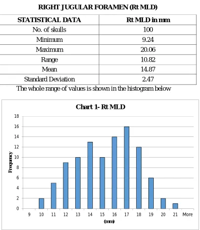
TABLE 1-MEDIOLATERAL DIAMETER OF
RIGHT JUGULAR FORAMEN (Rt MLD)
STATISTICAL DATA Rt MLD in mm
No. of skulls 100
Minimum 9.24
Maximum 20.06
Range 10.82
Mean 14.87
Standard Deviation 2.47
The whole range of values is shown in the histogram below
0 2 4 6 8 10 12 14 16 18
9 10 11 12 13 14 15 16 17 18 19 20 21 More
42
TABLE 2-MEDIOLATERAL DIAMETER OF LEFT JUGULAR
FORAMEN (Lt MLD)
STATISTICAL DATA Lt MLD in mm
No. of skulls 100
Minimum 8.34
Maximum 19.68
Range 11.34
Mean 13.76
Standard Deviation 2.37
The whole range of values is shown in the histogram below
0 2 4 6 8 10 12 14 16 18 20
8 9 10 11 12 13 14 15 16 17 18 19 20 More
43
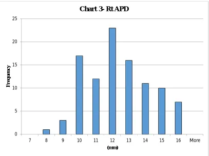
TABLE 3-ANTEROPOSTERIOR DIAMETER OF RIGHT
JUGULAR FORAMEN (Rt APD).
STATISTICAL DATA Rt APD in mm
No. of skulls 100
Minimum 7.59
Maximum 15.89
Range 8.3
Mean 11.90
Standard Deviation 1.93
The whole range of values is shown in the histogram below
0 5 10 15 20 25
7 8 9 10 11 12 13 14 15 16 More
F
r
e
q
u
e
n
c
y
44
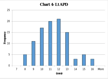
TABLE 4-ANTEROPOSTERIOR DIAMETER OF LEFT
JUGULAR FORAMEN (Lt APD)
STATISTICAL DATA Lt APD in mm
No. of skulls 100
Minimum 7.29
Maximum 15.23
Range 7.94
Mean 10.88
Standard Deviation 1.82
The whole range of values is shown in the histogram below
0 5 10 15 20 25
7 8 9 10 11 12 13 14 15 16 More
F
re
q
u
en
c
y
45
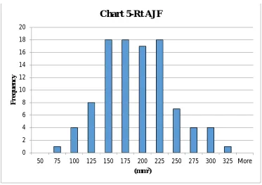
TABLE 5-AREA OF RIGHT JUGULAR FORAMEN (Rt AJF)
STATISTICAL DATA Rt AJF in mm2
No. of skulls 100
Minimum 70.13
Maximum 305.41
Range 235.28
Mean 179.23
Standard Deviation 49.39
The whole range of values is shown in the histogram below
0 2 4 6 8 10 12 14 16 18 20
50 75 100 125 150 175 200 225 250 275 300 325 More
F r eq u e n c y
(mm2)
46
TABLE 6-AREA OF LEFT JUGULAR FORAMEN (Lt AJF)
STATISTICAL DATA Lt AJF in mm2
No. of skulls 100
Minimum 78.38
Maximum 294.81
Range 216.43
Mean 151.65
Standard Deviation 43.58
The whole range of values is shown in the histogram below
0 5 10 15 20 25 30
75 100 125 150 175 200 225 250 275 300 More
F
r
e
q
u
e
n
cy
(mm2)
(A) - Right sided dominance
(B) - Left sided dominance
(C) - No side dominance
47
SIDE DOMINANCE OF JUGULAR FORAMEN – Table 7& Fig.20.
69% 25%
6%
Chart 7-Side Dominance of JF
Right dominance Left dominance No side dominance
Size Right
dominance
Left
dominance No dominance
Number 69 25 6
48
TABLE 8-WIDTH OF RIGHT JUGULAR FOSSA (Rt FJW)
STATISTICAL DATA Rt FJW in mm
No. of skulls 100
Minimum 5.02
Maximum 13.34
Range 8.32
Mean 7.90
Standard Deviation 1.80
The whole range of values is shown in the histogram below
0 5 10 15 20 25
5 6 7 8 9 10 11 12 13 14 More
F
re
q
u
en
c
y
(mm)
49
TABLE 9-WIDTH OF LEFT JUGULAR FOSSA (Lt FJW)
STATISTICAL DATA Lt FJW in mm
No. of skulls 100
Minimum 4.3
Maximum 12.11
Range 7.81
Mean 7.40
Standard Deviation 1.66
The whole range of values is shown in the histogram below
0 5 10 15 20 25 30
4 5 6 7 8 9 10 11 12 13 More
F
r
e
q
u
e
n
c
y
(mm)
50
TABLE 10-DEPTH OF RIGHT JUGULAR FOSSA (Rt FJD)
STATISTICAL DATA RFJD in mm
No. of skulls 100
Minimum 5.06
Maximum 21.79
Range 16.73
Mean 11.20
Standard Deviation 3.07
The whole range of values is shown in the histogram below
0 2 4 6 8 10 12 14 16 18 20 F r e q u e n c y (mm)
51
TABLE 11-DEPTH OF LEFT JUGULAR FOSSA (Lt FJD)
STATISTICAL DATA Lt FJD in mm
No. of skulls 100
Minimum 5.32
Maximum 17.78
Range 12.46
Mean 10.05
Standard Deviation 2.85
The whole range of values is shown in the histogram below
0 5 10 15 20 25
5 6 7 8 9 10 11 12 13 14 15 16 17 18 More
F
re
q
u
en
c
y
(mm)
52
TABLE 12-COMPARISON BETWEEN MEDIOLATERAL
DIAMETER (MLD), ANTEROPOSTERIOR DIAMETER (APD)
AND AREA OF RIGHT AND LEFT JUGULAR FORAMINA BY
PAIRED T TEST.
Parameter
(n=100) Side Mean SD SE
t value
p value
MLD
Right 14.87mm 2.47mm 0.25
3.22 0.0015 Left 13.76mm 2.37mm 0.24
APD
Right 11.90mm 1.93mm 0.19
3.85 0.0002 Left 10.88mm 1.82mm 0.18
Area
Right 179.23mm2 49.39mm2 4.94
4.19 0.04 Left 151.65mm2 43.58mm2 4.36
53
TABLE 13-COMPARISON BETWEEN WIDTH OF JUGULAR
FOSSA (FJW) AND DEPTH OF JUGULAR FOSSA (FJD) OF
BOTH SIDES BY PAIRED T TEST FOR STATISTICAL
SIGNIFICANCE
Parameter
(n=100) Side Mean(mm) SD(mm) SE
t value
p value
FJW
Right 7.90 1.80 0.18
2.08 0.04
Left 7.40 1.66 0.17
FJD
Right 11.20 3.07 0.30
2.74 0.007
Left 10.05 2.85 0.28
54
TABLE 14-COMPARISON BETWEEN MEDIOLATERAL
DIAMETER (MLD) AND ANTEROPOSTERIOR DIAMETER
(APD) OF JUGULAR FORAMEN OF THE SAME SIDE FOR
[image:86.612.122.526.182.376.2]CORRELATION.
TABLE 15-COMPARISON BETWEEN FJW AND FJD OF THE SAME SIDE FOR CORRELATION
Sl No. Parameter
(n=100) Mean (mm) SD (mm) Correlation coefficient value 1.
Rt FJW 7.90 1.80
0.196
Rt FJD 11.20 3.07
2.
Lt FJW
7.40 1.66
0.322
Lt FJD 10.05 2.85
Sl No. Parameter
(n=100) Mean (mm) SD (mm) Correlation coefficient value 1.
Rt MLD 14.87 2.47
0.471
Rt APD 11.90 1.93
2.
Lt MLD 13.76 2.37
0.407
55
With 100 pairs of observations, critical values of correlation coefficient at various levels of significance are given below.
Level of significance Critical value 0.05 0.195
0.01 0.254 0.001 0.321
Fig.21 : Dome of Jugular Fossa.
(A) Bilateral dome - both equal in depth (B) Bilateral dome - right deeper
(C) Bilateral dome - left deeper
(E) Unilateral dome on left side
(D) Unilateral dome on right side
56
PRESENCE OF DOMED JUGULAR FOSSA ON RIGHT SIDE
AND LEFT SIDE
[image:89.612.124.534.309.676.2]A dome in the roof of the Jugular Fossa was present bilaterally in 69% of skulls. It was observed unilaterally in 22% on the right side and 5% on the left side. A dome in the roof of the Jugular Fossa was absent bilaterally in 4% of skulls (Fig.21)
TABLE 16-INCIDENCE OF DOMED JUGULAR FOSSA WITH
SIDE DISTRIBUTION
Domed Jugular Fossa Right side
(n=100 sides)
Left side (n=100 sides)
Number
91 74
Percentage 91% 74%
91 74 0 10 20 30 40 50 60 70 80 90 100
Rt side Lt side
P e rc e n ta g e
Chart 12- Domed Jugular Fossa: side distribution
57
TABLE 17-INCIDENCE OF DOMED JUGULAR FOSSA (n= 100
SKULLS)
69% 22%
5% 4%
Chart 13- Domed Jugular Fossa
B/L present U/L present - R side U/L present - L side B/L absent
Domed Jugular Fossa
Bilaterally present
Unilaterally
present Bilaterally
absent Right side Left side
Number 69 22 5 4
58
TABLE 18- PRESENCE OF SEPTATE JUGULAR FORAMEN ON
RIGHT AND LEFT SIDES.
Presence of one septum- Bipartite JF
Right side (n=100 sides)
Left side (n=100 sides)
Number 32 29
Percentage 32% 29%
20 22 24 26 28 30 32 34
R side L side
32 29 P e r c e n ta g e
Chart 14- Septate JF (Bipartite) with side distribution
(A) - Septum absent bilaterally
(B) - Septum present unilaterally
(C) - Septum present bilaterally
59
TABLE 19- INCIDENCE OF SEPTATE JF, BIPARTITE TYPE
(n=100 SKULLS) Fig.22
Bilaterally present (n=100 skulls)
Unilaterally present
(n=100 skulls) Bilaterally
absent (n=100 skulls)
Right side Left side
21 11 8 59%
21% 11% 8% 59%
The presence of two septa (tripartite Jugular Foramen) was observed bilaterally in one skull (1%). Fig.23
21%
11%
8% 59%
1%
Chart 15- Septate JF
B/L Bipartite
U/L Bipartite- R side
U/L Bipartite- L side
B/L septum absent
60
TABLE 20-TYPE OF SEPTUM IN THE JUGULAR FORAMEN
WITH SIDE DISTRIBUTION
0 2 4 6 8 10 12 14 16 18 Complete Incomplete 17 15 16 13 P e r c en ta g e
Chart 16- Type of septum: side distribution
R side L side
Type of septum in Bipartite JF Right side
(n=100 sides)
Left side (n=100 sides)
Complete
Number 17 16
Percentage 17% 16%
Incomplete
Number
15 13
Percentage
[image:95.612.127.536.381.630.2](A) Bilateral incomplete septum (B) Bilateral complete septum
(C) Bilateral septum
incomplete septum complete septum
(D) Unilateral incomplete septum
61
TABLE 21- TYPE OF SEPTUM IN THE JUGULAR FORAMEN
(n=100 SKULLS) Fig.24.
Type of septum Bilaterally
present
Unilaterally present
Right side Left side
Complete
Number 7 10 9
Percentage 7% 10% 9%
Incomplete
Number 4 11 9
Percentage 4% 11% 9%
Tripartite Jugular Foramina, present bilaterally in one skull were each partitioned by one complete septum and one incomplete septum (Fig.23)
7% 10% 9% 4% 11% 9% 0% 2% 4% 6% 8% 10% 12%
B/L Present U/L Present- R side U/L Present- L side
Chart 17-Type of septum
1- occipital condyle 2 - anterior condylar canal opening
3 - styloid process 4 - mastoid process 2
3
4
1
Fig. 25 : Accessory opening in the medial part of
62
TABLE 22- PRESENCE OF ACCESSORY OPENING IN THE
WALLS OF JUGULAR FORAMEN ON RIGHT AND LEFT SIDES
Accessory opening was found in 7% on the right side and 4% on the left side. All the accessory openings were on the medial part of the posterior wall of JF. (Fig.25)
7 4 0 1 2 3 4 5 6 7 8
R side L side
P e r c e n ta g e
Chart 18- Accessory opening with side distribution
Accessory opening
Presence of accessory opening
Right side (n=100 sides)
Left side (n=100 sides)
Number 7 4
63
TABLE 23- INCIDENCE OF ACCESSORY OPENING IN THE
WALLS OF JUGULAR FORAMEN (n=100 SKULLS)
Presence of accessory
opening
Bilaterally present
Unilaterally Present
Right side Left side
Number 2 5 2
Percentage 2% 5% 2%
2%
5%
2%
0% 1% 2% 3% 4% 5% 6%
B/L present U/L present- R side U/L present- L side
(A) Leading to posterior condylar canal
1 - Occipital condyle 2 - Anterior condylar canal 3 - Foramen Magnum
(B) Leading to posterior margin of Foramen Magnum 1
2
3
64
TABLE 24- SITE WHERE THE ACCESSORY
OPENING LEADS TO
Site where the accessory opening
leads to.
Posterior condylar canal
Posterior margin of Foramen Magnum
Number 10 1
Percentage 91% 9%
Out of the 11 accessory openings, ten (91%) led to the posterior condylar canal (PCC)-Fig.26A.One accessory opening (9%) on the right side led to a canal which opened at the posterior margin of foramen magnum(FM)- Fig.26B.
91% 9%
Chart 20- Other end of the Accessory Opening
PCC
65
DISCUSSION
Findings of the present study were compared with those of similar studies conducted in India and abroad.
MEDIOLATERAL DIAMETER (MLD) OF THE JUGULAR
FORAMEN
OE Idowu34 (2004) studied 40 JF of 20 Nigerian skulls and stated
that the mean MLD on the right (Rt MLD) and left (Lt MLD) were 13.90 mm and 14.11 mm respectively.
Ekinci et al15 (2009) conducted a study on 70 skulls of Turkish
population and reported that Rt MLD was 16.0mm and Lt MLD was 15.5 mm.
Namita A Sharma et al33 (2011) studied the Jugular Foramina in
50 dry skulls and found that Rt MLD and Lt MLD were 15.59+/- 2.64mm and 13.83+/- 4.94mm respectively.
Ketu Chauhan et al28 (2011) analysed 50 dry skulls and reported
66
Anitha MR et al3 (2013) in their study of 100 adult dry skulls
observed that the mean Rt MLD and Lt MLD were 15.21mm and 13.39mm respectively.
Shifan Khanday et al48 (2013) studied 648 JF of 324 skulls and
found that mean Rt MLD was 14.6 mm and mean Lt MLD was 13.9 mm.
N. Himabindu et al32 (2015) studied the JF of 110 adult dry skulls
and reported that mean Rt MLD was 14.6mm and mean Lt MLD was 12.69mm.
In the present study, the mediolateral diameter of right JF ranged from 9.24mm to 20.06mm with mean as 14.87+/- 2.47mm. The mediolateral diameter of left JF ranged from 8.34mm to 19.68mm with mean as 13.76+/- 2.37mm. The mean RMLD and LMLD of the present study coincided with the values of all these studies (Table 25) except OE Idowu et al (RMLD 13.9mm and LMLD 14.11mm, with the value on right side being smaller than the value on left side) and Ekinci et al (RMLD 16.0mm and LMLD 15.5mm). This could be due to racial differences in the skulls.
67
IJV is larger than the left in most of the individuals. The larger superior sagittal sinus continues in succession as right transverse sinus, right sigmoid sinus and right IJV, on the other hand the smaller inferior sagittal sinus continues in succession as straight sinus, left transverse sinus, left sigmoid sinus and left IJV.7,61
TABLE 25SHOWING STUDIES WITH COMPARABLE VALUES
OF MEDIOLATERAL DIAMETER (MLD) OF THE JF.
Sl No. Authors Rt MLD(mm) Lt MLD(mm)
1. Namita A Sharma et al
(2011) 15.59 13.83
2. Ketu Chauhan et al (2011) 13.46 13.10
3. Anitha MR et al (2013) 15.21 13.39
4. Shifan Khandey et al
(2013) 14.6 13.9
5. N. Himabindu et al (2015) 14.6 12.69
6. Present Study 14.87 13.76
15.59
13.46
15.21
14.6 14.6 14.87
13.83
13.1 13.39 13.9 12.69 13.76
0 2 4 6 8 10 12 14 16 18 Namita A Sharma et al
(2011)
Ketu Chauhan et al (2011)
Anitha MR et al (2013)
Shifan Khandey et al
(2013)
N. Himabindu et al (2015)
Present Study
Chart 21- MEDIOLATERAL DIAMETER (MLD)