Early prediction of coagulopathy in
acquired bleeding disorders
-Revisited using Rotational
thromboelastometry
Experience in a tertiary hospital in South
India
Acknowledgement
First of all I thank God Almighty with whom everything is possible.
I would like to take this opportunity to express my deep sense of gratitude and respect towards my teacher and guide Dr. Sukesh Chandran Nair, Professor, for his inspiration, invaluable guidance, constant encouragement, and expert suggestions.
I am thankful to Dr. Joy Mammen, Professor and Dr Rajesh Kannangai, Professor for their advice in statistical analysis of this study.
I am deeply indebted to Dr Anup Joseph Devasia, Senior PG Registrar, for his constant encouragement, valuable suggestions and healthy criticisms.
I sincerely express my gratitude to my dearest colleague Dr. Tulasi Geevar and Dr Vishnu for all the help, consideration and cooperation given to me during this study period.
My thanks are due to all the staffs of Department of Transfusion Medicine and Immunohaematology, especially Miss.Remya V, for the generous help they have provided throughout the duration of my course. I thank all others who have assisted me in some form or the other in the preparation of this work.
Salutation to my dearest mother, husband, brother, sister and my beloved daughter who were a constant inspiration at every stage of this work.
Certificate
This is to certify that the dissertation ―Early prediction of coagulopathy in acquired bleeding
disorders – Revisited using Rotational Thromboelastometry ‖ is a bonafide work of Dr Soonam John towards the M.D. (Immuno Haematology and Blood Transfusion) Examination of the Tamil Nadu Dr M.G.R. University, Chennai to be held in 2015.
SIGNATURE:
Dr. Sukesh Chandran Nair
Professor, Department of Transfusion Medicine & Immunohaematology, Christian Medical College, Vellore, 632004, India
Dr. Joy J. Mammen
Professor and Head of Department, Transfusion Medicine & Immunohaematology Christian Medical College, Vellore, 632004, India
Dr Alfred Daniel
Declaration
Certificate
This is to certify that the dissertation titled ―Early prediction of coagulopathy in acquired
bleeding disorders – Revisited using Rotational Thromboelastometry ‖ which is submitted by me in partial fulfilment towards M.D. (Immuno Haematology and Blood Transfusion) Examination of the Tamil Nadu Dr M.G.R. University, Chennai to be held in 2015 comprises only my original work and due acknowledgement has been made in text to all material used.
SIGNATURE:
Soonam John
CONTENTS
AIMS AND OBJECTIVES……….1
INTRODUCTION………2
REVIEW OF LITERATURE……….6
MATERIALS AND METHODS………37
RESULTS……….58
DISCUSSION………99
LIMITATIONS………103
CONCLUSION………104
ABBREVIATION………105
BIBLIOGRAPHY………106
Title of the abstract: EARLY PREDICTION OF COAGULOPATHY IN ACQUIRED
BLEEDING DISORDERS – REVISITED USING THROMBOELASTOMETRY
Department: TRANSFUSION MEDICINE AND IMMUNOHAEMATOLOGY
Name of the candidate: DR SOONAM JOHN
Degree and subject : M.D. IMMUNO HAEMATOLOGY AND BLOOD TRANSFUSION
(BRANCH XXI)
Name of the guide: DR SUKESH CHANDRAN NAIR
Objectives
This study was done to establish a correlation between rotational thromboelastometryparameters
and standard coagulation profile in the context of acquired bleeding disorders.
Background
Acquired bleeding disorders are a major cause of mortality, both in the developed and
developing countries. An acute haemorrhage should be managed immediately with blood
products, factor concentrates or antifibrinolytics. Investigations to detect coagulopathies
typically include baseline screening tests like prothrombin time, activated partial thromboplastin
time, platelet count and fibrinogen level. These tests have a long turnaround time which
frequently lead to a blinded approach towards blood product support leading to under or over
transfusion. In contrast, rotational thromboelastometry (ROTEM) which assesses haemostasis
AIMS AND OBJECTIVES
Methods
A total of 138 subjects - 70 patients who presented with acquired bleeding disordersand 68 subjects diagnosed to be normal on the basis of a complete coagulation work up were included as the cases and controlsrespectively. All samples were subjected to standard coagulation profile by automated methods and ROTEM analysis which included Clotting Time, Clot Formation Time, Alpha Angle, Maximum Clot Firmness and Maximum Lysis.
Results
The Maximum Clot Firmness shows a very good correlation with serum fibrinogen levels (k value - 0.807; p<0.000; Sensitivity - 88%; Specificity - 92%), and good correlation with platelet count (k value - 0.793; p< 0.000; Sensitivity - 86%, Specificity-92%), whereas Clot Formation Time showed moderate correlation with activated partial thromboplastin time, platelet count and fibrinogen levels.Alpha angle had a moderate correlation with platelet count and fibrinogen.Clotting time had a poor correlation with prothrombin time and activated partial thromboplastin time.
Discussion and Conclusion
whereas standard coagulation profile required an average of 45minutes. In view of the good correlation to the standard coagulation profile, it appears that Rotational Thromboelastometryresults can be safely used to implement early transfusion therapy for haemorrhage.
INTRODUCTION
Bleeding disorders comprise a varied group of diseases due to the malfunction of blood vessels, coagulation proteins and platelets. The clinical spectrum varies from a mild bleeding diathesis to severe hemorrhage which can lead to death. It can be due to either quantitative or qualitative abnormalities of coagulation proteins or platelets. Mild bleeding symptoms like easy bruising, epistaxis, are more common than severe life threatening hemorrhage. The symptoms may sometimes reveal an underlying malignant bone marrow pathology.
Bleeding-history assessment tools can be used to assess the patient. A high bleeding score indicates a significant bleeding disorder but there can be an overlap of scores for individuals with mild bleeding disorder and without any bleeding symptoms.(1) The bleeding histories are subjective and therefore significant symptoms may sometimes be ignored and considered as normal while negligible or minor symptoms be given undue consideration.
Acquired bleeding disorders occur due to depletion, reduced synthesis, or inhibition of platelets and coagulation factors. The causes include disseminated intravascular coagulation, Vitamin K deficiency or antagonists, acquired inhibitors to coagulation factors, liver transplantation, renal causes, use of anticoagulant and traumatic bleeding. Disseminated intravascular coagulation can occur in various clinical conditions like septicemia, acute promyelocytic leukemia, abruption placenta, trauma. The most common condition resulting from a coagulation factor inhibitor is acquired Hemophilia A. The acquired platelet function disorders include drug usage,(Aspirin, Clopidogrel, Prasugrel) food additives, myeloproliferative neoplasm and Paroxysmal Nocturnal Hemoglobinuria.
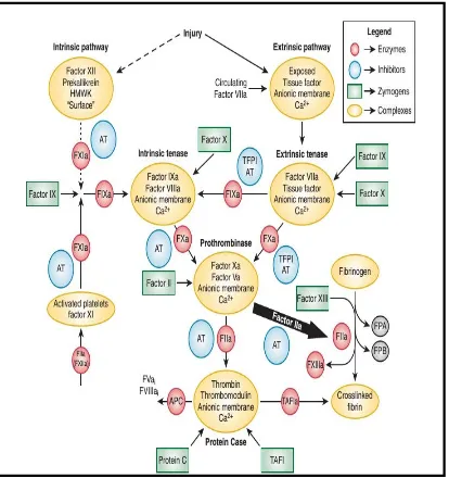
Overview of hemostasis
Figure 1- The coagulation pathway
partial thromboplastin time, serum fibrinogen, platelet count, help in exposing the cause of bleeding. Management depends on the history and laboratory tests. The treatment decision includes administration of vitamin K, blood, plasma, cryoprecipitate or platelets transfusion and blood derivatives like factor concentrates.
Acquired bleeding disorders are the most frequent causes of hemorrhage encountered in the practice of hematology. The various type of disorders include drug induced hemorrhage, acquired factor deficiencies, disseminated intravascular coagulation, acquired coagulation factor abnormalities like acquired Hemophila A, acquired von Willebrands disease, immune thrombocytopenia, non–drug induced, acquired platelet function disorders, vitamin K deficiency, liver and renal disease, hypothyroidism, surgical bleeding, scurvy, hypothyroidism and Cushings syndrome. The most common cause is a drug-induced defect. (2)
An acute bleed should be managed immediately with blood products, factor concentrates antifibrinolytics before the results of coagulation test are available. The investigations for a bleeding problem includes prothrombin time, activated partial thromboplastin time, platelet count and fibrinogen level. Abnormalities, if detected, require further evaluation to determine if the cause is a fibrinogen disorder, platelet disorder or coagulation factors deficiencies.
REVIEW OF LITERATURE
History
In 1628, William Harvey described the circulatory system.(3) In 1825, the first successful human-to-human blood transfusion was reported by James Blundell, an obstetrician and physiologist at Guy’s Hospital in London who transfused women with postpartum
haemorrhage.(4) During World War I and II blood transfusion was given to bleeding soldiers to combat the war injuries.(5)
Ratnoff and Davies proposed the coagulation pathway, which was composed of a series of proteolytic reactions starting with factor XII and terminating in the formation of clot. (6) The prothrombin time was introduced by Dr Armand Quick in 1935 which was used to assess coagulopathy in Hemophilia and was later used in acquired bleeding disorders.(7) In 1936, Warner et al developed a two-stage prothrombin assay, based on the fact that plasma, serum factors and tissue factor are required for the conversion of prothrombin to thrombin. (8) The partial thromboplastin time was illustrated by Langdell and was used to assess Hemophila. The platelet count estimation was done with the help of automated platforms since the early 60’s.(9)
Homeostasis
The hemostatic system or the coagulation cascade was based on the waterfall hypothesis of Ratnoff and Davies(9) and MacFarland,(10) who independently reported a series of proteolytic reactions beginning with factor XII activation and ceasing with the thrombin converting fibrinogen to form a thrombus. The shortcoming of this hypothesis was that factor XII deficiency was not associated with bleeding.(11) The cofactors for factor XII activation; prekallikrein and high- molecular-weight kininogen deficiencies also were not associated with hemorrhage.(12)
Over the decades these pathways have been modified The newer concept of coagulation represents a complex process of procoagulant and fibrinolytic actions which occur simultaneously.
The Vitamin K–dependent protein family
Vitamin K–dependent factors are produced in the liver. These factors act as either procoagulant or anticoagulant. These include factors II,VII, IX, X and the anticoagulants factors protein C, protein S, and protein Z. (13) Vitamin K is essential for the synthesis of these clotting factors which undergoes γ-carboxylation.(14) This alteration adds a negative charge to the molecules
that enables proteins to interact with Calcium and surface of membrane. Vitamin K– dependent protein complexes are composed of a serine protease enzyme, a cofactor, and a negatively charged membrane surface. The four vitamin K–dependent complexes include the extrinsic tenase complex ( FVIIa and tissue factor), the intrinsic tenase complex (FIXa–FVIIIa), the prothrombinase complex (FXa–FVa), and the anticoagulant protein Case complex (thrombin–thrombomodulin).
Cofactor proteins
2. Soluble plasma-derived procofactors - factor V, factor VIII and von Willebrand factor
.(6)
Tissue factor
A cofactor for factorVIIa in the extrinsic tenase complex. The release of tissue factor to the circulation initiates the procoagulant pathway. (15)
Thrombomodulin
A protein expressed on the surface of vascular endothelial cells. It acts as a cofactor for the activation of protein C. (16) The formation of protein C by the thrombin–thrombomodulin complex causes inactivation of factor Va and factor VIIIa.(17) This complex has an antifibrinolytic action by activating the fibrinolysis inhibitor, thrombin activatable fibrinolysis inhibitor (18)
Factor VIII
The factor VIII also known as the Antihemophilic factor circulates in plasma in complex with von Willebrand factor(19) The von Willebrand Factor–free factor VIIIa forms a complex with factor IXa, Calcium and platelet membrane resulting in the formation of intrinsic tenase complex.
The Intrinsic Accessory Pathway Proteins
understood.(21) This pathway play an important role in disseminated intravascular coagulation (DIC) associated with the systemic inflammatory response syndrome and also in cardiopulmonary bypass where there is contact between blood and synthetic surfaces.(22) The factor XI is an intersection point for the two pathways. Individuals with factor XI deficiency have a inconsistent bleeding phenotype proving that there is a role for factor XI in hemostasis. (23) The propagation phase of thrombin generation is carried out by FXI a.(24)
The clot formation inhibitors in the blood include antithrombin, heparin cofactor II, tissue factor pathway inhibitor and protein C inhibitor.
The integrity of vasculature is necessary for normal hemostasis. The endothelium that lines the interior of blood vessels, plays an important role by providing a structural barrier to blood regulates blood pressure and deposits an intricate basement membrane and extracellular matrix. Massive bleeding can occur due to abnormalities of the endothelial cells.(25) Studies by Ware and Seegers had identified that phospholipid is required for coagulation.(26) The damaged vascular tissue activated platelets produce phospholipids. The endothelial damage release von Willebrand factor that causes platelet aggregation and adhesion to the sub endothelium leading to thrombus formation.
Platelets are small, cell fragments, derived from megakaryocytes contribute to procoagulant events and fibrinolytic process. The platelets contain secretory granules: α-granules, lysosomes,
binding sites (27) Factor Xa bind to effector cell protease receptor-1 molecules which are present on activated platelets.(28)
The presence of receptors on the platelet surface thrombin, adenosine diphosphate, and thromboxane A2 to channel other platelets into the hemostatic. The close contact between platelets promote stabilization of the hemostatic plug (29)
Clot proteins
Fibrinogen helps in hemostasis to prevent hemorrhage. It supports inter platelet aggregation. Fibrinogen controls thrombin activity to form a fibrin clot. Fibrinogen consists of polypeptide chains with the NH2 termini cross linked to each other. Fibrinogen increases during inflammation as it is an acute phase reactant.(30)
Fibrinolysis or the removal of blood clots includes primary fibrinolysis and secondary fibrinolysis. The primary fibrinolysis is a normal process while secondary fibrinolysis is the due to a medicine or a medical disorder. Plasminogen is the precursor of the plasmin, which is the primary catalyst of fibrin degradation.(31) The activation of plasminogen occur through three pathways: (a) the intrinsic activator system (b) the extrinsic activators (c)an activator system involving fibrinolytic drugs. The plasmin degrades fibrin producing degradation products.
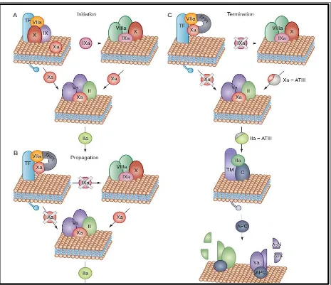
Initiation
the vascular system is perturbed the extrinsic tenase complex which is the tissue factor–factor VIIa and the calcium complex , comes into play.(33) The extrinsic tenase complex activates low levels factor X and factor IX to Xa and factor IXa.(34) The time interval in which factor Xa generates thrombin is called as the initiation phase of blood coagulation.(35) During the initiation phase, circulating blood cells and procoagulant proteins are activated, and a initial fibrin network is formed.(36)
Amplification
Thrombin also activates factor XI to Xia , initiating the accessory pathway that promotes factor IX activation.(37) The aggregated platelets and fibrin due to the thrombin formation are the major constituents of the initial vascular plug formation. The cofactor factor VIIIa combines on activated platelets with the factor IXa to form the intrinsic tenase complex which activates factor X. (38) Factor Xa formed from factor X combines with factor Va on the platelet membrane to form the prothrombinase complex which generates thrombin. (24) The prothrombinase and the intrinsic tenase complex are protected from inhibition by antithrombin.
Propagation
The coagulation mechanism is susceptible to factor V and platelets and is affected in case of congenital deficiencies, thrombocytopenia, abnormal platelet function or pharmacologic interventions.(41) Nearly 95% of thrombin is produced during the propagation phase after formation of fibrin clot.(42)
Termination
When blood flow stops due to the formation of a fibrin clot, the TFPI, antithrombin, heparin cofactor II, α2-macroglobulin, α1-antitrypsin, and protein C inhibitor, inhibit the various reactants as they dissociate from their respective complexes.(43) In the intact blood vessel surrounding the newly formed clot, procoagulant enzymes are rapidly reduced under normal circumstances. The thrombin, factor IXa, and factor Xa are repressed by the excess antithrombin molecules. The thrombin bind with thrombomodulin and is converted to an anticoagulant enzyme from a procoagulant state.(44) This complex activates protein C, which causes decrease of intrinsic factor tenase and prothrombinase. . These intravascular mechanisms function against occlusion of the blood vessel.
Elimination and Fibrinolysis
The hemostatic clot is composed of aggregated platelets and fibrin. There are some plasma proteins and blood cells which are trapped within the clot. Finally clot disintegration occurs with the help of plasmin which maintains hemostatic balance. The tissue plasminogen activator generates plasmin at the fibrin surface and governs fibrin homeostasis, and u-PA generates pericellular plasmin.(45) The t-PA and plasminogen bind to the fibrin surface which results in augmented plasminogen activation. Fibrinolysis is regulated primarily by PAI-1 (46) PAI-2, α2-antiplasmin, and TAFI. Antiplasmin is the primary inhibitor of plasmin. The α2-antiplasmin,
activation.(47) The elimination phase begins with tissue repair by dissolving the fibrin–platelet clot generated earlier in hemostasis. The plasmin clears the fibrin clot and also removes damaged tissue. If a sufficient hemostatic plug is not formed to arrest blood flow, it can result in pathologic hemorrhage.
Acquired bleeding disorders
Acquired bleeding disorders are commonly seen in the practice of hematology. The symptoms vary from mild bruises to life threatening hemorrhage. The clinical, family, social, and medication history site, frequency and the age of onset aids in the diagnosis of the type of disorder. These disorders can be due to consumption, reduced synthesis, inhibitors to either coagulation factors or platelets. The treatment depends on the cause of bleeding and includes blood component transfusion, factor concentrates administration of vitamin K and anti fibrinolytic agents.
Disseminated Intravascular Coagulation
bleeding manifestations and intracranial bleeds are relatively rare. The occlusion in the vasculature of organs by microthrombi results in necrosis causing multiple organ failure.(50)
CLINICAL CONDITIONS THAT CAUSES DIC
Sepsis
Trauma , Burns or heat stroke
Malignancy – Solid tumours, AML M3
Obstetric conditions – Amniotic fluid embolism, Placental abruption, HELLP
Vascular abnormalities- Aortic aneurysms, Kasabach-Merritt syndrome
Allergic reactions
Immunological reactions
Table 1 - Causes of Disseminated Intravascular Coagulation
Pathogenesis
Clinical manifestations
Disseminated Intravascular Coagulation in Infectious Disease
Infections are the most common causes of DIC. Septicemia is the most common clinical condition associated with disseminated intravascular coagulation. Mostly all microorganisms can cause disseminated intravascular coagulation and bacterial infection is most frequently related to the development of this syndrome. The triggers of the activation of coagulation in septicemia patients are cell membrane components of the microorganism which includes lipopolysaccharide , bacterial exotoxins which induce a generalized inflammatory response causing release of cytokines.(51)
Newborns and post splenectomy patients are prone to DIC.(52) Hemorrhage can be further aggravated by thrombocytopenia. Bacterial infections are more commonly associated with the syndrome. Viruses , protozoa , and fungi can cause DIC.(53) In septic patients can present with thromboembolic disease or multiple organ dysfunction.(48)Patients may present with severe bleeding in some cases.(54) Purpura fulminans is lethal form of DIC which affects infants and children in which the skin over the extremities undergo hemorrhagic necrosis due to diffuse thrombi in small blood vessels . This disorder occur due to meningococcemia,streptococcal disease or staphylococcus.(55)
Disseminated Intravascular Coagulation in Trauma and Burns.
prompt medical care can reduce the risk of DIC. The massive release of Tissue factor to the blood circulation, hemorrhagic shock, cytokines are possibly the factors that initiate DIC .TIC is due to dysfunction of inflammation, anticoagulation, and cellular systems. The underlying mechanisms of TIC have not been fully revealed. In addition dilutional coagulopathy, hypothermia, and acidosis also contributes to TIC. (56) Surgical excision of burn wounds causes severe bleeding leading to coagulopathy .(57)
Disseminated Intravascular Coagulation in Obstetric and Gynecological Complications
Postpartum hemorrhage (PPH) is significant problem in obstetrics and a leading cause of maternal deaths worldwide. The incidence of PPH is approximately 5%-20% of labors (58) with the highest rates in developing countries. Postpartum hemorrhage is associated with disseminated coagulopathy. The activation of coagulation system results in reduction of platelets and coagulation factors, aggravating bleeding due to consumption.(59)
The majority of cases due to obstetric pathology. The major obstetrical cause of postpartum hemorrhage is uterine atony which can lead to coagulation failure. (60) Placental abruption causes perinatal death and can lead to DIC. (16) Antenatal women with hypertension at highest risk. DIC can occur in such situations due to of enormous amounts of tissue factor in the blood circulation. The leakage of thromboplastin-like product from placenta which occurs in abruption leads to disseminated intravascular coagulation.(61)
revealed fetal debris and the meconium, in the amniotic fluid causes thrombus formation and fibrinolysis, leading to the occlusion of the pulmonary arteries causing breathlessness, cyanosis, shock, acute corpulmonale, left ventricular dysfunction and convulsions. These symptoms are followed by severe bleeding in some patients. (62)
Disseminated Intravascular Coagulation in Malignancy
Patients with solid tumors are susceptible to DIC that can exacerbate hemorrhage. The risk factors include old age, stage of the disease, chemotherapy , infection, liver metastases .(63) In acute promyelocytic leukemia, increased fibrinolysis causes hemorrhage. Studies in mice model suggested that annexin II have a role in acute promyelocytic leukemia coagulopathy.(64) The blasts overexpress annexin II, a receptor for tissue plasminogen activator (tPA), and plasminogen, and increases plasmin generation. The brain and lungs are the most common sites affected which can lead to fatal hemorrhage.(65)
Solid tumor cells express procoagulant molecules like tissue factor , which forms a complex with factor VII and activate factors IX and X, an endopeptidase which activates factor X.(66) . Nearly 15% to 20% of acute lymphoblastic leukemia, present with disseminated intravascular coagulation.(63)
Disseminated Intravascular Coagulation With Vascular Disorders
Disseminated Intravascular Coagulation With Toxic Reactions or Snake Bites
The venom of vipers and rattlesnakes, can produce a coagulopathy similar to that of DIC.(69) The venoms of these snakes contain enzymes that function by the following mechanism:
a) release fibrinopeptide A
b) activate prothrombin even in the absence of calcium
c) activate factors X and V
d) degrade fibrinogen
e) cause platelet aggregation
f) inhibit platelet aggregation
g) Damage endothelial cells and cause bleeding, tissue ischemia, and edema.
The victims of snake bites rarely have excessive hemorrhage.(6)
Liver disease and orthotopic liver transplantation
Low platelet counts is frequent in liver disease and the platelet counts range from 30 - 100× 109/L.(70) Platelet aggregation also occurs which causes prolonged bleeding time. (71) The increase of tissue plasminogen activator in plasma and decrease of the naturally-occurring inhibitors of plasmin results in hyperfibrinolysis (72)
naturally occurring anticoagulants.(76) Plasma hyperfibrinolysis has been reported in patients with chronic liver disease (36)
In liver transplantation age, severity of liver disease, hemoglobin and plasma fibrinogen values determine the bleeding crisis. During the hepatectomy and the anhepatic phase, hemorrhage occurs due to decrease in clotting factors either due to surgical bleeding or the primary disease. The coagulopathy exacerbates on infusion of crystalloid, colloid, or blood products.(77) At the time of graft reperfusion, further depletion of all coagulation factors, antithrombotic factors occurs which further aggravate bleeding. (37)
Renal disease
Bleeding diathesis have been reported in patients with uremia especially when they undergo surgery and other invasive procedures. Life -threatening conditions like pericardial tamponade, intracranial bleeding, and gastrointestinal bleeding are also seen in these conditions.(78) The patients have a platelet dysfunction and an abnormal platelet–vessel wall interaction.(79) Various studies have shown that contact of fibrinogen to ADP-stimulated platelets is impeded and the interaction between vWF and the GPIIb-IIIa is disturbed.(80)
Vitamin K deficiency
Vitamin C deficiency (Scurvy)
This is a rare condition especially in developed countries, when patient presents with perifollicular hemorrhages and gum bleeding (83). There are case reports of scurvy patients presenting with hemarthrosis.(84)
Massive transfusion
Massive transfusion is defined as 1) transfusion of more than 10 red cell concentrate units, which is nearly the total blood volume of an average adult patient, within 24 h 2) transfusion of more than four red cell concentrates in 1 h with expectation of continued need for blood product support 3) replacement of fifty percent of the total blood volume by blood products within 3 h.(85–87) The hemostatic defects in such patients have multifactorial pathogenesis causing early trauma induced coagulopathy leading to further transfusion support.(88) Coagulopathy can be aggravated by the infusion of crystalloids and blood products The transfusion of red cell units without additional clotting factors or platelets causes further impairment of hemostasis due to dilutional coagulopathy , thrombocytopenia ,acidosis , hypocalcaemia and hypothermia from refrigeration.(89,90)
Acquired coagulation factor inhibitors
Acquired factor V inhibitor is another rare entity in the elderly, that presents with hemorrhagic manifestations. The probable causes for the inhibitor development include exposure to bovine thrombin, antibiotics, surgery and malignancy. (95) Acquired FX deficiency presents with variable symptoms, and there are various case studies showing association with amyloidosis.(96,97)
Hypothyroidism
There are case reports on association of hypothyroidism with acquired von Willebrand disease presenting with bleeding manifestations .The coagulation defects were corrected with the management of hypothyroidism.(98,99)
Cushing syndrome
Easy bruisability and menstrual disturbances are features of Cushing syndrome or treatment with systemic or topical glucocorticoids.(100)
Drug -induced platelet dysfunction
Aspirin, NSAIDs, other platelet function inhibitors are common causes of drug induced bleeding.(101,102)
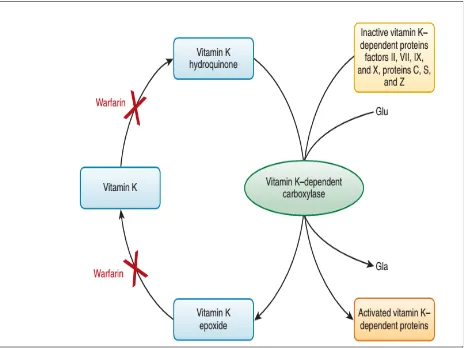
Anticoagulation
includes reversal of anticoagulant, vitamin K injection. In emergency situations fresh frozen plasma and coagulation factor concentrates can be used. (69)
Figure 3 – Mechanism of action of Vitamin K antagonists.
LABORATORY ASSESSMENT
STANDARD COAGULATION PROFILE
APTT can be very shortened in early acute DIC due to the presence of activated coagulation factors.(103) Fibrinogen depletion occurs in severe DIC but can be high or normal since it is an acute phase reactant.(104) In severe cases of DIC, microangiopathic hemolytic anemia may be
seen .Disseminated intravascular coagulation can be diagnosed on the basis of the following: a) An underlying cause like sepsis, trauma, burns.
b) Platelet count of less than 100,000 per cubic millimeter/ rapid decline in the platelet count. c) Prolonged prothrombin time and the activated partial-thromboplastin time
d )The presence of fibrin-degradation product. e) Depleted plasma fibrinogen. (48)
Low plasma fibrinogen levels of less than 50 mg/dL are associated with severe hemorrhage in DIC. Fragmented red blood cells or schistocytes will be seen in DIC cases. Toxic changes in leukocytes will be present in the smear of sepsis patients.(105)
[image:33.612.75.401.456.664.2]
LIMITATIONS OF STANDARD COAGULATION PROFILE
The PT and aPTT tests identify only partially if any coagulation defects are present. These tests also give a high number of false positive and false negative results.(106)In obstetrics pre-procedural coagulation screening is usually not recommended. A complete bleeding and medication history is considered more effective than doing coagulation profile. If there is a hemostatic impairement like placental abruption , coagulation profile is done.(107)
THROMBIN GENERATION TIME
Thrombin which acts as a procoagulant and anticoagulant is an important enzyme of the coagulation cascade. The thrombin generation is one of the prime steps in coagulation. The thrombin generation test is suitable for its assessment. This test detects both pro- and anticoagulant processes, so it can be applied for the detection of thrombosis and bleeding. Anticoagulant therapy can be monitored by this test. This assay can be used to assess bleeding severity , monitoring treatment and inhibitor by passing therapy in Hemophilia cases.(108)
THROMBOELASTOGRAPHY
coagulopathy Thrombelastography can diagnose fibrinolysis and can be used to decide on anti-fibrinolytic drugs, cryoprecipitate and fibrinogen concentrate therapy. (109)
Figure 5- Normal thromboelastography
ROTATIONAL THROMBOELASTOMETRY
Background
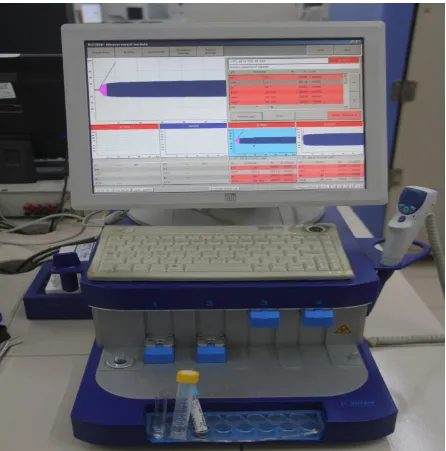
Figure 6- Thromboelastometry ( ROTEM) machine
Technology
whole blood sample and a pin suspended on a ball bearing mechanism initially oscillates through 4° 75′ every 6 sec through application of a constant force. As the strength of the clot
increases the rotation of the pin is hindered it is detected optically using a image sensor system. ROTEM device is capable of analyzing 4 samples simultaneously. ROTEM provides automated pipetting. (111)
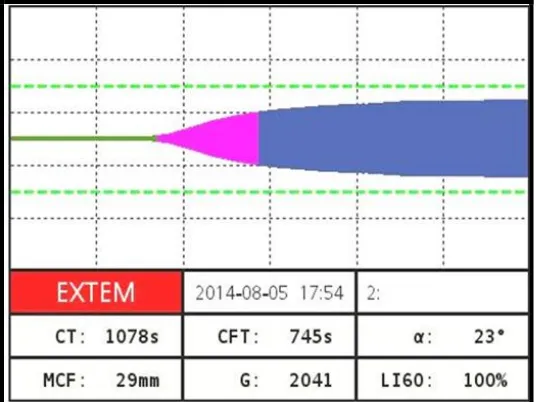
Parameters (Figure 6)
Clotting Time (CT)
It is the time from the beginning of the test by adding the clot activator until the time when amplitude of 2 mm is achieved.
Clot Formation Time (CFT)
It is the time between 2 mm amplitude and 20 mm amplitude of the clotting signal.
Alpha Angle (α)
It is defined as the angle between the middle axis and the tangent to the clotting curve through the 2 mm amplitude point. It describes the kinetics of clotting.
Maximum Clot Firmness (MCF)
It is the measure for the firmness of the clot and therefore the clot quality. It is the maximum amplitude that is reached before the clot is dissolved by fibrinolysis and the clot firmness falls again.
Maximum Lysis (ML)
ROTEM Assays
INTEM Contact activation.
Reagent - phospholipid and ellagic acid as activators Provides information similar to that of the APTT.
EXTEM Tissue factor activation.
Reagent contains tissue factor as an activator. Provides information similar to that of the PT.
APTEM Aprotinin for inhibiting fibrinolysis
Used in conjunction with EXTEM FOR assessing fibrinolysis.
HEPTEM Contains lyophilized heparinase for neutralizing unfractionated heparin Used in conjunction with INTEM to assess heparin effect.
FIBTEM Utilizes cytochalasin D, an actin polymerization inhibitor to block the platelet contribution to clot formation.
Used in conjunction with EXTEM reagent for qualitative analysis of the fibrinogen contribution to clot strength independent of platelets.
The use of rotational thromboelastometry in transfusion support assessment allows targeted hemostatic approach. This strategy reduces blood usage as compared to a blinded approach to transfusion on the basis of laboratory tests with long turnaround times. The usual turnaround time for PT, aPTT, fibrinogen concentration and platelet count is generally long (nearly 45- 60 min) to determine the therapy in severely bleeding patients. The initial variables of ROTEM are available within 15–20 min.(112)
APPLICATION IN CLINICAL CONDITIONS
CARDIAC SURGERY
Hemorrhage following cardiac surgery is a common clinical crisis. There are various studies on the use of ROTEM for analysis of hemorrhage which appear to reduce the transfusion requirements as compared to standard coagulation profile. Studies have shown that rotational thromboelastometry can predict thrombocytopenia and hypofibrinogenemia in cardiac surgery using the clot amplitude after 5 minutes and the turnaround time of ROTEM was less than the conventional laboratory tests. (112) Studies in cardio thoracic patients have shown that ROTEM helps in detection of causes for postsurgical hemorrhage (113) Another study showed that thromboelastometry can be used in the study of the coagulation profile of cardiac surgery patients in post perfusion period. This gives information regarding hemostasis disorders and aids in the management.(114)
dose-dependent manner. This combination of fibrinogen and platelets improved the clotting time and strength. The addition of platelets promoted platelet aggregation in a dose-dependent manner while fibrinogen had no effect. The combination of fibrinogen and platelets improved platelet aggregation less than when the platelets were used alone. In cardiac surgery patients, the use of platelet and fibrinogen concentrates on blood samples had an additive effect on clot formation as compared to the individual components. These results had an impact on clinical transfusion protocols.(115)
TRAUMA
Trauma induced coagulopathy is caused by systemic anticoagulation and hyperfibrinolysis which occurs due to endothelial injury and protein C activation following hemorrhage from trauma sites, fluid replacement, hypothermia and tissue acidosis following hypo perfusion .(116) Some studies have shown that ROTEM analysis was better in comparison to standard coagulation tests in diagnosing trauma induced coagulopathy.(117) In some centers, ROTEM analysis is proved to be useful in diagnosing Trauma induced coagulopathy and was used in transfusion.(117) Hyperfibrinolysis associated with trauma can be diagnosed with the help of ROTEM.(118) The A10 variable correlated with platelet count and fibrinogen concentration and detection help in earlier goal-directed transfusion therapy and may allow modification of existing transfusion algorithms.(119)
OBSTETRICS
specific coagulation deficiencies in these patients. This test is useful in hypofibrinogenemia and hyperfibrinolysis .The coagulopathy of PPH is can be due to dilutional, surgical, amniotic fluid embolus, and placental abruption causes and the ROTEM requires nearly 24 hours monitoring.(120)A study by Theusinger et al showed that maximum clot firmness in all channels were associated with fibrinogen and platelets levels, Clotting time significantly to aPTT and maximum clot firmness significantly to fibrinogen. Factor VIII also co related well with all ROTEM parameters except with Clotting time and Clot formation time. (121)
LIVER TRANSPLANTATION
In orthotopic liver transplantation, intraoperative bleeding is a major problem. The underlying liver pathology, the replacement of large blood losses, and the altered metabolism of coagulation factors during the prehepatic, anhepatic, and neohepatic phases of liver transplantation create a complicated hemostatic environment.(98) ROTEM assays can differentiate hypofibrinogenemia from thrombocytopenia based on the channels used.(122) A study by Blasi et al concluded that clot thickness at 10 minutes can be used to taken decisions regarding transfusions.(123)
The most important pathology in poly trauma patients are fibrinogen deficiency, poor fibrinogen polymerization and hyperfibrinolysis. A study by Tauber et al showed that when compared with standard coagulation profile, ROTEM parametersdetects trauma induced coagulopathy. The results can be obtained within a short time and reflects the in vivo situation in poly trauma patients. These patients predominantly showed hypofibrinoginemia and defective fibrin polymerization. In this study they found that a Maximum Clot Firmness of less than 7 mm, corresponds to a fibrinogen level of less than 150 mg/dl and those subjects had a higher mortality. Prothrombin time, fibrinogen concentrations and platelet count co- related with MCF. There was a high mortality rate for hyperfibrinolysis in this study, it and this can be reduced by early administration of tranexamic acid. (126)
OTHER CLINICAL CONDITIONS
A study by Mittermayr et al used Thromboelastometry heparin-protamine management. The variable used was CT. The ratio of CT in INTEM channel to CT-HEPTEM channel was used to differentiate the effects of heparin excess from those of protamine excess. When the ratio was more than 1 it is due to excess heparin while ratio equal to 1 was due to excess protamine.(131)
Graphical presentations in various clinical conditions
Figure 7 - A normal thromboelastogram
Figure 9 – Thromboelastogram with prolonged CT,CFT and decreased MCF and alpha angle
Figure 10 – Thromboelastogram with shortened CT and CFT
Figure 11-Thromboelastogram with prolonged CFT and decreased
MCF and alpha angle
Figure 12 – Thromboelastogram with prolonged CT, CFT, decreased MCF and alpha angle as in severe coagulopathy
MATERIALS AND METHODS
This was a prospective study of patients who presented with acquired bleeding disorders in Christian Medical College, Vellore. The study was conducted on seventy newly admitted consecutive bleeding patients in the wards, the emergency and intensive care units of Christian Medical College Hospital for a period of two years from 01 January 2013. A detailed history, diagnosis, complete blood count profile, transfusion requirements, condition at discharge were collected with the help of a prepared pro forma.
STUDY POPULATION
TEST SAMPLES
CONTROL SAMPLES
In our hospital, the Department of Transfusion Medicine and Immuno hematology manages the Clinical Pathology Lab, Blood Bank, HLA lab and Special Test Lab for Coagulation work up. Patients who are referred from outside hospitals and out patients from other clinical departments who had presented with minor bleeding symptoms in the past that did not require any medical , surgical or transfusion interventions or no bleeding symptoms and had come for complete coagulation work up in Special test Lab were selected as the control group. On the basis of a complete coagulation work up, which included, complete blood count profile, standard coagulation profile, coagulation factor assays, Ricof levels and Platelet aggregometry studies, this category was diagnosed to have a normal coagulation profile. The first 68 consecutive samples during the study period were selected as the control samples.
Inclusion criteria:
Samples with abnormal thromboelastogram.
Exclusion criteria:
1. Paediatric cases – The normal reference range for prothrombin time, activated partial
thromboplastin time, platelet count and serum fibrinogen levels are different from the adult ranges. There are no well defined pediatric reference ranges for rotem variables.
2. Repeat sample collected within fifteen days - Interventions like transfusion,
Detailed history including type of bleeding with duration, diagnosis , treatment details like transfusion support with components, antifibrinolytics, vitamin K, factor concentrates were taken in detail.
Setting where data was collected
Special test lab, Department of Transfusion medicine and Immuno haematology, Christian Medical college, Vellore.
Sampling strategy employed
All consecutive samples with abnormal thromboelastometry within the given time period were included as test samples. Consecutively selected 68 samples with normal coagulation profile from patients with minor acquired bleeding disorders were considered as controls. Selection was dependent on the reference standard which is the coagulation profile. Samples with abnormal coagulation profile were considered as cases and normal samples as controls. Data was collected from participants prospectively (before performing the diagnostic tests)
automated methods using Sysmex machine and Coulter. All samples including cases and controls were subjected to thromboelastometric analysis using the ROTEM machine.
STANDARD OPERATING PROCEDURE
ROTEM
Purpose
To check the coagulation state of a blood sample.
Principle
Sample
Citrated whole blood (platelet rich plasma can also be used)
Materials:
Citrated whole blood, Disposable cups and pins, 100-1000µl, 5-40µl pipettes, 0.2M Calcium Chloride, Diluted thromboplastin (1/2000), Personnel Protective Equipment (PPE)
Sample preparation
Blood Sample: Blood is preferably drawn via a 21G needle directly into vacuette Greiner tube containing 3.2% sodium citrate with minimal stasis. The temperature of the sample may influence the measurement results. Measure the blood sample directly after sampling. If this is not possible, preheat the blood sample for 5-10 min before measurement in the sample preheating station of the ROTEM delta.
Reagent preparation
Preparation of Tissue factor:
Dilute 10µl of recombiplastin into 490µl of Imidazole buffer in tube 1.
Add 50 µl of the contents in tube 1 to 950 µl of Imidazole buffer in tube 2 and mix well.
Add 500µl of the contents in tube 2 to 500 µl of imidazole buffer in tube 3 and mix well.
Procedure
Switch on the ROTEM system. Activate the ROTEM system with the main switch on the back of the device. Push the blue on/off button on the right hand side of the instrument. Log in to the system. Touch the screen if the screen saver is active. Select user. Enter password Measurement module screen will open.
Measuring Cell Preparation
Take the cup with the pin in it from the storage box. Push the pin in the cup onto the axis chosen for measurement. The status line under the channel is grey by default. In case the axis has been moved heavily when attaching the pin, the channel becomes inactive and the status line turns blue during the time of initialization. Place the cup with its opening facing upwards into the appropriate preheated cup holder. Leave the cup holders always in the temperature controlled area. Push and fix the cup in the cup holder using the MC Rod. The cup must fit tightly. Enter Patient Data: Touch one of the four channels. In the upper part of the screen entry fields for patient data {Patient ID, Patient name (first name, last name, and comment} are displayed. Touch the respective entry field. Enter patient data.
Procedure
Add 20µl of 0.2M CaCl2 pre warmed cup. Add 30µl of diluted recombiplastin to a separate plastic vial. Mix 500µl of well mixed whole blood the tissue factor. Add 320µl of the mixture to the CaCl2 and mix well. Place the cup holder onto the measuring position using the guiding rods. The cup holder is kept in measuring position by magnets. Press the ―Start manual‖ icon the
Interpretation
During measurement, the large Thromboelastogram is shown at the left upper side of the screen. In the upper right part of the screen, the current measurement results of the test parameters are shown.
ROTEM Parameters
Clotting Time (CT), Clot Formation Time (CFT), Alpha Angle (α ) Maximum Clot Firmness
(MCF) and Maximum Lysis (ML)
Clotted sample can interfere with the results.
PROTHROMBIN TIME (PT)
Purpose
To look for the overall efficiency of extrinsic pathway of coagulation and monitoring of oral anticoagulant therapy.
Platform - Sysmex CS 2000i
Principle
Limits of detection - 5 to 180 Sec
Primary sample-
Citrated Plasma sample.
Type of container
Light blue top vacutainer and 1ml mini collect Greiner Vacutte for pediatric purposes.
Reagents and Materials
Dade Innovin: product is reconstituted as per product insert/instruction sheet. This reagent already contains CaCl2. Patient plasma, control plasmas as required.
Procedure
From Main Menu - Press 'Reagent' icon. The reagent carousal and diluent table is displayed. On the screen, select one reagent position within a rack. This will be highlighted in blue, then press 'Change/Add' button. Wait while the carousal is rotating (the led will turn red). Once the carousal comes to a stop, the LED will turn green. Now unlock cover and remove the rack. Place the PT reagent (Dade Innovin) bottle into the rack with barcodes facing outwards. Use the appropriate adapters to keep smaller reagent containers.
Place the rack on the right side of the sample tray ensuring that right edge of the rack is placed under guard rail of the sampler. Press 'Start' icon on the main menu. Select the tests to be performed from the grey test selection box. The selected tests will be highlighted with a blue dot. Press 'OK' button to return to the Order screen. Press 'Save' to confirm the order of the tests. Press 'Start' icon to commence the analysis. Input the appropriate rack number when the sampler dialogue box appears.
To view sample results, press the ' Joblist' icon on the main menu or the taskbar. The Joblist screen will be displayed. The status of the test is displayed in the far left hand column. The Test results can be viewed by scrolling across the screen using 'backward' and 'forwards' arrows at the bottom of the screen. Samples that have not been analyzed will be shown as a 'tick' along with a blue pending box. The result for any sample that requires reviewing will have an asterisk next to it. The status bar will display a yellow box stating 'Review'. The 'Review' or 'On hold' status will provide alerts regarding the flags attached to that particular result. The tests may be put on hold if a new lot no. of reagent has been used but not calibrated. The status bar on the left will be clear for analysis that has been completed with no errors. The numerical values will be shown under the appropriate test column. Any result that has a red Error box attached will display ***.* instead of numeric result. They could be due to events such as rinse solution running out, reagent depletion, probe crash etc.
set to appear. If the test you require is not present then scroll using 'up/down' buttons. Double click on the Coagulation curve using the mouse or the touch screen, It will display the detailed sample screen. Click the red 'Detail' box to display the error message, For HIL errors. The system will flag Hemolysis. lcteric and Lipemia are present. If lipemia occurs, the system may perform wavelength switch to minimize interference, this occurs without intervention. For both hemolysis and icteric samples, sample will need to be visually checked before result is validated. To validate the result, close the error dialogue box. Then close the detailed sample joblist screen. Press 'Validate' button.
QC protocol
A normal control ( pooled normal plasma [PNP]) and an abnormal control plasma ( Coag-path from Stago) is included with every batch of patient plasma tested, or every few hours if testing a large number of plasmas throughout the day. Normal control and abnormal control plasma are run for 10 days and the mean and 2SD is calculated . The control values should fall within +/-2SD.
Possible interference
Improper centrifugation, plasma from badly haemolysed blood, highly lipemic sample and blood not tested within four hours of collection.
Result interpretation
The results are expressed automatically on the ―worklist‖ page as seconds. The results are always
interpreted with INR (International Normalized Ratio).
ISI
INR = Prothrombin Time (PT) of test plasma (sec)
Mean Normal Prothrombin Time (MNPT) (sec)
ISI = International Sensitivity Index.
INR= International normalized ratio.
Ideally, each laboratory should establish its own Mean Normal Prothrombin Time (MNPT) obtained by testing at least 20 normal plasmas in that laboratory’s PT assay, and taking the mean
PT. Selected instrument specific ISI values are usually provided for each batch of Innovin.
Always use the necessary PPE for all procedures done in the laboratory. Consider every biological sample as a potential bio hazard.
ACTIVATED PARTIAL THROMBOPLASTIN TIME (APTT)
Platform used
Sysmex CS 2000i
Purpose
Principle
This is a clot based assay which measures the clotting time of the plasma after the activation of contact factors but without adding tissue thromboplastin and so indicates the overall efficiency of intrinsic pathway. For the activation of contact factors, the plasma is first pre- incubated for a set period of time with a contact activator such as Kaolin or elagic acid. During this phase of the test, factor XIIa is produced which cleaves factor XI to XIa but coagulation does not proceed beyond this in the absence of calcium. After recalcification, F XIa activates factor IX and coagulation cascade proceeds. Since these tests are performed on platelet poor plasma a standardized phospholipid is included in the reagent.
Limits of detection
8 to 180 Sec
Primary sample
Citrated Plasma sample. Light blue top vacutainer and 1ml mini collect Greiner Vacutte for pediatric purposes.
Reagents and Materials: SynthAsil (IL), CaCl2 provided in the APTT reagent kit, Patient plasma, control plasmas as required.
Procedure
Now unlock cover and remove the rack. Place reagent bottles, Synth Asil and CaCl2 into the rack with barcodes facing inside. Use the appropriate adapters to keep smaller reagent containers. In case of using cups adapters, they are placed so that the flat edge is facing outwards. Place the reagent rack back into the reagent table, replace the lid and lock it. A message will appear prompting reagent barcode reading. Press 'OK'. The reagent carousal will start running and will read the barcodes within the rack selected. Once the barcodes are read the reagent screen will appear. The reagents that have been previously read by the analyzer will be displayed in its relevant position number with the name of the reagent underneath. A red question mark is displayed if the reagent placed in an adapter cannot be identified as there is no barcode or non-readable barcode. If the reagent has been previously placed onboard, then highlight the red question mark and press' Edit Reagent Info'. Place the cursor inside the reagent box and a drop down menu appears. Now select the appropriate reagent from the list. Click on the lot number box and select the lot number from selection list. Enter the type of container by selecting from the drop down menu. Press 'OK' to save. Place the samples in the rack with the barcodes visible facing the front of the rack.2. Place the rack on the right side of the sample tray ensuring that right edge of the rack is placed under guard rail of the sampler. lf the rack is in the correct position, the barcodes on the samples are facing towards the analyzer. The rack position numbers are from l on left to I0 on the right. Press
before result is validated. To validate the result, close the error dialogue box. Then close the detailed sample joblist screen. Press 'Validate' button.
QC protocol
A normal control (e.g. pooled normal plasma [PNP]) and an abnormal control plasma (e.g. Coag-path from Stago) should also be included with every batch of patient plasma tested, or every few hours if testing a large number of plasmas throughout the day. Run normal control and abnormal control plasma for 10 days and calculate the mean and +/-2SD. The control values should fall within +/-2SD.
Possible interference
Improper centrifugation of patient sample and badly hemolysed blood may give an artifactual coagulation results that do not accurately represent the coagulation status of the patient under investigation. Hemolysed blood may suggest a traumatic blood collection and you may need to request a repeat sample collection. Blood not tested within four hours of collection.
Result interpretation: Report APTT results in seconds. As a rough guide the aPTT of normal plasma should be 25 to 35 sec. In our lab any value more than the assigned reference range is considered as abnormal and needs further evaluation. Always use the necessary PPE for all procedures done in the laboratory. All samples must be processed within four hours of collection.
Fibrinogen
Platform
CS 2000i
To estimate the amount of fibrinogen in citrated plasma sample.
Principle
Clot based assay.
Dilutions of standard normal plasma with known fibrinogen content are prepared in Imidazole buffer. The clotting time is measured after the addition of thrombin. A graph of clotting times against the fibrinogen concentration is constructed. The clotting time is proportional to the concentration of fibrinogen and the 1/10 dilution is taken to represent the value in the standard preparation. The test plasma is diluted 1/10 and the result read from the standard line.
Limits of detection
30-1400 mg/dL.
Primary sample
Citrated Plasma sample. Light blue top vacutainer and 1ml mini collect Greiner Vacutte for pediatric purposes.
Reagents and Materials
Standard or reference plasma with known fibrinogen concentration. Thrombin > 30 u/ml (concentration may vary according to source) Imidazole buffer (glyoxalin) pH 7.35
Procedure
reconstituted Fibrinogen reagent in Reagent table A. Imidazole Buffer in a 5 ml container is to be placed in STAT diluent table. Use the appropriate adapters to keep smaller reagent containers. In case of using cups adapters, they are placed so that the flat edge is facing outwards. Place the reagent rack back into the reagent table, replace the lid and lock it. A message will appear prompting reagent barcode reading. Press 'OK'. The reagent carousal will start running and will read the barcodes within the rack selected.
Calibration
The required reagents and calibrator are loaded on to the CS-2000i analyzer. The calibrators will need to be poured into 4ml cups and placed in the reagent table with suitable adapter.Once the barcodes of the reagents have been read, the system is ready to run the calibration for the assay and generate calibration curve. Click 'Order' icon from the main menu. In the Order screen, select the 'Switch Order' button. Select the 'Holder Calib. Curve Order' box. Click on the required 'Gray button' on the bottom of the screen to select the required assay for the calibration curve. Once selected, the assay box will have a blue dot in it. A small screen appears, click into the top box labeled 'Reagent lot'. Another box titled select reagent lot will appear. Select the required lot from drop down list and press 'OK' Select the appropriate lot no. for the current calibration. CS analyzer can hold up to 10 separate calibration curve per assay at a time. If no lot numbers are available in the drop down box it means that no reagents are registered on the reagent table for this particular assay. Click in the 'Lot' box. The screen will show the calibrator lots that are available and select the required lot number. Then Click in the
depletion, probe crash etc. For both hemolysis and icteric samples, sample will need to be visually checked before result is validated. To validate the result, close the error dialogue box. Then close the detailed sample joblist screen. Press 'Validate' button.
QC protocol
A normal control (e.g. pooled normal plasma [PNP]) and an abnormal control plasma (e.g. Coag-path from Stago) should also be included with every batch of patient plasma tested, or every few hours if testing a large number of plasmas throughout the day. Run normal control and abnormal control plasma for 10 days and calculate the mean and +/-2SD. The control values should fall within +/-2SD.
Possible interference
Improper centrifugation of patient sample and badly haemolysed blood may give an artifactual coagulation results that do not accurately represent the coagulation status of the patient under investigation. Haemolysed blood may suggest a traumatic blood collection and you may need to request a repeat sample collection. Blood not tested within four hours of collection.
Result interpretation
The normal range should be established locally but is usually close to 150-450mg/ml. For most
Always use the necessary PPE for all procedures done in the laboratory. All samples must be processed within four hours of collection.
Platelet Count
Principle
Perform total Platelet count on a Neubauer chamber.
Primary sample
K2EDTA blood.
Sample and equipment requirement:
Platelet diluting fluid, a. 1% formalin, 3% sodium citrate, 1-2 drops brilliant cresyl blue ,Sahali pipette ,Whole blood, Microscope (45X),Neubauer chamber
Procedure
Mix the blood specimen carefully. Using sahali pipette dilute the blood 1: 100 with platelet diluting fluid. Mix well, after 5 mins charge it on a counting chamber. Place the filled chamber under petridish with a moist filter paper. Let it stand for 10 to 15 minutes. Place the counting chamber carefully on the stage of microscope. Focus the area under 45 X. Keep the condenser down, reduce the light by adjust the diaphragm. Platelet will appear like highly refractile particle. Count the platelets on the central RBC square or any of the WBC squares.
QC protocol:
Specimen diluted
IV line, aged and lysed specimen interfere with the results.
Result interpretation
Platelets = Number of cell counted x dilution factor x depth factor Area counted
= Number of cell counted x 100 x 10 1
Table -2 . Statistical analysis plan for calculating sensitivity and specificity of Rotational
Thromboelastometry compared to Standard coagulation profile
Sensitivity = a/a+c Specificity=d/b+d
A fixed number of cases and controls are sampled between the patients with acquired bleeding disordrs and the normal subjects. When controls are easier to obtain, more controls can be considered than cases (Janes et al, 2005).
2X2 TABLE FOR ANALYSIS
STANDARD COAGULATION PROFILE
(GOLD STANDARD)
TOTAL
POSITIVE NEGATIVE
ROTEM (TEST)
POSITIVE A B a + b
NEGATIVE C D c + d
RESULTS
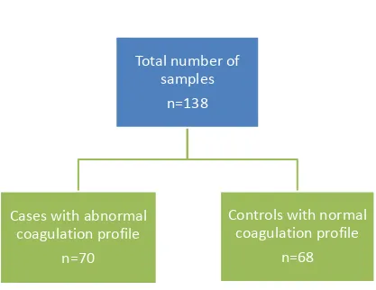
We studied a total of 138 samples which included 70 cases and 68 controls from January 2013- August 2014.
Results:
Total number of patients studied -138
[image:67.612.75.492.297.619.2]Cases (altered standard coagulation profile) -70 Controls (normal coagulation profile) – 68
Figure 13- Flow chart of cases and controls category
Total number of
samples
n=138
Cases with abnormal
coagulation profile
n=70
Controls with normal
coagulation profile
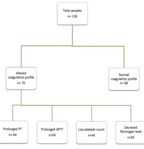
In the seventy cases, certain cases had one, two or three altered tests( Prothrombin time, activated Partial thromboplastin Time, Platelet count and Serum Fibrinogen levels) , but none had all the four coagulation parameters altered.
Table 3 - Distribution of altered coagulation profile parameters Total number of patients studied - 138
Number of cases who had an altered complete standard coagulation profile – 70
Number of control who had a normal complete standard coagulation profile- 68
Number of cases who had prolonged Prothrombin time – 64
Number of cases who had prolonged Activated Partial Thromboplastin Time – 54
Number of cases who had low platelet count ( less than 1,00,000/cumm) -44
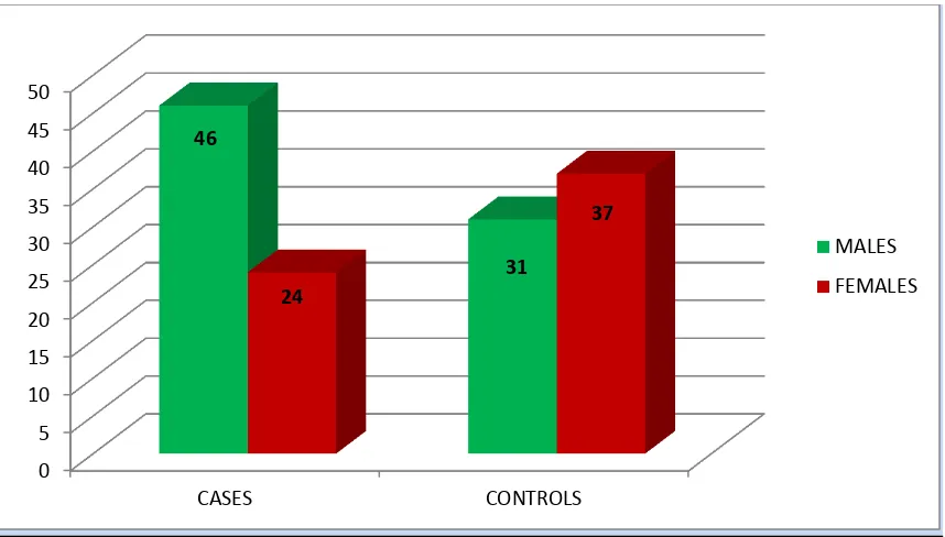
The baseline characteristics of cases and controls are given in table no 4..The mean age of cases was 41.3±16.2 years and that of controls was 33.3 ±13.7 years. Females accounted for 34.3% of the cases and 54.4% of the controls (Figure 15). In both cases and control group, a majority of cases were seen in the age group 21 -30 years. (Figure 16)
Characteristic Total n(%) Cases
n(%)mean±sd
Controls
n(%)mean±sd
Total Number 138 70 68
Age (years) 37.3 ± 15.5 41.3± 16.2 33.3±13.7
Gender Male 77(55.8%) 46 (65.7%) 31(45.6%)
Female 61 (44.2%) 24 (34.3%) 37 (54.4%)
Table 4 - Baseline Characteristics
Figure 15- Sex distribution among cases and controls
Figure 16- Distribution of age in cases and controls
0 5 10 15 20 25 30 35 40 45 50 CASES CONTROLS 46 31 24 37 MALES FEMALES 7 17 12 9 14 9 2 10 29 14
5 5 4
1 0 5 10 15 20 25 30 35
< 20 yrs 21- 30 yrs 31- 40 yrs 41- 50 yrs 51- 60 Yrs 61 -70 yrs 71-80 yrs
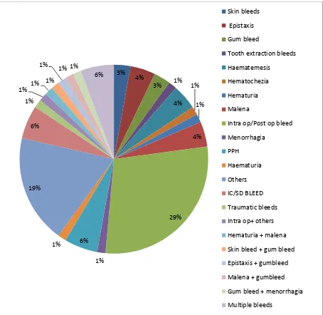
Figure 17- Distribution of bleeding manifestations among cases.
The bleeding manifestations ranged from minor mucosal bleeds like epistaxis to life threatening operative hemorrhages requiring transfusion support. A major proportion of our cases had severe intraoperative and postoperative bleeds leading to coagulopathy. Twelve percent of the total cases had more than one bleeding manifestation.
3% 4%
3% 1%
4% 1% 1% 4% 29% 1% 6% 1% 19% 6% 1% 1% 1% 1%
1% 1% 1%
6%
Skin bleeds
Epistaxis
Gum bleed
Tooth extraction bleeds
Haematemesis
Hematochezia
Hematuria
Malena
Intra op/Post op bleed
Menorrhagia PPH Haematuria Others IC/SD BLEED Traumatic bleeds
Intra op+ others
Hematuria + malena
Skin bleed + gum bleed
Epistaxis + gumbleed
Malena + gumbleed
Gum bleed + menorrhagia