CLINICAL STUDY OF PREPERITONEAL MESH REPAIR IN BILATERAL AND RECURRENT INGUINAL HERNIAS
A dissertation submitted to TAMILNADU DR.M.G.R MEDICAL
UNIVERSITY,CHENNAI, TAMILNADU in partial fulfillment for the
degree of
M.S.GENERAL SURGERYDEPARMENT OF GENERAL SURGERY TIRUNELVELI MEDICAL COLLEGE
TIRUNELVELI – 627 011
THE TAMIL NADU
Dr. M.G.R. MEDICAL UNIVERSITY CHENNAI
DECLARATION BY THE CANDIDATE
I hereby declare that this dissertation titled “A CLINICAL STUDY OF PREPERITONEAL MESH REPAIR IN BILATERAL AND RECURRENT INGUINAL HERNIAS” is a bonafide and genuine research work carried out under the guidance of DR.B.M.PABITHA DEVI MS, Senior Assistant Professor, Department of General Surgery, and under the supervision
of Chief Dr. V. PANDY MS, Professor, Department of General Surgery,
Tirunelveli Medical College Tirunelveli, TAMILNADU
Date: Signature of the Candidate
Place: TIRUNELVELI Dr .S. NAMBIRAJAN
CERTIFICATE BY THE GUIDE
This is to certify that the dissertation titled A CLINICAL STUDY OF PREPERITONEAL MESH REPAIR IN BILATERAL AND RECURRENT INGUINAL HERNIAS” is a bonafide research work done by Dr. S.NAMBIRAJAN in partial fulfillment of the requirement for the
M S degree in GENERAL SURGERY.
Date: Signature of the Guide
Place: Tirunelveli Dr.B.M.PABITHA DEVI, MS, Senior Assistant Professor, Department of General Surgery, Tirunelveli Medical College, Tirunelveli.
TAMILNADU.
CERTIFICATE BY THE HEAD OF DEPARTMENT
This is to certify that this dissertation titled “A CLINICAL STUDY OF PREPERITONEAL MESH REPAIR IN BILATERAL AND RECURRENT INGUINAL HERNIAS” is a bonafide work done by Dr S.NAMBIRAJAN under the guidance of DR.B.M.PABITHA DEVI. MS, Senior Assistant Professor, Department of Surgery, Tirunelveli Medical
College, Tirunelveli
Date: PROF.DR.K.RAJENDRAN. MS
Place: Tirunelveli
Professor and head of the department, Department of General Surgery,
Tirunelveli Medical College, Tirunelveli.
CERTIFICATE BY THE DEAN
This is to certify that Dr. S.NAMBIRAJAN has prepared this
dissertation entitled “A CLINICAL STUDY OF PREPERITONEAL
MESH REPAIR IN BILATERAL AND RECURRENT INGUINAL HERNIAS” in the Department of General Surgery, Tirunelveli Medical College , under the guidance of DR.B.M.PABITHA DEVI MS ,Senior Assistant Professor in Surgery in compliance with the regulations of the
TAMILNADU DR.M.G.R MEDICAL UNIVERSITY,CHENNAI,
TAMILNADU for the award of degree of Master of Surgery in General
Surgery.
Date: DR.SITHY ATHIYA MUNAVARAH M.D.,
DEAN
Place: Tirunelveli TIRUNELVELI MEDICAL COLLEGE
COPYRIGHT DECLARATION BY THE CANDIDATE
I hereby declare that THE TAMILNADU DR M G R MEDICAL
UNIVERSITY Chennai, shall have the rights to preserve, use and disseminate
this dissertation / thesis in print or electronic format for academic / research
purpose.
Date: Signature of the Candidate Place: TIRUNELVELI DR. S.NAMBIRAJAN
ACKNOWLEDGEMENT
It is my privilege to thank .DR.B.M.PABITHA DEVI MS, Senior
Assistant Professor of General Surgery, who helped me in choosing the subject
for this study and guided me at every stage.
It is my honour and privilege to thank Prof. DR.V. PANDY, MS, Professor of General Surgery, whose valuable suggestions and timely advice
were of immense help to me throughout all phases of this study.
I express my gratitude towards Prof. Dr. K.RAJENDRAN, M S ,Head of the Department of General Surgery, Tirunelveli Medical College,
Tirunelveli, for his valuable suggestions and guidance.
I thank my Professors and Chiefs Dr. V.PANDY M.S, Dr.
MAHESWARI M.S, Dr. VARADHARAJAN M.S, DR. ALEX ARTHUR
EDWARDS M.S, Dr. SRIDHAR M.S,Dr.SULAIMAN M.S.
I am thankful to our Assistant Professors Dr. Senthil Arumugam MS, Dr.
S. Amalan M.S, Dr Sathik M.S for their encouragement and guidance.
I am also thankful to all our Assistant Professors Dr Kamalin Viji MS,
Dr. Rajkumar MS, Dr. Josephine Pudhumai Selvi MS, Dr. SivanuPandian MS,
Dr. Nagalakshmi MS,Dr. Irene Aruna Edwin MS,Dr.Raja MS,Dr.Bethcy
Prinscilla MS,Dr.Vinodh Kumar MS,Dr. Nirmal Kumar MS,Dr Sugirtharaj
DNB,Dr. Karthiyayini MS,Dr.Rajmohan MS,Dr.Rakesh Fernando MS,Dr.Balaji
I thank our honourable Dean, DR.SITHY ATHIYA MUNAVARAH
M.D., TIRUNELVELI MEDICAL COLLEGE, TIRUNELVELI
I am very thankful to my colleagues particularly
Dr.Muthukumaraswamy Dr.Irphan, Dr.Jeyakumar, Dr.Karthik,
Dr.JohnAmalan, Dr.Patchaipandy, Dr.Ponmuthu, Dr.Pratheep ,Dr.Rathinavelu
who helped me in preparing this Dissertation.
My Sincere thanks to all the patients without whose co-operation, this
study would have not been possible and last, but not the least, I express
gratitude to my parents and my friends without whose co-operation, this study
would have not been possible.
ABSTRACT
BACKGROUND OF STUDY
Groin hernia is the one of most common performed surgery. There are
many methods of hernia repair. Pre peritoneal mesh repair is one of innovative
repair for bilateral and recurrent inguinal hernias.
OBJECTIVES
To study the effectiveness of Pre peritoneal mesh repair for bilateral and
recurrent inguinal hernia with respect to wound healing duration, duration of
hospital stay, post-operative complication and recurrence rate.
METHODS
All the materials for this study has been taken from 76 patients who got
admitted to Tirunelveli Medical College hospital attached to Tamilnadu DR M
G R Medical university, Chennai for the treatment of Groin hernia for one
and half years.
All the patients included in the study underwent surgical management for Groin
hernia. No patient in the study group underwent conservative management.
Patients underwent Preperitoneal mesh repair operative procedure for bilateral
RESULTS
In our study bilateral and recurrent inguinal hernias were more common in age groups of 51 to 70 yrs.
1. 98.68% of our patients were males and 1.31% were females.
2. Bilateral inguinal hernias were common in the study than recurrent hernias.
3. All patients were operated in spinal anesthesia.
4. Time taken for patients to get back to normal activity was 2-3 days.
5. Post operative pain was mild in our study.
The post operative complication rate was minimal and during the brief follow
up there were one recurrence.
CONCLUSION
Open preperitoneal mesh repair with suprapubic pfannensteil incision has
found to have short duration of surgery, less peri operative complication and
cost effectiveness to the patient. Still surgeons experience and orientation
KEY WORDS
Groin hernia; Preperitoneal mesh repair; cost effective; recurrence; return
to normal activity.
CONTENTS
SL NO. CONTENTS PAGE NO.
1. INTRODUCTION 1
2. AIMS OF THE STUDY 3
3. REVIEW OF LITERATURE 4
4. METHODOLOGY 71
5. OBSERVATION & RESULTS 73
6. DISCUSSION 85
7. CONCLUSION 91
8. SUMMARY 93
8. BIBLIOGRAPHY 95
9. ANNEXURES
I PROFORMA
II CONSENT
III KEY TO MASTER CHART
LIST OF TABLES
1. Clinical type of hernia 73
2. Mode of presentation 74
3. Age at presentation 75
4. Sex distribution 76
5. Duration of illness 76
6. Risk and predisposing factors 77
7. Occupation 78
8. Associated with smoking 79
9. Post operative pain 80
10. Time taken for procedure 81
11. Complication of hernia repair 82
LIST OF FIGURES
SL NO.
FIGURES
PAGE NO.
1. Embryology of testicular Descent. 9 2. Anterior Abdominal wall - Superficial Dissection. 14 3. Anterior Abdominal wall - Deep Dissection. 14 4. Inguinal Canal – Anterior view. 23 5. Inguinal Canal - Posterior view. 24 6. Inguinal Canal and Spermatic cord. 26
7. Cross section of inguinal canal. 26
8. Modified Gilbert classification. 42
9. Nyhus classification. 42
10. Modified Bassini’s repair. 51
11. Shouldice repair. 52
12. Lichtenstein’s repair. 54
13. Plug repair. 56
1
INTRODUCTION
Abnormal protrusion of part or whole of viscus through the wall
that contains it.
“A protrusion of any viscus from its proper cavity is denominated
a hernia. The parts are generally contained in a bag by a membrane
with which the cavity is naturally invested”
-Sir Astley Cooper 1804
Inguinal hernias are among the most common problems
encountered by the surgeon. Total 15% of surgical procedures are done
are groin hernia repairs. Groin hernias account to nearly 75% of all
abdominal wall hernias. Present hernia repair techniques involve
anterior approach with mesh fixation like litchenstein repair, gilberts
technique which was sutureless and mesh rutkow's hernioplasty with
mesh plug, these techniques involve dissection of cord structures with
attendant complications like nerve entrapment, testicular atrophy,
orchitis and chronic groin pain.
Inguinal hernia surgery has continued to evolve from tissue
2
have been explained in both anterior and posterior preperitoneal
approach. Though laparoscopic repair is popular in preperitoneal
method, this method is still under debate because of long duration of
surgery, need for general anesthesia, and associated complications1.
Laparoscopic surgery requires well equipped instruments and cost
effective to the patient is more.
Open preperitoneal mesh repair with suprapubic pfannensteil
incision has found to have short duration of surgery, less peri
operative complication and cost effectiveness to the patient2. Need of
present study is to review the effectiveness and complication of
3
AIMS AND OBJECTIVE OF THE STUDY
A. Age and sex distribution, occupation, duration of symptom,
mode of presentation of bilateral and recurrent inguinal hernia
in adults. To study the feasibility open preperitoneal mesh
repair for bilateral and recurrent inguinal hernias
B. To study the peri operative complication of open preperitoneal
mesh repair.
C. To study the post-operative complication with respect to pain,
hematoma, wound infection and recurrence.
4
REVIEW OF LITERATURE
HISTORICAL ASPECTS OF INGUINAL HERNIA
In the entire history of surgery no subject has been as
controversial as the repair of groin hernia.
--C.B.MCVay
PERIOD OF ASEPTIC SURGERY/THE LISTERIAN ERA(19th and 20th centuries) 8
In 1867, Joseph Lister, Professor of orthopedic surgery at
Glasgow infirmity presented his first paper on antiseptic surgery
performed under carbolic acid spray
In 1869, McEwen recognized the important and the role of
transversalis fascia in the repair of hernia. McEwen obliterated the
inguinal canal with mattress sutures.
In 1871, Marcy, published the first article in United States on
antiseptic herniorraphy, using carbolised catgut ligature. In 1877,
Czerny introduced excising sac at superficial ring by pulling it,
5
Kocher introduced transplantation of the sac anterolaterally
with external oblique aponeurosis suturing.
Lucas-championniere introduced antisepsis to France. In 1885,
he incised the aponeurosis of external oblique muscle, opening the
inguinalcanal and imbricate the roof in the closure.
1880-90, has rightly been termed as "The Decade of Inguinal
Hernia", for the significant contributions made towards hernia
surgery by Lucas championniere, Marcy and Bassini.
The credit for modern herniology should be given to Marcy of
United States. Marcy (1837-1924), was the first to indicate the
importance of the high ligation of the hernial sac and closure of
dilated inguinal ring as essential steps in the inguinal hernia repair.
Edoardo Bassini (1844-1924) of Pavia, Italy, revolutionized the
treatment of inguinal hernia by the introduction of a technique
designed to restore the conditions in the area of hernial orifice, which
6
He initiated the use of transversalis fascia, rectus sheath and
interrupted silk sutures. He used to do bilateral hernia repairs and
surgery for cryptorchidisim in the same sitting.
Bassini advocated inguinal canal reconstruction
physiologically, recreating the deep and superficial openings with
anterior and posterior walls. He sutured the conjoined transverses
abdominis and internal oblique to the inguinal ligament with
continuous silk sutures. His triple layer included transversalis fascia,
which was divided from pubis to an inch beyond the internal ring. He
emphasized closing the floor from below upwards to restore the
valve-like mechanism.
William S Halsted (1852-1922) independently developed a similar procedure with few differences, which included the laying
open of all three musculoaponeurotic layers, reforming the internal
ring after strengthening the posterior wall and transplantation of the
cord to a subcutaneous position and debulking the cord. This was
called the Halsted I procedure, to distinguish it from the Halsted II
7
(also known as Ferguson-Andrew's procedure), where cord was kept
along as per Ferguson and external aponeurosis imbricated as per
Andrews. Bassini's procedure was adopted widely.
In 1940, McVay and Anson pointed out that, the rectus fascia, a
portion of the tranversalis fascia that inserts into the lateral border of
rectus muscle, was strong enough to prevent incisional herniation.
Darn Repairs 9
To obtain tensionless or tension free posterior wall, natural tissue
or biologic or synthetic materials were used to darn the posterior
inguinal wall.
McArthur in 1901 used the pedicled strips of external oblique
aponeurosis woven between the conjoint tendon and inguinal
ligament.
Gallie and Lemesurier in 1921, published papers on using
fascia lata strips as sutures woven into the muscles and the inguinal
ligament and the tissues of the posterior wall of the inguinal canal--
8
Ogilvy in 1936, practiced floss silk lattice repairs with non
absorbable material which was followed by Maingot. Pratt, in 1948,
used steel wire followed by Koontz, who used tantalum gauze in
1950.
In 1948, Moloney introduced the forerunner of the modern nylon
darn technique.
Patch Graft Repairs 10 .
Expended polytetrafluroethylene (ePTFE) has been adopted for
both the external and pre-peritoneal approach, with good results.
Recently, a bilayered patch device(Hernia system) for inguinal hernia
has been introduced
Pre-Peritoneal Repairs 6
Thomas Annandale of Edinburgh presented for the first time in
1876, the concept of the pre- peritoneal approach.
This approach was strongly recommended by 'Nyhus' in 1960.
The foremost proponent of today's pre-peritoneal approach is
Stoppa (1968), who recommends it, especially for problematic cases
9
carried out, and in which tissues have become scarred and weakened
and the normal anatomy is destroyed.
Laparoscopic Inguinal Hernia Repair
Ger and his colleagues, in 1982, through laparoscope used
Michel staple applied with a Kocher clamp to close peritoneal
opening of the hernia sac.
EMBRYOLOGY11, 12
. The inguinal canal is formed by the processus vaginalis
penetrating the embryological structures between the skin and
peritoneum. This inturn leads to the descent of the testis, however the
10
Fig 1: Embryology of testicular Descent
11 INGUINAL REGION
The testis originally lies on the posterior wall of the
abdomen at the level of the upper lumbar vertebrae on the medial side
of the mesonephrons attached by a peritoneal fold called mesorchism.
The descent or migration of testis into its corresponding scrotal
chamber is accomplished by following the lead of the fibro muscular
band—gubernaculums testis. It arises mainly within a peritoneal fold
called the plica inguinalis, which stretches from the inguinal region to
the lower end of mesonephros. The gubernaculums attains greatest
development about the sixth month and is attached above to the lower
end of the testis and below, it pierces through the abdominal wall in its
passage to the bottom of the scrotal pouch, thereby forming the
inguinal canal.
Along with it a process of peritoneum, the processus vaginalis
descends into the scrotum dragging with it thin fascial prolongations
of the layers of the abdominal wall. Thus the processus vaginalis
receives covering from the aponeurosis of the external oblique and
12
The blind extremity of the processus vaginalis gets invaginated
for the reception of the descending testis. As the migration of the
testis proceeds, the gubernaculums shortens and eventually atrophies,
but some trace of gubernaculums persists at the bottom of the
scrotum, below tunica vaginalis forms the scrotal ligament fixing the
testis to the bottom of the scrotal pouch.
The cavity of the upper part of the processus vaginalis
disappears near the end of the 8th month. In female, the
gubernaculums extends from the lower poles of the ovaries to the
labium majus through the inguinal canal. This part atrophies and is
represented by the ligament of the ovary while the lower part which
is developed within the plica inguinalis is represented by the round
ligament of the uterus, extending from the side of uterus to the
labium majus. A pouch of peritoneum is called the canal of nuck,
accompanies the gubernaculums into the labium majus, similar to
that of processus vaginalis.
ANATOMY 13, 14, 15
The part of the anterior abdominal wall below the level of
13
of the pelvis opens anteriorly as well as superiorly. The pubis and
superior pubic (Cooper's) ligament are medial. The epigastric vessels
and transversalis fascia condensation at the internal ring are lateral. The
anterior femoral sheath, inguinal ligament and iliopubic tract form the
inferior border and the transverses abdominis aponeurosis and its arch
forms the superior border.
Skin
The deep ring is located approximately 2 cm above the skin
crease between the thigh and the abdomen and midway between
anterior superior iliac spine and pubic symphysis.. The skin of groin
is innervated by the ilioinguinal, iliohypogastric and genital branch of
genitofemoral nerves.
Subcutaneous Tissues of Groin
It divided into superficial fatty layer (Camper's fascia) and deeper membranous layer (Scarpa's fascia), which continues into perineum as the Colle’s fascia.
. It is accompanied by superficial epigastric artery, branches of
superficial circumflex iliac vessels laterally and external pudendal
14
Fig 2: Anterior Abdominal Wall- Superior Dissection
15
Musculoaponeurotic structures External Oblique Muscle and Aponeurosis.
It arises from the lower 8 ribs (5 to 12) and its fibers are
directed downwards, forwards, andmedially. Above, the aponeurosis
inter digitates with the serratus anterior muscle and continues as a
sheet of fascia over both the muscles. The most posterior fibers run
vertically downwards and insert into the anterior half of iliac crest.
Between the last ribs and iliac crest, a free border forms the lateral
boundary of lumbar triangle. From the anterior superior iliac spine to
the Pubic spine, the aponeurosis forms a free border which is called
inguinal ligament. The superficial inguinal ring is a triangular
opening in the external oblique aponeurosis, 1 to 1.5 cm lateral to the
pubic tubercle. The opening is formed by the splitting of external
oblique.
Internal Oblique Muscle and Aponeurosis
The internal abdominal oblique muscle lies between the
external oblique and the transverses abdominis muscle. It originates
16
line on the iliac crests and from the posterior lamella of the
lumbodorsal fascia through which it gains attachment to the lumbar
spines. Fibers are directed upwards, forwards and medially. The
anterior lamella accompanies the external oblique aponeurosis to
form the anterior rectus sheath and the posterior lamella accompanies
the aponeurosis of the transverses abdominis to form the posterior
rectus sheath.
Transverses Abdominis Muscle and Aponeurosis
This is the most internal of the three flat muscles of the
abdominal wall. The muscle arises from the iliopsoas fascia, from the
inner lip of the anterior 2/3rd of the iliac crest, the lumbodorsal fascia
between the iliac crest and 12th rib and from the inner surfaces of the
cartilages of the lower 6 ribs where it inter digitates with the
diaphragm. It passes medially in a transverse manner around the
lateral aspect of the abdomen on to the anterior abdominal wall.
a. The continuous portion is the extension of the main muscle and aponeurosis, the lower border of which arches above and
medial to cord structures and are called Transverses abdominis
17
insertion into the pubic tubercle and the crest is called falx
inguinalis. In 3% of cases the falx receives contribution from
the internal oblique aponeurosis also thereby forming the
conjoined tendon.
b. The discontinuous portion lies below the transverses arch, forms the posterior wall of the inguinal canal, medial to the
internal ring. One fourth of these fibers show marked
individual variations and most often is deficient, represented
only by the transversalis fascia, thereby forming a critical weak
spot in the posterior wall of the inguinal canal. The inferior
most edge of this layer is formed by the "iliopubic tract" a
collection of aponeurotic fibers. This tract arises laterally from
the inner lip of the iliac crest, the anterior superior iliac spine
and the iliopectineal arch. The fibers traverse medially
separating away from the inguinal ligament and present
beneath the deep inguinal ring. This ligament forms at least one
border of a defect in an indirect, direct or femoral hernia and
18 Transversalis Fascia
This is a portion of the endoluminal fascia that encloses
the abdominal cavity and peritoneum. The portion which invests the
transverses muscle and aponeurosis is called Transversalis fascia. It
is continuous with the lumbar, iliac, psoas, obturator and rectus
fascia. At the deep inguinal ring there is a tubular projection of this
fascia internal spermatic fascia that extends outwards in a blunt
funnel like fashion to cover the ductus deferens and the spermatic
vessels. However, the blunt funnel is not perfectly conical, but is
skewed and the axis of the funnel is less oblique than the axis of the
vessels through the deep inguinal ring. The redundant transversalis
fascia in the medial side of the deep ring is called 'Transversalis
fascia sling'.
Rectus Sheath
The posterior rectus sheath is lacking in any tendinous
structure from the semicircular line to the pubis. Above this point,
which is located midway between the umbilicus and the pubis,
19
muscles reinforce the posterior sheath. In the groin aponeurosis of all
the three flat muscles contribute to the anterior sheath.
Peritoneum
In the groin as elsewhere, the peritoneum is a thin elastic
membrane that serves only to provide a lubricating surface for its
contained viscera. Because of the elastic character of the peritoneum
it does not act in the prevention of hernia.
The Conjoint Tendon (Falx Inguinalis)
The transverse fibers of the transverses muscle proceed to
their insertion in the rectus sheath and the linea alba, while the lower
fibers course downward medially and caudally, sometimes to fuse with
fibers of internal oblique as they insert into the anterior pubis and
iliopectineal line. Only when the aponeurosis of the transverses and the
internal oblique are fused some distance lateral to the rectus sheath, the
term conjoined tendon is used.
The transverses muscle contributes 80% of the conjoint
tendon. The conjoint tendon lies lateral to the rectus muscle and
20
its insertion deep to the inguinal and lacunar ligaments. The
spermatic cord or round ligament of uterus lies anterior to it while
passing through the superficial inguinal ring.
John E. Skandalakis et al (1993)14, state that
1. The conjoined tendon rarely exists.
2. The distinction between the ligament of Henle and conjoined tendon is one of anatomic nicety and of little practical
significance, provided the distinction is recognized.
3. The term "conjoined area" may be applied correctly to the region which covers the ligament of Henle, the transverses
abdominis aponeurosis, the inferomedial fibers of the internal
oblique (musculature or aponeurosis), the reflected inguinal
ligament and the lateral border of the rectus tendon and sheath.
Cooper's Ligament (Iliopectineal Ligament)
Cooper's ligament is remarkably constant in form and extent
and represents the strongly reinforced periosteum of the superior
ramus of the pubis. On the superior and internal aspect of superior
21
the periosteum is supplemented by a considerable quantity of dense
fibrous tissue so that it usually becomes 2 cm or even 3 cm thick. The
fibers are adherent to and directed parallel to the superior ramus of
the pubis. This fibrous reinforcement gradually fades away near
midline on the internal surface of body of pubis. Laterally, it
continues posteriorly along the brim of the true pelvis, becoming
progressively thinner until it can no longer be distinguished from
periosteum of ileum. Cooper's ligament is particularly important in
the surgical correction of femoral hernias and large direct inguinal
hernias, because it forms a solid anchor along the inferior or posterior
aspect of these hernial defects, through which sutures in be placed
with confidence that they will hold.
Inguinal Ligament (Ligament of Poupart)
It is the lower, thickened portion of external oblique
aponeurosis extending from the anterior superior iliac spine to the
pubic tubercle. This is also called Poupart’s ligament. Its edge is
22
The lower edge of the inguinal ligament is loosely bound to the
fascia lata by the Innominate fascia. This fascia also serves to bind
together the collagenous fibers of aponeurosis and inguinal ligament.
Medially, the inguinal ligament gets inserted on the pubic tubercle
and fans downward to the superior pubic ramus as the lacunar
ligament. The medial attachment of the inguinal ligament is
continuous with the insertion of the aponeurosis into the linea alba.
Ligament of Gimbernat
The Gimbernat’s ligament is a triangular fascial extension of
the inguinal ligament, before its insertion to the pubic tubercle.
Cremaster muscle
The cremaster consists of a number of loosely arranged muscle
fasciculi lying along the spermatic cord
Inguinal Canal
It begins at the site of emergence of the spermatic cord through
the transverses aponeurosis (internal ring) and ends at the pubic
tubercle. It is oblique and 3.75 cm long, slanting downwards and
23
extends from the deep to the superficial inguinal ring. The boundaries
are: Anteriorly: the muscular fibers of the internal oblique in 1/3rd lateral boundary, external oblique aponeurosis in medial 2/3rd,
[image:39.612.129.460.208.418.2]superficial fascia ,skin.
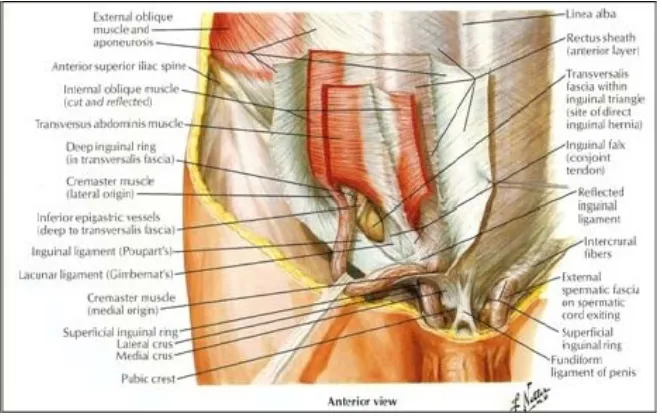
Fig :4. Inguinal Canal- Anterior view
24
Fig 5 : Inguinal Canal- Posterior view
Superiorly: The arched fibers of internal oblique and transverses aponeurosis.
Inferiorly: The inguinal ligaments and it continuation, lacunar ligament.
Hasselbach's Triangle
It is bounded medially by the lateral border of the rectus sheath,
laterally by the inferior epigastric vessels and below by the inguinal
25
These boundaries have to be redefined to include only those
structures that are in contact with the posterior inguinal wall in the
same plane. Redefined boundaries are: Rectus sheath with or without
falx inguinalis, inferior epigastric vessels and iliopubic tract and
Cooper's ligament.
Structures passing through the inguinal canal
Spermatic cord: Originates at the deep ring and consists of
a.Arteries: Testicular, cremasteric and artery to vas.
b.Veins: Corresponding veins, mainly testicular (pampiniform plexus).
c.Nerves: Genital branch of genitofemoral nerve, cremasteric nerve,
and Sympathetic plexus derived from Para aortic and pelvic plexus.
d.Lymphatics of the testes.
e.Vas deferens and areolar connective tissue.
26
cremasteric fascia (Internal oblique muscle and fascia) and external
[image:42.612.190.468.142.396.2]spermatic fascia (External oblique muscle and fascia).
Fig 6: Inguinal Canal and Spermatic Cord
[image:42.612.184.468.466.655.2]27 Blood Vessels
The external iliac artery gives off two major branches, before
crossing beneath Poupart's ligament, where it becomes the femoral
artery. These tributaries, the (deep) circumflex iliac and the (inferior)
epigastric vessels, are not vital. The latter, serves as the medial
border of the deep ring, or the lateral border of the direct triangle. Its
course can be followed topographically by an imaginary line
connecting a point midway between the umbilicus and the pubis. The
inferior epigastric artery gives off two branches, the external
spermatic cremasteric and the pubic branch. The main inferior
epigastric artery runs vertically upwards in the preperitoneal space to
enter and ramify with the rectus abdominis muscle forming collateral
connections.
The cremasteric vessel exits along the medial aspect of the deep
inguinal ring
Nerves
The muscles of the abdominal wall are innervated by the
28
the groin are the ilioinguinal, the iliohypogastric and the
genitofemoral
Pre peritoneal space:
Pre peritoneal space (extra peritoneal) is easily cleavable
space situated between peritoneum internally and transversalis fascia
externally.
Significant parts of preperitoneal space includes
myopectineal orifice of Fruchaud, the prevesical space of Retzius, the
space of Bogros and the retroperitoneal peri urinary space.
Myopectineal orifice of Fruchaud :
The myopectineal orifice is beneath the arching lower
boarder of transeversus abdominis and internal oblique muscle is
bounded laterally by iliopsoas muscle and medially by lateral edge of
rectus abdominis muscle and inferiorly by pubic pectin.
Myopectineal orifice represents the potentially weak area in
abdominal wall that permits the inguinal and femoral hernias.
29 Space of Retzius :
The space of Retzius extends from muscular floor of the
pelvis to the level of the umbilicus. Anteriorly the bodies of the
pelvic bones, medial portions of pubic rami and posterior lamina of
rectus sheath bound it to the level of arcuate lines of Douglas.
In this pelvis the prevesical fascia and lateral pillars of
bladder and the covering of pelvic peritoneum bound the space
posteriorly. More superiorly vesicoumbilical fascia and the
peritoneum provide a posterior wall of the space. Space of Retzius is
closed laterally along the line of fusion provided by inferior
epigastric vessels and the tissue that encloses them.
Space of Bogros:
This space extends upwards in to retroperitoneal area and
some workers state that it continues medially with space of Retzius
but is separated by line of fusion along the inferior epigastric vessels.
Though there are no major structures pass through this space but the
wall contain important nerves and vessels which have to be protected
30
CONTENTS OF PREPERITONEAL SPACE OF INGUINOFEMORAL REGION
1. VASCULAR
ARTERIES VEINS
External iliac and its branches External iliac vein
Deep circumflex iliac artery Deep circumflex iliac vein
Inferior epigastric artery Inferior epigastric vein
Bendavid vein
2. NERVES
Ilio inguinal nerve Femoral nerve
Iliohypogastric nerve Lateral cutaneous nerve of thigh
Genito femoral nerve L1, L2, L3 ventral rami
31
AETIOLOGY AND PATHOPHYSIOLOGY OF INGUINAL HERNIA
The aetiology of an inguinal hernia is mainly multifactorial.
Evolution16
Groin hernias all share the common feature of emerging
through the myopectineal orifice of Fruchaud, The absence of
posterior rectus sheath below the arcuate line and only a rather
substantial transversalis fascia unsupported by muscles or
aponeurosis, resisting the intra abdominal pressure.
CONGENITAL FACTORS
a. Patent processus vaginalis:. The entire processus vaginalis
may remain patent or only part of it, giving rise to indirect
hernia, scrotal hydrocele, and encysted hydrocele of cord or
hydrocele of canal of Nuck in female. The presence of patent
processus vaginalis does not necessarily indicate that hernia is
present or does it mean that one will necessarily develop in
32
b. Females are particularly free of direct inguinal hernia. The
narrowness of the interval between the transverses arch and the
inguinal ligament is an important factor protecting women
against direct hernia. On the other hand, musculoaponeurotic
attachments in women are such that they frequently develop
femoral hernia.
c. ANATOMICAL FACTORS
Shutter mechanism:
The accepted explanation for this is the physiologic
"Shutter mechanism" which is activated when the abdominal muscles
contract to increase intra abdominal pressure. As the internal oblique
and transversus abdominis muscles contract, their lower fibers
forming "the conjoined tendon" also sharply contracts and as the
fibers shorten, the arch straightens out and descends to come to lie
close to or on the inguinal ligaments and so covers and protects the
33 Raised intra abdominal pressure
The balance between the resistance of the abdominal wall and
the intra abdominal pressure may upset even in a fit young man who
is suddenly called upon to lift an extremely heavy weight.
Integrity of the fascia transversalis
These factors include connective tissue disorders like
Marfan's, Ehler'Danlos and Hurler - Hunter syndromes and
mesenchymal metabolic defects causing a deficiency of collagen and
structural abnormalities of the collagen fibers, predisposing to groin
hernia.
Cigarette smoking
It is found that the substances in cigarette smoke
inactivate antiproteases in lung tissues and so upset the
protease/anti-protease system which is responsible for destruction of elastin and
collagen of the rectus sheath and fascia transversalis and predispose
34 Physical exertion
The etiology of groin herniation has been strongly related
to manual work strains of lifting and strong muscular or athletic
exertion.
General contributing factors
Others like weakening of muscle and fascia by advancing
age, lack of physical exercise, obesity and multiple pregnancies.
Pulmonary disease like COPD and emphysema, prostatisim, chronic
constipation, diverticular disease, genito-urinary causes like cystitis,
cystocele, and urethrocele contribute to formation of groin hernia.
RECURRENT GROIN HERNIA
The recurrence rate after initial repair of a groin hernia
varies from 1% to 30%.
Early recurrence (within 2-3 years of repair)
The early group of recurrence is mainly caused by failure
on the part of surgeon and by infection. Some of the causes are as
35 1. Experience of the surgeon: 2. Tension on the repair
3. Infection
4. Suture material: 5. Suturing techniques.
6. Other factors anatomical indicators in recurrence
• Unrecognized hernias
• dissection/ sacligation is incomplete.
• deep ring is closed inadequately
• Inadequate reconstruction of the internal ring
General factors: General Conditions:
Malnutrition, jaundice, prolonged infection chronic debilitating
diseases, malignant diseases and long-term steroid therapy influence
on the success of groin hernia repair by influencing wound healing
36 Age:
The recurrence rate is lower following repair of primary of
recurrent inguinal hernia in older age groups than in younger patients.
Smoking:
Ascites:
Metabolic defects:
Local factors:
Repeated repairs:
Defects grow larger with each attempt at repair and tissue
become progressively stiff and unyielding.
Femoral hernia:
Failure of post wall following Lothissen approach for femoral
hernia result in recurrence.
Size of hernia:
37 Emergency repair:
Because of wide deep ring and friable tissue with common
postoperative infection recurrence rates are high.
Type of repair:
The answer is the method that the surgeon knows well and does
best.
Incision:
Poor exposure leads to inadequate repair and recurrence.
Missed hernias:
Posterior wall buttress:
Failure to construct this reinforcement of the posterior wall of
the inguinal canal.
Medial recurrence:
Occurs if medial anchoring has not been sufficient.
38
COMPONENTS OF INGUINAL HERNIA 18, 19 The Sac: Different parts of the Hernial Sac
A. Mouth: This is path between the sac interior and the abdominal cavity B. Neck: This is narrowest section between the mouth and the body of sac.
C.Body: It lies between the neck and the Fundus.
D.Fundus: This is the blind end or the distal most part of the sac.
E.Contents of Hernia: These can be almost any abdominal viscous, except the liver.
Coverings:
Coverings of an indirect inguinal hernia are (from inside out) as follows:
• Extra peritoneal fatty tissue
• Internal spermatic fascia
• Cremasteric fascia
• External spermatic fascia
• Two layers of superficial fascia and
39
In case of a direct hernia the coverings are as follows:
• Extra peritoneal fatty tissue
• Fascia transversalis
• Conjoint tendon
• External oblique aponeurosis
• 2 layers of superficial fascia and
• skin
CLASSIFICATION OF INGUINAL HERNIAS 17-19, 21-24 Anatomical classification 18, 19
Classification according to descent of the sac
• Bubonocele: The persistent patent process's vaginalis is obliterated at external ring and hernia is constrained till it.
• Funicular: The persistent patent process's vaginalis is obliterated above the epididymis. When the sac is occupied the
contents of the sac can be separately felt from testis.
40
Classification depending on the contents of hernia
Omentocele Omentum
Richter’s hernia Circumferential portion of
intestinal wall
Littre’s hernia Meckel’s diverticulum
Enterocele Intestine
Cystocele Urinary bladder
• Other varieties
Sliding/hernia en-glissade Caecum,urinary bladder
Maydl’s hernia W – shaped loop of intestine
Amyand s hernia Appendix
Saddle/pantaloon hernia Hernia on either side of epi
41 CLINICAL CLASSIFICATION 19
This is based on the clinical presentation of hernia.
Reducible hernia Contents are reducible
Irreducible hernia Adhesions within the contents,contents to sac,constricted neck
Obstructed hernia Irreducibility+obstruction
Strangulated hernia Irreducibility+obstruction+gangrenous ischemia
42
Fig 8: Modified Gilberts classification
43
EUROPEAN HERNIA SOCIETY CLASSSIFICATION: Primary or Recurrent(P or R)
Lateral, Medial or Femoral(L,M or F)
Defect size in finger breaths(assumed to be 1.5cms)
A primary indirect, inguinal hernia with 3cm defect size would be PL2.
CLINICAL FEATURES 25, 26
“Clinical diagnosis is an art and mastery of an art has no end you can always be a better diagnostician”.
- L.Clendening(1884-1943)
Symptoms
Lump or swelling in the groin, though some patients may describe a
sudden pain and bulge that occurred while lifting or straining. Some
patients complain of a dragging sensation and particularly with indirect
44 Systemic Symptom.
The four cardinal symptoms of intestinal obstruction, colicky abdominal pain, vomiting, abdominal distension and absolute
constipation. In late cases of strangulation where gangrene has set in,
patient can present with features of peritonitis more so if perforation of
bowel has occurred.
Signs
On inspection in standing position a bulge or swelling will be
seen in groin. This might disappear in lying down position if the
hernia is reducible spontaneously in direct hernia. Impulse on
coughing is present in reducible hernia. Loss of rugosities of scrotal
skin in large inguino-scrotal hernias is seen. Visible peristalsis is seen
in Enterocele. Malgaigne's bulges are seen in lax abdominal wall.
An indirect hernia is sausage or pear shaped and lies parallel to
the inguinal ligament. After reduction it reappears more laterally and
runs down above the inguinal ligament towards the scrotum. A direct
45
go down to scrotum. After reduction it reappears in a forward
direction.
On palpation
Reducing the hernia by manipulation is called “taxis” and it is
performed in lying down position of the patient. As the hernia is
reduced following features are noted:
a.Gurgling sound felt in Enterocele.
b.In Enterocele first part take longer to reduce and in Omentocele later part takes longer.
c.Impulse on coughing is felt.
Internal ring occlusion test: Internal ring is occluded and patient is asked to cough. If a bulge is seen medial to the occluding finger , it is
direct hernia, if not indirect.
Finger invagination Test: After reduction of the hernia, this test may be performed to palpate the hernial orifice. The skin is
invaginated from the bottom of the scrotum by little finger, which is
pushed up to palpate the pubic tubercle. The finger is then rotated and
46
triangular slit, which admits only the tip of a finger. The patient is
asked to cough.
Normally by pinchcock action, the finger will be squeezed by
approximation of two pillars. A palpable impulse will confirm the
diagnosis of hernia. When the finger enters the ring, it goes directly
backwards in direct hernia and it goes upwards, backwards and
outwards in indirect hernia. The finger is again rotated so that the
pulp of the finger looks backwards. The patient is again asked to
cough. If the impulse is felt on finger's pulp, it is direct hernia, if on
the finger's tip, then it's indirect.
Three Finger Test or Zieman's Technique: This test can be done only when there is no obvious swelling or after the hernia has been
completely reduced. In this test the finger is placed at the site of
internal ring, another at the external ring and one on saphenous
opening. The patient is asked to cough, when impulse is felt at the
ring, then it is indirect hernia. If impulse is felt at external ring it is
direct hernia and if impulse is felt at saphenous opening it is femoral
hernia. Percussion note over the swelling: Tympanic over Enterocele
47
Auscultation: Bowel sounds will be heard in Enterocele.
Examine the scrotum for thickened spermatic cord, testis
whether absent or atrophic or have a hydrocele. An incarcerated
hernia is soft and non-tender, but irreducible. A strangulated hernia is
tense, swollen, tender and irreducible and becomes red, edematous an
inflamed.
Examine for stricture urethra and prepuce for phimosis and
external urethral meatus for pinhole meatus. Per rectal examination is
done for benign enlargement of prostate or any growth.
Abdominal Examination: For any abdominal lump, ascites, and divarication of recti.
Respiratory system: To rule out COPD and Koch's.
DIAGNOSIS 17 Physical examination
Physical examination is the best way to determine the presence or
absence of an inguinal hernia. The diagnosis may be obvious by
simple inspection when a visible bulge is present. The differential
48
hernias require digital examination of the inguinal canal. This is best
done in both the lying and standing position.
PREOPERATIVE INVESTIGATIONS
Careful history taking and thorough physical examination and
investigations are of paramount importance for the assessment of
patients for surgery.
1. Laboratory investigations
• Complete blood examination for Hb%, BT, CT and blood sugar, urea and serum
creatinine.
• Urine analysis for Albumin, Sugar and microscopic study.
2.X-ray Chest to rule out TB, COPD, emphysema, malignancy of lungs.
3.Electrocardiogram and Echocardiogram to rule out atherosclerotic
49
4.USG abdomen to rule out any prostatomegaly, tumors of colon,
kidney, liver, ascites and size of prostate and post voidal residual
urine.
5. Roentengen studies in Hernia.
• Plain X-ray abdomen in intestinal obstruction, incarcerated
hernia and Ritcher’s hernia.
• Barium Enema in sliding hernia.
• Herniography-Positive contrast peritoneography used for the
diagnosis of hernias in the inguinofemoral region and the pelvis
is called herniography.
SURGICAL MANAGEMENT OF INGUINAL HERNIA9, 11, 17, 27-34 Non-operative treatment 42
The term “watchful waiting” is used to describe this
non-operative treatment recommendation. It is only applicable in
asymptomatic, minimally symptomatic hernias.
A truss is a mechanical appliance consisting of a belt with a pad
that is applied to the groin after spontaneous or manual reduction of
hernia. The purpose is to maintain reduction and to prevent
50
Truss is associated with complication. Atrophy of the spermatic
cord occurs in some cases and an eventual repair is made more
difficult by the constant mechanical pressure in the groin area that
renders the tissue more difficult to dissect due to atrophy and fibrosis.
Operative Treatment of inguinal hernia 27-29, 43, 44
Operative treatment –
(a)Herniotomy
(b)Herniorrhaphy
(c)Hernioplasty
a) Herniotomy 27-29 – transfixation of sac's neck and ligation, and then excision.
INDICATIONS
(a)In infants and children with preformed sac
(b)In young adults (very good inguinal musculature )
b) Herniorrhaphy – Here there is additional repair of the posterior wall of inguinal canal by opposing conjoint tendon to
51
repair are usually non absorbable material example- prolene or
silk.
Indications :
1) In indirect hernia( exception children < 18 years )
2) In patients with quite good musculature.
Types
(1) Modified Bassini’ s repair 35
Strengthening of the posterior wall of the inguinal canal by
interrupted stitch of the lower margin of the conjoint tendon to the
inner margin of the inguinal ligament behind the cord. And the most
medial suture should be passed through periosteum of pubic tubercle.
52
FIGURE 10: MODIFIED BASSINI’ S REPAIR
(2) The Shouldice repair 43, 44
The Shouldice technique is pure tissue hernia repair.hernial sac
is ligted at the internal inguinal ring. The transversali's fascia is
incised from the deep inguinal ring to pubic tubercle. The inferior
epigastri'c vessels are preserved. The inferior flap of the transversalis
fascia, is sutured continuously to the posterior aspect of the superior
flap of the transversalis fascia until the internal ring is encountered.
At the internal ring, the second layer is the re-approximation of the
superior edge of the transversalis fascia to the inferior fascial margin
and the shelving edge of the inguinal ligament.A third suture is
started at the tightened inguinal ring, joining the internal oblique and
transversus abdominis aponeurosis to external oblique aponeurotic
fibers just superficial to the inguinal ligament. This layer is continued
to the pubic tubercle where it reverses upon itself to create a fourth
53 (3) The Marcy's repair
(4) Halsted operation with subcutaneous transplantation of cord (Halsted I)
(5) The Ferguson operation (6) The Andrews operation
(7) Halsted II (Ferguson – Andrews operation) (8) Nylon darn inguinal hernia repair
HERNIOPLASTY 31
In 1984 Lichtenstein introduced tension free hernioplasty. It is herniotomy and post wall reinforcement by either autogenous material or heterogeneous material.
[image:69.612.152.514.68.216.2]
54 Indications of hernioplasty-
(1) indirect hernia – with poor tone of muscles
(2) direct hernias
(3) recurrent hernias
(4) Patient who do strenuous jobs or suffering from chronic cough
etc.
(1) LICHTENSTEIN TENSION – FREE HERNIOPLASTY:
A 5cm skin incision which starts from the pubic tubercle and extends laterally within Langer’s line is made 2cm above and
parallel to the medial half of inguinal ligament is made. External oblique
aponeurosis is opened. The cord with its cremasteric covering is
separated from the floor of inguinal canal and pubic bone. In case of
direct hernias, the large sacs are inverted with absorbable suture. A
thorough exploration of groin is necessary to rule out co- existing
femoral hernia.
A sheet of 6 x 11 cm of mesh is used for reinforcement
55
the medial corner of inguinal canal. The rounded corner is sutured
with non absorbable monofilament suture material to the anterior
rectus sheath above the public bone and overlapping the rectus sheath
by 1 to 1.5 cm. This is a crucial step in the repair, because failure to
cover this bone with the mesh can result in recurrence. external
[image:71.612.140.506.270.550.2]oblique aponeurosis is then closed.
FIGURE 12: LICHTENSTEIN TENSION – FREE HERNIOPLASTY
56 2) Plug Repair of Inguinal Hernia 47
Indication – Recurrent inguinal hernias and femoral hernias
The external aponeurosis is exposed only in the region of the
location where hernia can be seen. Spermatic cord is dissected only
upto hernial sac and is is pushed back into the preperitoneal tissue.
The plug is then prepared by coiling one or two polypropylene strips
with a dimension of 20x 2cm. It must fit snugly into the hernial
orifice. The cylinder is fixed in hernial annulus by 6 prolene sutures
placed above cylinder, the external aponeurosis, wound closed in
layers.
FIGURE 13: PLUG REPAIR OF INGUINAL HERNIA
57 4) Preperitoneal hernia repair 36
It is the hernial repair done anteriorly.
Advantages-
1) It provides visualization of all areas of inguinal herniation as
well as the confounding structures including the element of
sliding hernias are possible.
2) It minimizes bowel and bladder injury.
Laparoscopic Inguinal Hernia Repair 8, 36 Two techniques are used. 1. TAPP – Transabdominal preperitoneal repair
[image:73.612.234.439.453.582.2]2. TEP– Totally extraperitoneal approach.
FIGURE 14 : LAPAROSCOPIC HERNIA REPAIR
58 Advantages-
1) Less postoperative pain.
2) Myopectineal orifice can be approached bilaterally.
3) Previous anterior herniorrhaphy/ recurrent hernia
COMPLICATIONS OF INGUINAL HERNIA REPAIR 17, 37-40, 48
(I) Chronic groin pain after inguinal herniorrhaphy Postoperative pain (also called inguinodynia) including
1) Nerve injury
2) Nerve entrapment.
3) Tissue damage.
4) Mesh mispositioned.
5) Contracted,and harden'd mesh reactions (Meshoma's).
6) Infection/sepsis.
7) Recurrent hernia.
8) Deep ring narrowing .
59
10) Post herniorrhaphy syndromes: somatic/ visceral /
neuropathic .
(II) Wound infection in hernia repair 48
(III) Cord and testicular complications
1) Hydrocele
2) Hematocele
3) Complications involving vas deference
dysejaculation/transection .
4) Genitofemoral/ ilioinguinal nerve injury.
5) Damage to testes vascularity.
6) Ischemic orchitis and testicular atrophy
7) Testicular pain
8) Infertility
BIOMATERIALS 49-54
Mesh materials 49, 50, 51
60
NONABSORBABLE ABSORBABLE
Polypropylene Polyglactin 910
Polyvinyl Polyglycolic acid
polytetrafluoroethylene
Polyamide
Polyethylene terephtalate
Type of Anaesthesia
Regional anaesthesia is used routinely,it can also be
performed using local anaesthesia with intravenous sedation.
GIANT PROSTHETIC REINFORCEMENT OF THE VISCERAL SAC
The patients in this series had predominantly recurrent and
Bilateral inguinal hernias. In bilateral GPRVS, the peritoneum of both
groins is reinforced with a single prosthesis inserted in the
61
Usually a chevron shaped mesh is used.
CLINICAL STUDY OF PREPERITONEAL MESH REPAIR IN BILATERAL AND RECURRENT INGUINAL HERNIAS:
SURGICAL TECHNIQUE
The technique developed by Stoppa was used with certain modifications.
62
INCISION: Suprapubic pfannensteil incision is made.
63
MESH USED: Chevron shaped mesh with width 2cm less than the two anterior superior illiac spines.In our procedure a 15x15cm mesh was
[image:79.612.131.537.162.466.2]used.
Fig: chevron shaped mesh
PROCEDURE:
The incision ranges 8 to 10 cm from the midline
laterally, above the level of the internal ring. The intent is to expose the
lateral aspect of the rectus sheath and divide it and the oblique muscles
64
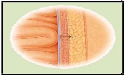
[image:80.612.131.537.76.408.2]expose the transversalis fascia, allowing it to be incised.
FIG: Preperitoneal space
The peritoneum is left intact to maintain the procedure within the
65
FIG:Parietal wall retracted to expose preperitoneal space
Wide dissection is then performed posterior to the rectus sheath and
inferior epigastric vessels and continues laterally to beyond the anterior
superior iliac spine. Inferiorly, the peritoneum is dissected to the
66
Fig: Retzius retropubic space and bogras space traced
The Retzius retropubic space and Bogros, are dissected from the
posterior portion of the rectus abdominis muscle proceeding behind the
epigastric vessels, and advanced into retroinguinal space and the
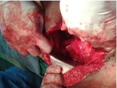
67
During reduction of hernial sacs, the spermatic cord and the gonadal
vessels are parietalized and then separated direct and indirect sacs
were reduced.
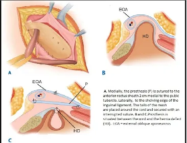
Fig: cord structures and gonodal vessels parietalised
.In case of large indirect sacs transfixation and ligation of sac was
done.The chevron shaped mesh ( Ethicon) is placed in the
68
[image:84.612.128.537.99.402.2]and indirect defects. The mesh is placed by stretching transversally.
Fig: Mesh placed in the preperitoneal space
The assistant retracts the parietal wall while the surgeon depresses the
peritoneal sac with the left hand, pulling toward him or her to open the
preperitoneal space. The mid-portion of the mesh is fixed to superior
pubic rami and pubic symphysis, laterally fixed to the illiopsoas
69
[image:85.612.129.530.69.256.2]Closed suction drainage is placed in position.
FIG:Postoperative picture with suction drain in position
Oral fluids started after 8 hours and patient mobilised early and advised
to carry on his normal day today activities.
[image:85.612.130.533.363.620.2]70 Indications and Contra-indications
We treated all types of hernias with this technique, mostly type
II, IIIA and IV according to the Nyhus classification. So in our series
there is not only treatment of bilateral inguinal hernias but also of
recurrent inguinal hernias as well.
Recurrences after preperitoneal mesh repairs are usually treated
with prosthetic repairs through an inguinal approach.
Technical problems
Problems during the learning period of the operation were
difficulty in identifying the fascia transversalis. The fascia has to be
incised and when it is opened the peritoneum will bulge out. Without
tearing this tiny fascia, dissection must be continued medially for the
identification of the epigastric vessels which stay attached to the
abdominal wall.
The cord structures must always be identified and parietalised.
Insufficient freeing of the sac from cord will result in a complete
incorporation of the sac within the mesh in the lateral iliac fossa, and
71
of the mesh, we have to be sure that the retractors are properly placed
exposing the MPO. The bladder must always be emptied before the
operation. A distended bladder makes dissection of the preperitoneal
space hazardous and could result in an unexpected vesical
perforation. so patients should be catheterised pre operatively itself.
MATERIALS AND METHODS
This is a prospective study done in medical college hospital attached to Tirunelveli Medical college with patients with bilateral
inguinal hernias and recurrent inguinal hernias undergoing
preperitoneal mesh repair for a period of 1.5 years under the guidance
of Dr.B.M.Pabitha Devi M.S.
During my study I attended to 76 cases of bilateral
inguinal hernia and recurrent inguinal hernias. To keep a proper
record a proforma was planned which was completed in each case.
Cases were included in the study based on the following criteria:
• Bilateral and recurrent inguinal hernias.
72
Following patients were not included in the study:
• Complicated inguinal hernia
• Patients not willing to participate
All cases were evaluated by documenting history taking, physical
examination and laboratory investigations.
Patients were explained about the type of surgery and anesthesia.
Each patient was explained bout the advantage of the said surgery,
short duration of surgery, less peri operative complication and cost
effectiveness.
Following data were collected with respect to
• Duration of surgery
• Intra operative complication
• Post operative monitoring – pain, wound infection, time of
ambulation
• Monitoring for recurrence
73
OBSERVATIONS
The following observations were done in the study
1) Clinical types
Among the 76 cases of bilateral inguinal hernia and recurrent inguinal hernias
Type No .of patients Percentage
Indirect 16 21%
Direct 60 79%
Our study shows that 79% were direct, 21% were indirect of all bilateral and recurrent inguinal hernias in study.
74
2) Mode of presentation
Symptoms No. Of patients Percentage
Groin swelling 42 55
Swelling with pain 34 45
In our study groin swelling was most common presentation followed by pain with swelling.
75
3) Age at presentation
Age groups(yrs) No. of patients Percentage
21-30 4 5%
31-40 5 7%
41-50 12 16.%
51-60 34 45%
61-70 21 27%
In our study most of patients presented in 51-60 yrs of age followed by 60-70yrs of age.
0 5 10 15 20 25 30 35 40
21-30 31-40 41-50 51-60 61-70
No. of patients
76
4) Sex distribution
Sex No. of patients Percentage
Male 75 98.68%
Female 01 1.31%
In our study most of the patients were male patients.
5) Duration of illness
Duration No. of patients Percentage
Less than 6 months 10 33.3%
6 months -1 year 15 50%
>1 year 5 16.7%
0 10 20 30 40 50 60 70 80
Male Female
No. of patients
77
In our study most of the patients were presented 6months to 1 year of illness followed by 6 months.
6)Risk and predisposing factors
Risk factors Patients Percentage
Prostatisim 3 10%
Constipation 2 6.6%
Coughing 6 20%
Heavy weight lift 15 50%
Smoking 21 70%
Obesity 6 20%