Copyright © 2004, American Society for Microbiology. All Rights Reserved.
Hepatitis C Virus Subgenomic Replicons in the Human Embryonic
Kidney 293 Cell Line
Samir Ali,† Charles Pellerin,† Daniel Lamarre, and George Kukolj*
Department of Biological Sciences, Boehringer Ingelheim Canada Ltd. Research and Development,Laval, Que´bec, Canada H7S 2G5 Received 31 July 2003/Accepted 23 September 2003
Hepatitis C virus (HCV) infects liver cells and its replication in other cells is incompletely defined. Human hepatoma Huh-7 cells harboring subgenomic HCV replicons were used in somatic cell fusion experiments with human embryonic kidney 293 cells as a means of examining the permissiveness of 293 cells for HCV sub-genomic RNA replication. 293 cells were generally not permissive for replication of Huh-7 cell-adapted replicons. However, upon coculturing of the two cell lines, we selected rare replicon-containing cells, termed
293Rep cells, that resembled parental 293 cells. Direct metabolic labeling of cells with33P in the presence of
actinomycin D and Northern blotting to detect the negative strand of the replicon demonstrated functional RNA replicons in 293Rep cells. Furthermore, Western blots revealed that 293Rep cells expressed the HCV nonstructural proteins as well as markers of the naïve 293 cells but not Huh-7 cells. Propidium iodide staining and fluorescence-activated cell sorting analysis of 293Rep cells revealed that clone 293Rep17 closely resembled naïve 293 cells. Transfection of total RNA from 293Rep17 into naïve 293 cells produced replicon-containing 293 cell lines with characteristics distinct from those of Huh-7-derived replicon cell lines. Relative to Huh-7 replicons, the 293 cell replicons were less sensitive to inhibition by alpha interferon and substantially more sensitive to inhibition by poly(I)-poly(C) double-stranded RNA. This study established HCV subgenomic replicons in nonhepatic 293 cells and demonstrated their utility in expanding the study of cellular HCV RNA replication.
Infection with hepatitis C virus (HCV), the causative agent for non-A, non-B hepatitis (8), is a serious worldwide health problem. The number of infected individuals is estimated to exceed 170 million (38), and ⬎80% of contracting patients become chronically infected (2, 34). The disease is character-ized by symptoms of varying severity and may progress to cirrhosis of the liver and hepatocellular carcinoma (17). The etiologic agent is an enveloped positive-sense RNA virus be-longing to theFlaviviridaefamily (33). Translation of the 9.6-kb genome produces a polyprotein that is co- and posttranslation-ally cleaved into individual viral proteins, after which structural proteins are processed by host signal peptidases and nonstruc-tural proteins are cleaved by virally encoded proteases (re-viewed in reference 32).
The lack of robust cellular systems for virus replication has been overcome, in part, by the development of selectable, dicistronic, subgenomic replicons derived from the HCV ge-notype 1 strains (3, 4, 14, 23, 31). These RNA replicons harbor the viral 5⬘ and 3⬘ untranslated regions and use the HCV internal ribosome entry site (IRES) to express the neomycin phosphotransferase gene in place of the structural proteins in the first cistron; the second cistron translates the nonstructural region from the encephalomyocarditis virus IRES. RNA rep-licons have been established in the human hepatoma Huh-7
cell line and display sustained replication in the cytoplasm of these cells (1, 13, 31). Adaptive mutations in the nonstructural region improve the frequency with which replicons can be established (22), yet full-length viral genomes that encode these adaptive mutations fail to infect chimpanzees (7).
The Huh-7 cell line is permissive for adapted HCV replicons in vitro, although variability in the permissiveness for replicons has been observed for these cells and, recently, for two other cell lines (5, 21, 31, 39). One manner by which the permissive-ness of Huh-7 cells and the nonpermissivepermissive-ness of other cell types can be explored is through somatic cell fusion experi-ments. Heterokaryons generated by somatic cell fusion can combine the characteristics of two distinct cell types. Such heterokaryons may be used to differentiate between permissive and nonpermissive phenotypes of cells and to illustrate either the presence of essential or absence of inhibitory cellular fac-tors that account for the phenotype of cells that host viral genome replication (16, 24, 35, 37).
For this study, we examined fusions between replicon-con-taining Huh-7 cells and human embryonic kidney 293 cells and demonstrated that 293 cells are less permissive than Huh-7 cells for HCV RNA replication. In rare instances, we isolated colonies of cells harboring subgenomic replicons that displayed all of the characteristics of naïve 293 cells. Moreover, the replicon RNA extracted from these cells was passaged successfully into naïve 293 cells. The replicon-containing 293 cells displayed responses to treatment with alpha interferon (IFN-␣), as well as to poly(I)-poly(C), that were distinct from those of Huh-7 cells. The 293 cell replicons represent a novel system for in-vestigating the biology of HCV RNA replication and may over-come some of the limitations inherent to the Huh-7 cell line. * Corresponding author. Mailing address: Department of Biological
Sciences, Boehringer Ingelheim Canada Ltd. Research and Develop-ment, 2100 rue Cunard, Laval, Que´bec, Canada H7S 2G5. Phone: (450) 682-4640. Fax: (450) 682-4642. E-mail: gkukolj@lav.boehringer ingelheim.com.
† S.A. and C.P. both made distinct contributions to this work.
491
on November 8, 2019 by guest
http://jvi.asm.org/
MATERIALS AND METHODS
Cell culture and generation of neomycin- and zeocin-resistant stable cell lines.
Cell lines used for this study were grown in Dulbecco’s modified Eagle’s medium (DMEM) supplemented with 10% fetal bovine serum (FBS; Sigma or HyClone). Huh-7 cell lines resistant to neomycin (Huh-7Neo) or zeocin (HuH-7Zeo) were
generated by electroporation of naïve Huh-7 cells with plasmid DNA encoding the specified resistance gene followed by selection with neomycin or zeocin, respectively, for 3 weeks. Resistant cell lines were continuously grown in the presence of neomycin (500g/ml) (Geneticin; Invitrogen) or zeocin (100g/ml) (Invitrogen) in the growth medium. S22.3 cells are a Huh-7-derived cell line and contain a neomycin resistance gene encoded by an adapted HCV subgenomic replicon that was made with a construct resembling I377/NS2-3⬘(23). The cell line
was continuously grown in DMEM–10% FBS containing neomycin (500g/ml). Zeocin-resistant Flp-In 293 cells (293Zeo; Invitrogen) were maintained in
DMEM–10% FBS containing zeocin (100g/ml).
PEG fusion.Zeocin-resistant cells (0.5⫻106to 1⫻106) were cocultured with
an equivalent number of neomycin-resistant cells in 6-well plates. After 20 h of incubation, cells were incubated with 0.5 ml of DMEM, without FBS, containing 50% polyethylene glycol (PEG) 3000-3700 (Sigma) solution for 5 min (at 37°C, 5% CO2), rinsed three times with phosphate-buffered saline, and incubated with
3 ml of DMEM–10% FBS overnight. The following day, cells were transferred to 100-mm-diameter plates and medium containing neomycin (500 g/ml) and zeocin (100g/ml) was added. Medium was changed twice a week for 3 weeks. Plates were used to isolate colonies for expansion into cell lines or were fixed with 1% glutaraldehyde and stained with crystal violet.
33P labeling of replicons in the presence of actinomycin D.Cells were grown
to 50 to 80% confluency in DMEM–10% FBS in 6-well plates. The cells were rinsed with phosphate-free DMEM–2% FBS (Wisent Inc.) supplemented with 2 mML-glutamine and incubated in 1 ml of medium containing 1g of
actino-mycin D (actinoactino-mycin D-mannitol; Sigma) per ml for 3 h. Following incubation, 1 ml of labeling medium containing 5g of actinomycin D per ml and 80Ci of
33P radionuclide (NEN) was added and cells were incubated for 16 h. Total
cellular RNA was extracted by use of the Qiagen RNeasy kit, denatured for 5 min at 75°C in buffer (1⫻morpholinepropanesulfonic acid [MOPS], 2.2 M formaldehyde, 50% formamide, 40g of ethidium bromide per ml), and then loaded onto a 0.6% denaturing agarose gel. The gel was dried, exposed to a storage phosphor screen, and scanned with a Storm 860 phosphorimager scanner (Molecular Dynamics). In experiments in which the HCV protease inhibitor (36) was used, the cells were first cultured in the presence of the inhibitor for 3 days prior to33P metabolic labeling.
Northern blot analysis.Total cellular RNA was extracted (RNeasy; Qiagen) and quantified by measuring optical density at 260 nm. Twenty micrograms of total RNA from each cell line was loaded in a 0.8% glyoxal-dimethyl sulfoxide gel (Northern Max-Gly; Ambion). Following electrophoresis, RNA was transferred onto a Hybond-N⫹nylon membrane (Amersham Pharmacia Biotech) and then cross-linked to the membrane by UV irradiation. The hybridization probe was generated by using the T7 promoter-based Riboprobe in vitro transcription system (Promega) on a 1,620-nucleotide region, spanning the NS5A and NS5B segments of an HCV 1b clone, in the presence of 50Ci of [␣32P]GTP. The blot
was hybridized by use of ULTRAhyb solution (Ambion) and then was exposed to a phosphor screen. Detection was performed with a Storm 860 phosphorim-ager scanner (Molecular Dynamics). As a control, the full-length negative strand of the HCV genome, cloned into the pCR3 vector (Invitrogen), was transcribed with the Riboprobe in vitro transcription system (Promega) and loaded on the gel.
PI staining and FACS analysis.Cells were digested with trypsin, washed with phosphate-buffered saline, and resuspended at a concentration of 106cells/ml in
propidium iodide (PI) staining buffer (0.1% sodium citrate, 0.3% NP-40, 0.05 mg of PI per ml, 0.02 mg of RNase A per ml). After a 10-min incubation on ice, 2
⫻106cells were centrifuged, resuspended in 2 ml of fresh PI staining buffer, and
kept on ice. The DNA content of single cells was then determined by fluores-cence-activated cell sorting (FACS) analysis using a Beckman Coulter cytometer.
Western blot analysis.Whole-cell extracts obtained by lysing cells in Laemmli buffer were resolved by sodium dodecyl sulfate–8% polyacrylamide gel electro-phoresis, transferred to a nitrocellulose membrane, and then blocked in 5% nonfat milk in Tris-buffered saline–Tween buffer. Blots were incubated with either E1A monoclonal antibody (NeoMarkers),␣-fetoprotein (AFP) polyclonal antibody (NeoMarkers), albumin polyclonal antibody (Sigma), NS3 polyclonal antibody (27), or NS5A monoclonal antibody (Maine Biotech Services). Mem-branes were then incubated with the appropriate mouse- or rabbit-specific anti-bodies conjugated with horseradish peroxidase, developed by use of LumiLight
Western blotting solution (Roche), and exposed to film (Hyperfilm; Amersham Pharmacia Biotech).
Immunofluorescence.Approximately 105cells were grown on LabTek 4-well
chamber slides (Nalge Nunc International) that were precoated with 150g of poly-D-lysine per ml. At 1 day postseeding, cells were fixed with 4%
paraformal-dehyde, permeabilized with 0.2% Triton X-100, and then incubated with either a polyclonal antibody against NS3 protein (27) or a monoclonal antibody against NS5A (Maine Biotech Services), and bound antibodies were detected with Al-exaFluor 488 goat-anti-rabbit or AlAl-exaFluor 488 goat anti-mouse antibody, re-spectively (Molecular Probes). Cells were visualized with a fluorescence detec-tion microscope (Olympus).
Amplification and cloning of replicon RNA by RT-PCR and in vitro transcrip-tion of RNA.Total RNA extracted from cells (RNeasy; Qiagen) was used for reverse transcription (RT)-PCRs (One-Step RT-PCR; Qiagen) that amplified theKpnI/SalI,SalI/SalI, andSalI/KpnI fragments of the HCV nonstructural and 3⬘untranslated region segments. The three fragments were first cloned into the pCRII vector (Invitrogen) for sequence verification and then excised by restric-tion digesrestric-tion with the appropriate enzymes and assembled into a complete subgenomic replicon by subcloning into pET-9a vector (Novagen). Replicon RNA was synthesized by use of the T7 promoter-based RiboMax large scale RNA production system (Promega).
RNA transfection of cells.Total cellular RNA or in vitro synthesized RNA was transfected into cells by use of either Lipofectamine 2000 or DMRIE-C trans-fection reagents (Invitrogen). Lipofectamine 2000 transtrans-fection was performed by seeding either 2.2⫻106293 cells or 1⫻106Huh-7 cells in 60-mm-diameter
plates overnight in DMEM–10% FBS (HyClone). On the day of transfection, 20
g of RNA and 40l of transfection reagent (each premixed in 300l of OPTI-MEM I medium [Invitrogen] for 5 min) were combined and incubated for 20 min at room temperature, and then the 600-l reaction was added to cells for 18 h. The cells were then detached with trypsin and seeded into two 150-mm-diameter plates in the presence of 0.25 mg (Huh-7 cells) or 0.4 mg (293 cells) of neomycin (Invitrogen) per ml. DMRIE-C transfection was performed by seeding 7.5⫻105293 cells in 6-well plates overnight. Four micrograms of RNA was
added to a mixture of 1 ml of OPTI-MEM I reduced serum medium and 12l of DMRIE-C reagent and was incubated with the cells for 6 h. Following incubation, the medium was changed to DMEM–10% FBS and the cells were incubated for 18 h. At that point, cells were split and plated in 100-mm-diameter plates in medium containing 0.35 mg of neomycin per ml. Selection was per-formed over a period of 3 weeks.
Replicon RNA inhibition assays and real-time RT-PCR.S22.3 cells (8⫻103)
or 293rep17 cells (15⫻103) were seeded in 96-well plates in DMEM–10% FBS
for 16 h, which was replaced with medium containing the indicated concentration (refer to particular experiments) of poly(I)-poly(C) (Sigma), IFN-␣(Sigma), or an HCV NS3 protease inhibitor previously described as compound 4C (C39H46N4O8; molecular weight, 698.81) (36). After 72 h of incubation, total
cellular RNA was extracted with the Qiagen RNeasy 96 kit, quantified by use of RiboGreen (Molecular Probes) on the Wallac Victor2Vsystem (Perkin-Elmer),
and used in quantitativeTaq-Man real-time RT-PCRs (Applied Biosystems) performed with the ABI Prism 7700 sequence detection system (Applied Bio-systems). Primers used for PCR were 5⬘-CCGGCTACCTGCCCATTC-3⬘ as forward primer and 5⬘-CCAGATCATCCTGATCGACAAG-3⬘ as reverse primer. The probe used was 5⬘-(FAM)-ACATCGCATCGAGCGAGCACG TAC-(TAMRA)-3⬘, which hybridizes within the neomycin phosphotransferase sequence of the HCV replicon RNA (9). The cycling conditions used for RT-PCR were as follows: 2 min at 50°C, 30 min at 60°C, and 5 min at 95°C, followed by 20 s at 94°C and 1 min at 55°C for 40 cycles. The replicon copy number, given as copies per microgram of total RNA, was interpolated from predetermined (in vitro synthesized) HCV replicon RNA concentration standards.
RESULTS
Somatic cell fusion between S22.3 and 293 cells.HCV
sub-genomic replicons have been previously adapted for human hepatoma Huh-7 cells (3, 15, 22, 23, 31). These subgenomic RNAs replicate persistently and efficiently in Huh-7 cells. Our attempts to introduce in vitro-transcribed replicon RNA into nonhepatic cell types were unsuccessful. We tried to introduce Huh-7-adapted replicons into human embryonic kidney 293 cells through transfection of in vitro-synthesized RNA by elec-troporation or by using multiple cation-based transfection
on November 8, 2019 by guest
http://jvi.asm.org/
agents; however, no neomycin-resistant 293 cell colonies with the replicon were obtained. A possible interpretation of this result is that the cells were nonpermissive for HCV replicons because either (i) the cells express a dominant activity or factor that prevents replication or (ii) the cells lack an essential ac-tivity that is necessary for replication (29).
To investigate the potential nonpermissiveness of non-Huh-7 cells, we examined heterokaryon formation between replicon-containing Huh-7 cells (clone S22.3) and human em-bryonic kidney 293 cells through cell fusion. Successful
forma-tion of heterokaryons between 293 and S22.3 cells would sug-gest that the nonpermissiveness of 293 cells is recessive and that they lack an activity provided by Huh-7 cells for HCV RNA replication (Fig. 1A). Alternatively, failure to generate heterokaryons would be consistent with the hypothesis that 293 cells express a dominant inhibitor of HCV RNA replication. We used 293 cells that have a stably integrated chromosomal copy of the zeocin resistance gene (293Zeo). As controls, we also constructed a Huh-7Neocell line, which has the neomycin resistance gene stably integrated, and a Huh-7Zeo cell line, FIG. 1. Neomycin- and zeocin-resistant colony formation with Huh-7- and 293-derived cell lines. (A) Schematic of somatic cell fusion between replicon-containing NeorS22.3 cells and Zeor293 cells. The generation of heterokaryons from S22.3 and 293 cells would indicate that 293 cells
are recessive for an activity present in Huh-7 cells that is essential for RNA replication; the lack of clones would indicate that the restriction of the 293 cells is dominant over HCV RNA replication. (B) Cellular fusions performed with Huh-7Neoand Huh-7Zeo, two distinct Huh-7-derived
cell lines with chromosomally integrated markers (i); Huh-7Neoand 293Zeocells, each with the respective chromosomally integrated marker (ii);
S22.3 and Huh-7Zeo, two homologous cell lines with distinct RNA and DNA selectable markers (iii); and S22.3 and 293Zeo, two heterologous cell
lines with RNA and DNA selectable markers (iv). Cellular fusions were performed by coculturing the cells for 20 h and then either leaving cells untreated (⫺PEG) or treating them with a 50% PEG solution for 5 min (⫹PEG). Cells were then transferred to 100-mm-diameter plates and selection was done with neomycin and zeocin for 21 days. Colonies obtained were stained with crystal violet and counted (numbers in lower right corners).
on November 8, 2019 by guest
http://jvi.asm.org/
which has the zeocin resistance gene stably integrated. Cell-to-cell fusions were enhanced by the brief addition of the fusogen PEG, whereas in the controls (⫺PEG), the two cell types were simply cocultured without the addition of PEG (Fig. 1B, panel iv). After an overnight incubation, cells were replated in 10-cm-diameter plates with a selection medium containing 100g of zeocin per ml and 500g of neomycin per ml, and selection was maintained for 21 days. PEG-mediated cellular fusions between S22.3 and 293Zeocells (Fig. 1B, panel iv) produced no doubly resistant colonies in repeated attempts (n ⫽ 4). In parallel, control fusions were performed with combinations of control cell lines that have chromosomally integrated markers (Fig. 1B, panels i, ii, and iii). The control fusion of Huh-7Neo with Huh-7Zeo(homologous cell lines with different chromo-somally integrated resistance markers) resulted in no colony formation without the addition of PEG and in 112 colonies for the PEG-treated sample (Fig. 1B, panel i). Coculturing of the heterologous Huh-7Neoand 293Zeocells (both with DNA chro-mosomal markers) in the absence of PEG resulted in 11 col-onies, and PEG treatment enhanced this result to 55 colonies (Fig. 1B, panel ii). Hence, Huh-7 cells and 293 cells, with appropriate markers, could be used to select for potential heterokaryons that harbor markers from both cells, and the addition of PEG increased the efficiency of colony formation. In order to examine the proficiency for such experiments with neomycin-resistant Huh-7 cells (S22.3) that encode the marker on an RNA replicon, we cocultured S22.3 cells with Huh-7Zeo cells that have a chromosomal zeocin resistance marker and demonstrated relatively efficient colony formation (303 colo-nies [Fig. 1B, panel iii]). Therefore, PEG can also be used to enhance the selection of potential heterokaryons with RNA and DNA markers in replicon-permissive cell lines, although PEG treatment reduced the number of viable cells. Through-out these experiments, we obtained significant variations in colony formation (up to fivefold) between repeated experi-ment sets; however, the relative efficiency of colony formation among the samples was consistent with the experiment pre-sented in Fig. 1B.
Invariably, PEG-enhanced fusion between S22.3 and 293Zeo replicon cells repeatedly failed to generate neomycin- and zeo-cin-resistant colonies. However, coculturing of the two cell lines without PEG treatment, followed by neomycin and zeocin selection, produced rare resistant colonies. In the first trial, we obtained three clones (clones 5, 6, and 7). Upon repeating the coculturing experiment four times, we obtained four other clones (clones 15, 16, 17, and 18). The result presented (Fig. 1B, panel iv) is from a PEG-independent coculture experiment by which no resistant colonies were obtained. Therefore, the frequency of obtaining doubly antibiotic-resistant colonies was low and independent of PEG-enhanced fusion. The neomycin-and zeocin-resistant colonies, termed 293Rep cells, were ex-panded into cell lines.
293Rep cells contain the HCV subgenomic replicon.In
or-der to assess whether 293Rep cells contain the HCV sub-genomic replicon acquired through the coculturing process with S22.3, cellular RNA was metabolically labeled with33P in the presence of actinomycin D. Two representative cell lines resulting from coculturing experiments, 293Rep5 and 293Rep17, respectively, were chosen for the metabolic labeling experiment. In addition, parental Huh-7 and 293 cells and our
original replicontaining S22.3 cells were included as con-trols. Metabolically labeled RNA from clones 293Rep5 and 293Rep17 revealed an RNA band that was identical in size to the replicon band found for S22.3 cells (Fig. 2A), suggesting that the neomycin-resistant 293Rep5 and 293Rep17 cell lines contain a functional replicon. No RNA was detected in nega-tive control naïve 293 cells or Huh-7 cells.
Further confirmation that 293Rep cells harbor the sub-genomic replicon was obtained from a Northern blot specifi-cally probing for negative-strand RNA. Blots with RNA iso-lated from 293Rep5 and 293Rep17 revealed replicon RNA bands consistent with the S22.3 positive control (arrow in Fig. 2B). The replicon was not detected in the control samples from naïve 293 and Huh-7 cells. Hence, 293Rep cells contained the subgenomic negative-strand replicative intermediate of the replicon RNA. Based on the intensities of standards loaded in lanes 1 to 3 (Fig. 2B), the negative-strand replicon copy num-ber is estimated to be in the range of 5⫻105replicon copies perg of total RNA for the 293Rep5 and 293Rep17 cell lines; this is approximately an order of magnitude lower than the quantity (as determined byTaq-Man real-time RT-PCR) of 1
⫻107plus-strand copies/g of total RNA estimated for these cell lines.
293Rep cell protein markers.293 cells and Huh-7 cells
ex-press distinct protein markers. 293 cells exex-press the adenovirus transforming E1A protein (12), whereas Huh-7 cells express FIG. 2. 293Rep cells contain the HCV subgenomic replicon. (A)33P metabolic labeling of replicon RNA in the presence of
acti-nomycin D. Total cellular RNA was extracted from cells treated with
33P and actinomycin D for 16 h and was resolved in a 0.6% denaturing
agarose gel. (B) Northern blots detecting the negative strand of HCV subgenomic replicons. Total cellular RNA was extracted and resolved in a glyoxal-dimethyl sulfoxide agarose gel. The RNAs were trans-ferred and hybridized with an HCV negative-strand-specific 32
P-la-beled RNA probe. The first three lanes are positive controls of in vitro-synthesized negative strand RNA, with 108, 107, and 106copies
used, as indicated.
on November 8, 2019 by guest
http://jvi.asm.org/
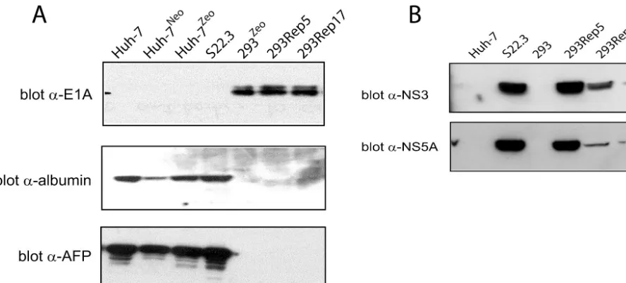
albumin and AFP markers (6). Western blot analysis was per-formed on total cellular lysates obtained from 7, Huh-7Neo, Huh-7Zeo, S22.3, 293, 293Rep5, and 293Rep17 cells to examine their protein expression patterns (Fig. 3A). Polyclonal antibodies specific for albumin or AFP, or monoclonal anti-bodies against E1A, showed that Huh-7 and its derived cell lines (S22.3, Huh-7Neo, and Huh-7Zeo) all expressed the liver cell markers albumin and AFP but lacked expression of the 293 cell marker E1A. In contrast, 293 cells, as well as the 293Rep cells, expressed the E1A marker but lacked expression of Huh-7 cell protein markers albumin and AFP. Western blots examining HCV protein expression were performed with a polyclonal antibody against NS3 protein or a monoclonal an-tibody against NS5A protein. 293Rep5 and 293Rep17 cells expressed both NS3 and NS5A proteins, similar to S22.3 cells, albeit at lower levels, particularly for 293Rep17 cells.
The subcellular localization of NS3 and NS5A was also ex-amined in 293Rep cells by immunofluorescence microscopy. Huh-7, S22.3, 293, 293Rep5, and 293Rep17 cells were fixed and then probed with either a polyclonal antibody against NS3 or a monoclonal antibody against NS5A (Fig. 4). 293Rep5 and 293Rep17 cells expressed both NS3 and NS5A proteins in the cytoplasm, similar to S22.3 cells. In contrast, naïve Huh-7 and 293 cells lacked this cytoplasmic staining. Notably, the gross morphology of 293Rep cells, characterized by size and shape, resembled that of naïve 293 cells. Moreover, 293 and 293Rep cells lacked the distinctive lipid droplets that are readily de-tectable in Huh-7 and S22.3 cells (arrows in Fig. 4).
Genetic content of 293Rep17 cells. The gross karyotypic
content of 293Rep cells relative to the parental 293 and Huh-7 cells was examined by FACS analysis of PI-labeled samples. Huh-7, S22.3, 293, 293Rep5, 293Rep17, and Huh-7Neo/293Zeo (a control hybrid cell line resulting from the fusion of Huh-7 and 293 cells) cells were analyzed. PI staining of parental 293
and Huh-7 cells (Fig. 5A), as well as of S22.3 cells (Fig. 5B), revealed the two peaks characteristic of diploid cells in the G1 stage and the G2/M phase of the cell cycle. The PI staining pattern of the Huh-7Neo/293Zeofusion cells (Fig. 5C) reflected the polyploid characteristic of the hybrid cell line; cells in G1 were sorted in a fashion consistent with tetraploidy (note that the G1 peak for this hybrid cell line coincides with the G2/M peak for parental cells). PI staining of 293Rep5 cells (Fig. 5E) suggested that these cells were not equivalent in DNA content to 293 cells, containing additional genetic material that may have been acquired during the heterologous cell fusion. In contrast, PI staining of 293Rep17 cells (Fig. 5D) resembled the pattern observed for naïve 293 cells; the pair of peaks (repre-senting cells in G1and G2/M) for 293Rep17 cells were coinci-dent with those of the 293 cell sample. 293Rep17 appeared to bear a greater similarity to parental 293 cells.
Transfection of RNA from 293Rep17 cells into naïve 293
cells.In order to determine whether the 293Rep cell replicons
were uniquely adapted for replication in 293 cells, we isolated total cellular RNA from 293Rep5, 293Rep17, and S22.3 cells and attempted to reintroduce the RNA by cationic lipid trans-fection into naïve 293Zeocells. Transfection of total RNA from S22.3 cells resulted in no colony formation. Similarly, transfec-tion of total RNA from 293Rep5 cells also failed to establish colonies. In contrast, transfection of total RNA isolated from 293Rep17 cells into naïve 293 cells produced 76 neomycin-resistant colonies.
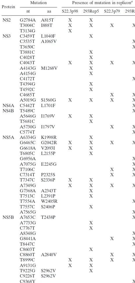
[image:5.603.70.514.70.271.2]We sequenced the RT-PCR-amplified products from the replicon RNA that was isolated from 293Rep5 and 293Rep17 cells. The sequences were compared to those of the replicons of S22.3 cells, at passage 98 or 79, that represent different lineages (diverging at passage 20) which were used for cocul-turing with 293 cells in the fusion experiments described above. The sequences of all of the replicons were compared in turn to FIG. 3. 293Rep cells express 293- and HCV-specific proteins. (A) Western blot analysis of 293Rep cells. Equivalent concentrations of total cellular proteins, obtained from cell lysates and separated by sodium dodecyl sulfate-polyacrylamide gel electrophoresis, were immunoblotted with either anti-E1A monoclonal antibody or anti-AFP polyclonal antibody. The anti-AFP blot was stripped and then reblotted with anti-albumin polyclonal antibody. (B) Western blot analysis of proteins from 293Rep cells. Total cellular proteins, prepared as for panel A, were immunoblotted with either anti-NS3 polyclonal antibody or anti-NS5A monoclonal antibody.
on November 8, 2019 by guest
http://jvi.asm.org/
the sequence of the previously published (AJ238799) HCV Con1 replicon (23). Table 1 lists the nucleotide mutations as well as amino acid substitutions in the replicons. 293Rep5 and 293Rep17 cells each contain unique mutations that are dis-persed throughout the NS2-NS5B region. Most of the muta-tions resulting in amino acid substitumuta-tions were within the NS5A region. We also sequenced four replicon clones derived from 293 cells that were transfected with early-passage 293Rep17 total RNA and only identified minor deviations (i.e.,
⬍6 nucleotide transitions in the nonstructural region) from the 293Rep17 sequence. These predominantly silent mutations (encoding no more than 2 amino acid substitutions) were
unique to each clone and were not found in other 293 repli-cons.
[image:6.603.95.489.67.510.2]The NS2-NS5B regions of 293Rep5 and 293Rep17 replicons were reverse transcribed, amplified, cloned, and assembled individually within a standard I377/NS2-3⬘dicistronic replicon construct (23). In vitro-synthesized replicon RNA was trans-fected into naïve 293 and Huh-7 cells and the frequency of colony formation was evaluated after a course of neomycin selection. Transfection of in vitro-transcribed replicon RNA that encodes Huh-7 cell-adapted mutations into naïve Huh-7 cells produces colonies in the range of 2⫻106to 3⫻106CFU perg of RNA (3, 15, 22). We observed that transfection of in FIG. 4. Detection of HCV proteins in 293Rep cells by immunofluorescence. Fixed and permeabilized cells were incubated with either a polyclonal antibody against the NS3 protein (A) or a monoclonal antibody against the NS5A protein (B) and appropriate secondary antibodies. IF, immunofluorescence with the indicated primary antibody; DAPI, 4⬘,6⬘-diamidino-2-phenylindole DNA staining; LM, phase-contrast light microscopy of the same cells. Arrows define the characteristic cytoplasmic lipid droplets of Huh-7 cells.
on November 8, 2019 by guest
http://jvi.asm.org/
vitro-transcribed 293Rep5 or 293Rep17 replicon RNA into naïve Huh-7 cells also displayed the characteristics of moder-ately adapted replicons by producing ⬎103 CFU per g of RNA (data not shown). However, transfection of either 293Rep5 or 293Rep17 in vitro-transcribed RNA into naïve 293 cells produced no colonies harboring a replicon. Transfections of naïve 293 cells with total cellular 293Rep17 RNA, per-formed in parallel, consistently (n ⫽ 3) produced 60 to 80 colonies and indicated that our transfection procedure for 293 cells functioned properly. Thus, the cloned and in vitro-tran-scribed 293Rep17 RNA, though active in Huh-7 cells, failed to establish 293 colonies and differed in functionality from the RNA directly isolated from 293Rep17 cells.
Transfection of total RNA isolated from 293Rep17 cells into
[image:7.603.48.279.70.448.2]naïve 293Zeo cells produced clones that were expanded into cell lines. We characterized some of these cell lines to confirm the presence of replicon RNA and the expression of viral proteins. Metabolic labeling with33P in the presence of acti-nomycin D was performed with Huh-7, 293, S22.3, 293Rep 5, 293Rep17 cells and the LR1, DM1, DM3, DM4, DM6, DM8, LIP3, LIP5, and LIP7 second-generation replicon cell lines (Fig. 6A). Labeled replicon RNA was detected in 293Rep cells as well as in all of the cell lines obtained from the transfection of 293Rep17 total RNA into naïve 293 cells (LR1, DM1, DM3, DM4, DM6, DM8, LIP3, LIP5, and LIP7).
FIG. 5. PI staining and FACS analysis of cell lines. Huh-7, 293, S22.3, 293Rep5, and 293Rep17 cells were digested with trypsin and incubated in staining buffer containing PI for 10 min. DNA content was determined with a FACS analyzer. (A) Overlay of the FACS profiles of parental Huh-7 (black) and 293 (blue) cells depicting ca-nonical G1and G2/M phase peaks. (B) Overlay of FACS profile of
S22.3 cells (red) onto the profile of panel A. (C) Overlay of FACS profile of Huh-7Neo/293Zeohybrid cells (red) onto the profile of panel
A, revealing different G1and G2/M phase peaks. (D) Overlay of FACS
[image:7.603.303.535.94.579.2]profile of 293Rep17 (red) cells onto the profile of panel A. (E) Overlay of FACS profile of 293Rep5 (red) cells onto the profile of panel A.
TABLE 1. Nucleotide and amino acid mutations found in 293Rep replicons relative to the Con1 replicon sequence
Protein Mutation Presence of mutation in replicon
a
nt aa S22.3p98 293Rep5 S22.3p79 293Rep17
NS2 G2784A A815T X X
T3004C I888T X X X X
T3134G X
NS3 C3459T L1040F X
C3535T A1065V X
T3650C X
T3881C X
C4028T X
C4043T X X X X
A4143G M1268V X
A4154G X
C4172T X
T4394G X
T4592C X
C4685T X X
A5019G S1560G X X X X
NS4A C5442T L1701F X
NS4B T5489C X
A5646G I1769V X X
T5681C X
A5730G I1797V X X
C5774T X
NS5A A6334G K1998R X
G6465C G2042R X X X X
G6618A V2093I X X
T6805C L2155P X
G6956A X
A7075G E2245G X
T7106C X X
C7314T P2325S X X
T7347C S2336P X X
A7349G X X X X
G7368A A2343T X
T7513C L2391P X
T7554A W2405R X
T7557C S2406P X
A7565G X
NS5B A7653C T2438P X
A7733G X
C7767T X
A8348G X
G8441A X X
T8447C X
C8603T X
C8860T A2840V X X
T8999C X X X X
A9131G X X
T9225G S2962V X
C9226T S2962V
C9368Y X
aX, mutation is present.
on November 8, 2019 by guest
http://jvi.asm.org/
Western blot analysis (performed as described for Fig. 3) of total cell lysates from two of the selected second-generation replicon-containing 293 cell lines, LR1 and DM1, revealed proteins detected by NS3 and NS5A antibodies, albeit at lower levels of expression than in S22.3 cells (Fig. 6B); negative control Huh-7 and 293 cells did not express these proteins. Furthermore, by immunofluorescence, both NS3 and NS5A were detected in the cytoplasm of LR1 cells (Fig. 6C), as previously observed for 293Rep17 and S22.3 cells (Fig. 4A and B).
Distinct biological characteristics of replicon-containing
293 cells. HCV subgenomic replicons in 293 cells present a
distinct cellular system for the study of HCV RNA replication. In a preliminary effort, we examined the characteristics of these replicon-containing 293 cells in response to various in-hibitors and compared them to Huh-7 replicon cells. The effect of a specific HCV NS3 protease inhibitor, 4C, previously dem-onstrated to inhibit HCV subgenomic replication in S22.3 cells (36) was examined with 293Rep17 cells (Fig. 7A). Cells were incubated for 3 days in the presence of protease inhibitor 4C concentrations ranging from 0.1 to 1M. Huh-7 and 293 cells were also included as controls. Metabolically labeled RNAs indicated that treatment of S22.3 cells as well as 293Rep17 cells with 0.1M protease inhibitor significantly reduced33 P-labeled replicon levels, whereas 0.33 and 1M protease inhib-itor concentrations eliminated the labeled replicon from both of these cell types. Moreover, the 50% inhibitory concentration (IC50), as determined by quantitativeTaq-Man RT-PCR
mea-surement (27, 36) for 293Rep17 replicon RNA (IC50⫽0.05⫾ 0.013M) (data not shown), was similar to the value reported for Huh-7 replicon cell lines (27, 36).
We next examined the effect of treating replicon-containing 293 cells with biological reagents that induce antiviral re-sponses. S22.3, 293Rep17, and LR1 cells were treated with IFN-␣at concentrations ranging between 0.023 and 80 U/ml (Fig. 7B). Treatment of S22.3 cells with IFN-␣reduced repli-con levels, and a 50% reduction relative to untreated repli-controls (IC50) was achieved at a concentration of 0.33⫾0.12 U/ml (n
⫽ 9), consistent with previously reported values for IFN-␣
[image:8.603.89.492.68.349.2]using this cell line (27). However, treatment of replicon-con-taining 293 cell lines 293Rep17 and LR1 required significantly higher (14-fold) concentrations of IFN-␣to achieve 50% inhi-bition; the IC50was 4.8⫾1.2 U/ml (n⫽8) for 293Rep17 cells and 7.2⫾4.9 U/ml (n⫽5) for the LR1 cell line (Fig. 7B). The 293Rep5 hybrid cell line displayed a similar sensitivity to IFN-␣, with an IC50 of 4.5 ⫾ 3.0 U/ml (n ⫽ 3) (data not shown). We also assessed the response of replicon-containing 293 cells to treatment with the double-stranded RNA mimic polyinosine-poly(C) [poly(I)-poly(C)] (28). This agent is pro-posed to stimulate the activation of cellular antiviral responses through the Toll-like receptor 3 (TLR-3) (18), although the detailed mechanism of activation and the upstream link to TLR-3 remain unknown. We treated the replicon-containing S22.3, 293Rep17, and 293LR1 cells with doses of poly(I)-poly(C) ranging from 0.9 to 2,000g/ml and also measured the decline in replicons by usingTaq-Man real-time RT-PCR (Fig. FIG. 6. Analysis of 293 cell lines generated by transfection of total RNA from 293Rep17 cells. (A)33P labeling of total RNA in the presence
of actinomycin D. Selected 293 cell clones (LR1, DM1, DM3, DM4, DM6, DM8, LIP3, LIP5, and LIP7) were metabolically labeled with33P and
actinomycin D for 16 h and visualized as described for Fig. 2. (B) Western blot with anti-NS3 or anti-NS5A antibody as described for Fig. 3. (C) Immunofluorescence detection of viral proteins in replicon-containing LR1 and naïve 293 cells was performed as described for Fig. 4.
on November 8, 2019 by guest
http://jvi.asm.org/
7C). Treatment of the S22.3 cell line had little effect on repli-con levels, even at the highest repli-concentration used, repli-consistent with previous reports (19). In contrast, the replicon-containing 293Rep17 and LR1 cell lines responded effectively even to relatively low doses of poly(I)-poly(C) treatment. The IC50of poly(I)-poly(C) for the 293Rep17 cells was 36⫾11g/ml (n⫽
8), and the IC50for LR1 cells was 77⫾29g/ml (n⫽5).
DISCUSSION
In an attempt to examine and extend the tropism of HCV replicons, we conducted a series of somatic cell fusion exper-iments between replicon-containing human hepatoma Huh-7 cells (S22.3) and human embryonic kidney 293 cells. From the fusion experiments, we determined that homologous fusion of Huh-7 cells as well as heterologous fusion of Huh-7 and 293 cells could be achieved through selection for two antibiotic resistance markers. S22.3 cells were proficient in fusion exper-iments with Huh-7 cells harboring a DNA-integrated zeocin resistance marker. The selection of heterokaryons from S22.3 replicon cells and Huh-7Zeo cells, which carry the resistance marker in the nucleus, occurred with a higher efficiency than heterokaryons from the homologous Huh-7Zeoand Huh-7Neo cells, which both have their respective resistance marker inte-grated in the chromosome. The higher efficiency may reflect a facilitated fusion process between a nuclear marker and a cytoplasmic RNA marker relative to the conventional cell fu-sion that involves two nuclear DNA markers. Alternatively, as subgenomic replicon RNA can be produced and released into the extracellular medium by Huh-7 cells in ill-defined RNase-resistant particles (30), the zeocin-RNase-resistant cells may have ac-quired the replicon independently of direct cell fusion.
In contrast to the successful fusion between Huh-7 cells and 293 cells, fusion of S22.3 with 293 cells did not produce viable colonies under the same conditions. One interpretation from these results is that 293 cells restrict replication of the Huh-7 cell-adapted replicons and that the restrictive phenotype of 293 cells is dominant over the permissive phenotype of Huh-7 cells. This phenotype of 293 cells may be due to the expression of an inhibitor that is absent (or differentially expressed) in Huh-7 cells. Due to the fact that we observed a difference between 293 and Huh-7 cells in responding to treatment with activators of antiviral responses (discussed below), we suspect that 293 cells may encode a replicon inhibitor that is less active in (or absent from) Huh-7 cells. Other groups have observed that Huh-7 cells are not uniformly permissive for HCV replicons, and variations in replication efficiency have been reported for different cell passages or for “replicon-cured” Huh-7 cells (4, 5, 21, 25). Whether a common inhibitory activity accounts for restriction in 293 and some Huh-7 cells is unknown, and the components of a potential repressive pathway(s) and their role in HCV subgenomic replication remain to be determined.
The nonpermissive phenotype of 293 cells for Huh-7 cell-adapted replicons was suggested from the repeated attempts that failed to produce cell fusions between 293Zeo and S22.3 cells. When replicon-containing S22.3 cells were simply cocul-tured with 293 cells in the absence of PEG, we isolated rare neomycin- and zeocin-resistant colonies. We suspect that a PEG-independent fusion occurred between the two cell types or that 293Zeocells acquired an extracellular form of the RNA marker produced by S22.3 cells (30) (see above).
[image:9.603.48.268.71.481.2]Analysis of the DNA content of the replicon-containing 293Rep cells, 293Rep5 and 293Rep17, by PI staining and FACS supported both proposals. The analysis revealed that 293Rep5 cells contain more than the normal diploid, but less than tetraploid, DNA content observed for either parental 293 or Huh-7 cells. This suggests that 293Rep5 cells originated from a fusion of the two cocultured parental cell types and then FIG. 7. Inhibition of subgenomic RNA replicon in 293 cells.
(A) An HCV NS3 protease inhibitor (4C) decreases intracellular rep-licon levels. S22.3 and 293Rep17 cells were cultured with the protease inhibitor and labeled with33P and actinomycin D; total cellular RNA
was resolved as described for Fig. 2. Protease inhibitor was tested at 0, 0.1, 0.3, and 1M concentrations. (B) Replicon cells were cultured in the presence of different doses of IFN-␣, ranging from 0.023 to 50 U/ml, for 72 h. Total cellular RNA was extracted and the replicon copy number was quantified byTaq-Man real-time RT-PCR to obtain the percent inhibition of treated samples with respect to untreated control levels. Circles, S22.3 cells; squares, 293Rep17 cells; triangles, LR1 cells. (C) Inhibition of replicon in cells as described for panel B, but cultured in the presence of different doses (0.9 to 2,000g/ml) of poly(I)-poly(C). The plotted percentages of inhibition represent aver-age values (with error bars) from at least three determinants.
on November 8, 2019 by guest
http://jvi.asm.org/
lost chromosomes but maintained the replicon. A similar anal-ysis on 293Rep17 cells, on the other hand, revealed that the DNA content of this cell line was indistinguishable from that of naïve 293 cells. Hence, either of the two proposed methods of replicon acquisition could apply for 293Rep17 cells. Both 293Rep5 and 293Rep17 cells expressed the E1A protein, a marker for the adenovirus Ad5 immortalized 293 cells (20), and lacked expression of the albumin and AFP protein mark-ers of Huh-7 cells; the latter marker may not represent an ideal marker for Huh-7 cells, since its expression is commonly lost in somatic cell hybrids (26). Replicon RNA was detected in these cells by metabolic labeling with33P and by Northern blots that confirmed the presence of the negative-strand replicative in-termediate. Both Western blots and immunofluorescence de-tected HCV nonstructural proteins in 293Rep cell lines. Our data supported the proposal that 293Rep17 has a 293 cell background and provided us with the means to isolate replicon HCV RNA that may have adapted to 293 cells.
Transfection of total cellular RNA isolated from the 293Rep17 cells into naïve 293 cells produced colonies. Notably, transfection of total RNA from 293Rep5 and 293Rep17 cells into naïve Huh-7 cells produced neomycin-resistant colonies, whereas only the total RNA from 293Rep17 cells produced replicon-containing colonies in naïve 293 cells. The consensus sequence of 293Rep17 replicon RNA differs from that of 293Rep5 replicon RNA by 41 nucleotides that encode five amino acid substitutions within the nonstructural region, some of which may represent adaptive mutations required for rep-lication in 293 cells.
Though the in vitro-transcribed RNA that was generated from the 293Rep17 DNA clone encoded all of the nonstruc-tural region mutations found in the total RNA isolated from 293Rep17 cells, only the latter efficiently established replicons in naïve 293 cells. We currently have not identified the cause of this difference, and a similar discrepancy was also noted by Zhu and colleagues (39) in replicons isolated and cloned from HeLa cells and a murine liver cell line. We can eliminate the possibility that the cloning of the replicon had inadvertently incorporated lethal mutations, as the in vitro-transcribed RNA derived from the 293Rep17 clone replicated and produced colonies when transfected into naïve Huh-7 cells. One possible explanation is that the nonstructural region adaptive muta-tions, though functional in Huh-7 cells, are insufficient to con-fer replication in 293 cells and additional mutations within or beyond this region are necessary; we have sequenced the 5⬘and 3⬘ untranslated regions of 293Rep17 but have not identified mutations that induce efficient 293 cell colony formation with in vitro-transcribed RNA. Alternatively, unidentified accessory RNA molecules, coisolated with the replicon from 293Rep17 cells, may play a role in RNA replication. A third possibility is that the replicon isolated directly from 293Rep17 cells has epigenetic modifications (lacking in vitro-transcribed RNA) that are essential. These possibilities are currently being ex-plored.
Preliminary characterization of the replicon-containing 293 cells revealed differences relative to Huh-7 cells. For example, replicon sensitivity to IFN-␣ differed between the two cell lines; the IC50was 14-fold higher for 293 cells than for S22.3 cells or our other Huh-7 adapted replicon cell lines. We also observed a significantly different response to the
double-stranded RNA mimic poly(I)-poly(C). There was a substantial reduction of HCV replicon RNA levels in 293 cells following poly(I)-poly(C) treatment, whereas we observed only a limited effect in Huh-7 cells, as previously reported (19). This differ-ence between the two cell types may be due to differdiffer-ences in the expression of a receptor for poly(I)-poly(C), such as TLR-3, or a downstream signaling component (10), such as IRF-3, which is proposed to regulate subgenomic RNA repli-cation (11).
We have isolated 293 cells that replicate HCV subgenomic RNA. These cells can be used to investigate specific cellular inducers or effectors of HCV RNA replication beyond Huh-7 cells and to expand the available cell culture models to inves-tigate the biology of HCV.
ACKNOWLEDGMENTS
We thank Martine Brault, Arnim Pause, and Louise Pilote for pro-viding the Huh-7zeo cell line; Christiane Bousquet for nucleic acid
sequencing; Martin Poirier and Youla Tsantrizos for supplying the protease inhibitor; and Jacques Archambault and Michel Liuzzi for valuable discussions. We are grateful to Michael Cordingley for his support and encouragement of this work.
S.A. is a recipient of a CIHR/Rx&D postdoctoral fellowship.
REFERENCES
1. Ali, N., K. D. Tardif, and A. Siddiqui.2002. Cell-free replication of the hepatitis C virus subgenomic replicon. J. Virol.76:12001–12007. 2. Alter, H. J., and L. B. Seeff.2000. Recovery, persistence, and sequelae in
hepatitis C virus infection: a perspective on long-term outcome. Semin. Liver Dis.20:17–35.
3. Blight, K. J., A. A. Kolykhalov, and C. M. Rice.2000. Efficient initiation of HCV RNA replication in cell culture. Science290:1972–1974.
4. Blight, K. J., J. A. McKeating, J. Marcotrigiano, and C. M. Rice.2003. Efficient replication of hepatitis C virus genotype 1a RNAs in cell culture. J. Virol.77:3181–3190.
5. Blight, K. J., J. A. McKeating, and C. M. Rice.2002. Highly permissive cell lines for subgenomic and genomic hepatitis C virus RNA replication. J. Vi-rol.76:13001–13014.
6. Breborowicz, J., and T. Tamaoki.1985. Detection of messenger RNAs of alpha-fetoprotein and albumin in a human hepatoma cell line by in situ hybridization. Cancer Res.45:1730–1736.
7. Bukh, J., T. Pietschmann, V. Lohmann, N. Krieger, K. Faulk, R. E. Engle, S. Govindarajan, M. Shapiro, M. St. Claire, and R. Bartenschlager.2002. Mutations that permit efficient replication of hepatitis C virus RNA in Huh-7 cells prevent productive replication in chimpanzees. Proc. Natl. Acad. Sci. USA99:14416–14421.
8. Choo, Q. L., G. Kuo, A. J. Weiner, L. R. Overby, D. W. Bradley, and M. Houghton.1989. Isolation of a cDNA clone derived from a blood-borne non-A, non-B viral hepatitis genome. Science244:359–362.
9. Dhanak, D., K. J. Duffy, V. K. Johnston, J. Lin-Goerke, M. Darcy, A. N. Shaw, B. Gu, C. Silverman, A. T. Gates, M. R. Nonnemacher, D. L. Earn-shaw, D. J. Casper, A. Kaura, A. Baker, C. Greenwood, L. L. Gutshall, D. Maley, A. DelVecchio, R. Macarron, G. A. Hofmann, Z. Alnoah, H. Y. Cheng, G. Chan, S. Khandekar, R. M. Keenan, and R. T. Sarisky.2002. Identifica-tion and biological characterizaIdentifica-tion of heterocyclic inhibitors of the hepatitis C virus RNA-dependent RNA polymerase. J. Biol. Chem.277:38322–38327. 10. Doyle, S., S. Vaidya, R. O’Connell, H. Dadgostar, P. Dempsey, T. Wu, G. Rao, R. Sun, M. Haberland, R. Modlin, and G. Cheng.2002. IRF3 mediates a TLR3/TLR4-specific antiviral gene program. Immunity17:251–263. 11. Foy, E., K. Li, C. Wang, R. Sumpter, Jr., M. Ikeda, S. M. Lemon, and M.
Gale, Jr.2003. Regulation of interferon regulatory factor-3 by the hepatitis C virus serine protease. Science300:1145–1148.
12. Gaynor, R. B., D. Hillman, and A. J. Berk.1984. Adenovirus early region 1A protein activates transcription of a nonviral gene introduced into mammalian cells by infection or transfection. Proc. Natl. Acad. Sci. USA81:1193–1197. 13. Gosert, R., D. Egger, V. Lohmann, R. Bartenschlager, H. E. Blum, K. Bienz, and D. Moradpour.2003. Identification of the hepatitis C virus RNA repli-cation complex in Huh-7 cells harboring subgenomic replicons. J. Virol.
77:5487–5492.
14. Gu, B., A. T. Gates, O. Isken, S. E. Behrens, and R. T. Sarisky.2003. Replication studies using genotype 1a subgenomic hepatitis C virus repli-cons. J. Virol.77:5352–5359.
15. Guo, J. T., V. V. Bichko, and C. Seeger.2001. Effect of alpha interferon on the hepatitis C virus replicon. J. Virol.75:8516–8523.
on November 8, 2019 by guest
http://jvi.asm.org/
16. Hart, C. E., C. Y. Ou, J. C. Galphin, J. Moore, L. T. Bacheler, J. J. Wasmuth, S. R. Petteway, Jr., and G. Schochetman.1989. Human chromosome 12 is required for elevated HIV-1 expression in human-hamster hybrid cells. Sci-ence246:488–491.
17. Hoofnagle, J. H.1997. Hepatitis C: the clinical spectrum of disease. Hepa-tology26:15S–20S.
18. Kadowaki, N., S. Ho, S. Antonenko, R. Waal Malefyt, R. A. Kastelein, F. Bazan, and Y. J. Liu.2001. Subsets of human dendritic cell precursors express different Toll-like receptors and respond to different microbial an-tigens. J. Exp. Med.194:863.
19. Lanford, R. E., B. Guerra, H. Lee, D. R. Averett, B. Pfeiffer, D. Chavez, L. Notvall, and C. Bigger.2003. Antiviral effect and virus-host interactions in response to alpha interferon, gamma interferon, poly(I)-poly(C), tumor ne-crosis factor alpha, and ribavirin in hepatitis C virus subgenomic replicons. J. Virol.77:1092–1104.
20. Lassam, N. J., S. T. Bayley, and F. L. Graham.1979. Tumor antigens of human Ad5 in transformed cells and in cells infected with transformation-defective host-range mutants. Cell18:781–791.
21. Lohmann, V., S. Hoffmann, U. Herian, F. Penin, and R. Bartenschlager.
2003. Viral and cellular determinants of hepatitis C virus RNA replication in cell culture. J. Virol.77:3007–3019.
22. Lohmann, V., F. Korner, A. Dobierzewska, and R. Bartenschlager.2001. Mutations in hepatitis C virus RNAs conferring cell culture adaptation. J. Virol.75:1437–1449.
23. Lohmann, V., F. Korner, J. Koch, U. Herian, L. Theilmann, and R. Barten-schlager.1999. Replication of subgenomic hepatitis C virus RNAs in a hepatoma cell line. Science285:110–113.
24. Mariani, R., B. A. Rasala, G. Rutter, K. Wiegers, S. M. Brandt, H. G. Krausslich, and N. R. Landau.2001. Mouse-human heterokaryons support efficient human immunodeficiency virus type 1 assembly. J. Virol.75:3141– 3151.
25. Murray, E. M., J. A. Grobler, E. J. Markel, M. F. Pagnoni, G. Paonessa, A. J. Simon, and O. A. Flores.2003. Persistent replication of hepatitis C virus replicons expressing the-lactamase reporter in subpopulations of highly permissive Huh7 cells. J. Virol.77:2928–2935.
26. Opdecamp, K., C. Szpirer, and J. Szpirer.1991. Major chromatin changes accompany extinction of alpha-fetoprotein gene in hepatoma⫻fibroblast hybrids. Somat. Cell Mol. Genet.17:49–55.
27. Pause, A., G. Kukolj, M. Bailey, M. Brault, F. Do, T. Halmos, L. Lagace, R. Maurice, M. Marquis, G. McKercher, C. Pellerin, L. Pilote, D. Thibeault,
and D. Lamarre.2003. An NS3 serine protease inhibitor abrogates replica-tion of subgenomic hepatitis C virus RNA. J. Biol. Chem.278:20374–20380. 28. Peters, K. L., H. L. Smith, G. R. Stark, and G. C. Sen.2002. IRF-3-dependent, NFkappa B- and JNK-independent activation of the 561 and IFN-beta genes in response to double-stranded RNA. Proc. Natl. Acad. Sci. USA99:6322–6327.
29. Pietschmann, T., and R. Bartenschlager.2003. Tissue culture and animal models for hepatitis C virus. Clin. Liver Dis.7:23–43.
30. Pietschmann, T., V. Lohmann, A. Kaul, N. Krieger, G. Rinck, G. Rutter, D. Strand, and R. Bartenschlager.2002. Persistent and transient replication of full-length hepatitis C virus genomes in cell culture. J. Virol.76:4008–4021. 31. Pietschmann, T., V. Lohmann, G. Rutter, K. Kurpanek, and R. Barten-schlager.2001. Characterization of cell lines carrying self-replicating hepa-titis C virus RNAs. J. Virol.75:1252–1264.
32. Reed, K. E., and C. M. Rice.2000. Overview of hepatitis C virus genome structure, polyprotein processing, and protein properties. Curr. Top. Micro-biol. Immunol.242:55–84.
33. Robertson, B., G. Myers, C. Howard, T. Brettin, J. Bukh, B. Gaschen, T. Gojobori, G. Maertens, M. Mizokami, O. Nainan, S. Netesov, K. Nishioka, T. Shini, P. Simmonds, D. Smith, L. Stuyver, and A. Weiner.1998. Classi-fication, nomenclature, and database development for hepatitis C virus (HCV) and related viruses: proposals for standardization. International Committee on Virus Taxonomy. Arch. Virol.143:2493–2503.
34. Shimotohno, K.2000. Hepatitis C virus and its pathogenesis. Semin. Cancer Biol.10:233–240.
35. Simon, J. H., N. C. Gaddis, R. A. Fouchier, and M. H. Malim.1998. Evidence for a newly discovered cellular anti-HIV-1 phenotype. Nat. Med.4:1397– 1400.
36. Tsantrizos, Y. S., G. Bolger, P. Bonneau, D. R. Cameron, N. Goudreau, G. Kukolj, S. R. LaPlante, M. Llinas-Brunet, H. Nar, and D. Lamarre.2003. Macrocyclic inhibitors of the NS3 protease as potential therapeutic agents of hepatitis C virus infection. Angew. Chem. Int. Ed. Engl.42:1356–1360. 37. van Olphen, A. L., and S. K. Mittal.2002. Development and characterization
of bovine⫻human hybrid cell lines that efficiently support the replication of both wild-type bovine and human adenoviruses and those with E1 deleted. J. Virol.76:5882–5892.
38. Wasley, A., and M. J. Alter.2000. Epidemiology of hepatitis C: geographic differences and temporal trends. Semin. Liver Dis.20:1–16.
39. Zhu, Q., J. T. Guo, and C. Seeger.2003. Replication of hepatitis C virus subgenomes in nonhepatic epithelial and mouse hepatoma cells. J. Virol.
77:9204–9210.