CopyrightX 1976 AmericanSociety for Microbiology Printed in U.S.A.
Restriction
and Modification
in
Bacillus subtilis: Inducibility
of
aDNA
Methylating Activity
in
Nonmodifying
Cells
URSULA GUNTHERT,* BRIGITTE PAWLEK, JOAN STUTZ, AND THOMAS A. TRAUTNER
Max-Planck-Institutfur molekulare Genetik,Berlin-Dahlem,Germany
Received for publication 16 April 1976
The nonrestricting/nonmodifying strainBacillus subtilis 222 (r-m-) can be
induced to synthesize a DNA-modifying activity upon treatment with either
mitomycinC (MC)orUVlight. This is shown by the following facts. (i)Infection
ofMC-pretreated 222 cells with unmodified SPP1 phage yields about 3%
modi-fiedphage thatareresistanttorestriction in B. subtilis R (r+m+). The induced
modifying activity causes the production ofa small fraction of fully modified
phage inaminority class ofMC-treatedhostcells. (ii) The MC-pretreatedhost
cells contain a DNA cytosine methylating activity: both bacterial and phage
DNAshave elevated levels of 5-methylcytosine. (iii) The MC-induced
methyla-tionofSPP1DNA takes placeatthe recognitionnucleotidesequencesof
restric-tionendonuclease R from B. subtilis R. (iv)CrudeextractsofMC-pretreated222
cells have enhanced DNAmethyltransferase activities, with asubstrate
speci-ficity similar tothatfound in modification enzymespresent in (constitutively)
modifying strains.
Modification is a process of base- and
se-quence-specific DNA methylation, which leads
toresistance of such modified DNA against the
action ofrestriction enzymes recognizing the
same basesequence.
Inaprevious communication (6), we
demon-strated that modification of Bacillus subtilis and phage SPP1 DNAs against endonuclease
Bsu R restriction iscorrelated with enhanced
levels of 5-methylcytosine (5MC) in such DNAs. We conclude that the modification is
causedby methylation of two of the four
cyto-sine (C) residues in the endoR -Bsu R
restric-tion sequence 5'-G-G-C-C (2), which is known
to occur 80 times per SPP1 genome (3). This
modification-specified methylation represents inboth modified SPP1 andB. subtilis DNAs a
majorportion of the total DNA methylation (6),
in contrast to the reverse situation observed
with Escherichia coli restriction/modification
systems. TheanalyzedBsu *R-specific
methyla-tion is a constant feature of modified SPP1
phage (SPP1 R) DNA or bacterial DNA from
cells with a restricting/modifying (r+m+) phe-notype.
However, Arwert and Rutberg (1) discovered
that phages 4105 and SPO2 grown in
nonre-stricting/nonmodifying (r-m-)B. subtilis cells
that had been pretreated with mitomycin C
(MC) became partially resistant to Bsu-R
re-striction. Ifthis MC-induced resistance to
re-striction werealso due to DNAmethylation, it
wouldimplythat normal cells, which are
phe-notypically nonmodifying, carryagene(s)for a
modification-type methyltransferase. This
im-plicationwould leadtothequestion ofwhy such geneticinformation is only expressed after MC
induction. Inthis paper we investigatethe fol-lowing questions: Is MC-induced resistance to
restriction also caused by methylation? Is the specificity of an MC-provoked
methyltransfer-ase identical to that of the methyltransferase
active inrestricting andmodifyingtypeR cells
ofB. subtilis?
MATERIALS AND METHODS
Bacteriaand phages. (i) Nonrestricting/nonmodi-fying strainsincluded: B. subtilis168(trpC2)(14),B. subtilis222 (argtrpC2) (15),B.subtilis MCB (trpC2) (13); (ii) restricting/modifying strains included: B. subtilis 5GR (metnicuratyrrib),B.subtilis R(15). Phageswere SPP1 (9)and SPOl (8).
Media and plates. MIIIM medium, used for the growth ofbacteria, for softagar, and for the prepa-ration oflysates ofSPOl was a modified Spizizen
(14)medium supplementedappropriately for strain requirements (4). LTTplates wereas described by
Spatz and Trautner (12). TY medium was as de-scribedby Rottlander and Trautner(10).
Chemicals. Chemicals used in this study were:
[2-3H]adenine (specific activity, 18 Ci/mmol),
[6-3H]uridine (specific activity, 10 Ci/mmol),
S-adeno-syl-L-[methyl-3H]methionine (specific activity, 7.5
Ci/mmol) (all radiochemicals fromthe Radiochem-ical Centre, Amersham), MC (Calbiochem), and Aquasol(NewEnglandNuclearCorp.).
Conditions for induction of modification.(i)MC. Thefollowingprocedurewasadoptedtogivea
maxi-mal degree of resistance to restriction. Strain 222 188
on November 10, 2019 by guest
http://jvi.asm.org/
was grown at37°C to adensity of 5x 10⁸ cells/mlin TY medium. MC wasadded to a final concentration of 2
A.g/ml,
and incubation was continued for a fur-ther 30 min. The cells were thenwashed free of MC by two cycles of centrifugation and resuspended in the original volume of TY. Such cells served as the sourceof bacterial DNAand crude extract of meth-yltransferase. For one-step growth experiments, SPP1 wasadded to such cells at an input multiplic-ity of10. After 15 min, the mixture was diluted10-fold, nonadsorbed phages were removed by
filtra-tion,and the infected cells were resuspended in five timesthe volume of theadsorption mixture of TY. Incubationcontinued at 37°C. Samples wereplated onnonrestricting(MCB)and restricting hosts (5GR) after various times. An analogous procedure was
followed in single-burst experiments except that after removal ofunadsorbed phages dilutions in-tended to give less than oneinfected center per tube were made in prewarmed test tubes. After 3 h of incubation at 37°C, the contents ofeach tube were plated on the appropriate indicator strains. Mass lysates ofSPP1 grown on cellspretreated with MC
asabove (called SPP1*MC)wereharvestedat4to6
hafteraddition ofphagesto amultiplicity of
infec-tion(MOI)of0.1.
(ii) UV.Cells of strain222grown asaboveinTY were washedand resuspended in Spizizen (14) salt solution to adensityofabout 108 cells/ml. Samples wereremoved after varying times of UV irradiation (Sterisol F1140 Lamp, Hanau). After removal of small portions from each irradiatedsampletoassay bacterial survival, the remainder wascentrifuged,
resuspendedinfresh TY medium,andinfected with
nonmodified SPP1 phages (SPP1*-O) at an input multiplicity of 1. After 4 h of incubation, the lysates were clarified by centrifugation and phage titers
weredetermined on strains MCB (r-m-) and 5GR (r+m+). All operations with cells exposed to UV irradiation were carried outinthe dark.
Radioactive labeling of DNA. In all determina-tions of DNA methylation in vivo, we have used radioactive base labeling instead of the commonly
usedmethylgrouplabeling(6).
(i) Bacterial DNA. Strains 222 (r-m-) and 5GR (r+m+) were grown in 50 ml of minimal medium (MIIIM) (4) to adensityof2x 108 to 3 x 10⁸cells/ml. Cells were then incubated with [2-3H]adenine (1
,uCi/ml)and[6-3H]uridine (2
ACi/ml)
foronegener-ation.Theyweresubsequently exposedtoMC inthe presence oflabel (controlswithout addition of MC werealso analyzed)understandardconditions. The DNA wasextracted asdescribed previously (6).
(ii) Phage DNA.Strain222cells at acelldensity
of 5 x 10⁸cells/mlinminimal mediumweretreated
with MC understandard conditions (controls were
withoutaddition of MC). After washing the cellsand resuspending theminfresh minimal medium, they wereinfected withSPP1or SPOl phages (multiplic-ity of infection of 0.1), and 10
MiCi
each of[2-3H]adenine and [6-3H]uridine per ml were added.
Complete lysis occurred after 4 to 6 h. The phage were then isolated and plated to establish the re-strictionratio,and their DNAswerepreparedas de-scribedpreviously(6).
DNA preparations for transformation, transfec-tion, and in vitro methylation. DNAs of SPP1 -0, SPP1-R, SPP1 MC, and SPOl were prepared by phenol extraction of concentrated, CsCl-purified phage stocks. Bacterial DNA was isolated as de-scribedby Bron et al. (3). Transfection assays were performed as described by Bron et al. (3). Nucleotide base analysis by a coupled two-dimensional cellu-lose and silica gel thin-layer chromatography was performed as described by U. Gunthert (Ph.D. the-sis, Univ.of Tubingen, Tubingen, FederalRepublic
ofGermany, 1975)and Gunthert et al. (Proc. Natl.
Acad. Sci. U.S.A., in press).
Treatment of DNA with endoR -Bsu R and aga-rosegel electrophoresis of DNA was as described by Bron etal. (3). The enzyme preparation used was a purified DEAE-cellulose fraction as described by Bron et al. (3).
Preparation of crude cell extracts. Untreated bacteria and bacteria exposed to MC under standard conditions were concentrated by centrifugation. A total of 5 x 10¹¹ cells were suspended in 5 ml of TMA buffer (10 mM Tris-hydrochloride, 10 mM
MgCl2,22 mMNH4C1, 1 mM dithiothreitol, pH 7.5)
and lysed in the French pressure cell press. The lysates were centrifuged at4°Cfor 90min at 100,000 xg, and thesupernatant was taken as the source of the methylating enzymes. All cell extracts were adjusted to a protein concentration of 10 mg/ml (7) and treated with DNase I (Worthington) (50
g.g/ml)
for 30 min at 37°C.
DNA methyltransferase assay. The reaction mix-turecontained, in a total volume of 100pLI:0.6Mmol ofTris-hydrochloride (pH 7.5), 0.7 ,umolof NH4CI,
0.1 ,umol ofMgCl2, 0.05 ,umol of dithiothreitol, 1.3
,/mol
ofEDTA, 10 ,ug of RNase (DNase free), 5 ,ugof DNA, 100Mgof protein extract,and 0.67 nmolofS-adenosyl-L-[methy1-3H]methionine(5,Ci).The
mix-ture was incubated in Eppendorfmicrotest tubes (1.5ml) for 90 min at 37°C. The reaction was stopped byaddition of 100 u1 of10%sodiumdodecyl sulfate, andthe mixturewasheated for 10 min at60°C. One hundred microliters of sonicated salmon sperm DNA (5mg/ml) wasaddedas carrierDNA, andthe mix-ture wasthendeproteinized with 360 ,lI of
chloro-form-isoamylalcohol (24:1) and 60 ,M1of 5 Msodium
perchlorate. The aqueous phase wascollected and the nucleicacidswereprecipitatedby the addition of
500 ulof5Nperchloric acid (PCA). After10 min at
0°C, the precipitate was centrifuged and washed
oncewith 500 MA1of 0.5 N PCA. Residual RNA was hydrolyzed with 500 M1 of 1 N NaOH for 20 min at 60°C. The DNA was again precipitated byaddition of 50
,lM
of 10 N HCI and 1,000ul ofabsoluteethanolandplacingthe reaction mixture at-20°Cfor 1to2
h. After centrifugation, the pelleted DNA was washed once with 70% ethanol and thenhydrolyzed
in 500 ,ul of 0.5 N PCA for 20 min at 90°C. The hydrolysate was counted in Aquasol scintillation fluid.
RESULTS
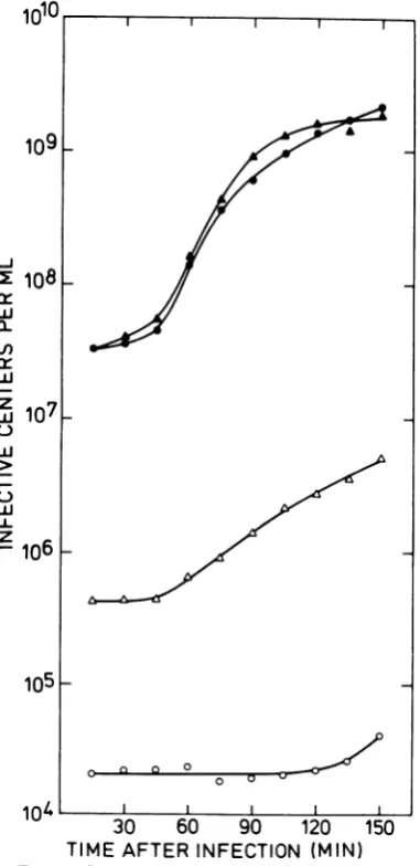
Induction of restriction resistance. (i) MC.
Arwert andRutberg(1)demonstrated thatMC
20, 1976
on November 10, 2019 by guest
http://jvi.asm.org/
treatment ofnonmodifyingB. subtilis cells be-fore infection with phage
0105
andSPO2 signif-icantly increased the restriction ratio (ratio of phage titers on the restricting host to that on thenonrestricting host) of the resulting lysates. This effect of MC is also observed with the virulent phage SPP1: Fig. 1 shows one-step growth curves and development of restriction resistance of SPP1 after infection ofuntreated and MC-pretreated cells. After one cycle of growth, the restriction ratio was 2 x 10⁻⁵ in lysates from untreated cells and 2.5 x 10⁻³in lysates from MC-pretreated cells. Restriction enhancement is also observed in mass lysates1010
>X
,
108
w
0_
w
-
107
0 w
w
U.
Z
106
105
H
1n4l
30 60 90 120 150
TIME AFTER INFECTION (MIN)
FIG. 1. One-step growth curves of SPP1 on
un-treated andMC-pretreated222cells. Infectivecenters from untreated cellsplated onMCB (0) and5GR (0), andfrom MC-pretreated cellsplatedon MCB
(r-m-) (A) andon5GR (r+m+) (A).
(Table
1). Under suchconditions,
therestric-tion ratio has consistently been higher than thatobserved inone-stepexperiments.
The experiments inFig. 1 and Table 1 were
performed
using MCat aconcentration of2,g/
ml. MC atconcentrations of less than1
g.g/ml
is not as effective, whereas concentrations of greater than 4jig/ml
leadtoveryslowgrowth
of the cells and low phage yields even after removal of MC. The restriction ratio was the same using cellstreated for various times
be-tween 15 and 90 min, although phage yields
were reduced 10-fold with exposure times of more than 60 min. In contrast to results
re-portedby Arwert and Rutberg (1) for SPO2 and 4105, wedo notfindasdrasticaneffect of the timeelapsed between removal of MC andphage infectiononthephage yield (Fig. 2). At 90 min after removal of MC, phage infection and growth isnodifferentfrom thezerotime infec-tion.Thedifferencebetween the restriction
ra-tiosofthetwopairsof curves showninFig.2 is
within the range of normal statistical varia-tion.
The MC-mediated enhancement to
restric-tion resistance might result from the produc-tionof SPP1 phages with type R modification. The extent ofmodificationinsuch phages could
be at any level between partial and complete
modification (see subsequent section). Such modified phages could arise from MC-pre-treated cells in any situation between these
extremes: (i) all modifiedphagesare produced
in a few bursts, (ii) every infected cell yields modifiedphages with theprobability of the
re-striction ratio observed in MClysates. A single-burst experimentwasperformedtoanalyzethis
situation. Cells pretreated with MC and in-fected with SPP1 were plated on strains 5GR and MCB to obtain the restriction
ratio,
and diluted and distributed into test tubes sothat each tube received ontheaverage0.6infective centers when plated with the nonrestricting host (setA in Table2). To compensate for the restriction of phages on the restricting host,each tube of the sets to be plated with this
indicator (setB in Table 2) received 50 or 100
times as many infective centers. If modified
TABLE 1. EffectsofMConrestrictionofSPP1 Titer on
Phage Restriction
Phage MCB 5GR ratioa
(r- m-) (r+m+)
SPP1*0 2.0.1010 4.5*105 2.3 10-5
SPP1-MC 2.2-109 5.3*107 2.4.10-2
a Stocks of SPP1-Oand SPP1 * MCwereplatedon
MCB and 5GRtoestablish their restriction ratio. cl 0 0
0 U
on November 10, 2019 by guest
http://jvi.asm.org/
[image:3.508.69.260.233.626.2]MODIFICATION
iU-
-109
I
108k
106
k
105
30 60 90 120 150
TIME AFTER INFECTION (MIN)
FIG. 2. One-step growth curves ofSPP1
estab-lishedattimes 0min(A) and(A)and 90 minafter removal of MC (a) and (0). PlatingswereonMCB
(r-m-) (filled symbols) and on 5GR (r+m+) (open
symbols).
phage were produced exclusively according to
situation (ii) above, onewould expectan
aver-agenumberof modifiedplaquesintubes of sets
Bthatwould begiven by theexpectednumber
of nonmodifiedphagespertubeof set B
multi-plied by the restriction ratio. This value is 20
forsetB, and40 for setB2.Takingintoaccount
a sixfold lower plating efficiency of modified phage on strain5GR (Table 2), this would give average numbers of
modified
phage per tube of3 and 6 for sets B, and B2, respectively, which
arenotcompatible with the observed numbers of tubes without phage (Table 2). These ob-served numbers are, however, in agreement
with the prediction madeon the basis of
situa-tion (i) above, according to which a few modi-fied
phage-producing
cellsshould followa Pois-son distribution in tubes ofset B. Thecalcu-latedzero classvaluesofsuchadistributionfor
B, are p(o) =
e-28s5x
10-2) e-042 = 0.66 and for B2,p(o)
= e-55(l.5X10-2) e-0.82 =0.44,
ingood agreement with the observed values of
74%
(B1)
and 61%(B2). Whether
bursts with modified phages also containnonmodified par-ticles cannotbe analyzedwith this type ofex-periment.
(ii) UV irradiation. In Fig. 3, we have plot-ted the survival of strain222cells asafunction
0
10-1
-_a
1.)
S
w
Da
10-2 .> 1/)
UV dose Isec]
FIG. 3. Influenceof UV irradiation of222 cellson
therestrictionratioofSPP1 propagatedonsuch
bac-teria.
TABLE 2. Single-burstexperimenta
Relative
Set Platingbacteria concn of in- No. (%)of tubes No. (%)of tubes ofbursts/ Avgburst fectedcells! scored withoutphage tube sizeobserved
tube
A MCB (rm-) 1 104 (100) 59 (57) 0.55 42
B, 50 72 (100) 53 (74) 0.31 11
5GR(r+m+)
B2 100 72 (100) 44 (61) 0.51 3
aInfective centers plated before lysis gave titers of 2.50 x 10⁷ and 3.81 x 10⁵ onstrains MCB and 5GR, respectively; this correspondsto arestriction ratioof 1.5 x 10⁻².
I I Il
107
L1J
-(.)
LLJ
uj
z C)
L) LIL z
VOL. 20, 1976
1
I ~ Il
on November 10, 2019 by guest
http://jvi.asm.org/
[image:4.508.51.245.66.408.2] [image:4.508.255.450.291.513.2] [image:4.508.52.450.565.653.2]of UV dose and the restriction ratio of SPP1
mass lysates produced on cells that had been
irradiated for the times indicated. It is obvious
that increasing UV dosesinduceincreasing
re-striction resistance in SPP1 0 phages
propa-gated on cells exposedto UV irradiation. The
maximum level of UV-induced restriction
re-sistance is similar to that observed after MC
induction.
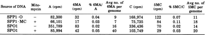
Methylation levels in bacterial and phage
DNAs isolated after MC treatment of cells.To
establish whether MC-enhanced resistance to
restrictionwasdue tomethylation-linked
modi-ficationwehavestudied the effects of
preincu-bation of cells with MConthecontentof
meth-ylated bases in bacterial DNAs (Table 3) and
phage DNAs (Table 4). Molecular weights and
guanine plus cytosine contents of DNAs
ana-lyzedaregiven in reference 6.
Instrain 222(r-mi),there isnodifferencein
the N-6-methyladenine (6MA) content in
treated and nontreated cells, butaremarkable
increase inthe 5MC contentoccursafter
expo-sure to MC. In contrast to untreated cells in
whichonly 0.13% of C is methylated, the5MC
content in pretreated cells is increased more
than 10-fold to 1.42%. This isveryclose tothe
level of methylation observed in DNA from
cells of the normally modified strains (6). We
conclude that MC treatment ofr-m- cells
in-ducesamethyltransferase that methylatesC to
5MC. We have also analyzed the effect ofMC
pretreatment on the DNA of the modifying
strain 5GR. Here we observe asmall increase
in 5MCcontent. In addition, 0.2% of the
ade-ninesarepresentas6MA.
The effects of MCtreatmentofstrain 222on
phage DNAs (Table 4) indicate, for SPP1, no
change in 6MA content. There is, however, a
small and significant difference of0.04% 5MC/
C between phages obtained from treated and
untreated cells (0.07 and0.11%5MC/C,
respec-tively). Thismayreflectaslightenhancement
of MC in all SPP1 MC phages or complete
methylation ofa small subpopulation in such
stocks.
MC treatment of host cells does not greatly
alter the cytosine methylation of SPOl DNA
(Table 4). Thus this DNA, which isa poor
sub-strate for the modification-specific
methyl-transferase in r+m+ strains (6), is also a very
poorsubstrate foranyinduced
methyltransfer-ase.
Distribution of 5MC in SPP1-MC DNA. The
biological tests described before indicated that
onlya small proportion of SPP1-MC could be
modified. Inagreement,chromatographic
anal-ysesof SPP1*MC DNA indicateanincrease in
the cytosine methylation of 0.04% upon
infec-tion ofMC-treated host cells. Ifa molecule of
methylated phage DNA from an SPP1 *MC
stock were as fully methylated as DNA from
SPP1 R (5MC/C = 1.26% [6]), this would
indi-catethat 3% of the DNAmoleculesweretotally
modified andtherest not atall. Alternatively,
the additional 0.04% methylation could be
dis-tributedcontinuously amongthe population of
DNAmoleculestocreatephages with
[image:5.508.69.463.460.529.2]interme-diate levels of modification. To distinguish be-tween these possibilities, we have subjected
TABLE 3. EffectsofMConin vivo methylation ofbacterial DNAa
6MA ~~Avgno.of Avgno.of
Source of mito- A (cpm) 6MA %6MA/ A perof 5MC %5MC/ 5Mg per
DNA mycin (cpm) A
pernCocmm
(cpm) C gnm222(rm-) - 122,644 95 0.08 2,100 156,538 203 0.13 2,450
222(rWmi) + 67,640 57 0.08 2,100 48,949 696 1.42 27,000
5GR(r+m+) - 178,469 23 0.01 <250 434,791 7,455 1.69 32,000
5GR(r+m+) + 24,922 49 0.20 5,100 106,651 2,020 1.90 36,000
aThe numbers give the sum of two separatechromatographic analyses. Thefluctuation between these
determinationswasless than10%.The lower limit of detection is0.01%methylatedbase per main base.
TABLE 4. Effects of MConthe in vivomethylation of phageDNAa
Source of DNASoureofDNAMito-myci AA (cpm)(pm) (cpm)6MA % 6MA/A Avg no. of6MApier C(cpm) (cpm)5MC %5MG/C Avg5MGno.perof
mycin
~~~~~~~~genome
genomeSPP1.O - 82,300 32 0.04 9 168,974 122 0.07 11
SPP1MC + 66,101 17 0.03 7 75,735 84 0.11 18
SPOl - 351,789 83 0.02 16 336,436 70 0.02 14
SPOl + 85,994 42 0.05 40 103,749 29 0.03 20
a Numbers give the sum of two separate chromatographic analyses. The fluctuation between these
determinations was less than 10%. The lower limit of detection is 0.01% methylated base per main base.
on November 10, 2019 by guest
http://jvi.asm.org/
[image:5.508.69.463.577.645.2]INDUCIBILITY OF
SPP1 MC DNA to complete endoR Bsu R
digestion and
analyzed
the digest by agarosegel electrophoresis (Fig. 4).
This
analysis
re-veals the presence of a small portion of
SPP1 -MC DNA which is completely resistant
to enzyme treatment, whereas the remaining
material follows essentially the degradation
patternof nonmodified DNA.
Although
wecan-notexclude the possibility thatasmall fraction of DNAshowsintermediary modification, MC-inducedmodification affects onlya minor
frac-tion of molecules, and these are completely
modified.
This result is further substantiated by an
analysis of the inactivation kinetics of
trans-fecting SPP1* MC DNAby treatmentwith the endoR-Bsu R restriction enzyme. A general small increase in the 5MC content would still
not protect all SPP1 molecules from
degrada-2 34 5
tion by the restriction enzyme. One should, however, expect areductioninthe slope of the inactivation curve indicating decreased
sensi-tivityof the DNA to restriction. On the basis of
ahighly discontinuous distribution of
modifica-tion, one would expect a biphasic curve; i.e.,
themajorityofmoleculeswould followthe
inac-tivation curve ofSPP1-O DNA, but a minor
fraction would be completelyresistant. The
re-sults showninFig.5 supportthe latter
expecta-tion. Theresistant biological activity amounts
to 0.1%. Takinginto accountthat SPP1
trans-fection shows a quadratic dose response (13),
thisplateau level would be expectedif 3%of the
100
10
1-0
-5 cri
.U
0 n
0
L/)
0.1
0.01
FIG. 4. Agarose gel electropherograms ofvarious SPP1 DNAsafterlimitdigestionwith endoR Bsu R
restrictionenzyme.Tracks containthefollowing
por-tions: 1, 0.1 Mgof SPP1 *R DNA; 2,1 ,ug of SPP1 *0 DNA; 3,1 Mgof SPP1 MC DNA; 4, 1 Mg of
undi-gested SPP1 MC DNA; 5, 0.05 pg of undigested
SPP1-ODNA.
Time of enzyme treatment [minI
FIG. 5. Inactivationof biologicalactivityof
trans-fectingSPP1 DNA by endoR -Bsu R restriction
en-zyme. SPP1 -0 DNA (A), SPP1 -R DNA (0), and SPP1 -MC DNA (A) were incubated for the times indicated with the DEAE-cellulose enzyme fraction
(1/800 dilution) of the endoR -Bsu R enzyme. The enzyme reaction was stoppedatthe indicated times
by heating corresponding portions for 10 min at
68°C. Such DNA was assayed oncompetent strain 222cells by transfection. DNA concentration in the
reactionmixtures was5
Mg/ml.
One hundred percentvaluesof biologicalactivity were(infectivecentersper
milliliter)2.7x10⁵(A), 6.7 x 10⁴(0),and 8.0 x 10⁴
(A)
20, 1976
on November 10, 2019 by guest
http://jvi.asm.org/
[image:6.508.262.443.234.539.2] [image:6.508.65.239.280.599.2]SPP1*MCmolecules carried complete modifica-tion.
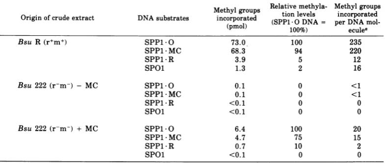
Levels of methyltransferase activity in
crudecell extracts. Themethylating activities
ofcrudeextractsfrom B. subtilis strains R and
222 (untreatedorpreincubated with MC) were
analyzed with the DNAs of phage SPP1 0,
SPP1 MC, SPP1 R, and SPOl as substrates
(Table 5). As expected, methyltransferase
ac-tivity is high inextractsofthemodifying strain
R incomparison with extractsofthe
nonmodi-fying strain 222. However, MCtreatmentof222
cells enhances thelevel of methyltransferaseat
least 30-fold. Thefinal methyltransferase level
obtained in the total extract is about 10% of
that found in Rstrains. This activity sufficesto
produce the degrees of methylation observed in DNAs from MC-pretreated cells. Extracts from MC-treated 222 and untreated R cells show
very similar substrate specificities: with both
extracts, the highest incorporation of methyl
groups is into SPP1 0 DNA, slightly less is
incorporated into SPP1 * MC DNA, andvery
lit-tle is incorporated into the SPP1 R and SPOl
DNAs. The difference in the capacity of
SPP1*0and SPP1*MC DNAstoacceptmethyl
groups is compatible with our other results
(Fig. 4 and 5), indicating that only a small
fraction of the SPP1 MC molecules carry
R-specific modification. The lowcapacityof SPOl
DNA tobe methylated by crude extracts is in
agreement with DNA analyses of methylated
bases, which showed that this DNA is not a
substrate for R-specific modification in vivo
either (6). The estimated amount of
incorpo-rated methylgroupsintoanSPP1*0 DNA
mol-ecule by the R strain extracts is somewhat
more (235) than the number observed in the
naturally modified SPP1*Rmolecule, which is
190 (6). Weattributethis, and alsothe fact that
slight methylationofSPP1 R DNA stilloccurs
by the"homologous" Renzyme,totheuseofin
vitro conditions.
DISCUSSION
MCtreatment, and possibly also UV
irradia-tion, induce in nonmodifyingB. subtilis cells
the formation ofaDNA methyltransferase
ac-tivity. The specificities of the MC-provoked
methyltransferase and the modification
en-zymefound inextractsofrestricting/modifying
cells are verysimilar. The important question
of whether thesetwoactivitiesaredueto
iden-tical enzymes must await their purification.
Suchstudiesarepresentlybeing undertaken.
Methylation of C to 5MC in bacterial DNA
after MC treatment approaches the level
ob-served in DNA from modifyingtype R strains.
Methylation in a stock of SPP1-MC is not
nearly as high. In two respects, MC-provoked
methylation of SPP1 DNA is nonrandom: (i)at
least some modified phages originate from
largerbursts ofafew MC-treated cells (Table
2). Wecannot determine whether cells
releas-ingmodified phages in addition produce
sensi-tive phage; (ii)atleast the majority of modified
phagesarecompletelymodified; i.e., there isno
indication of the presence of DNA molecules
with intermediate modification (Fig. 4 and 5).
The restriction ratio of 2.4 x 10⁻² in
SPP1 MC stocks (Table 1) can readily be
ac-counted forby thepresenceinthepopulation of
2to3%completelymodified phages. This value
is ingoodagreementwith (i) thelevel of
resid-ualtransfecting activity of SPP1 * MC DNA
ex-posed to degradation with the endoR Bsu R
TABLE 5. DNA methyltransferase activities in crude cellextracts.
Methylgroups Relativemethyla- Methylgroups
Originofcrude extract DNA substrates incorporated (SPP1O DNA = perDNA
mol-(pmol) 100%) eculea
Bsu R(r+m+) SPP1-O 73.0 100 235
SPP1*MC 68.3 94 220
SPP1*R 3.9 5 12
SPOl 1.3 2 16
Bsu 222(r-m-) - MC SPP1 O 0.1 0 <1
SPP1 MC 0.1 0 <1
SPP1 *R <0.1 0 0
SPOl <0. 1 0 0
Bsu 222 (r-m-) + MC SPP1 0 6.4 100 20
SPP1 MC 4.7 75 15
SPP1 *R 0.7 10 2
SPOl <0.1 0 0
aThemeasuredspecific activityof
S-adenosyl-L-[methyl-:'H]-methionine
is3,250cpm/pmol.on November 10, 2019 by guest
http://jvi.asm.org/
[image:7.508.68.461.490.657.2]restriction enzyme(Fig.5), (ii)thedegradation
pattern of SPP1 MC DNAobserved after limit
digestion with endoR-Bsu R (Fig. 4), (iii) the extent of DNA methylation (Table 4), and is
(iv) compatible with the degree to which
SPP1 -MC DNA canbe furthermethylated by
crudecellextracts (Table 5).
The MC inducibility ofa modification-type
methyltransferase activity in phenotypically nonmodifyingcells implies that the genetic
in-formation for suchanenzyme(s)mustbe
pres-entinthese cells. Wearepresently
investigat-ing whether such a genetic determinant(s) is
identical to those mapped intype R cells (15).
The question arises, why is such information
expressedaftertreatmentwith agentsknownto
causeprophage induction?It isconceivable, as
pointed out by Arwert and Rutberg (1), that
genesfor modification(andpossibly restriction) enzymes are carriedby anunexpressed
defec-tive inducible prophage in r-m- cells which
becomemanifest after induction. The induction
ofmethyltransferases afterphage T2 infection
orinductionofXhas beenreported by Wainfan
etal. (16). Inoursystemthe inductionof
mod-ifying activity and the presence of defective
prophagePBSX (11)werecorrelated byArwert
and Rutberg (1). However, subsequently A.
Garro (personalcommunication) observed that
modifying activitycould also be induced in cells
with noninducible PBSX prophage. Although
thisfinding makes the connection between
in-ductionof PBSX and methyltransferase
some-whatmore complicated, it does not affect the
generality of the concept of a correlation
be-tween enzyme and prophage induction,
espe-cially in view of the existence of many other
defectiveprophages in B. subtilis (5).
ACKNOWLEDGMENTS
WethankW.ArberandourcolleaguesinBerlin, partic-ularlyM.Achtman,for their criticism of themanuscript,B. Behrensforperformingtheagaroseelectrophoresis experi-ment,andR.Thompsonforacquaintinguswiththis tech-nique.
LITERATURE CITED
1. Arwert, F., and L. Rutberg. 1974. Restriction and modi-fication in B.subtilis. Induction of a modifying activ-ity in Bacillussubtilis 168. Mol. Gen. Genet. 133:175-177.
2. Bron, S., and K. Murray.1975. Restriction and modifi-cation in B. subtilis. Nucleotide sequence recognized by restriction endonuclease R Bsu R from strain R. Mol. Gen.Genet. 143:25-33.
3. Bron, S., K. Murray, and T. A. Trautner. 1975. Restric-tion andmodification in B. subtilis. Purification and general properties of a restriction endonuclease from strain R. Mol. Gen.Genet. 143:13-23.
4. Esche, H., M. Schweiger, and T. A. Trautner. 1975.
Geneexpressionof bacteriophage SPP1. I. Phage
di-rectedproteinsynthesis. Mol. Gen. Genet. 142:45-55. 5. Garro,A.J., and J.Marmur. 1970. Defective
bacterio-phages. J. Cell. Physiol. 76:253-264.
6. Gunthert,U., J. Stutz, and G. Klotz. 1975. Restriction and modification in B. subtilis. The biochemical basis of modification against endo R Bsu R. Mol. Gen. Genet. 142:185-191.
7. Lowry,0.H., N.J.Rosebrough, A. L.Farr,and R. J. Randall. 1951. Protein measurement with the Folin phenol reagent. J. Biol. Chem. 193:265-275. 8. Okubo, S., B. Strauss, and M. Stodolsky. 1964. The
possiblerole of recombination in the infection of com-petent B. subtilis by bacteriophage DNA. Virology 24:552-562.
9. Riva,S., M. Polsinelli, and A. Falaschi. 1968. A new phage of B. subtilis with infectious DNA having sepa-rablestrands.J. Mol. Biol. 35:347-356.
10. Rottlander,E.,andT. A. Trautner. 1970. Genetic and transfectionstudies with B. subtilis phage SP50. I. Phage mutants withrestricted growth on B.subtilis
strain168.Mol. Gen. Genet. 108:47-60.
11. Seaman, E., E. Tarmy, and J. Marmur. 1964. Inducible phages ofBacillussubtilis.Biochemistry 3:607-613. 12. Spatz, H. Ch., and T. A.Trautner. 1970.One way to do
experiments ongene conversion? Mol. Gen. Genet. 109:84-106.
13. Spatz, H. Ch., and T. A. Trautner. 1971. The role of recombination in transfection of B. subtilis. Mol. Gen. Genet. 113:174-190.
14. Spizizen, J. 1958. Transformation ofbiochemically
defi-cient strains of Bacillus subtilis by
deoxyribonu-cleate. Proc. Natl. Acad. Sci. U.S.A. 44:1072-1078. 15. Trautner, T. A., B. Pawlek, S. Bron, and C.
Anagnos-topoulos. 1974. Restriction and modification in B.
subtilis. Biological aspects. Mol. Gen. Genet. 131:181-191.
16. Wainfan, E., P. R. Srinivasan, and E. Borek. 1965.
Alterations in thetransfer ribonucleic acid
methyl-asesafterbacteriophageinfectionorinduction.
Bio-chemistry4:2845-2848. VOL. 20, 1976