Volume 2012, Article ID 768720,16pages doi:10.1155/2012/768720
Research Article
Allosteric Modulation of Beta1 Integrin
Function Induces Lung Tissue Repair
Rehab AlJamal-Naylor,
1Linda Wilson,
2Susan McIntyre,
3Fiona Rossi,
4Beth Harrison,
3Mark Marsden,
4and David J. Harrison
31Avipero Ltd., 5th Floor, 125 Princes Street, Edinburgh EH2 4AD, UK
2School of Biomedical Sciences, The University of Edinburgh, Hugh Robson Building, George Square, Edinburgh EH8 9XD, UK 3Division of Pathology, Institute of Genetics and Molecular Medicine, The University of Edinburgh, Western General Hospital,
Edinburgh EH4 2XU, UK
4MRC Centre for Inflammation Research, The Queen’s Medical Research Institute, The University of Edinburgh,
47 Little France Crescent, Edinburgh EH16 4TJ, UK
Correspondence should be addressed to Rehab AlJamal-Naylor,[email protected] Received 16 August 2011; Revised 21 October 2011; Accepted 31 October 2011
Academic Editor: Chi Hin Cho
Copyright © 2012 Rehab AlJamal-Naylor et al. This is an open access article distributed under the Creative Commons Attribution License, which permits unrestricted use, distribution, and reproduction in any medium, provided the original work is properly cited.
The cellular cytoskeleton, adhesion receptors, extracellular matrix composition, and their spatial distribution are together fundamental in a cell’s balanced mechanical sensing of its environment. We show that, in lung injury, extracellular matrix-integrin interactions are altered and this leads to signalling alteration and mechanical missensing. The missensing, secondary to matrix alteration and cell surface receptor alterations, leads to increased cellular stiffness, injury, and death. We have identified a mono-clonal antibody againstβ1 integrin which caused matrix remodelling and enhancement of cell survival. The antibody acts as an allosteric dual agonist/antagonist modulator ofβ1 integrin. Intriguingly, this antibody reversed both functional and structural tissue injury in an animal model of degenerative disease in lung.
1. Introduction
Tissue regeneration comprises dedifferentiation of adult cells into a stem cell state and the development of these cells into new remodelled tissue, identical to the lost one. Tissue repair is defined as replacement of normal tissue by fibrous tissue and integrins are crucial in these processes.
Integrins are membrane spanning proteins facilitating the two-way communication between the inside and outside of a cell. Integrins have the capacity to bind a multitude of molecules both inside and outside of the cell. The binding of these molecules results in the transmission of information into and out of the cell, which can influence a host of dif-ferent cellular functions, including the cells metabolic activ-ity.
Of the many types of integrin receptors, theβ1 integrin is by far the most ubiquitous allowing cells to detect a vast array of stimuli ranging between toxins, protein hormones,
neurotransmitters, and macromolecules. There have been numerous publications documenting a potential role ofβ1 integrin in tissue development and repair in several tissue types (reviewed in [1]). It is clear that β1 integrin plays a crucial role during postnatal skin development and wound healing, with the loss of epithelialβ1 integrin causing exten-sive skin blistering and wound healing defects. More recently, there has been active interest in the cosmeceutical develop-ment of β1 integrin targeting formulations. One such ex-ample is following the discovery of fucoidans from Fucus
vesiculosus and its effect on skin scarring and ageing [2,3] which was later found to be mainly attributed to alpha2 and β1 integrin [4].
its ligand on the cell surface, as well as the spatial and geo-metric arrangement and movement) [5]. Recent evidence has demonstrated that both affinity and avidity of integrins are strongly related to the size of the focal adhesion clusters [6,7]. Overall, integrins have three main possible conforma-tions of the extracellular domain; a low affinity, bent con-formation; extended conformation with closed headpiece representing an intermediate affinity state; the ligand-bind-ing-induced high-affinity extended form, with an open head-piece [8–10].
Altered conformation of integrins rather than expression levels have been reported both during physiological and pathological remodelling processes which include neurite outgrowth, fibrosis, asthma, cancer, and wound healing amongst many [11–14]. To successfully develop disease mod-ifying therapy, it should be beneficial to rescue or replenish dying or dead cells by activating inherent repair processes other than simply stem cell regeneration. In other words, altering the interaction of the cells with other cells and their abnormal surroundings to promote their survival and con-tinued function may alleviate chronic, ongoing cell loss, a hallmark of many progressive degenerative diseases. We hy-pothesised that tissue repair might be achieved by rescueing cells from death by mechanically dampening the signals cells receive from their abnormal environment. One key cell sur-face receptor for adhesion isβ1 integrin. We considered that conformational modulation ofβ1 integrin may cause alter-ation in cytoskeletal organizalter-ation and cell stiffness leading to increase susceptibility to oxidative stress and death and thus that prevention of these changes may have therapeutic bene-fit. Here, we show how we have identified a specificβ1 inte-grin targeting modality using a monoclonal antibody, which we have demonstrated, both protects from tissue injury and facilitates repair. The antibody acts as an allosteric dual agonist/antagonist modulator of β1 integrin and resulted in increased matrix remodelling and enhancement of cell survival.
2. Results
2.1. The Effects ofβ1 Integrin Modulation on Elastase-Induced Signalling. As a model of tissue remodelling in disease, we
investigated the activity ofβ1 integrin using an in vitro model of elastase-induced injury. A coculture of primary, adult human lung fibroblasts was overlayered with NCI-H441 lung cells, under cyclic mechanical stimulation, and subjected to elastase treatment.
To investigate the involvement ofβ1 integrin activation in elastase-induced signalling, we used three different mon-oclonal antibodies againstβ1 integrin. The first was the ad-hesion blocking clone, JB1a, which is said to target primarily the amino acids 82–87 comprising part of the hybrid domain [15]. We also used the adhesion blocking clone, AIIB2, which binds amino acid residues 207–218 within the A-domain [16], and K20, widely reported to have no functional effects which binds the hybrid/EGF repeat region [17].
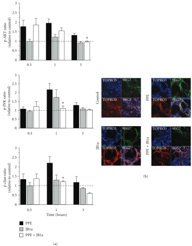
Addition of elastase to cell culture induced an increased phosphorylation of signalling proteins known to act down-stream of β1 integrin (Figure 1(a)). During the course of
injury, pAKT levels increased, followed by transient increases in phospho-cJUN and phospho-JNK (Figure 1(a)). No sig-nificant changes were detected for 12 other phosphoproteins at the sampled time points.
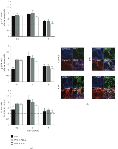
Whenβ1 integrin was bound by antibody clone JB1a in the absence of injury, there was no significant effect on down-stream signalling. However, when JB1a was added during elastase-induced injury, it abrogated all elastase-induced changes in phosphorylation of signalling proteins. This effect was not seen with either AIIB2 or K20 (Figure 2(a)).
2.2. The Effects of Elastase-Induced Injury onβ1 Integrin Activ-ity and Localisation. We next examined the effects of elastase on ligand-binding activity of β1 integrin using the ligand competent specific anti-β1 integrin antibody 9EG7. Ligand competent state can be any of the intermediate physiological conformations or the fully activated extended conformation [19]. Elastase caused an increase in ligand competent/active β1 integrin expression as evident from the staining pattern inFigure 1(b). Modulation ofβ1 integrin, using JB1a, abro-gated the elastase-induced increase in ligand-competentβ1 integrin (Figure 1(b)). However, the anti-β1 integrin clone K20 potentiated the elastase-induced increase in the ligand-competent conformation (Figure 2(b)).
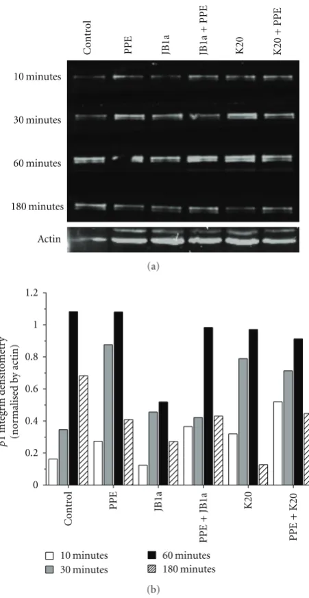
The effects of elastase and JB1a on the level of ligand-competent receptor were not simply a result of change in cell surface expression (Figure 1(c)). However, preliminary meas-urements showed that elastase increased the cytosolic frac-tion-associated β1 integrin which might be attributed to recycling or degradation. To address that we conducted a time course analyses ofβ1 integrin in membrane fractions. Elastase induced a change inβ1 integrin recycling, an effect inhibited by JB1a but not K20 (Figure 3). Further evidence of elastase-inducedβ1 integrin activation was the increase of caveolin-1 phosphorylation after two hours of exposure to elastase; changes once again inhibited by JB1a (Figure 4).
2.3. The Conformational Effects of JB1a on β1 Integrin. To
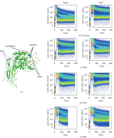
further determine the pharmacological mode of action of JB1a action, we questioned whether the effect seen with JB1a is due to its effect onβ1 integrin chain allostery. We first esti-mated the location of the epitope of JB1a on the basis of the theoretical 3-dimentional structure of β1 integrin (Figure 5(a)). We then conducted FRET studies using nonadherent Jurkat cells. FITC-labelled LDV cyclic peptide was used to label the head of alpha4β1 integrin, and the lipophilic dye R18 was used to label the cell membrane. LDV-FITC acts as a donor and R18 as an acceptor [20]. FACS was used for the FRET acquisition and measurement. JB1a caused a confor-mational activation when added at baseline and inhibited the full conformational activation induced by the divalent cation, Mn2+. The JB1a-induced change resulting in FRET
efficiency was indicative of an intermediate partially extend-ed conformation when comparextend-ed to Mn2+and other known
3
2.5
2
1.5
1
0.5
0
0.5 1 3
0.5 1 3
3
2.5
2
1.5
1
0.5
0
3
2.5
2
1.5
1
0.5
0
0.5 1 3
∗
∗
∗
Time (hours)
PPE
JB1a PPE + JB1a
p-JNK
ratio
(r
elati
ve
to
cont
rol)
p-AKT
ra
tio
(r
elati
ve
to
cont
rol)
p-cJ
un
ra
tio
(r
elati
ve
to
cont
rol)
PP
E
JB1a
Cont
ro
l
TOPRO3 TOPRO3
TOPRO3 TOPRO3
TOPRO3 TOPRO3
TOPRO3 TOPRO3
9EG7 9EG7
9EG7 9EG7
9EG7
9EG7
9EG7
9EG7
PP
E
+
JB1a
(a)
[image:3.600.100.497.71.579.2](b)
Figure 1: The effects of PPE-induced injury and JB1a treatment on (a) activation of signalling downstream of beta1 integrin during mecha-nical stretch (asterisks denote statistical significance withP <0.05 in comparison to PPE) and (b) beta1 integrin conformational activation indicated by increase in staining using the anti-β1 integrin antibody 9EG7 recognising the ligand competent receptor in comparison to staining using nonconformation dependent antibody (K20).
2.4. Conformational Modulation ofβ1 Integrin Inhibits Elas-tase-Induced Changes in Cell Membrane Composition. Using
mixed epithelial-mesenchymal in vitro cultures, we found that elastase increased neutral sphingomyelinase activity transiently; an effect inhibited byβ1 integrin binding by anti-body JB1a (Figure 6(a)). No effect on acid sphingomyelinase was detected under the same conditions.
2.5. Conformational Modulation ofβ1 Integrin Inhibits Elas-tase-Induced Changes in Actin Polymerisation and Cellular Impedance. Using our in vitro culture system, we tracked
3
2.5
2
1.5
1
0.5
0
3
2.5
2
1.5
1
0.5
0
3
2.5
2
1.5
1
0.5
0
0.5 1 3
0.5 1 3
0.5 1 3
Time (hours)
PPE
p-JNK
ra
tio
(r
elati
ve
to
cont
rol)
p-AKT
ra
tio
(r
elati
ve
to
cont
rol)
p-cJ
un
ra
tio
(r
elati
ve
to
cont
rol)
PP
E
Cont
ro
l
TOPRO3 TOPRO3
TOPRO3
TOPRO3
TOPRO3 TOPRO3
TOPRO3
TOPRO3
9EG7 9EG7
9EG7
9EG7
9EG7 9EG7
9EG7
9EG7
K20
(a)
(b)
PPE + AIIB2
[image:4.600.100.508.72.586.2]PPE + K20
Figure 2: The effects of PPE-induced injury and targeting beta1 integrin using AIIB2 and K20 clones on (a) activation of signalling down-stream of beta1 integrin during mechanical stretch (asterisks denotes statistical significance withP <0.05 in comparison to PPE) and (b) beta1 integrin conformational activation indicated by increase in staining using the anti-β1 integrin antibody 9EG7 recognising the ligand competent receptor in comparison to staining using nonconformation dependent antibody (K20).
doi:10.1155/2012/768720). Formation of F-actin from mo-nomeric G-actin is energy dependent, and, under ATP depletion conditions, there is a net conversion of monomeric G-actin to polymeric F-actin. In cocultures, elastase reduced the levels of ATP, but this response was inhibited by JB1a (Figure 6(c)).
Actin 10 minutes 30 minutes 60 minutes 180 minutes PP E JB1a C o nt ro l K20 JB1a + P P E K20 + P PE (a) 1.2 1 0.8 0.6 0.4 0.2 0 C o nt ro l PP E JB1a K20 10 minutes 30 minutes 60 minutes 180 minutes P PE + JB 1 a PP E + K 2 0 β 1 int eg ri n d ensit o met ry (nor malised b y ac tin) (b)
Figure 3: The effects of PPE-induced injury (0.6 U/mL) and tar-geting beta1 integrin using JB1a (1 ug/mL) in comparison to K20 (1 ug/mL) clone on the kinetics of beta1 integrin levels on cell mem-brane in vitro using human lung coculture. (a) Western blot of the cell membrane expression ofβ1 integrin over time. Protein extracts were loaded at equal protein concentration (25μg). (b) Densitomet-ric analyses of the blot corrected using actin as an internal control.
2.6. Conformational Modulation ofβ1 Integrin Inhibits Elas-tase-Induced Caspase Activation. We then investigated the
effect of elastase on caspase activation and the role ofβ1 inte-grin in elastase-induced cell death. Elastase induced caspase activation after 3-hour exposure and led to detachment in-duced apoptosis of epithelial cells (anoikis) (Figures7(a) and S1–S3). Modulation ofβ1 integrin using JB1a prevented cas-pase activation. However, the potently inhibitory anti-β1 in-tegrin antibody 6S6, which is also known to induce homo-typic aggregation, induced caspase activation (Figure 7(b)).
p-Cav Cav1 Actin C o nt ro l PP E
JB1a JB1a K20 AIIB2
+ P P E A II B 2+P P E K 2 0+P P E (a) 0.8 0.7 0.6 0.5 0.4 0.3 0.2 0.1 0 C o nt ro l PP E JB1a K20 AIIB2 PP E + JB1a PP E + K20 PP E + AIIB2 Ratio o f p Ca v1/C av (nor malised b y actin) (b)
Figure 4: The effects of PPE-induced injury (0.6 U/mL) and targeting beta1 integrin using AIIB2 (1 ug/mL) and K20 (1 ug/mL) clones on phosphorylated caveolin-1 levels in membrane fractions. (a) Representative blots fromn = 4. Loading controlled by total amount of protein (50μg). (b) Densitometric analyses of the blot corrected using actin as an internal control.
2.7. Conformational Modulation ofβ1 Integrin Reversed Elas-tase-Induced Emphysema in Mice. To investigate the
signifi-cance ofβ1 integrin in injury in a disease setting in which remodelling is a key component, we established a murine model of emphysema caused by intratracheal installation of elastase. Mice were instilled with elastase on day 1 and lung injury ensued. At later timepoints, they were treated with the anti-β1 integrin monoclonal antibody, JB1a which bindsβ1 integrin in mouse tissues [22], or vehicle, either once on day 14 (21 day group, 21 d) or on days 21 and 28 (35 day group, 35 d). In a subsequent investigation, severe emphysema was induced and JB1a and B44 clones were instilled on days 21 and 28 before lung function assessment on day 35. Both clones demonstrated cross-reactivity with murine β1 inte-grin (Figure 8).
[image:5.600.314.543.70.432.2]JB1a
ADMIDAS
Time
Basal Mn2+
105
104
103
102 0
105
104
103
102 0
105
104
103
102 0 105
104
103
102 0
105
104
103
102 0
105
104
103
102 0
105
104
103
102 0 105
104
103
102 0
0 200 400 600
Time 0 200 400 600
Time 0 200 400 600 Time
0 200 400 600
Time 0 200 400 600
Time 0 200 400 600
Time 0 200 400 600 Time
0 200 400 600 (a)
(b) Control
(c) JB1a
(d) K20
(e) B44
LD
V
-FIT
C
(MCF)
LD
V
-FIT
C
(MCF)
LD
V
-FIT
C
(MCF)
LD
V
-FIT
C
(MCF)
LD
V
-FIT
C
(MCF)
LD
V
-FIT
C
(MCF)
LD
V
-FIT
C
(MCF)
LD
V
-FIT
C
(MCF)
MIDAS
LIBS
Figure 5: (a) The location of JB1a epitope as mapped by Ni and Wilkins [15] produced using polyview 3D as described in [18]
http://polyview.cchmc.org/polyview3d.htmlFRET analyses demonstrating. (b) the baseline conformation of beta1 integrin and following
Mn2+-induced integrin activation. The effect of JB1a (c), K20 (d), and B44 (e) on integrin at baseline and on Mn2+-induced integrin
activation detected by FRET using the LDV-FITC small molecule and R18 in Jurkat cells. LDV binding is plotted as mean channel fluorescence (MCF) versus time.
group (Figure 9(a)). JB1a, given as a single intratracheal dose at this time point, reversed the loss of respiratory elastic recoil induced by elastase treatment (Figure 9(b)).
In addition to the reversal of functional characteristics, treatment with JB1a was associated by structural repair, assessed by histology and morphometry (Figure 9(c)). In elastase-treated lungs, apoptosis was demonstrated by the TUNEL assay at 21 and 35 days, even in the absence of
inflammation. This was prevented by JB1a treatment (Figure 9(d)). There was no change in cellular proliferation as assessed by immunostaining for Ki67. The efficacy of β1 integrin modulation using the clone JB1a was evident even in more severe injury when a higher dose of elastase was used. (Figure 10).
[image:6.600.106.499.70.547.2]0 10 20 30 40
Control PPE
×103
6 7 8 9 10
Time (minutes)
PPE + JB1a
Sphingom
ye
linase acti
vi
ty
(r
elati
ve uor
esc
enc
e units)
(a)
Control PPE
PPE + JB1a
(b)
Control PPE
Time (minutes)
PPE + JB1a 30
25
20
15
10
5
0
0 10 20 30 240 250 260 270 280 290
A
TP le
ve
ls
(r
elati
ve luminesc
ent units)
(c)
600
500
400
300
Control PPE JB1a
Time (hours)
PPE + JB1a 700
0 0.5 1 1.5 2 2.5 3 3.5 4
Im
pedanc
e (Ohms)
(d)
Figure 6: The effects of PPE-induced injury (0.6 U/mL) and JB1a treatment (1 ug/mL) in vitro using human lung coculture cultured on collagen-coated surfaces. The effects measured were on (a) neutral sphingomyelinase activity one on cultures subjected to mechanical stretch of 2–10% amplitude at 1 Hz (n =3), (b) F-actin using 3D reconstruction of images of human lung coculture after injury using elastase demonstrating the formation of F-actin (blue) and caspase 3/7 activation (red). Ganglioside GM1 for the cell membrane-green and its inhibition by JB1a done on cells cultured on glass (n=3), (c) ATP levels (n=3 and each included separate measurements of cells cultured in 8 wells in 96-well plates). (d) Cellular electrical impedance (n=3).
protocol of the 35 day group, B44 had no significant effect at a comparable dose (Figures11(a)and11(b)). The clone B44 bears the closest resemblance in its conformational effect to the JB1a from our FRET results. In parallel studies, we tested the potent inhibitory antibody, 6S6 known to induce homo-typic aggregation. Whilst 6S6 had no effect in control ani-mals, its effect on elastase-treated animals was detrimental and worsened injury corroborating it proapoptotic effect
in vitro.
3. Discussion
In this paper we have investigated the role ofβ1 integrin in lung injury and repair in emphysema. We demonstrated that
2
1.5
1
0.5
0
PP
E
JB1a
ZV
AD 6S6
∗ ∗
∗ ∗
1 hour 3 hours
PP
E
+
JB1a
PP
E
+
ZV
AD
PP
E
+
6S6
F
old
change
in
caspase
3/7
acti
vit
y
(c
ompar
ed
to
cont
rol)
Figure 7: The effects of PPE-induced injury (0.6 U/mL) and tar-geting beta1 integrin using JB1a (1 ug/mL) in comparison to 6S6 clone (1 ug/mL) and the broad spectrum caspase inhibitor, ZVAD-fmk, on caspase 3/7 activation in vitro using human lung coculture during mechanical stretch (n=3). Asterisks denotes statistical sig-nificance with∗P <0.05 and∗∗P <0.005 in comparison to PPE.
JB1a B44
M
ouse
H
uman
Figure 8: JB1a and B44 immunoreactivity with beta1 integrin
ver-ification on human tissues and their cross-reactivity with beta1 in-tegrin in mouse tissue. Images were collected using ×40 oil lens and Zeiss LSM510 CLSM microscope with nyquist settings. The re-sulting images were deconvolved, and three-dimensional images were reconstructed using Huygens software (Scientific Volume Im-aging (SVI), The Netherlands).
Our findings support the notion that cytomechanics are im-portant determinants of cell fate and effect repair.
Upon activation, integrin-linked kinase (ILK) binds to the cytoplasmic domain of theβ1 integrin subunit [23]. In turn, ILK activates multiples signalling pathways such as pro-tein kinase B (PKB/AKT) and inhibits glycogen synthase kinase-3β (GSK-3β) activity affecting transcription factor binding to their DNA sequences [23–25]. We demonstrated
that elastase-induced injury activated signalling downstream ofβ1 integrin and this effect was modulated by targetingβ1 integrin using the clone JB1a. Although JB1a is known as an inhibitory antibody, the effect on elastase-induced signalling was specific to JB1a since targetingβ1 integrin using the in-hibitory clone AIIB2 did not have the same effect nor did the clone K20. The elastase-induced activation ofβ1 integrin was corroborated by demonstrating that there was an increased detection of ligand-competent β1 integrin which was not caused by increased protein level but rather increased re-cycling. By contrast, the anti-β1 integrin clone K20 induced an increase in the ligand-competent conformation; an effect previously noted [26].
The separation of the alpha andβsubunit legs is a critical step in integrin activation to transform the bent structure to an extended conformation, thus allowing headpiece-ligand engagement [8]. Therefore, we questioned whether the effect seen with JB1a is due to its effect onβ1 integrin chain al-lostery. Indeed, targeting amino acid sequences within the same epitope of JB1a in the hybrid domain region using other antibodies has been reported to stabilise the physiological intermediate state of the receptor in a similar fashion as an allosteric antagonist [8]. We adopted an assay method used to detect conformational changes in integrin [20,27] and found that, under baseline conditions, JB1a had an activating effect whilst it acted as a conformational antagonist whenβ1 integrin was activated with manganese. We have examined 8 other clones and determined that the clones closest to JB1a in its conformational effect were the B44 and HUTS 21 clones; both of which bind to the second hybrid domain ofβ1 integrin (reviewed in [1]). Therefore, adding to the reported effects of JB1a, we have shown that it functions both as an agonist and antagonist.
We then sought to elucidate the significance of integrin activation in response injury. Receptor clustering is, in part, aided by interactions with cellular proteins such as caveolins cell membrane fluidity. The composition of cell plasma membrane directly affects β1 integrin function and mem-brane fluidity in response to other types of injury [28,29], reviewed in [1]. In using in vitro mixed epithelial-mesen-chymal cultures, we found that elastase increased neutral sphingomyelinase activity transiently; an effect inhibited by β1 integrin binding by antibody JB1a. The association of neutral sphingomyelinase has been shown recently in ciga-rette smoke models of lung injury [30].
0.8 0.7 0.6 0.5 0.4 0.3 0.2 0.1 0
0 5 10 15 20 25 30
PP E JB1a L ung function
1 14 21 (days)
V
o
lume
(mL)
Pressure (cmH2O)
Veh/Veh 21 d PPE/Veh 21 d PPE/JB1a 21 d
Veh/Veh 35 d PPE/Veh 35 d PPE/JB1a 35 d
(a) 40 30 20 10 0 ∗ ∗ PP E JB1a JB1a L ung function
1 21 28 35 (days)
V ehicle 21 d PP E 21 d PP E/JB1a 21 d V ehicle 35 d PP E 35 d PP E/JB1a 35 d Quasistatic elastanc e (cmH 2 O/mL) (b) 150 125 100 75 50 25 0 ∗∗ ∗∗ Lm ( μ m) V ehicle 21 d PP E 21 d PP E/JB1a 21 d V ehicle 35 d PP E 35 d PP E/JB1a 35 d (c) ∗ ∗ 6 5 4 3 2 1 0 T unel p ositi ve cel l (% of total n umber of ce lls) V ehicle 21 d PP E 21 d PP E/JB1a 21 d V ehicle 35 d PP E 35 d PP E/JB1a 35 d (d)
Figure 9: The effect of porcine pancreatic elastase (PPE, 0.2 U/g) on respiratory function in mice and its reversal using the anti-beta1 integrin antibody JB1a (3 mg/kg). (a) The effect of PPE on mean respiratory pressure-volume curves in mice from the 21days (21 d) and 35 days (35 d) after instillation and its reversal by JB1a (vehicle=Veh). (b) Reversal of PPE-induced increase in the quasistatic elastance between 5 and 9 cm H2O by JB1a treatment at different time points after injury. (c) Mean linear intercept (Lm) measurements from the 21 d and 35 d
groups.n=5-6 in 35 d groups andn=10 in 21 d groups. (d) TUNEL staining demonstrating the effect of JB1a treatment after PPE-induced lung injury. (c) quantification of TUNEL positive cells in lung tissue sections from 21 d and 35 d group following PPE-induced injury and JB1a treatment (n=5-6 per group). Asterisks denote statistical significance with∗P <0.05,∗∗P <0.005 and∗∗∗P <0.0005 in comparison to vehicle.
Integrin activation can occur via outside-inside and/or inside-outside signalling. We postulate from our results that outside-insideβ1 integrin signalling and activation are in-duced during injury, possibly as a result of extracellular matrix degradation (reviewed in [1]). Matrix integrity has been shown to play a key role in various injuries including emphysema [34] and amyloidβneurotoxicity [35]. Indeed, unpublished data from our laboratory have shown that β1 integrin allosteric modulation using JB1a, but not 6S6 or TS2/16, caused an increase in perlecan [36]; a change
partially sensitive to pretreatment with cycloheximide and the nonspecific metalloproteinase (MMPs) activator amino-phenylmercuric acetate (APMA). The changes in perlecan in response to JB1a were accompanied by an increase in tissue inhibitors of metalloproteinase-1 (TIMP1) initially and pro-MMP-9 subsequently.
0.8
0.7
0.6
0.5
0.4
0.3
0.2
0.1
0
0 5 10 15 20 25 30
Sham
Vo
lu
m
e
(m
L
)
PPE (0.3 U/g) PPE (0.3 U/g)/JB1a
PPE (0.2 U/g)/JB1a
PPE (0.2 U/g)
Veh (0.2 U/g) Veh (0.3 U/g) Pressure (cmH2O)
Figure 10: Dose-response of the effects of porcine pancreatic elas-tase (PPE, 0.2 U/g and 0.3 U/g) on respiratory function in mice and its reversal using the anti-beta1 integrin antibody JB1a. The effect of PPE on mean respiratory pressure-volume curves in mice from 35 days (35 d) after instillation and its reversal by JB1a (vehicle=Veh).
has been shown to cause apoptosis [30,41]. We have shown that, upon the onset of elastase-induced injury, neutral sphingomyelinase increased which may have contributed to β1 integrin activation; an effect inhibited by modulation of β1 integrin using JB1a.
Integrin activation is associated with increased engage-ment with the actin cytoskeleton [42,43]. More recently, actin polymerisation has been shown to be affected in ciga-rette smoke models [44]. We have investigated actin poly-merisation in our in vitro system during the course of elas-tase-induced injury and the effect ofβ1 integrin modulation on the process. We used live cell imaging of labelled mono-meric actin incorporation to ascertain de novo increase in the formation of actin aggregates since the phalloidin staining fails to demonstrate the newly formed aggregate. We were able to show an increase in actin aggregates during the course of elastase-induced injury. This effect was inhibited by mod-ulation ofβ1 integrin.
We then investigated how elastase-induced injury im-pacted on ATP. Under ATP depletion conditions, there is a net conversion of monomeric G-actin to polymeric F-actin resulting from an alteration in the ratio of ATP-G-actin and ADP-G-actin with the resultant F-actin forming dispersed aggregates [45]. We chose to characterise ATP dynamic changes in vitro following elastase-induced injury. We found not only the levels were reduced after prolonged exposure but preceding this reduction, abnormal fluctuations were detect-ed at the onset of exposure to elastase. These responses were inhibited by allosteric modulation ofβ1 integrin.
With changes in cell membrane composition and actin cytoskeleton, we sought to confirm if those changes have im-pacted on cellular mechanical properties. We have shown
that cellular impedance is altered during the course of elas-tase-induced injury and this effect was inhibited by modula-tion ofβ1 integrin. Although this measurement does not dis-tinguish between effects caused by changes in cellular com-position and cell-cell interaction, when taken together with evidence of alteration in actin polymerisation and cell mem-brane composition, it further supports our notion. There is strong evidence for the role of the state of the actin cyto-skeleton on cell survival and differentiation which mainly came from studies focused on thymosinβ4. Thymosin β4 functions mainly as a sequestering protein of actin mono-mers and promotes wound healing and cardiac repair by affecting cell survival [46].
Furthermore, we were able to show caspase activation in both end-point assays and real time. Elastase-induced cas-pase activation was inhibited by the modulation of β1 in-tegrin. However, complete inhibition ofβ1 integrin using 6S6 clone (potent inhibitor and inducer of homotypic aggrega-tion) has activated caspase.
Our findings support the hypothesis that cellular mecha-nics play a key part in cell fate and therefore affect repair. To investigate this in a disease setting in which remodelling is a key component, we established a murine model of emphy-sema caused by intratracheal instillation of elastase. Emphy-sema is an irreversible component of chronic obstructive pulmonary disease, a major cause of morbidity and mortality worldwide.
We hypothesised that, in irreversible moderately severe emphysema, β1 integrin becomes allosterically activated, with the corollary that only then might allosteric modulation become therapeutically beneficial. The expression of activa-tion epitopes ofβ1 integrin, hence a fully extended active conformation, in human disease is poorly understood due to the technical limitations. However, recently, the presence of ligand competentβ1 integrin in eosinophils from induced sputum samples of asthmatic patients was investigated and found to correlate with airway hyperresponsiveness [47]. Modulation of β1 integrin using JB1a reversed elastase-induced emphysema when administered at the two different time points after the onset and stabilisation of emphysema. It had no effect on vehicle instilled animals. This was confirmed by both unconscious lung function testing and structural analyses using the mean linear intercept. We have also previously determined that modulatingβ1 integrin function allows septation to proceed in damaged lungs by altering the pool of GATA-6 and TTF-1 expressing cells [48].
Although, the clone B44, which had the closest effect to JB1a-induced conformational effect, showed some effect on elastase-induced lung function abnormalities, it has de-creased lung compliance in normal animals. We have not yet determined whether the clone B44 has induced fibrosis or alteration in airway responsiveness to account for the ob-served functional effects.
0.8
0.7
0.6
0.5
0.4
0.3
0.2
0.1
0
0 5 10 15 20 25 30
Sham
V
o
lume
(mL)
Vehicle PPE
Veh/B44 PPE/B44 Veh/JB1a PPE/JB1a
Pressure (cmH2O)
(a)
Sham
V
ehicle
V
eh/B44
PP
E/B44
V
eh/JB1a
PP
E/JB1a
PP
E
Quasistatic elastanc
e
(cmH
2
O/mL)
60
50
40
30
20
10
0
∗ ∗ ∗
∗
(b)
Figure 11: The effects of porcine pancreatic elastase (PPE, 0.3 U/g) on respiratory function in mice and its reversal using the anti-beta1 integrin antibody JB1a (3 mg/kg) in comparison to the anti-beta1 integrin clone B44 (3 mg/kg). (a) The effect of PPE on mean respiratory pressure-volume curves in mice from 35 days (35 d) after instillation and its reversal by JB1a and not B44. (b) Reversal of PPE-induced increase in the quasi-static elastance between 5 and 9 cm H2O by JB1a treatment (n=6–10). Asterisks denote statistical significance with ∗P <0.05,∗∗P <0.005, and∗∗∗P <0.0005 in comparison to vehicle.
Figure 12: Selected frames from time lapse videos of epithelial-mesenchymal cultures during stretch (compressed videos) demonstrating
the formation of F-actin (blue) and caspase 3/7 activation (red) in reponse to elastase (PPE, 0.6 U/mL) and its inhibition by JB1a done on cells cultured on glass. Sytox green was used for cell tracking. (a) control, (b) PPE (0.6 U/mL), and (c) PPE + JB1a (1 ug/mL).
environment, thereby reducing the tendency of injury to cause increased cell stiffness, loss of energy, and ultimately death.
4. Material and Methods
4.1. Signalling. Adult human lung fibroblasts (ATCC,
CCD-8Lu) were seeded onto collagen-I-coated BioFlex 6-well
plates at 0.5×10⁶/well. The following day, NCI-H441 cells
K20 (1μg/mL, Santa Cruz). At the end of the stretch, the media was aspirated and protein extracted from the cell layer using Bio-Plex cell lysis kit (Bio-Rad). The protein concen-tration in the lysates was measured using BCA method. Lysates were analysed for phosphoproteins (50μg/sample) using Bio-Plex Phospho 15-Plex assay kit (Bio-Rad) for Akt, c-Jun, CREB, ERK1/2, GSK3, histone H3, HSP27, IκB, IRS-1, JNK, MEK1, P38 MAPK, Src, and STAT3 and 6. Measure-ments were according to manufacturer instructions.
4.2. β1 Integrin Imaging. Adult human lung fibroblasts
(CCD-8Lu, ATCC, Rockville, MD) were seeded onto col-lagen-I-coated glass coverslips. The following day, NCI-H441 (ATCC, Rockville, MD) was seeded on top of the fibroblasts at the same density. Cells were starved with media containing 0.1% FCS. The plates were subjected PPE at 0.3 U/mL alone or in combination with JB1a (1μg/mL) or K20 (1μg/mL) for 1 and 3 hours. The cells were then fixed using ice-cold 4% paraformaldehyde. The cells were blocked using SuperBlock (Pierce) and double immunostained using antibodies against ligand competentβ1 integrin (9EG7, BD Biosciences) and JB1a followed by Alexa 488 anti-rat and Alexa 555-anti-mouse, respectively, and nuclear staining with TO-PRO3. Images were acquired using Zeiss LSM 510 using×40 oil lens and raw images presented using LSM image browser.
4.3. Fluorescence Resonance Energy Transfer (FRET). The
human leukemia Jurkat (clone E6-1) cell line was purchased from ATCC (Rockville, MD). Octadecyl rhodamine B chlo-ride (R18) was from molecular probes. The FITC-conjugat-ed analog ofα4 specific peptide 4-((n -2-methylphenyl)urei- do)-phenylacetyl-L-leucyl-L-aspartyl-L-valyl-L-prolyl-L-al-anyl-L-alanyl-L-lysine (LDV-FITC) was synthesized at Com-monwealth Biotechnologies (Richmond, VA).
Cell- and bead-based fluorescence measurements were performed using BD LSRFortessa. The detailed analysis of LDV-FITC binding was described previously [20]. Cells were treated with a range of concentrations of the fluorescent ligand (typically 0–12 nM) in the presence of divalent cations (1 mM Mn2+), eventually choosing 4 nM for experiments.
Similar studies were done for R18 and 10 um concentra-tion achieved saturable binding. All experiments were per-formed in HEPES buffer (110 mM NaCl, 10 mM KCI, 10 mM glucose, 1 mM MgCl2, and 30 mM HEPES, pH 7.4)
con-taining 0.1% FCA. Jurkat cells were used at a density of 1 × 10⁶cells/mL. Kinetic analysis was done as described
previously [20]. Briefly, cells were preincubated in HEPES buffer with or without divalent cations for 10 min at 37◦C.
Samples were analyzed for 30 s to establish a baseline, then the fluorescent ligand LDV-FITC was added and FACS ad. Additional measurements were carried out in the presence of anti-β1 integrin antibodies at 1–10μg/mL. studies were done where the antibody was added 1 minute before the com-mencement of the measurements or 30 seconds after the ad-dition of LDV-FITC without any difference observed. Data were acquired up 600 seconds to a total of 200,000 events. The data were converted to mean channel fluorescence over time using FlowJo software (Tree Star, Inc., Oregon, USA).
4.4. Cell Fractionation
4.4.1. β1 Integrin. Adult human lung fibroblasts
(CCD-8Lu) were seeded onto collagen-I-coated culture dishes. The following day, NCI-H441 cells were seeded on top of the fibroblasts at the same density. Cells were starved with media containing 0.1% FCS. The plates were subjected PPE at 0.3 U/mL alone or in combination with JB1a (1μg/mL) or K20 (1μg/mL) for 1 and 3 hours. At the end of the ex-periment, media was aspirated and cell layer extracted using MEM-PER protein extraction kit (Pierce), protein assayed in the membrane fraction using the BCA methods, and 50μg separated onto 10% SDS-PAGE, transferred onto Hybond-ECL (GE Healthcare). All membranes were stained with Ponceau S (Sigma) to assess the quality of the transfer and loading and probed forβ1 integrin (JB1a) followed by HRP-labelled secondary antibody and developed using ECL-Plus (GE Healthcare) and exposed to Hyperfilm ECL (GE Health-care). Densitometric analyses was carried out using Image J (NIH).
4.4.2. Caveolin. In an additional set of experiments, cells
were cultured as described onto collagen-I-coated Bioflex plates and subjected to stretch at 2–10% for 10, 30 minutes, 1 or 3 hours. PPE was added at 0.3 U/mL alone or in bination with PPE was added at 0.3 U/mL alone or in com-bination with JB1a (1μg/mL), AIIB2 (1μg/mL), or K20 (1μg/mL). At the end of the stretch, the media was aspirated, and protein extracted from the cell layer was fractionated using the compartmental protein extraction kit (CNMCS, Biochain). Protein content was measured using the BCA methods. Lysates were separated onto 10% SDS-PAGE using Biorad’s Mini Protean 3 Dodeca electrophoresis cell which allows running 12 gels simultaneously to ensure validity for densitometric analyses. The gels were transferred onto ECL-hybond ECL. All membranes were stained with Ponceau S (Sigma) to assess the quality of the transfer and loading. ECL membranes were probed forβ1 integrin using JB1a (gener-ous gift from John Wilkins, Manitoba) and m(gener-ouse actin anti-body (NH3, Abcam), or caveolin (rabbit anti-human, BD Biosciences) and phosphocaveolin 1 (BD Biosciences). Sec-ondary detection was done using 680 nm and 800 nm flu-orescent antibodies (LiCor), and images of the blots were acquired using LiCor system. Densitometric analyses was carried out using ImageJ (NIH).
4.4.3. Sphingomyelinase Activity. Adult human lung
fibrob-lasts (CCD-8Lu) were seeded onto collagen-I-coated BioFlex 6 well plates at 0.5 × 10⁶/well. The following day,
4.4.4. Time-Lapse Studies. Cells were cultured as described
in methods at 50,000 cell/membrane onto collagen I Bioflex membranes using silicone gaskets of 10 mm diameter. Cells were starved with media containing 0.1% FCS and Syto 16 (Molecular Probes). The media was removed, and Alexa-Fluor 647 labelled G-actin (100μg/membrane) from rabbit was loaded using Influx (Molecular Probes). The cells were loaded with PhiPhiLux-G2D2for visualisation of caspase
ac-tivation (OncoImmune). The membrane was then mounted onto the StageFlexer (FlexCell), placed on the stage of an upright Leica-TCS-NT confocal microscope system (Leica Microsystems GmbH, Heidelberg, Germany), and subjected to 2–10% cyclic stretch at 1 Hz for up to 6 hours. Images were collected simultaneously from 3 channels at 1-minute intervals, using the×10 lens. The resulting time-lapse movies were collated and analysed with Imaris software (Bitplane AG, Switzerland). At various time points during the study, the membrane was held static while serial optical sections were acquired the three fluorescent channels supplemented by the collection of the brightfield channel image.
4.4.5. Three-Dimensional Confocal Microscopy. NCI-H441
cells and human lung fibroblasts were cultured as described above onto collagen-I-coated glass coverslips at 20,000 cells within an area of 5 mm in diameter. The media was removed and Alexa-Fluor 647 labelled G-actin (30μg/coverslip) from rabbit was loaded using Influx (Molecular Probes). The cells were loaded with PhiPhiLux-G2D2 for visualisation of
cas-pase activation (OncoImmune) and FL-ganglioside 1 (GM1, Molecular probes) to visualise the plasma membrane. Images were collected through 4 separate channels (GM1:λ=488, caspaseλ =568, actin:λ = 647 and brightfield) using x63 water lens and Zeiss LSM510 CLSM microscope. The result-ing images were analysed with Imaris software (Bitplane AG, Switzerland). Three-dimensional images were reconstructed.
4.4.6. ATP Measurments. In a separate set of experiments
lung fibroblasts and epithelial cells were seeded onto 96 multi-well plates as described above. The cells were starved in media containing 0.1% FCS then in DMEM-glucose-free with 0.1% FCS for 45 minutes before treating with (i) PPE at 0.3 U/mL alone or (ii) PPE preceded by JB1a (1μg/mL). At the end of the experiment, ATP levels were measured using a bioluminescent ATP kit (Perkin Elmer).
4.4.7. Electrical Impedance. ECIS monitors the impedance of
small 250-micrometer diameter electrodes used as substrates for cell growth. When cells grow on the electrode, they impede current flow. Cells were layered as described before onto slides (8W10E, Applied Biophysics) which contain 8 wells each containing ten circular 250μm diameter active electrodes connected in parallel on a common gold pad. PPE was added at 0.3 U/mL alone or in combination with JB1a (1μg/mL). Impedance was monitored using ECIS controller model 1600 (Applied Biophysics).
4.5. Caspase Activation Measurements in Human Mesenchy-mal and Epithelial Cell Coculture. Adult human lung
fibrob-lasts (CCD-8Lu) were seeded onto collagen-I-coated BioFlex 6-well plates at 0.5×10⁶/well. The following day, NCI-H441
cells were seeded on top of the fibroblasts at the same density. Cells were starved with media containing 0.1% FCS. The plates were subjected to stretching at 0–5%, 0–10%, or 2– 10% sinusoidal stretch at 1 Hz for 6 hours. Control plates on plastic or bioflex plates without stretch were also included. PPE was added at 0.3 U/mL alone or in combination with JB1a (1μg/mL), 6S6 (1 mg/mL), or ZVAD-fmk at 10μM. At the end of exposure period which was 1, 3, and 6 hours, the media was aspirated and caspase 3 activity assayed using Caspase-Glo 3/7 (Promega) according to the manufacturers’ instructions.
4.6. Cross-Reactivity of β1 Integrin Antibodies. Human and
murine neuronal cells were fixed in formaldehyde. The cells were blocked using SuperBlock (Pierce) and immunostained using antibodies againstβ1 integrin (JB1a and B44, gift from John Wilkins, Manitoba) followed by Alexa 647 anti-mouse (Molecular Probes) and nuclear staining with 7-AAD (Mo-lecular Probes). Image z stacks were acquired using Zeiss LSM 510 with Nyquist calculator settings (http://www.svi.nl/ NyquistCalculator) using Plan-Neofluar 40x/1.30 Oil DIC lens and raw images deconuolved and presented in 3d using Huygens software (Scientific Volume Imaging SVI, The Netherlands).
4.7. PPE-Induced Air Space Enlargement Model in Mice.
The pressure-volume curve was obtained during infla-tion and deflainfla-tion in a stepwise manner by applying volume perturbation incrementally during 16 seconds. The pressure signal was recorded and the pressure-volume (P-V) curve calculated from the plateau of each step. The constant K was obtained using the Salazar-Knowles equation and reflects the curvature of the upper portion of the deflation P-V curve. Quasistatic elastance reflects the static elastic recoil pressure of the lungs at a given lung volume. It was obtained by cal-culating the slope of the linear part of P-V curve.
In an additional study, female C57/BL6 mice (6–8 weeks old) were instilled intratracheally with 0.3 U/g porcine pan-creatic elastase (Elastin products) as detailed above. At days 21 and 28, mice were treated intratracheally with either the anti-integrin antibody JB1a or B44 at 3 mg/kg in sterile PBS. Control group was instilled initially with PBS with either JB1a or B44 at days 21 and 28. An additional vehicle control group was instilled with PBS at days 1, 21, and 28. Lung func-tion was assessed as described above.
4.8. Histochemistry. After the measurements, the animals
were sacrificed and lungs were removed and formalin-fixed at a pressure of 25 cm H2O, paraffin-embedded, and sectioned
at 4μm thickness. Sagittal sections were used from each ani-mal for histological and immunohistochemical assessment of damage and morphometric analysis (mean linear intercept, Lm). Images from 10 fields per section were digitised using Image-Pro plus (version 5.1) and micropublisher 3.3 RTV camera connected to a Zeiss Axioskope with 10x objective. The field size was 0.83μm×0.63μm. Mean linear intercept was calculated from each field (horizontal and vertical) by dividing the length of the line by the number of alveolar intercepts.
4.9. Apoptosis Measurement. Terminal deoxyribonucleotidyl
transferase- (TdT-) mediated dUTP Nick End Labelling (TUNEL) was assessed in sections using the Red ApopTagTM Kit (Chemicon). Data for the quantification of positively stained apoptotic nuclei was acquired using the ×40 oil objective of a Zeiss 510 Axiovert confocal microscope system (Carl Zeiss Ltd, Welwyn Gardens City, Herts, UK). The stage-tiling utility was employed for the collection of 4×4 tiled images, equivalent to a total area of 0.921 mm×0.921 mm, imaged from a lung section of∼8 mm×8 mm (two tiles each from right and left lobes). Images of mainly alveolar tissue were constructed. The images were then converted to 8-bits grey scale, and Image J was used to count total number of cells. TUNEL positive cells were counted manually.
4.10. Statistical Analyses. All data were analysed using SPSS
for windows. Data were analysed using the general linear model and multivariate ANOVA with post hoct-test.
Disclosure
R. AlJamal-Naylor and D. Harrison are the inventors for the intellectual property detailing the therapeutic effects of the modulation of β1 integrin function owned by R.
AlJamal-Naylor and Robert J. Naylor (WO2005037313 (17th October 2003) and WO2008104808 (27th February 2007)). The inventors are cofounders and shareholders of AVIPERO Ltd which is developing anti-β1 integrin humanised leads for Parkinson’s disease, emphysema, and arthritis (http://www .avipero.com/). The funders had no role in study design, data collection and analysis, decision to publish, or preparation of the paper.
Authors’ Contributions
R. AlJamal-Naylor and D. Harrison contributed to the intel-lectual property, scientific input, planning and carrying out the experiments, and paper preparation. L. Wilson partici-pated in the confocal imaging and time lapse experiments in the investigation of mechanisms of injury and repair. B. Harrison carried out the preliminaryβ1 integrin clustering (immunofluorescence) and fractionation experiments. S. McIntyre performed the ATP experiments. F. Rossi contrib-uted to flow cytometry in the FRET studies. M. Marsden provided technical assistance in the pilot in vivo studies.
Acknowledgments
The authors also wish to thank Robert J. Naylor for the sci-entific and editorial guidance and James G. Martin for his valuable scientific advice. This work was supported in part by the Chief Scientist Office, Scottish Government. All work was funded by the Chief Scientific Office (Scotland) under Grants CZB4/129 and CZB4/602 in addition to endowment funding from the Division of Pathology of Edinburgh University.
References
[1] R. Al-Jamal and D. J. Harrison, “Beta1 integrin in tissue re-modelling and repair: from phenomena to concepts,” Pharma-cology and Therapeutics, vol. 120, no. 2, pp. 81–101, 2008. [2] T. Fujimura, Y. Shibuya, S. Moriwaki et al., “Fucoidan is the
active component of Fucus vesiculosus that promotes con-traction of fibroblast-populated collagen gels,” Biological and Pharmaceutical Bulletin, vol. 23, no. 10, pp. 1180–1184, 2000. [3] T. Fujimura, K. Tsukahara, S. Moriwaki, T. Kitahara, and Y.
Takema, “Effects of natural product extracts on contraction and mechanical properties of fibroblast populated collagen gel,” Biological and Pharmaceutical Bulletin, vol. 23, no. 3, pp. 291–297, 2000.
[4] T. Fujimura, S. Moriwaki, G. Imokawa, and Y. Takema, “Cru-cial role of fibroblast integrinsα2 andβ1 in maintaining the structural and mechanical properties of the skin,” Journal of Dermatological Science, vol. 45, no. 1, pp. 45–53, 2007. [5] M. Shimaoka and T. A. Springer, “Therapeutic antagonists
and conformational regulation of integrin function,” Nature Reviews Drug Discovery, vol. 2, no. 9, pp. 703–716, 2003. [6] J. C. Friedland, M. H. Lee, and D. Boettiger, “Mechanically
activated integrin switch controlsα5β1 function,” Science, vol. 323, no. 5914, pp. 642–644, 2009.
[8] B. H. Luo, K. Strokovich, T. Walz, T. A. Springer, and J. Takagi, “Allostericβ1 integrin antibodies that stabilize the low affinity state by preventing the swing-out of the hybrid domain,” Jour-nal of Biological Chemistry, vol. 279, no. 26, pp. 27466–27471, 2004.
[9] M. Shimaoka, J. Takagi, and T. A. Springer, “Conformational regulation of integrin structure and function,” Annual Review of Biophysics and Biomolecular Structure, vol. 31, pp. 485–516, 2002.
[10] J. Takagi, B. M. Petre, T. Walz, and T. A. Springer, “Global con-formational earrangements in integrin extracellular domains in outside-in and inside-out signaling,” Cell, vol. 110, no. 5, pp. 599–611, 2002.
[11] H. Xia, D. Diebold, R. Nho et al., “Pathological integrin sig-naling enhances proliferation of primary lung fibroblasts from patients with idiopathic pulmonary fibrosis,” Journal of Exper-imental Medicine, vol. 205, no. 7, pp. 1659–1672, 2008. [12] N. Koyama, J. Seki, S. Vergel et al., “Regulation and function of
an activation-dependent epitope of theβ1 integrins in vascular cells after balloon injury in baboon arteries and in vitro,” American Journal of Pathology, vol. 148, no. 3, pp. 749–761, 1996.
[13] M. W. Johansson, S. R. Barthel, C. A. Swenson et al., “Eosino-philβ1 integrin activation state correlates with asthma activity in a blind study of inhaled corticosteroid withdrawal,” Journal of Allergy and Clinical Immunology, vol. 117, no. 6, pp. 1502– 1504, 2006.
[14] S. Wright, N. L. Malinin, K. A. Powell, T. Yednock, R. E. Rydel, and I. Griswold-Prenner, “α2β1 andαVβ1 integrin signaling pathways mediate amyloid-β-induced neurotoxicity,” Neuro-biology of Aging, vol. 28, no. 2, pp. 226–237, 2007.
[15] H. Ni and J. A. Wilkins, “Localisation of a novel adhesion blocking epitope on the humanβ1 integrin chain,” Cell Adhe-sion and Communication, vol. 5, no. 4, pp. 257–271, 1998. [16] D. L. Brown, D. R. Phillips, C. H. Damsky, and I. F. Charo,
“Synthesis and expression of the fibroblast fibronectin recep-tor in human monocytes,” Journal of Clinical Investigation, vol. 84, no. 1, pp. 366–370, 1989.
[17] M. Ticchioni, C. Aussel, J. P. Breittmayer, S. Manie, C. Pelassy, and A. Bernard, “Suppressive effect of T cell proliferation via the CD29 molecule: the CD29 mAb 1 “K20” decreases dia-cylglycerol and phosphatidic acid levels in activated T cells,” Journal of Immunology, vol. 151, no. 1, pp. 119–127, 1993. [18] A. A. Porollo, R. Adamczak, and J. Meller, “POLYVIEW: a
flex-ible visualization tool for structural and functional annota-tions of proteins,” Bioinformatics, vol. 20, no. 15, pp. 2460– 2462, 2004.
[19] A. Chigaev, A. Waller, O. Amit, L. Halip, C. G. Bologa, and L. A. Sklar, “Real-time analysis of conformation-sensitive anti-body binding provides new insights into integrin conforma-tional regulation,” Journal of Biological Chemistry, vol. 284, no. 21, pp. 14337–14346, 2009.
[20] A. Chigaev, T. Buranda, D. C. Dwyer, E. R. Prossnitz, and L. A. Sklar, “FRET detection of cellularα4-integrin conformational activation,” Biophysical Journal, vol. 85, no. 6, pp. 3951–3962, 2003.
[21] X. Trepat, L. Deng, S. S. An et al., “Universal physical responses to stretch in the living cell,” Nature, vol. 447, no. 7144, pp. 592– 595, 2007.
[22] U. Cavallaro, J. Niedermeyer, M. Fuxa, and G. Christofori, “N-CAM modulates tumour-cell adhesion to matrix by inducing FGF-receptor signalling,” Nature Cell Biology, vol. 3, no. 7, pp. 650–657, 2001.
[23] G. E. Hannigan, C. Leung-Hagesteijn, L. Fitz-Gibbon et al., “Regulation of cell adhesion and anchorage-dependent growth by a newβ1-integrin-linked protein kinase,” Nature, vol. 379, no. 6560, pp. 91–96, 1996.
[24] M. D’Amico, J. Hulit, D. F. Amanatullah et al., “The in-tegrated-linked kinase regulates the cyclin D1 gene through glycogen synthase kinase 3βand cAMP-responsive element-binding protein-dependent pathways,” Journal of Biological Chemistry, vol. 275, no. 42, pp. 32649–32657, 2000.
[25] M. Delcommenne, C. Tan, V. Gray, L. Rue, J. Woodgett, and S. Dedhar, “Phosphoinositide-3-OH kinase-dependent regu-lation of glycogen synthase kinase 3 and protein kinase B/AKT by the integrin-linked kinase,” Proceedings of the National Academy of Sciences of the United States of America, vol. 95, no. 19, pp. 11211–11216, 1998.
[26] Y. Shikata, K. Shikata, M. Matsuda et al., “Integrins mediate the inhibitory effect of focal adhesion on angiotensin II-in-duced p44/42 mitogen-activated protein (MAP) kinase activ-ity in human mesangial cells,” Biochemical and Biophysical Re-search Communications, vol. 261, no. 3, pp. 820–823, 1999. [27] A. Chigaev, A. M. Blenc, J. V. Braaten et al., “Real time analysis
of the affinity regulation ofα4-integrin: the physiologically activated receptor is intermediate in affinity between resting and Mn2+ or antibody activation,” Journal of Biological Chem-istry, vol. 276, no. 52, pp. 48670–48678, 2001.
[28] R. Barsacchi, C. Perrotta, P. Sestili, O. Cantoni, S. Moncada, and E. Clementi, “Cyclic GMP-dependent inhibition of acid sphingomyelinase by nitric oxide: an early step in protection against apoptosis,” Cell Death and Differentiation, vol. 9, no. 11, pp. 1248–1255, 2002.
[29] D. K. Sharma, J. C. Brown, Z. Cheng, E. L. Holicky, D. L. Marks, and R. E. Pagano, “The glycosphingolipid, lactosyl-ceramide, regulatesβ1- integrin clustering and endocytosis,” Cancer Research, vol. 65, no. 18, pp. 8233–8241, 2005. [30] S. Filosto, S. Castillo, A. Danielson et al., “Neutral
sphingomy-elinase 2: a novel target in cigarette smoke-induced apopto-sis and lung injury,” American Journal of Respiratory Cell and Molecular Biology, vol. 44, no. 3, pp. 350–360, 2011.
[31] M. A. Fern`andez, C. Albor, M. Ingelmo-Torres et al., “Caveo-lin-1 is essential for liver regeneration,” Science, vol. 313, no. 5793, pp. 1628–1632, 2006.
[32] M. A. del Pozo, N. Balasubramanian, N. B. Alderson et al., “Phospho-caveolin-1 mediates integrin-regulated membrane domain internalization,” Nature Cell Biology, vol. 7, no. 9, pp. 901–908, 2005.
[33] R. D. Singh, E. L. Holicky, Z. J. Cheng et al., “Inhibition of caveolar uptake, SV40 infection, andβ1-integrin signaling by a nonnatural glycosphingolipid stereoisomer,” Journal of Cell Biology, vol. 176, no. 7, pp. 895–901, 2007.
[34] A. M. Houghton, P. A. Quintero, D. L. Perkins et al., “Elastin fragments drive disease progression in a murine model of emphysema,” Journal of Clinical Investigation, vol. 116, no. 3, pp. 753–759, 2006.
[35] S. Wright, C. Parham, B. Lee et al., “Perlecan domain V in-hibitsα2 integrin-mediated amyloid-βneurotoxicity,” Neuro-biology of Aging. In press.
[36] R. Al-Jamal and D. G. Harrison, “Tissue repair,” WO2005037313, 2003, http://v3.espacenet.com/publication Details/biblio?DB=EPODOC&adjacent=true&locale=en GB& FT=D&date=20050428&CC=WO&NR=2005037313A2&KC=
A2.
[38] S. J. Shattil, C. Kim, and M. H. Ginsberg, “The final steps of integrin activation: the end game,” Nature Reviews Molecular Cell Biology, vol. 11, no. 4, pp. 288–300, 2010.
[39] R. Pankov, T. Markovska, R. Hazarosova, P. Antonov, L. Ivan-ova, and A. MomchilIvan-ova, “Cholesterol distribution in plasma membranes ofβ1 integrin-expressing andβ1 integrin-defi-cient fibroblasts,” Archives of Biochemistry and Biophysics, vol. 442, no. 2, pp. 160–168, 2005.
[40] G. Pande, “The role of membrane lipids in regulation of inte-grin functions,” Current Opinion in Cell Biology, vol. 12, no. 5, pp. 569–574, 2000.
[41] I. Petrache, V. Natarajan, L. Zhen et al., “Ceramide upregu-lation causes pulmonary cell apoptosis and emphysema-like disease in mice,” Nature Medicine, vol. 11, no. 5, pp. 491–498, 2005.
[42] B. Butler, C. Gao, A. T. Mersich, and S. D. Blystone, “Purified integrin adhesion complexes exhibit actin-polymerization ac-tivity,” Current Biology, vol. 16, no. 3, pp. 242–251, 2006. [43] J. D. Whittard and S. K. Akiyama, “Activation ofβ1 integrins
induces cell-cell adhesion,” Experimental Cell Research, vol. 263, no. 1, pp. 65–76, 2001.
[44] N. Minematsu, A. Blumental-Perry, and S. D. Shapiro, “Ciga-rette smoke inhibits engulfment of apoptotic cells by macro-phages through inhibition of actin rearrangement,” American Journal of Respiratory Cell and Molecular Biology, vol. 44, no. 4, pp. 474–482, 2011.
[45] J. L. Daniel, I. R. Molish, L. Robkin, and H. Holmsen, “Nu-cleotide exchange between cytosolic ATP and F-actin-bound ADP may be a major energy-utilizing process in unstimulated platelets,” European Journal of Biochemistry, vol. 156, no. 3, pp. 677–684, 1986.
[46] I. Bock-Marquette, A. Saxena, M. D. White, J. M. DiMaio, and D. Srivastava, “Thymosinβ4 activates integrin-linked kinase and promotes cardiac cell migration, survival and cardiac re-pair,” Nature, vol. 432, no. 7016, pp. 466–472, 2004.
[47] M. W. Johansson, S. R. Barthel, C. A. Swenson et al., “Eosino-philβ1 integrin activation state correlates with asthma activity in a blind study of inhaled corticosteroid withdrawal,” Journal of Allergy and Clinical Immunology, vol. 117, no. 6, pp. 1502– 1504, 2006.
[48] R. Al-Jamal and D. J. Harrison, “Compounds and methods for the modulation of integrin function to mediate tissue repair,” WO2008104808, 2007, http://v3.espacenet.com/publication Details/biblio?DB=EPODOC&adjacent=true&locale=en GB &FT=D&date=20080904&CC=WO&NR=2008104808A2&KC =A2.
[49] E. C. Lucey, R. H. Goldstein, R. Breuer, B. N. Rexer, D. E. Ong, and G. L. Snider, “Retinoic acid does not affect alveolar sep-tation in adult FVB mice with elastase-induced emphysema,” Respiration, vol. 70, no. 2, pp. 200–205, 2003.