Revealed a Requirement of the N-Terminal Region of Dengue Virus
Capsid Protein in Virus Particle Formation
Marcelo M. Samsa, Juan A. Mondotte, Julio J. Caramelo, and Andrea V. Gamarnik
Fundación Instituto Leloir-CONICET, Buenos Aires, Argentina
Little is known about the mechanism of flavivirus genome encapsidation. Here, functional elements of the dengue virus (DENV) capsid (C) protein were investigated. Study of the N-terminal region of DENV C has been limited by the presence of overlapping
cis-acting RNA elements within the protein-coding region. To dissociate these two functions, we used a recombinant DENV RNA with a duplication of essential RNA structures outside the C coding sequence. By the use of this system, the highly conserved amino acids FNML, which are encoded in the RNA cyclization sequence 5=CS, were found to be dispensable for C function. In contrast, deletion of the N-terminal 18 amino acids of C impaired DENV particle formation. Two clusters of basic residues (R5-K6-K7-R9 and K17-R18-R20-R22) were identified as important. A systematic mutational analysis indicated that a high density of positive charges, rather than particular residues at specific positions, was necessary. Furthermore, a differential requirement of N-terminal sequences of C for viral particle assembly was observed in mosquito and human cells. While no viral particles were observed in human cells with a virus lacking the first 18 residues of C, DENV propagation was detected in mosquito cells, al-though to a level about 50-fold less than that observed for a wild-type (WT) virus. We conclude that basic residues at the N ter-minus of C are necessary for efficient particle formation in mosquito cells but that they are crucial for propagation in human cells. This is the first report demonstrating that the N terminus of C plays a role in DENV particle formation. In addition, our results suggest that this function of C is differentially modulated in different host cells.
D
engue virus (DENV) is a member of the genusFlavivirusin the familyFlaviviridae, together with other important patho-gens such as yellow fever virus (YFV), West Nile virus (WNV), Saint Louis encephalitis virus (SLEV), and Japanese encephalitis virus (JEV). DENV is the most significant mosquito-borne hu-man viral pathogen worldwide, infecting more than 50 million people each year (41). Despite the urgent medical need to control DENV infections, vaccines and antivirals are still unavailable.DENV has a plus-stranded RNA genome of about 11 kb. The single open reading frame encodes a polyprotein that is co- and posttranslationally processed by host and viral proteases. This processing yields three structural proteins (C, prM, and E) and at least seven nonstructural proteins (NS1, NS2A, NS2B, NS3, NS4A, NS4B, and NS5) (22). The coding sequence is flanked by highly structured 5=and 3=untranslated regions (UTRs) (11). In recent years, a number ofcis-acting RNA elements that modulate viral RNA amplification have been identified in the viral genome (for a review, see reference 38). Viral RNA synthesis is catalyzed by the RNA-dependent RNA-polymerase activity of the viral protein NS5 and requires interaction with a 5=-end promoter element (9, 10, 13). During flavivirus morphogenesis, the newly synthesized viral RNA is recruited by the capsid protein (C). The interaction of the viral genome with C presumably forms a nucleocapsid core that buds into the endoplasmic reticulum (ER) lumen, acquiring membranes together with the structural proteins E and prM (21, 23). The mechanism by which C interacts with the viral RNA during packaging is still unclear, and little is known about the structural requirements of flavivirus C proteins for viral encapsi-dation.
Mutagenesis analyses using different flaviviruses, such as YFV and tick-borne encephalitis virus (TBEV), have demonstrated a structural and functional flexibility of the C protein for viral
en-capsidation (19, 20, 31). The flavivirus mature C is a highly basic protein of 12 kDa that forms homodimers in solution (15, 39). The first and last⬃30 residues have a high density of positive charges, which have been proposed to interact with the viral RNA (16). Three-dimensional structures of the DENV and WNV C proteins have been solved by nuclear magnetic resonance (NMR) and crystallography, respectively (8, 24). These studies indicate that the monomer contains four alpha helices (␣1 to␣4). The region corresponding to the first 21 amino acids of DENV C is conformationally labile in solution and was cleaved in WNV C crystals (8). Thus, no structure was assigned to this region of C. The first 3 helices (␣1 to␣3) form a right-handed bundle that constitutes the monomer core, while␣4, the longest helix, extends away from the monomer core. The surface contributed by the dimers␣2-␣2=and␣1-␣1=is largely uncharged and is proposed to interact with membranes (24, 25). During flavivirus infection, the C protein is distributed between the nucleus (mainly in the nucle-olus), and the cytoplasm (6, 32, 33, 35, 40). It has been shown recently that the cytoplasmic fraction of the DENV C protein ac-cumulates around host organelles known as lipid droplets (LD) (32). In this regard, amino acids L50 and L54 of␣2 have been shown to be involved in the association of C with LD (32).
The coding sequence corresponding to the unstructured first
Received21 June 2011 Accepted21 October 2011 Published ahead of print9 November 2011
Address correspondence to Andrea V. Gamarnik, [email protected].
M.M.S. and J.A.M. contributed equally to this article.
Copyright © 2012, American Society for Microbiology. All Rights Reserved.
doi:10.1128/JVI.05431-11
on November 7, 2019 by guest
http://jvi.asm.org/
21 amino acids of C contains RNA elements essential for viral RNA replication (1, 2, 38). In all mosquito-borne flaviviruses, the cyclization sequence 5=CS overlaps with a coding sequence of four amino acids at the N-terminal region of C (12). In addition, a conserved hairpin structure (cHP) present in the coding sequence of C is necessary for efficient RNA replication (7). Therefore, in-vestigation of the role of the N-terminal amino acids of C in viral particle formation has been limited by the overlapping functions in this part of the genome. Here, to dissociate the two roles of the 5=end of the DENV genome, we used full-length recombinant DENV RNAs in which thecis-acting replication elements were duplicated outside the C coding region. Using this system, a dele-tion of the highly conserved FNML sequence of C (amino acids 13 to 16) resulted in a virus that replicated and encapsidated effi-ciently. However, deletion of the N-terminal 18 amino acids im-paired viral particle formation. This unstructured part of C con-tains two conserved clusters of basic amino acids. A systematic mutational analysis indicated that at least two basic amino acids in each cluster are necessary for infectious particle production in mammalian cells. Replacement of each of the eight basic residues at the N terminus of C showed no requirement of amino acids at specific locations for DENV encapsidation. In fact, replacement of the N-terminal 20 amino acids by a 10-amino-acid sequence, which only maintained the number of positive charges, largely rescued viral particle formation. Interestingly, the requirement for the N terminus of C was host dependent. While a dramatic reduc-tion in the level of particle formareduc-tion was observed in viruses lacking the basic amino acids at the N terminus of C in mamma-lian cells, viral particles were produced in mosquito cells. Infec-tious particle formation in C6/36 cells was reduced about 50-fold when the C protein lacked the N-terminal region. This observa-tion contrasted with the undetectable propagaobserva-tion of the same mutant in human cells.
MATERIALS AND METHODS
Cell lines.A baby hamster kidney cell line (BHK-21) was cultured in minimum essential medium (MEM) alpha supplemented with 10% fetal bovine serum. Raji cells (a nonadherent human B cell line expressing dendritic-cell-specific intercellular adhesion molecule 3-grabbing nonin-tegrin [DC-SIGN]) were cultured in RPMI 1640 medium supplemented with 10% fetal bovine serum. A549 cells (a human lung cell line) were cultured in Dulbecco’s modified Eagle’s medium–Ham F-12 medium supplemented with 10% fetal bovine serum. C6/36 HT mosquito cells fromAedes albopictus, adapted to grow at 33°C, were cultured in L-15 medium (Leibovitz’s) supplemented with 0.3% tryptose phosphate broth, 0.02% glutamine, 1% MEM nonessential amino acid solution, and 5% fetal bovine serum. Media were supplemented with 100 U/ml penicillin and 100g/ml streptomycin.
Construction of recombinant DENVs.Dengue virus mutants were generated using a DENV type 2 (DENV2) cDNA clone (pD2/IC-30P-A) (17), with an additional AflII restriction site just upstream of the polyprotein stop codon and a NotI restriction site at nucleotide 244 (pD2/ICAflII-NotI). The construction of a monocistronic reporter dengue virus (mDV-R) was described recently (32). To facilitate the construction of C mutants in the mDV-R cDNA clone (pD2/ ICRenilla2A.AflII-NotI), we generated an intermediate plasmid by di-gestion of pD2/ICRenilla2A.AflII-NotI with unique SacI-SphI restric-tion sites. The fragment obtained was cloned into pGEM-T (Promega) using the same restriction sites. The desired mutations were intro-duced into the intermediate plasmid by replacing the SacI-NotI frag-ment of the wild-type (WT) plasmid with the respective fragfrag-ment derived from overlapping PCR. The resulting intermediate plasmid
was digested with SacI-SphI, and the fragment obtained was cloned into pD2/ICRenilla2A.AflII-NotI. The sequences of oligonucleotides used to introduce mutations in C into the mDV-R clone are listed in Table 1. The overlapping PCRs were carried out using the external primers 478 and 488 (Table 1).
To generate DENV Mut Nt1, Mut Nt1.1, Mut Nt1.2, Mut Nt2, Mut Nt2.1, and Mut Nt2.2, overlapping PCRs were performed using external primers 101 and 422 and the internal primers listed in Table 1. These overlapping PCR products were digested and were cloned directly into pD2/ICAflII-NotI with unique SacI-NotI restriction sites. In the design of mutants Nt1, Nt1.1, and Nt1.2, the folding prediction of cHP RNA struc-ture (7) was taken into account to avoid altering hairpin formation.
The plaque morphologies of DENV mutants were characterized by plaque assays as described previously (1).
RNA transcriptions and transfections.DENV genomic RNA was ob-tained byin vitrotranscription using T7 RNA polymerase in the presence of an m7GpppA cap analog. The corresponding plasmids were linearized with XbaI and were purified by phenol-chloroform extraction. RNA in-tegrity was confirmed in 0.7% agarose gels. RNA transfections were per-formed with Lipofectamine 2000 (Invitrogen) according to the manufac-turer’s instructions. For reporter DENVs, 500 ng of RNA transcripts was transfected into BHK-21, A549, Raji, or C6/36 HT cells grown in 24-well plates. TheRenillaluciferase activity present in cell extracts was analyzed at the times indicated in the figures, according to the manufacturer’s in-structions (Promega). The supernatants collected at the times indicated in the figures were stored at⫺80°C. For indirect immunofluorescence (IF) assays and Western blotting (see below), 5g of RNA transcripts was transfected into BHK-21 and C6/36 HT cells grown in 60-mm-diameter tissue culture dishes.
Immunofluorescence assays.Transfected BHK-21 and C6/36 HT cells with WT or mutated DENV RNAs were grown in 60-mm-diameter tissue culture dishes containing 1-cm2coverslips. At various times posttransfection, the coverslips were removed, and the cells were fixed with methanol for 20 min at⫺20°C. To maintain cell viability for a long time, cells were trypsinized every 4 days, and one-fifth of the total cells and the supernatant were reseeded in a 60-mm-diameter tissue culture dish containing a new coverslip. For the detection of viral antigens, a 1:500 dilution of murine hyperimmune ascitic fluid against DENV type 2 in phosphate-buffered saline (PBS)– 0.2% gelatin was used. Alexa Fluor 488-conjugated rabbit anti-mouse immunoglobulin G (Molecular Probes) was employed to detect the primary antibody at a 1:500 dilution under the same conditions. Photomicrographs (magnification, ⫻200) were acquired with an Olympus BX60 microscope coupled to a CoolSnap-Pro digital camera (Media Cybernetics).
Western blotting.Transfected BHK-21 cells with WT or mutated DENV RNAs were grown in 60-mm-diameter tissue culture dishes. For the detection of C protein in BHK cell extracts, cells were harvested 24 and 48 h posttransfection, washed with PBS, and lysed using buffer L (50 mM Tris, 150 mM NaCl, 2 mM EDTA, 1% Triton X-100, 0.5 mM phenylmeth-ylsulfonyl fluoride) at pH 7.5. The lysates were centrifuged at 10,000⫻g for 10 min to clear cellular debris and were denatured in buffer S (5% sodium dodecyl sulfate [SDS], 50 mM Tris [pH 6.8], and 10% glycerol) at 70°C for 15 min. Samples were analyzed under denaturing conditions by 15% SDS-polyacrylamide gel electrophoresis (SDS-PAGE), and Western blotting was performed using a specific anti-C polyclonal antibody ob-tained in our laboratory and described previously (32). For the detection of secreted E protein in BHK cells, 5 ml of medium obtained 48 h post-transfection was ultracentrifuged at 4°C for 3 h at 140,000⫻g. The pel-leted virus was resuspended in 100l of buffer L, and 10l of each sample was denatured and analyzed under denaturing and nonreducing condi-tions by 10% SDS-PAGE. Western blotting was performed using the spe-cific anti-E monoclonal antibody (MAb) E18 (30).
Real-time RT-PCR.For the quantification of viral RNA by real-time reverse transcriptase PCR (RT-PCR), the supernatants of cells trans-fected/infected with WT or mutated viruses were extracted with TRI-zol (Invitrogen) at various times. We used an iCycler IQ system (Bio-Role of the N-Terminal Region of DENV Capsid Protein
on November 7, 2019 by guest
http://jvi.asm.org/
Rad) employing TaqMan technology. The primers and probe were targeted to amplify nucleotides 10419 to 10493 within the viral 3=
UTR. Each 50-l reaction mixture contained 3l of the RNA sample and final concentrations of 1⫻RT-PCR buffer (10 mM Tris-HCl [pH 8.4], 50 mM KCl, 0.01% [wt/vol] gelatin, and 10 mM dithiothreitol [DTT]), 2.5 mM MgCl2, 250M deoxynucleoside triphosphates, 100
nM primer 5= (5=-CCTGTAGCTCCACCTGAGAAG-3=), 100 nM
primer 3=(5=-CACTACGCCATGCGTACAGC-3=), 100 nM probe (5=
-/6-FAM
[6-carboxyfluorescein]/CCGGGAGGCCACAAACCATGG/36-TAMRA [6-carboxytetramethylrhodamine]/-3=), and 100 U Moloney
murine leukemia virus (M-MLV) RT (Promega). PCR amplification and detection were performed as described previously (1).
RESULTS
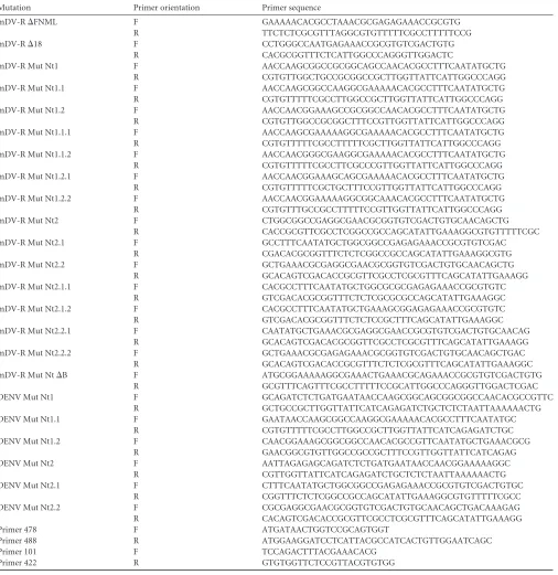
[image:3.585.43.549.78.599.2]The amino acids of the DENV capsid encoded within the cycli-zation sequence 5=CS are dispensable for viral particle forma-tion.DENV RNA replication requires inverted complementary sequences at the 5=and 3=ends of the genome. At the 5=end, the highly conserved 5=CS encodes amino acids FNML (Fig. 1A and
TABLE 1Oligonucleotide sequences and mutations introduced
Mutation Primer orientation Primer sequence
mDV-R⌬FNML F GAAAAACACGCCTAAACGCGAGAGAAACCGCGTG
R TTCTCTCGCGTTTAGGCGTGTTTTTCGCCTTTTTCCG
mDV-R⌬18 F CCTGGGCCAATGAGAAACCGCGTGTCGACTGTG
R CACGCGGTTTCTCATTGGCCCAGGGTTGGACTC
mDV-R Mut Nt1 F AACCAAGCGGCCGCGGCAGCCAACACGCCTTTCAATATGCTG
R CGTGTTGGCTGCCGCGGCCGCTTGGTTATTCATTGGCCCAGG
mDV-R Mut Nt1.1 F AACCAAGCGGCCAAGGCGAAAAACACGCCTTTCAATATGCTG
R CGTGTTTTTCGCCTTGGCCGCTTGGTTATTCATTGGCCCAGG
mDV-R Mut Nt1.2 F AACCAACGGAAAGCCGCGGCCAACACGCCTTTCAATATGCTG
R CGTGTTGGCCGCGGCTTTCCGTTGGTTATTCATTGGCCCAGG
mDV-R Mut Nt1.1.1 F AACCAAGCGAAAAAGGCGAAAAACACGCCTTTCAATATGCTG
R CGTGTTTTTCGCCTTTTTCGCTTGGTTATTCATTGGCCCAGG
mDV-R Mut Nt1.1.2 F AACCAACGGGCGAAGGCGAAAAACACGCCTTTCAATATGCTG
R CGTGTTTTTCGCCTTCGCCCGTTGGTTATTCATTGGCCCAGG
mDV-R Mut Nt1.2.1 F AACCAACGGAAAGCAGCGAAAAACACGCCTTTCAATATGCTG
R CGTGTTTTTCGCTGCTTTCCGTTGGTTATTCATTGGCCCAGG
mDV-R Mut Nt1.2.2 F AACCAACGGAAAAAGGCGGCAAACACGCCTTTCAATATGCTG
R CGTGTTTGCCGCCTTTTTCCGTTGGTTATTCATTGGCCCAGG
mDV-R Mut Nt2 F CTGGCGGCCGAGGCGAACGCGGTGTCGACTGTGCAACAGCTG
R CACCGCGTTCGCCTCGGCCGCCAGCATATTGAAAGGCGTGTTTTTCGC
mDV-R Mut Nt2.1 F GCCTTTCAATATGCTGGCGGCCGAGAGAAACCGCGTGTCGAC
R CGACACGCGGTTTCTCTCGGCCGCCAGCATATTGAAAGGCGTG
mDV-R Mut Nt2.2 F GCTGAAACGCGAGGCGAACGCGGTGTCGACTGTGCAACAGCTG
R GCACAGTCGACACCGCGTTCGCCTCGCGTTTCAGCATATTGAAAGG
mDV-R Mut Nt2.1.1 F CACGCCTTTCAATATGCTGGCGCGCGAGAGAAACCGCGTGTC
R GTCGACACGCGGTTTCTCTCGCGCGCCAGCATATTGAAAGGC
mDV-R Mut Nt2.1.2 F CACGCCTTTCAATATGCTGAAAGCGGAGAGAAACCGCGTGTC
R GTCGACACGCGGTTTCTCTCCGCTTTCAGCATATTGAAAGGC
mDV-R Mut Nt2.2.1 F CAATATGCTGAAACGCGAGGCGAACCGCGTGTCGACTGTGCAACAG
R GCACAGTCGACACGCGGTTCGCCTCGCGTTTCAGCATATTGAAAGG
mDV-R Mut Nt2.2.2 F GCTGAAACGCGAGAGAAACGCGGTGTCGACTGTGCAACAGCTGAC
R GCACAGTCGACACCGCGTTTCTCTCGCGTTTCAGCATATTGAAAGGC
mDV-R Mut Nt⌬B F ATGCGGAAAAAGGCGAAACTGAAACGCAGAAACCGCGTGTCGACTGTG
R GCGTTTCAGTTTCGCCTTTTTCCGCATTGGCCCAGGGTTGGACTCGAC
DENV Mut Nt1 F GCAGATCTCTGATGAATAACCAAGCGGCAGCGGCGGCCAACACGCCGTTC
R GCTGCCGCTTGGTTATTCATCAGAGATCTGCTCTCTAATTAAAAAACTG
DENV Mut Nt1.1 F GAATAACCAAGCGGCCAAGGCGAAAAACACGCCTTTCAATATGC
R CGTGTTTTTCGCCTTGGCCGCTTGGTTATTCATCAGAGATCTGC
DENV Mut Nt1.2 F CAACGGAAAGCGGCGGCCAACACGCCGTTCAATATGCTGAAACGCG
R GAACGGCGTGTTGGCCGCCGCTTTCCGTTGGTTATTCATCAGAG
DENV Mut Nt2 F AATTAGAGAGCAGATCTCTGATGAATAACCAACGGAAAAAGGC
R CGTTGGTTATTCATCAGAGATCTGCTCTCTAATTAAAAAACTG
DENV Mut Nt2.1 F CTTTCAATATGCTGGCGGCCGAGAGAAACCGCGTGTCGACTGTGC
R CGGTTTCTCTCGGCCGCCAGCATATTGAAAGGCGTGTTTTTCGCC
DENV Mut Nt2.2 F CGCGAGGCGAACGCGGTGTCGACTGTGCAACAGCTGACAAAGAG
R CACAGTCGACACCGCGTTCGCCTCGCGTTTCAGCATATTGAAAGG
Primer 478 F ATGATAACTGGTCCGCAGTGGT
Primer 488 R ATGGAAGGATCCTCATTACGCCATCACTGTTGGAATCAGC
Primer 101 F TCCAGACTTTACGAAACACG
Primer 422 R GTGTGGTTCTCCGTTACGTGTGG
on November 7, 2019 by guest
http://jvi.asm.org/
FIG 1DENV reporter system to investigate the structural requirements of C for viral particle formation. (A) Schematic representation of thecis-acting replication elements located at the 5=end of the DENV genome. The promoter stem-loop A (SLA), the cyclization sequence upstream of the AUG (5=upstream activator region [5=UAR]), the replication element cHP, and the cyclization sequence 5=CS are indicated. Below, the corresponding region of the DENV polyprotein with the amino acid sequence FNML encoded by 5=CS is shown. (B) Alignments of nucleotide sequences and amino acids corresponding to 5=CS and the N terminus of C, respectively, for different mosquito-borne flaviviruses. The amino acid sequence encoded by the 5=CS is shaded. (C) Replication of the mDV-R and WT viruses. (Top) Schematic representations of WT DENV and the monocistronic DENV reporter construct (mDV-R) showing the duplication of thecis-acting elements (CAE) and the locations of the luciferase and the viral proteins. (Bottom) (Left) Growth curves comparing the replication of the mDV-R and WT viruses. (Right) Plaque morphologies of the mDV-R and WT viruses. (D) Stability of the luciferase in the mDV-R genome. (Top) Schematic represen-tation of the experiment. Transfection of the mDV-R RNA was performed in BHK cells. The supernatant was harvested 3 days posttransfection and was used for successive passages in fresh cells every 4 days. (Bottom) Luciferase activity was measured in cytoplasmic extracts of cells infected with 100l of the supernatants of transfected cells obtained in each passage.
on November 7, 2019 by guest
http://jvi.asm.org/
[image:4.585.134.445.39.619.2]B). It is still not known whether this conserved sequence is necessary for DENV C function. To investigate the require-ment for the amino acids present at the N terminus of C with-out altering DENV genome replication, we used a full-length viral RNA in which the replicationcis-acting elements (CAE) (Fig. 1A) were duplicated (32). The CAE was fused to a lucif-erase gene, followed by the complete coding sequence of C and the rest of the structural and nonstructural viral proteins (mDV-R) (Fig. 1C). Transfection of the mDV-R RNA resulted in the production of infectious viral particles. The recombinant virus carrying the luciferase gene showed a delay in replication relative to that of the WT virus and a smaller plaque phenotype (Fig. 1C). In addition, the luciferase gene introduced into the viral genome was unstable. Luciferase activity decreased in cells infected with viral stocks obtained in successive passages (Fig. 1D). Therefore, to investigate the ability of a viral mutant to produce infectious particles, the mDV-R RNA was transfected, and viral particle production was evaluated in passage 1 (P1) (Fig. 1D).
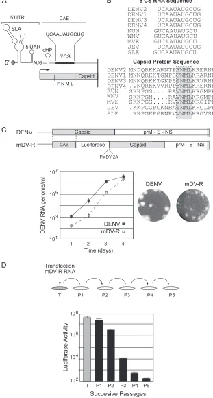
In the context of the recombinant construct, a deletion of the complete C coding region or the FNML sequence was designed. Viral RNAs corresponding to these deletion mutants (Mut⌬C and Mut⌬FNML) were transfected into BHK cells together with the WT and the replication-impaired mutant carrying a substitu-tion in the polymerase NS5 (Mut NS5).
Mut⌬C mDV-R showed luciferase levels at 4, 24, and 48 h posttransfection that were indistinguishable from the WT mDV-R levels, indicating efficient translation and RNA amplifi-cation (Fig. 2A, left). The Mut⌬FNML RNA also was translated and replicated similarly to WT RNA (Fig. 2A, left). These results confirmed that the C protein is dispensable for RNA synthesis and
indicated that the virus with the duplication of thecis-acting RNA elements was fully functional. To analyze the production of infec-tious viral particles, supernatants of transfected cells were col-lected as a function of time and were used to infect fresh BHK cells (infection assay). As expected, no luciferase activity was detectable in cells infected with the medium obtained from cells transfected with Mut⌬C RNA. In contrast, cells infected with supernatants obtained from cells transfected with Mut⌬FNML RNA showed WT luciferase levels (Fig. 2A, right), suggesting that the FNML sequence is not required for C function during viral particle for-mation. To further confirm this observation, viral propagation was analyzed by immunofluorescence (IF) as a function of time after transfection of BHK cells with the WT or Mut⌬FNML virus. At 72 h posttransfection, both viruses infected about 80% of the cell monolayer (Fig. 2B). While the coding sequence of amino acids FNML contains RNA structures essential for viral genome replication, the dissociation of this function from the C coding sequence indicated that these conserved amino acids are dispens-able for the production of infectious DENV particles.
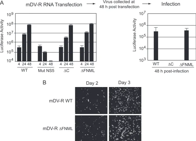
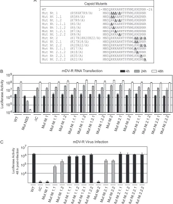
Two clusters of basic amino acids at the N terminus of C are necessary for DENV encapsidation.An important feature of the DENV C protein is the unusually high number of positive resi-dues. There are 26 basic and only 3 acidic amino acids per 100-residue subunit. The N-terminal region of DENV C is unstruc-tured in solution, and besides encoding the conserved FNML sequence, it contains eight basic residues distributed in two con-served clusters (R5-K6-K7-R9 and K17-R18-R20-R22) (Fig. 1B). To investigate the requirement of N-terminal amino acids of C for viral particle formation, we performed deletions and substitutions in the context of the mDV-R RNA.
Three mutants were designed. One mutant contained a
dele-FIG 2Dissociation ofcis-acting RNA elements from the capsid coding region indicates that the FNML sequence is dispensable for DENV propagation. (A) (Left) Luciferase activity as a function of time posttransfection in mDV-R RNA corresponding to the WT, the replication-impaired Mut NS5 mutant, a mutant with the C coding sequence deleted (⌬C), and a mutant with amino acids FNML deleted (⌬FNML). (Right) Luciferase activity measured at 48 h postinfection using 100 l of supernatants of transfected cells. (B) Images for IF assays showing DENV antigen-positive BHK cells transfected with WT mDV-R or mDV-R⌬FNML RNA.
on November 7, 2019 by guest
http://jvi.asm.org/
[image:5.585.124.460.68.311.2]tion of the first 18 amino acids of C (Mut⌬18), and the other two mutants carried replacements of the four basic residues in each cluster. The basic amino acids in Mut Nt1 (R5, K6, K7, and R9) and in Mut Nt2 (K17, R18, R20, and R22) were each replaced by Ala (Fig. 3A). Viral RNAs corresponding to the mutants (Mut
⌬18, Mut Nt1, and Mut Nt2) were transfected into BHK cells together with three controls: the WT, the encapsidation-impaired Mut⌬C construct, and the replication-impaired Mut NS5 con-struct.
The translation and replication of the viral RNAs correspond-ing to the mutants Mut⌬18, Mut Nt1, and Mut Nt2 were similar to those of the WT (Fig. 3A). During infection, the mature C protein is released from its membrane anchor sequence, which precedes prM, by the NS3/2B protease (3, 34, 43). To examine whether deletions or mutations at the N terminus of C affect pro-tein expression or processing, the mature viral propro-tein was
de-tected by Western blotting using cytoplasmic extracts of cells transfected with WT or C mutant RNAs (Fig. 3B). The WT and mutant proteins were readily detected between 24 and 48 h post-transfection, corresponding to the mDV-R RNA amplification step. The faster mobility of Mut⌬18 C was consistent with the deletion in this protein. Interestingly, infection of fresh BHK cells with supernatants of transfected cells showed that deletion of the N terminus of C or mutation of either of the two clusters of basic residues impairs viral particle formation (Fig. 3C).
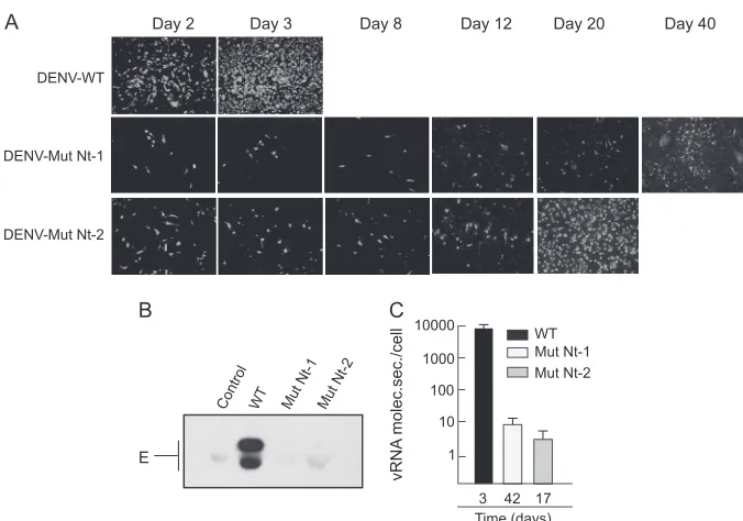
Although it was not possible to include a deletion of the N terminus of C in an infectious DENV clone, due to undesired effects on RNA synthesis, analysis of predicted RNA structures indicated that the Nt1 and Nt2 substitutions were possible. There-fore, to confirm the encapsidation defects of Mut Nt1 and Mut Nt2, the corresponding amino acid substitutions were introduced in the context of the DENV2 16681 clone. Viral RNAs of the WT, Mut Nt1, or Mut Nt2 were used to transfect BHK cells, and infec-tion was followed as a funcinfec-tion of time by IF. The fact that viruses without the luciferase gene grow faster in culture should be taken into account (compare the WT in Fig. 2B with the WT in Fig. 4A). At 2 days posttransfection, about 50% of the monolayer was pos-itive for the WT DENV antigen, while about 10% of cells were positive for the Mut Nt1 or Mut Nt2 antigen. At 3 days posttrans-fection, most of the monolayer was positive for the WT, while 10% of cells were positive for mutant DENV antigen (Fig. 4A). To study whether noninfectious particles or subviral particles were pro-duced and secreted in cells transfected with Mut Nt1 or Mut Nt2, cell supernatants were collected and were used to analyze the pres-ence of viral envelope protein E. While E protein from the WT virus was clearly detected as two bands, no E protein was observed at day 2 in the medium of cells transfected with Mut Nt1 or Mut Nt2 RNA (Fig. 4B).
To investigate whether the Mut Nt1 or Mut Nt2 virus propa-gated slowly, cells were passed every 4 days and were analyzed by IF. The IF signal increased gradually as a function of time for both mutants (Fig. 4A). At 16 days, infectious foci were observed throughout the monolayer. The propagation of the Mut Nt1 virus continued to increase, and about 60% of the monolayer was in-fected at day 40. With mutant Nt2, about 90% of the monolayer was infected at day 20. These results suggested either a slow prop-agation of these viruses or reversions by spontaneous mutations. To analyze these possibilities, viruses recovered from the superna-tants at 20 and 40 days posttransfection (for Mut Nt2 and Mut Nt1, respectively) were used to infect fresh cells and generate viral stocks. Simultaneously, the viral RNA was purified from the me-dium for sequencing analysis. Infection of fresh cells with the mu-tant viruses showed very slow propagation of both viruses, similar to that observed after RNA transfection. In addition, Mut Nt1 and Mut Nt2 failed to form plaques. Sequencing analysis of viral RNAs purified from the media indicated that the original substitutions were maintained in both mutants, and no additional amino acid changes were detected. The results indicate that viruses carrying mutations in either of the two clusters of basic amino acids at the N terminus of C propagate very slowly.
To determine the cause of the slow propagation phenotype of mutants Nt1 and Nt2, we determined the number of viral particles produced per cell when the monolayer was more than 80% DENV antigen positive for the mutants, using for comparison a similar 80% infection with a WT virus. The media of cells transfected with the WT, Mut Nt1, or Mut Nt2 were collected after 3, 42, and 17
FIG 3The N-terminal region of C is necessary for the formation of DENV infectious particles. (A) (Top) Schematic representation of amino acid changes introduced in the viral C protein. (Bottom) The translation and am-plification of viral RNAs in mDV-R-transfected cells was followed by measur-ing luciferase activity. (B) Expression and processmeasur-ing of C protein in trans-fected cells. Western blotting with DENV anti-C antibodies was performed using cytoplasmic extracts of cells transfected with the RNAs indicated above the gel. (C) Luciferase activity of BHK cell extracts infected with supernatants obtained after transfection as indicated in panel A.
Role of the N-Terminal Region of DENV Capsid Protein
on November 7, 2019 by guest
http://jvi.asm.org/
[image:6.585.43.283.66.440.2]days, respectively, and were used to quantify the viral genome by real-time RT-PCR. While by IF the monolayer appeared to be almost completely infected, cells infected with the WT virus re-leased 1,000 to 5,000 more viral RNA molecules than cells infected with Mut Nt1 or Mut Nt2 (Fig. 4C). This result supports the idea that cells infected with mutant Nt1 or Nt2 release very small amounts of viral particles into the medium, which explains the slow propagation of these viruses.
Infectious DENV particle formation requires basic residues but not a specific sequence at the N terminus of C.We observed above that amino acids at the N terminus of C play a crucial role in DENV particle formation (Fig. 3 and 4). To investigate whether specific amino acids in this region are necessary for C function, a systematic mutational analysis was performed in the context of the mDV-R virus. Mutants carrying replacements of basic residues in the two clusters, including changes of individual amino acids to alanine and combinations of such changes, were designed (Fig. 5A). The Nt1 mutant, which contained four substitutions, was divided into 6 new mutants: Mut Nt1.1 and Mut Nt1.2 (each car-rying double amino acid changes) and four mutants with individ-ual mutations, Nt 1.1.1, Nt 1.1.2, Nt 1.2.1, and Nt 1.2.2 (Fig. 5A). Similarly, 6 new viral mutants were designed carrying the substi-tutions originally included in Mut Nt2: Mut Nt2.1 and Nt2.2 (each carrying double amino acid changes) and four mutants with individual substitutions, Nt 2.1.1, Nt 2.1.2, Nt 2.2.1, and Nt 2.2.2 (Fig. 5A).
RNAs corresponding to a set of 14 mutants together with 3 controls (the WT, Mut⌬C, and Mut NS5) were transfected into BHK cells. Translation and RNA synthesis were evaluated by lu-ciferase activity as a function of time. The levels of lulu-ciferase at 4, 24, and 48 h posttransfection were similar to those observed for
WT RNA (Fig. 5B). To evaluate the amount of viral particles pro-duced, the medium was collected at 48 h posttransfection and was used to infect fresh cells. Interestingly, mutation of one of the eight basic residues to alanine showed only marginal effects on viral particle formation (Fig. 5C). The combination of two such muta-tions decreased infectious particle formation about 10-fold. Mut Nt1.1 was the only mutant that showed a reduction of about 30-fold (Fig. 5C). These results indicate that the dramatic phenotype observed with Mut Nt1 and Mut Nt2 cannot be attributed to the role of specific amino acids and that at least two of any of the basic residues in each cluster are necessary for efficient DENV particle formation.
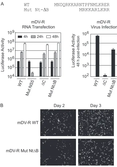
It is possible that the unstructured N-terminal region of C provides a positively charged environment necessary for protein function without a requirement for a specific sequence. To exam-ine this possibility, the N-terminal 20-amino-acid region of C was replaced by a 10-amino-acid sequence, which maintained only the number of positive charges (Mut Nt⌬B) (Fig. 6A). The Mut Nt⌬B RNA was transfected along with the controls (WT, Mut⌬C, or Mut NS5 RNA) into BHK cells. The translation and synthesis of Mut Nt⌬B RNA were as efficient as those of WT RNA (Fig. 6A). Interestingly, viral particle formation was largely rescued in this mutant (Fig. 6A, right). To confirm this observation, the propa-gation of the mDV-R Mut Nt⌬B virus was assessed by IF as a function of time. Propagation of the mDV-R Nt⌬B virus was ev-ident: the IF-positive signal increased between 48 and 72 h (Fig. 6B). The results indicate that a high density of basic residues is required, but at the same time, great amino acid flexibility is tol-erated in this region of C.
In order to search for revertant viruses, the mutants that showed reduced particle formation by use of the mDV-R system
FIG 4Impaired propagation of DENV with mutations in basic residues at the N terminus of C. (A) IF assays of BHK cells transfected with WT DENV2, mutant Nt1, or mutant Nt2 RNAs were performed at different times as indicated. (B) Secretion of envelope E protein from cells transfected with WT or mutated DENV RNAs. Western blotting of supernatants obtained 48 h after transfection of DENV WT, mutant Nt1, or mutant Nt2 RNAs, as indicated above the gel, was performed with specific anti-E antibodies. (C) Mutations at the N terminus of C decrease the amount of viral RNA released to the medium of transfected cells. The amounts of genomic viral RNA secreted into the supernatants of cells (vRNA molecules secreted/cell) transfected with WT DENV, mutant Nt1, or mutant Nt2 RNAs were determined 3, 42, and 17 days posttransfection, as indicated, by real-time RT-PCR using TaqMan.
on November 7, 2019 by guest
http://jvi.asm.org/
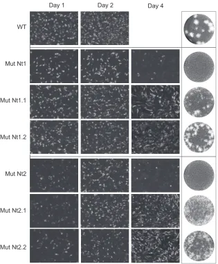
[image:7.585.122.460.62.299.2]were selected, and the respective amino acid substitutions were incorporated into the infectious DENV2 clone. The Nt 1.1, Nt 1.2, Nt 2.1, and Nt2.2 viruses were constructed, and the RNAs were transfected into BHK cells together with the WT control RNA. Although viral propagation was delayed, all four viruses replicated in cell culture (Fig. 7). While the WT infected the whole mono-layer in 2 days, viruses carrying the partial substitutions present in Nt1.1, Nt1.2, Nt2.1, and Nt2.2 showed DENV antigens in the whole monolayer between days 4 and 6. The initial delay of repli-cation of viruses Nt2, Nt2.1, and Nt2.2 (Fig. 7, day 1) suggests a possible defect in RNA replication. This observation highlights the usefulness of the mDV-R virus in dissociating RNA replication signals from the C coding sequence. The Nt1.1, Nt1.2, Nt2.1, and Nt2.2 viruses displayed small plaques that were more diffuse than those of the WT (Fig. 7, right). Recovered viruses were passed twice, and the viral RNA was purified for sequencing analysis. No spontaneous mutations were selected in the C coding sequence,
and the original mutations were maintained. The lack of success in rescuing revertant viruses in C might be explained by the failure to select the RNA with spontaneous mutations into particles.
Taking these results together, we conclude that the sequence of the N terminus of C tolerates substantial amino acid changes; however, positively charged residues in this region of C are crucial for DENV particle formation.
Requirement of the N-terminal sequence of C for DENV en-capsidation in human and mosquito cells.DENV cycles between mosquito and human hosts. To extend our studies of the struc-tural requirements of C during viral encapsidation in mosquito cells, we examined the replication of the N-terminal mutants of C in C6/36 cells. Replication of the input mDV-R RNA in mosquito cells was observed as a function of time between 24 and 72 h (Fig. 8A, compare WT with Mut NS5). Viral RNA amplification in these cells was delayed relative to that observed in BHK cells (com-pare Fig. 3A with Fig. 8A).
FIG 5Two clusters of basic amino acids at the N terminus of C are necessary for DENV particle formation in BHK cells. (A) Schematic representation of amino acid changes introduced into the viral C protein. (B) Luciferase activity showing the translation and amplification of mDV-R WT and mutant RNA. Mutants in which RNA replication (Mut NS5) or encapsidation (⌬C) is impaired are included as controls. (C) Luciferase activity measured in cell extracts 48 h after infection with 100l of supernatants obtained after transfection as indicated in panel B.
Role of the N-Terminal Region of DENV Capsid Protein
on November 7, 2019 by guest
http://jvi.asm.org/
[image:8.585.123.461.71.471.2]RNAs corresponding to WT mDV-R, Mut⌬18, Mut Nt1, Mut Nt2, Mut⌬C, and Mut NS5 were transfected into C6/36 cells (Fig. 8A). Luciferase levels between 4 and 72 h posttransfection were similar to those measured for WT RNA (Fig. 8A). To evaluate the production of infectious particles, the supernatant was collected at 72 h posttransfection and was used for infection assays. Notably, luciferase activity was detected after infection with the medium obtained from any of the three N-terminal mutants (Fig. 8B). Mut
⌬18 produced about 50-fold fewer viral particles than the WT, while the Nt1 and Nt2 mutations resulted in 30- and 10-fold re-ductions, respectively (Fig. 8B). Although the N-terminal se-quence of C appears to be important for viral particle formation, the phenotypes observed in mosquito cells were less drastic than those observed with the same mutants in mammalian cells.
To confirm this observation, the propagation of WT DENV and mutant Nt1 or Nt2 was examined by IF as a function of time (Fig. 8C). The propagation of the Mut Nt1 and Mut Nt2 viruses was delayed relative to that of the WT. With both mutants, how-ever, at day 5, 60 to 80% of the monolayer of C6/36 cells was infected (Fig. 8C). These viruses were used to infect fresh cells, and the recovered culture was used for sequencing analysis. As de-scribed above for mammalian cells, the original mutations intro-duced into C were maintained, and no additional mutations in the C coding sequence were selected.
In order to quantify the viral particles released from C6/36 cells, real-time RT-PCR to detect the viral genome was performed with supernatants at day 5 posttransfection. This analysis indi-cated that both mutants released about 100-fold less RNA per cell than the WT (Fig. 8C).
Because mutations and deletions within the N-terminal se-quence of C had drastic effects on the production of DENV par-ticles in BHK cells, while the effects of these mutations were much less pronounced in mosquito cells, viral particle formation by Mut
⌬18, Mut Nt1, and Mut Nt2 in different human cell lines was analyzed. A549 and Raji cells were transfected with Mut⌬18, Nt1, Nt2, or WT RNAs. As a control, the translation and replication of the input RNA were determined at 4, 24, and 48 h posttransfection (Fig. 8D). Infection assays using the supernatants of transfected cells indicated that deletion of the N terminus of C and mutations in both clusters of basic residues of the viral protein impair the formation of viral particles in human cells (Fig. 8E). As observed in BHK cells (Fig. 4), Mut⌬18 and Mut Nt1 failed to produce infectious particles in A549 and Raji cells. In the case of Mut Nt2, small but detectable amounts of viral particles were observed in both A549 and Raji cells (Fig. 8E). This observation was interest-ing, because in BHK cells, no Mut Nt2 particles were detectable.
These results indicate a requirement of the N terminus of C for efficient DENV particle formation in both mosquito and human cells. However, this function of C appears to be more important in human than in mosquito cells.
DISCUSSION
The mechanism by which the C protein recruits the viral genome during packaging is one of the most obscure steps of the flavivirus life cycle. Here we investigated structural requirements of DENV C protein for infectious particle formation. Because the viral ge-nome is compact and contains overlapping signals and functions, in this study, we dissociatedcis-acting RNA replication elements from the C coding sequence and found that basic residues within the unstructured N-terminal region of C are required for DENV particle formation. This is the first report of mutagenesis analysis of this region of the DENV C protein in the context of a fully replicating viral genome.
Mosquito-borne flavivirus C proteins contain two conserved features in the first⬃21 residues: (i) a highly conserved amino acid sequence, F(V/L)NML, and (ii) a large percentage of Lys/Arg (⬃30%) (Fig. 1B). We found that the F(V/L)NML sequence is necessary at the RNA level but that the amino acids are dispensable for C function (Fig. 2). In addition, the presence of positive charges at the N terminus of C was determined to be necessary for viral encapsidation.
A minimal amount of basic residues at the N terminus of DENV C was found to be crucial for viral particle formation (Fig. 5). It is possible that this region of the protein interacts directly with the viral genome.In vitroRNA binding experiments have previously shown RNA binding activity using the first 32 and the last 26 amino acids of Kunjin virus C protein (16). In contrast, investigators using a YFVtrans-packaging system have reported that the first 36 residues of YFV C protein were not essential for viral assembly (31). At this point, the different requirements for DENV and YFV encapsidation are not clear. It is possible that the
trans-packaging system, which does not provide equimolar amounts of viral structural and nonstructural proteins, is less sen-sitive to detect alterations in the C protein. However, we cannot
FIG 6The encapsidation defect caused by deletion of the first 20 residues of C can be rescued by a sequence that maintains basic residues. (A) (Top) Sche-matic representation of Mut Nt⌬B in which the first 20 amino acids were replaced by 10 residues that maintained the amounts of Lys and Arg in the WT protein. (Bottom) (Left) Luciferase activity measured as a function of time after transfection of BHK cells with WT mDV-R, Mut Nt⌬B, or control (Mut NS5 or⌬C) RNA. (Right) Luciferase activity measured in cell extracts 48 h after infection with supernatants obtained after transfection. (B) IF assays of transfected BHK cells with WT mDV-R or Mut Nt⌬B. IF assays were per-formed at days 2 and 3 as indicated.
on November 7, 2019 by guest
http://jvi.asm.org/
[image:9.585.60.264.64.356.2]rule out the possibility that the C proteins of these two flaviviruses have different requirements. The difference observed in the en-capsidation of DENV C mutants lacking the N terminus of C in mosquito versus human cells is particularly interesting (Fig. 8). Although DENV showed a requirement of basic residues at the N terminus of C for efficient encapsidation in mosquito cells, the remarkable reduction in viral particle formation in mammalian cells suggests a possible role of the N terminus of C in a host-virus interaction. It has been reported previously that the C protein of flaviviruses is involved in the induction of apoptosis and the breakdown of cellular tight junctions (26, 37, 44). A growing number of host proteins that interact with flavivirus C proteins have been reported (4, 5, 18, 28). For instance, by use of two-hybrid systems, several cellular proteins that interact with WNV C have been identified (29, 42). Less is known about possible inter-actions between DENV C and cellular proteins, but it is likely that C interacts with different host factors in infected mosquito and human cells.
Substitution of any one of the eight basic residues present at the N terminus of DENV C did not affect viral encapsidation
signifi-cantly, showing that there is no requirement for particular amino acids at specific positions of the protein. This finding was further supported by the encapsidation of Mut Nt⌬B, in which the re-placement of the first 20 amino acids by a sequence that only provides positive charges was functional (Fig. 6). The amino acid sequence flexibility found in DENV C is in agreement with previ-ous reports on TBEV and YFV (19, 31). Deletions of 4 to 21 resi-dues in the center of the TBEV C protein yielded viable viruses. A viral mutant with a 16-residue deletion was found to be highly attenuated but very immunogenic in adult mice. In another re-port, deletions of 19 to 30 residues of the hydrophobic region of TBEV C resulted in viruses with second-site mutations that in-creased protein hydrophobicity (20). These viruses were also at-tenuated but were capable of inducing a protective immune re-sponse in mice. Based on these studies, a strategy of designing live attenuated vaccines carrying deletions in the C protein was pro-posed.
In DENV-infected cells, C is distributed between the nucleus and the cytoplasm. Hydrophobic regions of C appear to be neces-sary for the association of the protein with ER membranes and LD
WT
Mut Nt1
Mut Nt1.1
Mut Nt1.2
Mut Nt2
Mut Nt2.1
Mut Nt2.2
Day 1 Day 2 Day 4
FIG 7Delay in the propagation of recombinant DENV2 with partial mutations in the two clusters of basic residues. (Left) IF assays of BHK cells transfected with WT DENV2, mutant Nt1, mutant Nt1.1, mutant Nt1.2, mutant Nt2, mutant Nt2.1, or mutant Nt2.2 RNAs. IF assays were performed at the times indicated above the images. (Right) Plaque phenotypes.
Role of the N-Terminal Region of DENV Capsid Protein
on November 7, 2019 by guest
http://jvi.asm.org/
[image:10.585.136.451.65.446.2](25, 32). Three putative nuclear localization signals have been pre-dicted in the DENV C protein (residues 6 to 9, 73 to 76, and 85 to 100) (6). It was originally reported that a bipartite site located between residues 85 and 100 was the site necessary for the nuclear localization of DENV C (40). In a different study, two signals, including amino acids 73 to 74 and 85 to 86, were found to be involved in the nuclear localization of C in DENV-infected cells; however, a lack of correlation between the nuclear localization of this viral protein and the growth properties of viral mutants was reported (33). Thus, whether C plays a role in the nuclei of in-fected cells is still unclear. Interestingly, for JEV, amino acids Gly42 and Pro43 were found to be necessary for the localization of
C protein to the nucleus. Mutations of these residues resulted in a reduction in the level of JEV pathogenesis in mice and lower titers in cell culture (27). Because Gly42 and Pro43 are conserved in different flavivirus C proteins, the question of whether these res-idues are necessary for the subcellular distribution of DENV C remains to be addressed.
Two clusters of basic residues at the N terminus of DENV C were found to be necessary for DENV particle formation (Fig. 5). Viruses with replacements of Arg and Lys residues in either of the two clusters were severely attenuated in mammalian cells (Fig. 3 and 4, Mut Nt1 or Mut Nt2). Cells infected with either of these viral mutants released much smaller amounts of viral particles
FIG 8Requirement of the N-terminal region of C for DENV particle formation in mosquito and human cells. (A) Luciferase activity showing translation and amplification of WT mDV-R or mutant⌬18, Nt1, or Nt2 RNA in C6/36 mosquito cells. RNA replication-impaired (Mut NS5) and encapsidation-impaired (⌬C) controls are included. (B) Luciferase activity measured in cell extracts 48 h after infection of BHK cells with supernatants obtained after transfection of WT and mutant RNAs into mosquito cells. (C) (Left) IF assays of C6/36 cells transfected with WT DENV2 16681, mutant Nt1, or mutant Nt2. IF assays were performed at 3 and 5 days, as indicated. (Right) The amount of viral RNA secreted into the medium of cells (vRNA molecules secreted/cell) collected 5 days posttransfection was determined by real-time RT-PCR. (D) Luciferase activity measured as a function of time after transfection of WT and mutated mDV-R RNAs into A549 and Raji cells. (E) Infectivities of viral particles obtained from human A549 and Raji cells. Luciferase activity was measured 48 h after infection of BHK cells with supernatants obtained from A549 or Raji cells 48 h posttransfection.
on November 7, 2019 by guest
http://jvi.asm.org/
[image:11.585.113.469.64.495.2]than cells infected with the WT virus. Interestingly, in different cell types, the Nt1 mutation was more drastic for viral propagation than the Nt2 mutation (Fig. 4A, 8B, and 8D). This observation is consistent with the 30-fold reduction in particle formation ob-served with the partial substitution of the first cluster in Mut Nt1.2 (Fig. 5C). It is possible that positive charges at the N terminus of DENV C are necessary for proper interaction with the viral ge-nome during particle morphogenesis. It is also possible that the N terminus of C participates in modulating viral RNA folding. An RNA chaperone activity for WNV, hepatitis C virus (HCV), and bovine viral diarrhea virus core proteins has been reported previ-ously (14). A hallmark of active RNA chaperone domains is the presence of highly basic and flexible protein segments (36). Be-cause the N terminus of C is intrinsically disordered, it is possible that it plays a role in modulating the architecture of the viral RNA during packaging.
In conclusion, a mutational analysis of the N-terminal region of C in a DENV construct with a duplication ofcis-acting RNA replication signals identified important structural determinants for virion production. We believe that understanding of the mo-lecular aspects of DENV encapsidation will aid in the rational design of new strategies to tackle this important human pathogen.
ACKNOWLEDGMENTS
We are grateful to Richard Kinney for the dengue virus cDNA clone. We also thank the members of A. V. Gamarnik’s laboratory for helpful dis-cussions and ideas about this work; special thanks go to Gabriel Iglesias. A.V.G. and J.J.C. are members of the Argentinean Council of Investiga-tion (CONICET).
This work was supported by grants from NIH (1R01AI095175-01) and Agencia Argentina de Promoción Científica y Tecnológica (PICT-2008-1141). M.M.S. and J.A.M. were awarded CONICET fellowships.
REFERENCES
1.Alvarez DE, DE Lella Ezcurra AL, Fucito S, Gamarnik AV.2005. Role of RNA structures present at the 3=UTR of dengue virus on translation, RNA synthesis, and viral replication. Virology339:200 –212.
2.Alvarez DE, Lodeiro MF, Luduena SJ, Pietrasanta LI, Gamarnik AV. 2005. Long-range RNA-RNA interactions circularize the dengue virus ge-nome. J. Virol.79:6631– 6643.
3.Amberg SM, Nestorowicz A, McCourt DW, Rice CM.1994. NS2B-3 proteinase-mediated processing in the yellow fever virus structural region: in vitro and in vivo studies. J. Virol.68:3794 –3802.
4.Bhuvanakantham R, Chong MK, Ng ML.2009. Specific interaction of capsid protein and importin-alpha/beta influences West Nile virus pro-duction. Biochem. Biophys. Res. Commun.389:63– 69.
5.Bhuvanakantham R, Li J, Tan TT, Ng ML.2010. Human Sec3 protein is a novel transcriptional and translational repressor of flavivirus. Cell. Mi-crobiol.12:453– 472.
6.Bulich R, Aaskov JG.1992. Nuclear localization of dengue 2 virus core protein detected with monoclonal antibodies. J. Gen. Virol.73(Pt 11): 2999 –3003.
7.Clyde K, Barrera J, Harris E.2008. The capsid-coding region hairpin element (cHP) is a critical determinant of dengue virus and West Nile virus RNA synthesis. Virology379:314 –323.
8.Dokland T, et al.2004. West Nile virus core protein; tetramer structure and ribbon formation. Structure12:1157–1163.
9.Filomatori CV, Iglesias NG, Villordo SM, Alvarez DE, Gamarnik AV. 2011. RNA sequences and structures required for the recruitment and activity of the dengue virus polymerase. J. Biol. Chem.286:6929 – 6939. 10. Filomatori CV, et al.2006. A 5=RNA element promotes dengue virus
RNA synthesis on a circular genome. Genes Dev.20:2238 –2249. 11. Gamarnik AV.2010. Role of the dengue virus 5=and 3=untranslated
regions in viral replication, p 55–78.InHanley KA, Weaver SC (ed), Fron-tiers in dengue virus research. Caister Academic Press, Norfolk, United Kingdom.
12. Hahn CS, et al.1987. Conserved elements in the 3=untranslated region of flavivirus RNAs and potential cyclization sequences. J. Mol. Biol.198: 33– 41.
13. Iglesias NG, Filomatori CV, Gamarnik AV.2011. The F1 motif of dengue virus polymerase NS5 is involved in promoter-dependent RNA synthesis. J. Virol.85:5745–5756.
14. Ivanyi-Nagy R, Lavergne JP, Gabus C, Ficheux D, Darlix JL.2008. RNA chaperoning and intrinsic disorder in the core proteins ofFlaviviridae. Nucleic Acids Res.36:712–725.
15. Jones CT, et al.2003. Flavivirus capsid is a dimeric alpha-helical protein. J. Virol.77:7143–7149.
16. Khromykh AA, Westaway EG.1996. RNA binding properties of core protein of the flavivirus Kunjin. Arch. Virol.141:685– 699.
17. Kinney RM, et al.1997. Construction of infectious cDNA clones for dengue 2 virus: strain 16681 and its attenuated vaccine derivative, strain PDK-53. Virology230:300 –308.
18. Ko A, et al.2010. MKRN1 induces degradation of West Nile virus capsid protein by functioning as an E3 ligase. J. Virol.84:426 – 436.
19. Kofler RM, Heinz FX, Mandl CW.2002. Capsid protein C of tick-borne encephalitis virus tolerates large internal deletions and is a favorable target for attenuation of virulence. J. Virol.76:3534 –3543.
20. Kofler RM, Leitner A, O’Riordain G, Heinz FX, Mandl CW. 2003. Spontaneous mutations restore the viability of tick-borne encephalitis vi-rus mutants with large deletions in protein C. J. Virol.77:443– 451. 21. Kuhn RJ, et al.2002. Structure of dengue virus: implications for flavivirus
organization, maturation, and fusion. Cell108:717–725.
22. Lindenbach BD, Thiel H-J, Rice CM.2007.Flaviviridae: the viruses and their replication, p 1101–1152.InKnipe DM, et al (ed), Fields virology, 5th ed, vol 1. Lippincott-Raven, Philadelphia, PA.
23. Lorenz IC, et al.2003. Intracellular assembly and secretion of recombi-nant subviral particles from tick-borne encephalitis virus. J. Virol.77: 4370 – 4382.
24. Ma L, Jones CT, Groesch TD, Kuhn RJ, Post CB. 2004. Solution structure of dengue virus capsid protein reveals another fold. Proc. Natl. Acad. Sci. U. S. A.101:3414 –3419.
25. Markoff L, Falgout B, Chang A.1997. A conserved internal hydrophobic domain mediates the stable membrane integration of the dengue virus capsid protein. Virology233:105–117.
26. Medigeshi GR, et al.2009. West Nile virus capsid degradation of claudin proteins disrupts epithelial barrier function. J. Virol.83:6125– 6134. 27. Mori Y, et al.2005. Nuclear localization of Japanese encephalitis virus
core protein enhances viral replication. J. Virol.79:3448 –3458. 28. Netsawang J, et al.2010. Nuclear localization of dengue virus capsid
protein is required for DAXX interaction and apoptosis. Virus Res.147: 275–283.
29. Oh W, et al.2006. Jab1 mediates cytoplasmic localization and degrada-tion of West Nile virus capsid protein. J. Biol. Chem.281:30166 –30174. 30. Oliphant T, et al.2005. Development of a humanized monoclonal
anti-body with therapeutic potential against West Nile virus. Nat. Med.11: 522–530.
31. Patkar CG, Jones CT, Chang YH, Warrier R, Kuhn RJ.2007. Functional requirements of the yellow fever virus capsid protein. J. Virol. 81: 6471– 6481.
32. Samsa MM, et al.2009. Dengue virus capsid protein usurps lipid droplets for viral particle formation. PLoS Pathog.5:e1000632.
33. Sangiambut S, et al.2008. Multiple regions in dengue virus capsid protein contribute to nuclear localization during virus infection. J. Gen. Virol. 89:1254 –1264.
34. Stocks CE, Lobigs M.1998. Signal peptidase cleavage at the flavivirus C-prM junction: dependence on the viral NS2B-3 protease for efficient processing requires determinants in C, the signal peptide, and prM. J. Virol.72:2141–2149.
35. Tadano M, Makino Y, Fukunaga T, Okuno Y, Fukai K.1989. Detection of dengue 4 virus core protein in the nucleus. I. A monoclonal antibody to dengue 4 virus reacts with the antigen in the nucleus and cytoplasm. J. Gen. Virol.70(Pt 6):1409 –1415.
36. Tompa P, Csermely P.2004. The role of structural disorder in the func-tion of RNA and protein chaperones. FASEB J.18:1169 –1175.
37. van Marle G, et al.2007. West Nile virus-induced neuroinflammation: glial infection and capsid protein-mediated neurovirulence. J. Virol.81: 10933–10949.
38. Villordo SM, Gamarnik AV.2009. Genome cyclization as strategy for flavivirus RNA replication. Virus Res.139:230 –239.
Role of the N-Terminal Region of DENV Capsid Protein
on November 7, 2019 by guest
http://jvi.asm.org/
39. Wang SH, Syu WJ, Hu ST.2004. Identification of the homotypic inter-action domain of the core protein of dengue virus type 2. J. Gen. Virol. 85:2307–2314.
40. Wang SH, et al.2002. Intracellular localization and determination of a nuclear localization signal of the core protein of dengue virus. J. Gen. Virol.83:3093–3102.
41. WHO Initiative for Vaccine Research.2011. Vector-borne viral infec-tions. WHO, Geneva, Switzerland.http://www.who.int/vaccine_research /diseases/vector/en/index1.html.
42. Xu Z, Anderson R, Hobman TC.2011. The capsid-binding nucleolar helicase DDX56 is important for infectivity of West Nile virus. J. Virol. 85:5571–5580.
43. Yamshchikov VF, Compans RW.1994. Processing of the intracellular form of the West Nile virus capsid protein by the viral NS2B-NS3 protease: an in vitro study. J. Virol.68:5765–5771.
44. Yang JS, et al.2002. Induction of inflammation by West Nile virus capsid through the caspase-9 apoptotic pathway. Emerg. Infect. Dis. 8:1379 –1384.