0022-538X/07/$08.00
⫹
0
doi:10.1128/JVI.02840-06
Copyright © 2007, American Society for Microbiology. All Rights Reserved.
Highly Conserved Configuration of Catalytic Amino Acid Residues
among Calicivirus-Encoded Proteases
䌤
Tomoichiro Oka,
1* Mami Yamamoto,
1Masaru Yokoyama,
2Satoko Ogawa,
1Grant S. Hansman,
1Kazuhiko Katayama,
1Kana Miyashita,
1Hirotaka Takagi,
3Yukinobu Tohya,
4Hironori Sato,
2and Naokazu Takeda
1Department of Virology II, National Institute of Infectious Diseases, Gakuen 4-7-1, Musashi-murayama, Tokyo 208-0011, Japan
1;
Center for Pathogen Genomics, National Institute of Infectious Diseases, Gakuen 4-7-1, Musashi-murayama, Tokyo 208-0011,
Japan
2; Division of Biosafety Control and Research, National Institute of Infectious Diseases, Toyama 1-23-1, Shinjuku-ku,
Tokyo 162-8640, Japan
3; and Department of Veterinary Microbiology, Graduate School of Agricultural and
Life Sciences, The University of Tokyo, 1-1-1 Yayoi, Bunkyo-ku, Tokyo 113-8657, Japan
4Received 22 December 2006/Accepted 11 April 2007
A common feature of caliciviruses is the proteolytic processing of the viral polyprotein catalyzed by the viral
3C-like protease encoded in open reading frame 1 (ORF1). Here we report the identification and structural
characterization of the protease domains and amino acid residues in sapovirus (SaV) and feline calicivirus (FCV).
The in vitro expression and processing of a panel of truncated ORF1 polyproteins and corresponding mutant forms
showed that the functional protease domain is 146 amino acids (aa) in SaV and 154 aa in FCV. Site-directed
mutagenesis of the protease domains identified four amino acid residues essential to protease activities: H
31, E
52,
C
116, and H
131in SaV and H
39, E
60, C
122, and H
137in FCV. A computer-assisted structural analysis showed that
despite high levels of diversity in the primary structures of the protease domains in the family
Caliciviridae
, the
configurations of the H, E, C, and H residues are highly conserved, with these residues positioned closely along the
inner surface of the potential binding cleft for the substrate. These results strongly suggest that the H, E, C, and H
residues are involved in the formation of a conserved catalytic surface of the SaV and FCV 3C-like proteases.
The family
Caliciviridae
is composed of four genera,
Sapo-virus
,
Lagovirus
,
Vesivirus
, and
Norovirus
, which include the
species
Sapporo virus
(SaV),
Rabbit hemorrhagic disease virus
(RHDV),
Feline calicivirus
(FCV), and
Norwalk virus
(NoV),
respectively (24). Caliciviruses infect a broad range of hosts,
including humans and animals, and cause a variety of diseases
and disorders, such as gastroenteritis, vesicular lesions,
respi-ratory infections, reproductive failure, and hemorrhagic
dis-ease (10).
Calicivirus is a nonenveloped virus, and its genome is a
linear, polyadenylated, positive-sense single-stranded RNA of
about 7.3 to 8.3 kb with either two or three open reading
frames (ORFs) (9). Calicivirus ORF1 encodes a polyprotein
that contains amino acid motifs including 2C-like nucleoside
triphosphatase (NTPase), VPg, 3C-like protease, and 3D-like
RNA-dependent RNA polymerase (polymerase) (11, 25). In
Sapovirus
and
Lagovirus
, the structural protein VP1 is encoded
in ORF1, whereas this protein is encoded in a separate ORF
(ORF2) in
Vesivirus
and
Norovirus
. Cotranslational proteolytic
processing of the ORF1 polyprotein is a common feature in the
caliciviruses and is performed with the 3C-like protease
en-coded in ORF1 (11). The calicivirus 3C-like protease cleaves
after the glutamic acid (E) or glutamine (Q) residue of the
specific site in the polyprotein (2, 4, 14, 20, 21, 30, 32, 33, 35,
39). The protease itself is released by an autocatalytic cleavage
in NoV (1, 2, 8, 15, 21, 32, 39) and RHDV (19, 25, 44), whereas
cleavage between the protease and polymerase does not occur
in FCV (7, 12, 40, 43) or SaV (7, 28–30).
The critical role of Cys in the calicivirus 3C-like protease
motif GDCG in the cleavage activity in NoV and RHDV has
been determined previously (4, 15, 20, 32, 34). In addition, the
active-site residues of the 3C-like proteases of the NoV Chiba
virus strain and the RHDV FRG strain have been identified by
site-directed mutagenesis (5, 14, 36, 46). Recently, the X-ray
crystal structures of the two 3C-like proteases of NoV (Chiba
and Norwalk strains) were determined (27, 46). In contrast,
although site-directed mutagenesis of the 3C-like proteases of
the SaV Mc10 and FCV Urbana strains showed that C in the
GDCG motif is crucial for the proteolytic processing activity
(28–30), the remaining amino acids that are important for the
activity have not been identified.
The aim of this study was to identify and structurally
char-acterize the functional protease domains and the amino acid
residues critical to the activities in SaV and FCV. For this
purpose, an in vitro coupled transcription-translation analysis
was performed with full-length or C-terminally truncated
forms of the ORF1 polyprotein with or without amino acid
mutations in the protease domain. In addition,
three-dimen-sional (3-D) structural models of the 3C-like protease domains
of the SaV Mc10, FCV F4, and RHDV FRG strains were
constructed and compared with the X-ray crystal structure of
the 3C-like protease of the NoV Chiba strain (27).
MATERIALS AND METHODS
Virus strains.The SaV Mc10 strain was isolated from a stool specimen from an infant hospitalized with acute gastroenteritis in Chiang Mai, Thailand (13).
* Corresponding author. Mailing address: Department of Virology
II, National Institute of Infectious Diseases, Gakuen 4-7-1,
Musashi-murayama, Tokyo 208-0011, Japan. Phone: 42-561-0771. Fax:
81-42-561-4729. E-mail: oka-t@nih.go.jp.
䌤
Published ahead of print on 25 April 2007.
6798
on November 8, 2019 by guest
http://jvi.asm.org/
The FCV F4 strain was isolated from a cat with respiratory symptoms in Japan (22).
Preparation of FCV F4.Crandell-Rees feline kidney cells (JCRB9035; Health Science Research Resources Bank, Japan) were grown in a 150-cm2flask
con-taining Eagle’s minimum essential medium (Sigma-Aldrich, St. Louis, MO) with 5% calf serum (JRH Bioscience Corp., Tokyo, Japan) and virus production serum-free medium withL-glutamine (Invitrogen, Carlsbad, CA). Approximately 104PFU of the virus was added to a monolayer of Crandell-Rees feline kidney
cells containing 10 ml of culture medium. The cells were incubated for 3 days at 37°C and were harvested when the cytopathic effect had reached 90%. After three cycles of freezing and thawing, the cell debris were removed by centrifu-gation and the supernatant was stored at⫺30°C until use.
Full-length cDNA clones.A plasmid designated pUC19/SaV Mc10 full-length containing a full-length SaV Mc10 genome with the T7 promoter, as well as a plasmid designated as pUC19/SaV Mc10 full-C1171A/ORF1 encoding a
1169
GDCG1172
-to-GDAG mutation in the protease, were expressed as previously described (29).
The FCV F4 genomic RNA was purified from the culture medium by the QIAamp viral RNA mini kit (QIAGEN, Hilden, Germany). FCV F4 cDNA was synthesized as previously described (16). The 5⬘ fragment corresponding to nucleotides (nt) 1 to 3785 was amplified with the sense primer 5⬘-GTAAAAG AAATTTGAGACAATGTCTC-3⬘and the antisense primer 5⬘-GTTTACAAA CTAATCCCTTGTAGC-3⬘. The middle fragment corresponding to nt 2990 to 6971 was amplified with the sense primer 5⬘-AATGCCAACAGAAAGCTTG A-3⬘and the antisense primer 5⬘-AGCACGCTAATGCGCACTAC-3⬘. The 3⬘ fragment corresponding to nt 6952 to 7681 was amplified with the sense primer 5⬘-GTAGTGCGCATTAGCGTGC-3⬘and the antisense primer 5⬘-CCCTGGG GTTAGACGCAAATGC-3⬘. These three fragments were cloned into the pCR-BluntII-Topo vector (Invitrogen), and the resulting constructs were designated FCV F4 5⬘/Topo, FCV F4 middle/Topo, and FCV F4 3⬘/Topo, respectively. Several amplification and cloning steps were performed to yield a full-length construct with a T7 RNA polymerase promoter at the 5⬘end and both a hepatitis D virus (HDV) ribozyme and a T7 terminator at the 3⬘ end, as described previously (15). To add the HDV ribozyme and the T7 terminator at the 3⬘end of the FCV F4 genome, the region from nt 6952 to 7681 was reamplified from the clone 3⬘/Topo with a sense primer (5⬘-GTAGTGCGCATTAGCGTGC-3⬘) and an antisense primer (5⬘-GAGGTGGAGATGCCATGCCGACCCT30CCCTGG
GGTTAGACGCAAATGC-3⬘). HDV ribozyme and T7 terminator sequences were amplified from the pT7HCV09Luc plasmid (45) with a sense primer (5⬘ -GGGTCGGCATGGCATCTCCACCTC-3⬘) and an antisense primer (5⬘-GAA CTAGTGGATCCGAGCTCAGATCTCCTTTCAGCAAAAAACCCCTCAA G-3⬘) that included a BglII site (underlined) and a SacI site (double underlined). These DNA fragments were joined by a primerless PCR as previously described (15), and the amplified fragment corresponding to nt 6952 to 7681 was purified from the gel by using the QIAGel extraction kit (QIAGEN). This DNA fragment was designated FCV F4 6952-7681 polyA-Rz-T7 term. Following these experi-ments, the FCV F4 region from nt 2990 to 6971 was reamplified with the sense primer 5⬘-AATGCCAACAGAAAGCTTGA-3⬘and the antisense primer 5⬘-A GCACGCTAATGCGCACTAC-3⬘. The amplified DNA fragment was joined with 6952-7681 polyA-Rz-T7 term in a primerless PCR. The joined amplified fragment was purified and cloned into the pCR-BluntII-Topo vector, and the construct was designated FCV F4 2990-7681 polyA-Rz-T7 term/Topo. This plas-mid was digested with KpnI and SacI (New England Biolabs, Beverly, MA), and the insert was cloned into a pUC19 vector (Toyobo, Osaka, Japan) which was previously digested with KpnI and SacI. The resultant plasmid was designated FCV F4 middle⫹3⬘polyA-Rz-T7 term/pUC19. Finally, the FCV F4 5⬘region corresponding to nt 1 to 3780 was reamplified from FCV F4 5⬘/Topo with a sense primer (5⬘-CAGGGGCCCGTCGACCTGGTAATACGACTCACTATAGTAAA AGAAATTTGAGACAATGTC-3⬘) that included a SalI site (underlined) and a T7 RNA polymerase promoter sequence (bold) and an antisense primer (5⬘-T TGGGCCATGCAGGTGAGCG-3⬘). The amplified DNA was purified and di-gested with SalI and KpnI (New England Biolabs) and cloned into SalI- and KpnI-digested middle⫹3⬘polyA-Rz-T7 term/pUC19. The resultant plasmid was designated FCV F4 T7-GG full-length-Rz-T7 term/pUC19 ver2 (pUC19/FCV F4 full-length). Sequence analysis confirmed that the cloned genome corresponded to the consensus sequence of FCV F4 (GenBank accession number D31836), with the exception of a silent mutation (T to C) at the nucleotide position 4075.
Site-directed mutagenesis.Site-directed mutagenesis was performed using the GeneTailor site-directed mutagenesis system (Invitrogen), with pUC19/ SaV Mc10 full-length (29) and pUC19/FCV F4 full-length as the templates. The site-directed mutagenesis primers are listed in Table 1. The resulting nine SaV Mc10 full-length mutant cDNA clones were designated as follows: pUC19/SaV Mc10 full-H1069A/ORF1 (where the H at amino acid [aa]
res-idue 1069 in the ORF1 product is changed to A), pUC19/SaV Mc10 full-H1075A/ORF1, pUC19/SaV Mc10 full-H1086A/ORF1, pUC19/SaV Mc10 full-E1107A/ORF1, pUC19/SaV Mc10 full-H1120A/ORF1, pUC19/SaV Mc10 full-H1136A/ORF1, pUC19/SaV Mc10 full-E1143A/ORF1, pUC19/ SaV Mc10 full-E1147A/ORF1, and pUC19/SaV Mc10 full-H1186A/ORF1. Similarly, 12 FCV F4 full-length mutant cDNA clones were constructed: pUC19/ FCV F4 full-H1079A/ORF1, pUC19/FCV F4 full-H1093A/ORF1, pUC19/FCV F4 H1099A/ORF1, pUC19/FCV F4 H1102A/ORF1, pUC19/FCV F4 full-H1110A/ORF1, pUC19/FCV F4 full-E1121A/ORF1, pUC19/FCV F4 full-D1125A/ ORF1, pUC19/FCV F4 full-E1131A/ORF1, pUC19/FCV F4 full-D1155A/ORF1, pUC19/FCV F4 full-E1164A/ORF1, pUC19/FCV F4 full-C1193A/ORF1, and pUC19/FCV F4 full-H1208A/ORF1. All of these full-length clones were verified by sequencing analysis to confirm that there were no additional mutations.
In vitro coupled transcription-translation assay.For an in vitro coupled tran-scription-translation, linear DNA fragments containing the T7 promoter were gen-erated by PCR with 100 ng of pUC19/SaV Mc10 full-length (29) and nine full-length mutant cDNA clones. DNA fragments corresponding to the entire SaV Mc10 ORF1-encoded region, aa 1 to 2278, were generated with the forward primer SaV Mc10-1F and the antisense primer SaV Mc10-2278R (Table 2). The C-terminally truncated SaV Mc10 ORF1-encoded mutant forms corresponding to the regions from aa 1 to 1246, 1 to 1240, 1 to 1234, 1 to 1229, 1 to 1223, 1 to 1218, 1 to 1212, 1 to 1206, 1 to 1205, 1 to 1204, 1 to 1203, 1 to 1202, 1 to 1201, 1 to 1200, and 1 to 1194 were generated with the forward primer SaV Mc10-1F and the antisense primers SaV Mc10-1246R, SaV Mc10-1240R, SaV Mc10-1234R, SaV Mc10-1229R, SaV 1223R, SaV 1218R, SaV 1212R, SaV 1206R, SaV Mc10-1205R, SaV Mc10-1204R, SaV Mc10-1203R, SaV Mc10-1202R, SaV Mc10-1201R, SaV Mc10-1200R, and SaV Mc10-1194R (Table 2). Similarly, linear DNA fragments corresponding to the entire and the C-terminally truncated FCV F4 ORF1-encoded regions and containing the T7 promoter were generated by PCR with 100 ng of the pUC19/FCV F4 full-length clone or with 12 full-length mutant cDNA clones. DNA fragments corresponding to the entire FCV ORF1-encoded region, aa 1 to 1763, were generated with the forward primer FCV F4-1F and the antisense primer FCV F4-1763R (Table 2). The truncated FCV F4 ORF1 mutant forms corresponding to the regions from aa 1 to 1419, 1 to 1345, 1 to 1267, 1 to 1245, 1 to 1240, 1 to 1235, 1 to 1230, 1 to 1225, 1 to 1224, and 1 to 1223 were generated with the forward primer FCV 1F and the antisense primers FCV 1419R, FCV 1345R, FCV F4-1267R, FCV F4-1245R, FCV F4-1240R, FCV F4-1235R, FCV F4-1230R, FCV F4-1225R, FCV F4-1224R, and FCV F4-1223R (Table 2).
In vitro T7 polymerase coupled transcription-translation was performed by using the TNT T7 Quick for PCR DNA kit (Promega, Madison, WI) as
previ-TABLE 1. Oligonucleotides used for the site-directed mutagenesis
Amino acid
changea Nucleotide sequence (5⬘–3⬘)b
SaV polyprotein H1069A CACATCTGGGGGTGATgccATTGGGTATGGTTG H1075A CATTGGGTATGGTTGTgccATGGGTAATGGGGTG H1086A GTTGTCACAGTTACAgccGTGGCCTCTGCGTCTG E1107A CAGGAAGACCGAGGGTgcaACCACCTGGGTGAAC H1120A CTTGGTCACTTGCCCgccTACCAGATCGGTGATG H1136A CTACTACTCGGCGCGCCTAgccCCTGTCACCACGC E1143A GTCACCACGCTTGCGgcgGGGACGTATGAG E1147A GCGGAGGGGACGTATgcgACACCCAATATCACG H1186A CGTTTGGTCGGACTGgccGCAGCCACATCAAC FCV polyprotein C1193A ACTCACCCTGGAGATgccGGGTTGCCGTATATTGATGAT H1079A GGGCCTGGCACTAAATTTgccAAAAATGCCATTGGATCAG H1093A GATGTGTGTGGGGAGgccAAGGGTTACTGCGTCCAC H1099A CACAAGGGTTACTGCGTCgccATGGGTCATGGGGTCTATG H1102A GGTTACTGCGTCCACATGGGTgctGGGGTCTATGCCTCTG H1110A TATGCCTCTGTGGCAgccGTGGTGAAAGGTGATTCCTAT E1121A GATTCCTATTTCTTGGGTgccAGGATCTTTGATGTAAAAACC D1125A GGTGAAAGGATCTTTgccGTAAAAACCAATGGTGAATTCTG E1131A GTAAAAACCAATGGTgccTTCTGTTGTTTCCGAAGCACT D1155A GGGAAACCAACCCGCgccCCATGGGGGTCGCCAGTTG E1164A TGGGGGTCGCCAGTTGCAACAgcgTGGAAACCAAAGGCCTATAC H1208A AGGGTTACCGGCTTAgccACTGGATCTGGTGGACCCAAA a
Amino acids are shown in the one-letter code. Letters before the numbers indicate the original amino acid residues, and letters after the numbers indicate the mutant amino acid residues.
b
Only the positive-sense oligonucleotide sequences are shown. The codons corresponding to the changed amino acids are indicated as lowercase letters.
on November 8, 2019 by guest
http://jvi.asm.org/
[image:2.594.301.542.81.294.2]ously described, except that a 25-l reaction volume and 3 h of incubation were used. The translation products were separated by sodium dodecyl sulfate-poly-acrylamide gel electrophoresis (SDS-PAGE), and the radiolabeled proteins were detected as previously described (29, 30).
Nucleotide and amino acid sequence analyses.Nucleotide sequence analysis was performed with the BigDye Terminator (version 3.1) cycle sequencing ready reaction kit (Applied Biosystems, Tokyo, Japan) and an automated sequencer (genetic analyzer 3130; Applied Biosystems). Nucleotide sequences were assem-bled with the program Sequencher, version 4.2.2 (Gene Codes Corp., Ann Arbor, MI). Nucleotide and amino acid sequences were analyzed with GENETYX Mac software, version 12.2.6 (Genetyx Corp., Tokyo, Japan).
Molecular modeling of SaV, FCV, and RHDV 3C-like proteases.The crystal structure of the NoV 3C-like protease (Protein Data Bank [PDB] code, 1WQS) (27) at a resolution of 2.80 Å was used as the template for the molecular modeling of the SaV, FCV, and RHDV 3C-like proteases. To minimize mis-alignments of the target and template sequences, multiple-sequence mis-alignments of 3C cysteine proteases, including rhinovirus 3C protease (PDB code, 1CQQ) (23), poliovirus 3C protease (PDB code, 1L1N) (26), and hepatitis A virus 3C protease (PDB code, 1QA7) (3), were used. The alignments were generated using MOE-Align in the Molecular Operating Environment (MOE) package (Chemical Computing Group, Inc., Montreal, Quebec, Canada). 3-D models of SaV Mc10, FCV F4, and RHDV FRG 3C-like proteases were constructed by the homology modeling technique using MOE-Homology in the MOE package as described previously (17, 18). The 3-D structures were thermodynamically opti-mized by energy minimization with the MOE package and an AMBER99 force field (31). A physically unacceptable local structure of the optimized 3-D model was further refined on the basis of Ramachandran plot evaluation using the MOE package. The quality of the models was assessed using the 3-D-structure evaluation program Verify3D (6, 47).
Strains for amino acid sequence alignments.The 16 SaV strains used for amino acid sequence alignments are as follows, with the nucleotide sequence accession numbers for the corresponding nucleotide regions given in parenthe-ses: Mc114 (AY237422), Manchester (X86560), Dresden (AY694184), N21 (AY237423), Nongkhai50 (AY646853), Chantaburi74 (AY646854), Mc10 (AY237420), Bristol (AJ249939), C12 (AY603425), Mc2 (AY237419), SK15 (AY646855), PEC Cowden (AF182760), PEC LL14 (NC 000940), Ehime1107 (DQ058829), Sw278 (DQ125333), and NK24 (AY646856). The 13 FCV strains used for amino acid sequence alignments are as follows, with the nucleotide sequence accession numbers for the corresponding nucleotide regions given in parentheses: F4 (D31836), F65 (AF109465), 2024 (AF479590), Urbana (L40021), F9 (M86379), CFI/68 (U13992), DD/2006/GE (DQ424892), UTCV-NH1 (AY560113), UTCV-NH2 (AY560114), UTCV-NH3 (AY560115), UTCV-H1 (AY560116), UTCV-H2 (AY560117), and USDA (AY560118).
RESULTS AND DISCUSSION
Identification of the C terminus of the SaV Mc10 3C-like
protease.
The SaV protease-polymerase (Pro-Pol) is a stable
product in both an in vitro translation system and an
Esche-richia coli
expression system (28–30). Chang et al. also
identi-fied Pro-Pol as a stable product in porcine SaV-infected cells
(7). Recently, we identified A
1056and E
1722as the N and C
termini, respectively, of the SaV Mc10 Pro-Pol (Fig. 1A) (28,
30). To define the amino acid residues essential for protease
activity, full-length forms and a series of 15 C-terminally
trun-cated forms of the ORF1 polyprotein were expressed in an in
vitro transcription-translation system (Fig. 1B; Table 1). The
expression was carried out with both wild-type (Pro
w) and
C1171A mutant (Pro
mut) forms of the protease, and the latter
were used as a negative control for the proteolytic processing
as previously described (29, 30).
At least seven proteins, p28, p32, p35 (NTPase), p46
(p32-VPg), p60 (VP1), p66 (p28-NTPase), and p120
(p32-VPg-Pro-Pol), were detected by direct SDS-PAGE analysis when the
ORF1 Pro
wform corresponding to aa 1 to 2278 was expressed,
and a major 250-kDa product (p250) corresponding to the
ORF1 polyprotein appeared when the full-length ORF1
Pro
mutform was expressed (29). If the functional protease
domain is eliminated, the cleavage patterns of the Pro
wforms
should be significantly changed. We first analyzed two
C-ter-minally truncated forms corresponding to aa 1 to 1246 and aa
1 to 1194. The proteolytic cleavage occurred in aa 1 to 1246 but
not in aa 1 to 1194 (Fig. 1B). The translated product size of the
Pro
wform of aa 1 to 1194 was identical to that of the Pro
mutform of aa 1 to 1194 (Fig. 1B), demonstrating that the C
terminus of the functional protease domain is positioned
downstream of aa 1194. Next, eight C-terminally truncated
forms of the ORF1 polyprotein, aa 1 to 1240, 1 to 1234, 1 to
1229, 1 to 1223, 1 to 1218, 1 to 1212, 1 to 1206, and 1 to 1200,
were expressed and analyzed. The proteolytic cleavage
oc-curred efficiently in the Pro
wforms of the C-terminally
trun-cated ORF1 polyproteins with aa 1 to 1240, 1 to 1234, 1 to
1229, 1 to 1223, 1 to 1218, 1 to 1212, and 1 to 1206 but not in
the form with aa 1 to 1200 (Fig. 1B), demonstrating that the C
terminus of the functional protease domain is positioned
be-tween aa 1200 and 1206. Finally, we expressed and analyzed
five C-terminally truncated forms of the ORF1 polyprotein, aa
1 to 1205, 1 to 1204, 1 to 1203, 1 to 1202, and 1 to 1201. The
cleavage occurred efficiently in the Pro
wforms of these
[image:3.594.41.283.88.400.2]C-terminally truncated ORF1 polyproteins (Fig. 1B). These
re-sults indicated that the C terminus of the functional protease
TABLE 2. PCR primers to prepare templates for in vitro
transcription-translation
Primer namea Sequence (5⬘–3⬘)
SaV primers
SaV Mc10-1F
GCTTCCAAGCCATTCTACCCAATAGAG
SaV Mc10-2278R
TTCTAAGAACCTAACGGCCCGG
SaV Mc10-1246R
TTCTCCTTCCACAGGGTTGGGC
SaV Mc10-1240R
GGGCCATGCAGGTGAGCGGTG
SaV Mc10-1234R
GTGGTAACGAGTCCCCGTGG
SaV Mc10-1229R
CGTGGGCATGCCGCCACAGTC
SaV Mc10-1223R
GTCGGGGCCTCGAACCACTGGTAG
SaV Mc10-1218R
CACTGGTAGACCCTTCCAAGC
SaV Mc10-1212R
AGCAAAAGCATTCTCCAC
SaV Mc10-1206R
CTTGGATGTTTTAGTCAC
SaV Mc10-1205R
GGATGTTTTAGTCACTCGCTGAGCAAGCTTGG
SaV Mc10-1204R
TGTTTTAGTCACTCGCTGAGCAAGCTTGG
SaV Mc10-1203R
TTTAGTCACTCGCTGAGCAAGCTTGG
SaV Mc10-1202R
AGTCACTCGCTGAGCAAGCTTGG
SaV Mc10-1201R
CACTCGCTGAGCAAGCTTGG
SaV Mc10-1200R
TCGCTGAGCAAGCTTGG
SaV Mc10-1194R
CTCACTGAATGACAATTTGGTG
FCV primers
FCV F4-1F
TCTCAAACTCTGAGCTTCGTG
FCV F4-1763R
TCAAACTTCGAACACATCACAGTG
FCV F4-1419R
CTCTTGCACCTTCTCAATGGG
FCV F4-1345R
TTCAGAGGAGATGTTCATGGG
FCV F4-1267R
AGTGCCTTTTGGTATGATCC
FCV F4-1245R
AGGTTTTGTTTCATCATACTT
FCV F4-1240R
ATACTTTTGAGGGGTTACAGATTTG
FCV F4-1235R
TACAGATTTGTTTTTCATATCAATG
FCV F4-1230R
CATATCAATGTGGATGTAGGGTAC
FCV F4-1225R
GTAGGGTACCACCAATTTTGCGCTTGG
FCV F4-1224R
GGGTACCACCAATTTTGCGCTTGG
FCV F4-1223R
TACCACCAATTTTGCGCTTGG
a
Primer names ending in F are those of forward primers that include the sequence 5⬘-GGATCCTAATACGACTCACTATAGGGAACAGCCACCAT G-3⬘with the T7 promoter (underlined) and an additional start codon (bold) at the 5⬘ends. Primer names ending in R are those of reverse primers (5⬘-T30TTA
-3⬘) that include poly(A) with a stop codon (bold) at the 5⬘ends.
on November 8, 2019 by guest
http://jvi.asm.org/
of SaV Mc10 is V
1201, and it seems that the polymerase domain
is not important for the protease activity. This amino acid
residue was conserved in the 14 human and 2 porcine SaV
strains listed in Materials and Methods (data not shown). The
Mc10 3C-like protease domain comprises 146 aa (A
1056to
V
1201), similar to the proteases of RHDV (143 aa) and NoV
(181 aa) (5, 36).
Identification of the active-site residues of the SaV Mc10
3C-like protease.
SaV Mc10 3C-like protease cleaves after the
E or Q residue of the specific site in the ORF1 polyprotein
(Fig. 1), and the C
1171in the GDCG motif was shown to be
critical to the protease activity (28–30). Furthermore, the
func-tional SaV Mc10 protease was found to be similar in size to
those of RHDV and NoV. These results suggested that the
catalytic amino acid residues of the SaV Mc10 protease would
also be similar to those of other caliciviruses. In RHDV and
NoV, the amino acid residues critical to protease activity have
been identified as H
27, D
44, C
104, and H
119and H
30, E
54, C
139,
and H
157, respectively, although it remains controversial
whether or not E
54has a critical role in NoV protease activity
(5, 14, 36, 46). The amino acid essential for the protease
activity will definitely be conserved among all SaV strains (at
least among human SaV strains) as it is in NoV and RHDV
(36) (data not shown). Nine amino acid residues (H
1069, H
1075,
H
1086, E
1107, H
1120, H
1136, E
1143, E
1147, and H
1186) within the
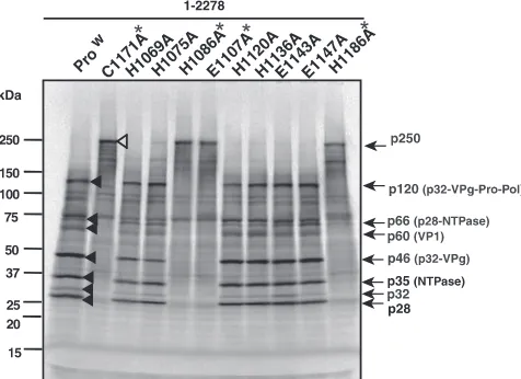
[image:4.594.302.540.440.613.2]FIG. 1. Identification of the C terminus of the SaV Mc10 protease. (A) Proteolytic cleavage map of the SaV Mc10 ORF1 polyprotein and the
processing intermediates (30). (B) SDS-PAGE of in vitro
35S-labeled wild-type (Pro
w) and C1171A mutant (Pro
mut) forms of the entire ORF1
polyprotein (aa 1 to 2278) and 15 C-terminally truncated polyproteins corresponding to aa 1 to 1246, 1 to 1240, 1 to 1234, 1 to 1229, 1 to 1223,
1 to 1218, 1 to 1212, 1 to 1206, 1 to 1205, 1 to 1204, 1 to 1203, 1 to 1202, 1 to 1201, 1 to 1200, and 1 to 1194. The protein bands corresponding
to either the Pro
wor Pro
mutform of the entire ORF1 polyprotein are indicated by filled and open arrowheads, respectively. The molecular sizes
of viral proteins are shown on the right, and size markers are shown on the left. Asterisks indicate two C-terminally truncated forms, corresponding
to aa 1 to 1200 and 1 to 1194, which show affected protease activity. Products of approximately 15 to 20 kDa would be the truncated Pro-Pol,
released from the truncated ORF1 polyprotein. A 60-kDa product of the C-terminally truncated form corresponding to aa 1 to 1200 was identified
as p32-VPg-truncated Pro-Pol.
FIG. 2. Identification of the amino acid residues critical to SaV
Mc10 protease activity. Shown are the results of SDS-PAGE of in vitro
35S-labeled wild-type (Pro
w) and C1171A mutant forms of the entire
ORF1 polyprotein (aa 1 to 2278) and nine other mutant forms, the
H1069A, H1075A, H1086A, E1107A, H1120A, H1136A, E1143A,
E1147A, and H1186A polyproteins. The protein bands corresponding
to either the Pro
wor C1171A form of the entire ORF1 polyprotein are
indicated by filled and open arrowheads, respectively. The molecular
sizes of viral proteins are shown on the right, and size markers are
shown on the left. Asterisks indicate four mutant forms, the C1171A,
H1086A, E1107A, and H1186A polyproteins, which display affected
protease activity.
on November 8, 2019 by guest
http://jvi.asm.org/
protease domain were selected based on the amino acid
align-ment of 14 human SaV strains, with one exception: D instead
of E
1147in a human SaV NK24 strain (data not shown). These
nine amino acid residues were changed to A by site-directed
mutagenesis (Table 1), and nine mutant forms of the ORF1
polyprotein,
the
H1069A,
H1075A,
H1086A,
E1107A,
H1120A, H1136A, E1143A, E1147A, and H1186A forms, were
expressed in an in vitro translation system. Two forms of the
ORF1 polyprotein, the full-length Pro
wand Pro
mut(C1171A)
forms, were used as positive and negative controls, respectively
(Fig. 2) (29, 30). Three mutant forms of the ORF1 polyprotein,
the H1086A, E1107A, and H1186A forms, each produced a
major 250-kDa product (Fig. 2), demonstrating that the
pro-teolytic processing of the ORF1 polyprotein was completely
blocked with these mutant forms. In contrast, the cleavage
products of six mutant forms, the H1069A, H1075A, H1120A,
H1136A, E1143A, and E1147A forms, were identical to those
of Pro
w, although their cleavages were slightly affected (Fig. 2).
From these results, we concluded that the amino acid residues
critical to SaV Mc10 3C-like protease activity are H
1086(31),
E
1107(52), C
1171(116), and H
1186(131). The first three of these
would form the catalytic triad (general base, anion, and
nu-cleophile, respectively), and the last one would correspond to
part of the binding pocket as previously described for other
calicivirus proteases (5, 27, 36, 46). Two amino acids, E
1107and
H
1186, essential to the SaV Mc10 protease activity are not
conserved in porcine SaV; (i) E
1107in Mc10 is D in the PEC
LL14 and Cowden strains, and (ii) H
1186in Mc10 is Y in the
PEC LL14 strain, whereas H
1186is conserved in the PEC
Cowden strain (data not shown).
Identification of the C terminus of the FCV F4 3C-like
protease.
The FCV protease-polymerase (Pro-Pol) has been
identified as a stable product in both infected cells and an in
vitro translation system (12, 40). Sosnovtseva et al. reported
that the entire Pro-Pol region is not essential for the
autocat-alytic polyprotein processing in the Urbana strain in vitro
translation system (42). However, the exact C terminus of the
functional protease domain has not been determined.
We generated two full-length cDNA clones, pUC19/FCV F4
full-length and pUC19/FCV F4 full-C1193A/ORF1, the latter
of which encodes a
1191GDCG
1194-to-GDAG mutation in the
protease. The expression was carried out with both Pro
wand
Pro
mut(C1193A) forms of the protease, and the latter were
used as a negative control for the proteolytic processing. Six
major cleavage products, p30, p32, p39 (NTPase), p43
(p30-VPg), p76 (Pro-Pol), and p120 (p30-VPg-Pro-Pol), were
de-tected when the Pro
wform corresponding to aa 1 to 1763 was
[image:5.594.85.501.69.363.2]expressed in an in vitro translation system (Fig. 3). These
FIG. 3. Identification of the C terminus of the FCV F4 protease. (A) Proteolytic cleavage map of the FCV F4 ORF1 polyprotein and processing
intermediates. The locations and designations of the proteins are adopted from the studies by Sosnovtsev et al. (40). (B) SDS-PAGE of in vitro
35S-labeled wild-type (Pro
w) and C1193A mutant (Pro
mut) forms of the entire ORF1 polyprotein (aa 1 to 1763) and 10 C-terminally truncated
polyproteins corresponding to aa 1 to 1419, 1 to 1345, 1 to 1267, 1 to 1245, 1 to 1240, 1 to 1235, 1 to 1230, 1 to 1225, 1 to 1224, and 1 to 1223.
The protein bands corresponding to either the Pro
wor Pro
mutform of the entire ORF1 polyprotein are indicated by filled and open arrowheads,
respectively. The molecular sizes of viral proteins are shown on the right, and size markers are shown on the left. Asterisks indicate two
C-terminally truncated forms, corresponding to aa 1 to 1224 and 1 to 1223, which display affected protease activity. Products of approximately 15
to 25 kDa would be the truncated Pro-Pol released from the truncated ORF1 polyprotein.
on November 8, 2019 by guest
http://jvi.asm.org/
cleavage products were identical to those of the Urbana strain
(40). A major 195-kDa product (p195) corresponding to the
ORF1 polyprotein appeared when the Pro
mutform was
ex-pressed (Fig. 3B), demonstrating that the C
1193is critical to the
FCV F4 3C-like protease activity, consistent with a previous
report (42).
To define the C terminus of the functional protease domain
of FCV F4, a series of 10 C-terminally truncated ORF1
polyproteins were expressed in an in vitro translation system
(Fig. 3B; Table 1). Both Pro
wand Pro
mutforms of each of
these regions were expressed, and the latter were used as a
negative control for the proteolytic processing. Eight
C-termi-nally truncated templates corresponding to aa 1 to 1419, 1 to
1345, 1 to 1267, 1 to 1245, 1 to 1240, 1 to 1235, 1 to 1230, and
1 to 1223 were first expressed and analyzed. The proteolytic
cleavage occurred in the Pro
wforms of the C-terminally
trun-cated ORF1 polyproteins corresponding to aa 1 to 1419, 1 to
1345, 1 to 1267, 1 to 1245, 1 to 1240, 1 to 1235, and 1 to 1230
but not in that corresponding to aa 1 to 1223. The translated
product size of the Pro
wform of aa 1 to 1223 was identical to
that of the Pro
mutform of aa 1 to 1223 (Fig. 3B),
demonstrat-ing that the C terminus of the functional protease domain is
positioned upstream of aa 1223. Next, we expressed and
ana-lyzed two additional C-terminally truncated forms, aa 1 to 1225
and 1 to 1224. The proteolytic cleavage occurred efficiently
when the Pro
wform of aa 1 to 1225 was expressed, whereas it
occurred partially when the Pro
wform of aa 1 to 1224 was
expressed (Fig. 3B). The C terminus of the functional protease
domain for FCV F4 was determined to be Y
1225. This amino
acid is conserved in 13 FCV strains (data not shown). Although
we did not identify the cleavage sites of the FCV F4 ORF1
polyprotein, the cleavage pattern and the sizes of the products
were consistent with the results of Sosnovtsev et al. (40).
Therefore, the size of the functional protease of FCV F4 would
be 154 aa (S
1072to Y
1225) when the cleavage site of the N
terminus of the Urbana strain Pro-Pol is considered (40). The
FCV F4 functional protease domain is similar in size to those
of other caliciviruses, including SaV.
Identification of the active sites of the FCV F4 3C-like
pro-tease.
The FCV 3C-like protease cleaves after the E residues of
the specific site in the ORF1 polyprotein (Fig. 3A), and the C
in the GDCG motif is critical to the protease activity (40–42).
The functional FCV F4 protease is similar in size to those of
SaV, RHDV, and NoV. Thus, the catalytic amino acid residues
of the FCV protease would also be similar to those of SaV,
RHDV, and NoV. Therefore, 11 amino acid residues (H
1079,
H
1093, H
1099, H
1102, H
1110, E
1121, D
1125, E
1131, D
1155, E
1164,
and H
1208) within the protease domain were selected and were
changed to A by site-directed mutagenesis (Table 1).
Eleven mutant forms of the ORF1 polyprotein, the H1079A,
H1093A, H1099A, H1102A, H1110A, E1121A, D1125A,
E1131A, D1155A, E1164A, and H1208A forms, were
ex-pressed in an in vitro translation system. Two forms of ORF1
polyprotein, the full-length Pro
wand Pro
mut(C1193A) forms,
were used as positive and negative controls for the proteolytic
processing (Fig. 4). H1110A did significantly affect the ORF1
polyprotein processing, whereas E1131A and H1208A affected
the processing partially (Fig. 4). That is, (i) p195, the entire
ORF1 polyprotein, was detected when the H1110A form was
expressed, (ii) p30 disappeared and p155 (likely the stable
intermediate of NTPase-p30-VPg-Pro-Pol) appeared when the
E1131A form was expressed, and (iii) p195 and p155
(NTPase-FIG. 4. Identification of the amino acid residues critical to FCV F4 protease activity. Shown are the results of SDS-PAGE of in vitro
35S-labeled
wild-type (Pro
w) and C1193A mutant forms of the entire ORF1 polyprotein (aa 1 to 1763) and 11 other mutant forms, the H1079A, H1093A,
H1099A, H1102A, H1110A, E1121A, D1125A, E1131A, D1155A, E1164A, and H1208A polyproteins. The protein bands corresponding to either
the Pro
wor C1193A form of the entire ORF1 polyprotein are indicated by filled and open arrowheads, respectively. The molecular sizes of viral
proteins are shown on the right, and size markers are shown on the left. Asterisks indicate four mutant forms, the C1193A, H1110A, E1131A, and
H1208A polyproteins, which show affected protease activity. The newly appearing product, p155, also marked with an asterisk, probably
corresponds to NTPase-p30-VPg-Pro-Pol.
on November 8, 2019 by guest
http://jvi.asm.org/
[image:6.594.113.472.69.295.2]p30-VPg-Pro-Pol) appeared whereas p43 (p30-VPg) and p30
disappeared when the H1208A form was expressed. Prolonged
incubation for up to 16 h did not change the processing
pat-terns of these templates (data not shown). In contrast, the
proteolytic processing of the ORF1 polyprotein was barely
affected when five mutant forms, the H1099A, H1102A,
E1121A, D1125A, and D1155A forms, were expressed (Fig. 4).
The p120 (p30-VPg-Pro-Pol) band disappeared when two
mu-tant forms, the H1079A and H1093A forms, were expressed.
However, the mutated amino acid residues in these forms are
not critical to protease activity, because other cleavage
prod-ucts were produced normally. Therefore, this product would be
a precursor intermediate. Combining these results, we
con-cluded that the amino acid residues important to FCV F4
3C-like protease activity are H
1110(39), E
1131(60), C
1193(122), and
[image:7.594.75.513.66.560.2]H
1208(137). The former three amino acid residues would form
FIG. 5. 3-D models of the calicivirus 3C-like proteases. (A to D) Structural models of calicivirus 3C-like protease domains of the SaV Mc10
(A), FCV F4 (B), and RHDV FRG (C) strains and crystal structure of the NoV Chiba strain protease (27) (D). (E) Superimposition of 3C-like
protease structures of the SaV Mc10, FCV F4, RHDV FRG, and NoV Chiba strains. The models were constructed with a homology modeling
technique by using programs in the MOE package. Ribbons represent the backbone of the 3C-like protease domain. Side chains of the catalytically
important amino acids identified in this study are shown as cyan sticks (H), red sticks (D and E), yellow sticks (C), and green sticks (H).
on November 8, 2019 by guest
http://jvi.asm.org/
the catalytic triad, and the last amino acid residue would
cor-respond to a part of the binding pocket as discussed for other
calicivirus proteases. A reverse genetics system has been
re-ported for FCV (37, 38, 40), and it would be interesting to
evaluate whether the E1131A and H1208A mutant forms are
also important in vivo.
Structural modeling of SaV and FCV 3C-like proteases.
Processing activities and specificities of the SaV and FCV
3C-like protease were similar when 3D-like RNA-dependent
RNA polymerase domains were sequentially deleted (Fig. 1
and 3), strongly suggesting that the structure of the protease
active site is self-determined and preserved in the part of the
Pro-Pol polypeptide independent of the Pol domain. To obtain
structural insights into the roles of the amino acid residues
critical to protease activity, 3-D models of the 3C-like protease
domains of the SaV Mc10, FCV F4, and RHDV FRG strains
were constructed and compared with the X-ray crystal
struc-ture of the 3C-like protease of the NoV Chiba strain (27).
Despite the low levels of amino acid sequence similarity of
proteases among these strains (about 20%), the overall
struc-tures were predicted to be similar and to retain structural
characteristics seen in the functional protease in general (Fig.
5A to D). The calicivirus proteases consist of N- and
C-termi-nal subdomains, which are separated by a large cleft, probably
for substrate binding. H and E in SaV, FCV, and NoV and H
and D in RHDV are located along the inner surface of the
N-terminal subdomain, whereas C and H in all viruses are
located along the inner surface of the C-terminal subdomain.
In contrast to the overall similarity, the conformations and
configurations of the local structures around the active site are
often different among the viruses, suggesting their potential
roles in determining protein substrate specificity.
Notably, the superimposition of the calicivirus protease
do-main structures showed that the thermodynamically favored
configurations of the amino acids H, E/D, C, and H are highly
conserved (Fig. 5E); the side chains of these amino acids
pro-trude from the main chains at almost identical positions, with
very similar configurations along the inner surface of the
po-tential binding cleft for the substrate. The C is a part of the
GDCG motif, a conserved and functionally important 3C-like
protease motif, and is surrounded closely by other amino acid
residues, H, E, and H. The side chain orientations are almost
identical to those of the protease. Higher-resolution X-ray
crystal structures from distinct NoV strains (46) supported the
conservation of the configuration of these catalytic amino acid
residues among calicivirus proteases. These findings are
con-sistent with the critical roles of these amino acids in the
pro-teolytic activity and suggest strongly that the H, E, C, and H
residues are involved in the formation of a conserved catalytic
surface of the SaV and FCV 3C-like proteases.
Conclusions.
The functional domains, amino acid residues
critical to the proteolytic processing activity, and structural
characteristics of SaV and FCV 3C-like proteases were
iden-tified. The molecular genetics study showed that the sizes and
catalytically important amino acids of the SaV and FCV
pro-teases are similar to those of RHDV and NoV propro-teases. The
computer-assisted structural study strongly suggested that
these amino acids are involved in the formation of a conserved
catalytic surface of the calicivirus 3C-like protease. In this
study, we studied both primary and 3-D structures of SaV and
FCV proteases, which are mutually closely related subjects and
should be understood in concert. In collaboration with other
resources, the data obtained in this study will provide
impor-tant bases to study the molecular function and inhibitors of
calicivirus proteases.
ACKNOWLEDGMENTS
We thank Y. Someya for his critical review of the manuscript.
This work was supported in part by grants for research on emerging
and reemerging infectious diseases, as well as food safety, from the
Ministry of Health, Labor, and Welfare of Japan and by a grant from
the Japan Health Science Foundation for Research on Health Sciences
Focusing on Drug Innovation.
REFERENCES
1.Asanaka, M., R. L. Atmar, V. Ruvolo, S. E. Crawford, F. H. Neill, and M. K. Estes.2005. Replication and packaging of Norwalk virus RNA in cultured mammalian cells. Proc. Natl. Acad. Sci. USA102:10327–10332.
2.Belliot, G., S. V. Sosnovtsev, T. Mitra, C. Hammer, M. Garfield, and K. Y. Green.2003. In vitro proteolytic processing of the MD145 norovirus ORF1 nonstructural polyprotein yields stable precursors and products similar to those detected in calicivirus-infected cells. J. Virol.77:10957–10974. 3.Bergmann, E. M., M. M. Cherney, J. McKendrick, S. Frormann, C. Luo,
B. A. Malcolm, J. C. Vederas, and M. N. James.1999. Crystal structure of an inhibitor complex of the 3C proteinase from hepatitis A virus (HAV) and implications for the polyprotein processing in HAV. Virology265:153–163. 4.Blakeney, S. J., A. Cahill, and P. A. Reilly.2003. Processing of Norwalk virus nonstructural proteins by a 3C-like cysteine proteinase. Virology308:216– 224.
5.Boniotti, B., C. Wirblich, M. Sibilia, G. Meyers, H. J. Thiel, and C. Rossi.
1994. Identification and characterization of a 3C-like protease from rabbit hemorrhagic disease virus, a calicivirus. J. Virol.68:6487–6495.
6.Bowie, J. U., R. Luthy, and D. Eisenberg.1991. A method to identify protein sequences that fold into a known three-dimensional structure. Science253:
164–170.
7.Chang, K. O., S. S. Sosnovtsev, G. Belliot, Q. Wang, L. J. Saif, and K. Y. Green.2005. Reverse genetics system for porcine enteric calicivirus, a pro-totype sapovirus in theCaliciviridae. J. Virol.79:1409–1416.
8.Chang, K. O., S. V. Sosnovtsev, G. Belliot, A. D. King, and K. Y. Green.2006. Stable expression of a Norwalk virus RNA replicon in a human hepatoma cell line. Virology353:463–473.
9.Clarke, I. N., and P. R. Lambden.2000. Organization and expression of calicivirus genes. J. Infect. Dis.181(Suppl. 2):S309–S316.
10.Clarke, I. N., and P. R. Lambden.1997. The molecular biology of calicivi-ruses. J. Gen. Virol.78:291–301.
11.Green, K. Y., T. Ando, M. S. Balayan, T. Berke, I. N. Clarke, M. K. Estes, D. O. Matson, S. Nakata, J. D. Neill, M. J. Studdert, and H. J. Thiel.2000. Taxonomy of the caliciviruses. J. Infect. Dis.181(Suppl. 2):S322–S330. 12.Green, K. Y., A. Mory, M. H. Fogg, A. Weisberg, G. Belliot, M. Wagner, T.
Mitra, E. Ehrenfeld, C. E. Cameron, and S. V. Sosnovtsev.2002. Isolation of enzymatically active replication complexes from feline calicivirus-infected cells. J. Virol.76:8582–8595.
13.Hansman, G. S., K. Katayama, N. Maneekarn, S. Peerakome, P. Khamrin, S. Tonusin, S. Okitsu, O. Nishio, N. Takeda, and H. Ushijima.2004. Genetic diversity of norovirus and sapovirus in hospitalized infants with sporadic cases of acute gastroenteritis in Chiang Mai, Thailand. J. Clin. Microbiol.
42:1305–1307.
14.Hardy, M. E., T. J. Crone, J. E. Brower, and K. Ettayebi.2002. Substrate specificity of the Norwalk virus 3C-like proteinase. Virus Res.89:29–39. 15.Katayama, K., G. S. Hansman, T. Oka, S. Ogawa, and N. Takeda.2006.
Investigation of norovirus replication in a human cell line. Arch. Virol.
151:1291–1308.
16.Katayama, K., H. Shirato-Horikoshi, S. Kojima, T. Kageyama, T. Oka, F. Hoshino, S. Fukushi, M. Shinohara, K. Uchida, Y. Suzuki, T. Gojobori, and N. Takeda.2002. Phylogenetic analysis of the complete genome of 18 Nor-walk-like viruses. Virology299:225–239.
17.Kinomoto, M., R. Appiah-Opong, J. A. Brandful, M. Yokoyama, N.
Nii-Trebi, E. Ugly-Kwame, H. Sato, D. Ofori-Adjei, T. Kurata, F. Barre-Sinoussi, T. Sata, and K. Tokunaga.2005. HIV-1 proteases from drug-naive West African patients are differentially less susceptible to protease inhibi-tors. Clin. Infect. Dis.41:243–251.
18.Kinomoto, M., M. Yokoyama, H. Sato, A. Kojima, T. Kurata, K. Ikuta, T. Sata, and K. Tokunaga.2005. Amino acid 36 in the human immunodefi-ciency virus type 1 gp41 ectodomain controls fusogenic activity: implications for the molecular mechanism of viral escape from a fusion inhibitor. J. Virol.
79:5996–6004.
19.Konig, M., H. J. Thiel, and G. Meyers.1998. Detection of viral proteins after infection of cultured hepatocytes with rabbit hemorrhagic disease virus. J. Virol.72:4492–4497.
on November 8, 2019 by guest
http://jvi.asm.org/
20.Liu, B., I. N. Clarke, and P. R. Lambden.1996. Polyprotein processing in Southampton virus: identification of 3C-like protease cleavage sites by in vitro mutagenesis. J. Virol.70:2605–2610.
21.Liu, B. L., G. J. Viljoen, I. N. Clarke, and P. R. Lambden.1999. Identification of further proteolytic cleavage sites in the Southampton calicivirus polypro-tein by expression of the viral protease in E. coli. J. Gen. Virol.80:291–296. 22.Makino, A., M. Shimojima, T. Miyazawa, K. Kato, Y. Tohya, and H. Akashi.
2006. Junctional adhesion molecule 1 is a functional receptor for feline calicivirus. J. Virol.80:4482–4490.
23.Matthews, D. A., P. S. Dragovich, S. E. Webber, S. A. Fuhrman, A. K. Patick, L. S. Zalman, T. F. Hendrickson, R. A. Love, T. J. Prins, J. T. Marakovits, R. Zhou, J. Tikhe, C. E. Ford, J. W. Meador, R. A. Ferre, E. L. Brown, S. L. Binford, M. A. Brothers, D. M. DeLisle, and S. T. Worland.1999. Structure-assisted design of mechanism-based irreversible inhibitors of human rhino-virus 3C protease with potent antiviral activity against multiple rhinorhino-virus serotypes. Proc. Natl. Acad. Sci. USA96:11000–11007.
24.Mayo, M. A.2002. A summary of taxonomic changes recently approved by ICTV. Arch. Virol.147:1655–1663.
25.Meyers, G., C. Wirblich, H. J. Thiel, and J. O. Thumfart. 2000. Rabbit hemorrhagic disease virus: genome organization and polyprotein processing of a calicivirus studied after transient expression of cDNA constructs. Vi-rology276:349–363.
26.Mosimann, S. C., M. M. Cherney, S. Sia, S. Plotch, and M. N. James.1997. Refined X-ray crystallographic structure of the poliovirus 3C gene product. J. Mol. Biol.273:1032–1047.
27.Nakamura, K., Y. Someya, T. Kumasaka, G. Ueno, M. Yamamoto, T. Sato, N. Takeda, T. Miyamura, and N. Tanaka.2005. A norovirus protease struc-ture provides insights into active and substrate binding site integrity. J. Virol.
79:13685–13693.
28.Oka, T., K. Katayama, S. Ogawa, G. S. Hansman, T. Kageyama, T.
Miyamura, and N. Takeda.2005. Cleavage activity of the sapovirus 3C-like protease in Escherichia coli. Arch. Virol.150:2539–2548.
29.Oka, T., K. Katayama, S. Ogawa, G. S. Hansman, T. Kageyama, H.
Ushijima, T. Miyamura, and N. Takeda.2005. Proteolytic processing of sapovirus ORF1 polyprotein. J. Virol.79:7283–7290.
30.Oka, T., M. Yamamoto, K. Katayama, G. S. Hansman, S. Ogawa, T.
Miyamura, and N. Takeda.2006. Identification of the cleavage sites of sapovirus open reading frame 1 polyprotein. J. Gen. Virol.87:3329–3338. 31.Ponder, J. W., and D. A. Case.2003. Force fields for protein simulations.
Adv. Protein Chem.66:27–85.
32.Seah, E. L., J. A. Marshall, and P. J. Wright.1999. Open reading frame 1 of the Norwalk-like virus Camberwell: completion of sequence and expression in mammalian cells. J. Virol.73:10531–10535.
33.Seah, E. L., J. A. Marshall, and P. J. Wright.2003.transactivity of the norovirus Camberwell proteinase and cleavage of the N-terminal protein encoded by ORF1. J. Virol.77:7150–7155.
34.Someya, Y., N. Takeda, and T. Miyamura.2005. Characterization of the norovirus 3C-like protease. Virus Res.110:91–97.
35.Someya, Y., N. Takeda, and T. Miyamura.2000. Complete nucleotide se-quence of the chiba virus genome and functional expression of the 3C-like protease in Escherichia coli. Virology278:490–500.
36.Someya, Y., N. Takeda, and T. Miyamura.2002. Identification of active-site amino acid residues in the Chiba virus 3C-like protease. J. Virol.76:5949– 5958.
37.Sosnovtsev, S., and K. Y. Green.1995. RNA transcripts derived from a cloned full-length copy of the feline calicivirus genome do not require VpG for infectivity. Virology210:383–390.
38.Sosnovtsev, S. V., G. Belliot, K. O. Chang, O. Onwudiwe, and K. Y. Green.
2005. Feline calicivirus VP2 is essential for the production of infectious virions. J. Virol.79:4012–4024.
39.Sosnovtsev, S. V., G. Belliot, K. O. Chang, V. G. Prikhodko, L. B. Thackray, C. E. Wobus, S. M. Karst, H. W. Virgin, and K. Y. Green.2006. Cleavage map and proteolytic processing of the murine norovirus nonstructural polyprotein in infected cells. J. Virol.80:7816–7831.
40.Sosnovtsev, S. V., M. Garfield, and K. Y. Green.2002. Processing map and essential cleavage sites of the nonstructural polyprotein encoded by ORF1 of the feline calicivirus genome. J. Virol.76:7060–7072.
41.Sosnovtsev, S. V., S. A. Sosnovtseva, and K. Y. Green.1998. Cleavage of the feline calicivirus capsid precursor is mediated by a virus-encoded proteinase. J. Virol.72:3051–3059.
42.Sosnovtseva, S. A., S. V. Sosnovtsev, and K. Y. Green.1999. Mapping of the feline calicivirus proteinase responsible for autocatalytic processing of the nonstructural polyprotein and identification of a stable proteinase-polymer-ase precursor protein. J. Virol.73:6626–6633.
43.Wei, L., J. S. Huhn, A. Mory, H. B. Pathak, S. V. Sosnovtsev, K. Y. Green, and C. E. Cameron.2001. Proteinase-polymerase precursor as the active form of feline calicivirus RNA-dependent RNA polymerase. J. Virol.75:
1211–1219.
44.Wirblich, C., H. J. Thiel, and G. Meyers.1996. Genetic map of the calicivirus rabbit hemorrhagic disease virus as deduced from in vitro translation studies. J. Virol.70:7974–7983.
45.Yap, C. C., K. Ishii, H. Aizaki, H. Tani, Y. Aoki, Y. Ueda, Y. Matsuura, and T. Miyamura.1998. Expression of target genes by coinfection with replica-tion-deficient viral vectors. J. Gen. Virol.79:1879–1888.
46.Zeitler, C. E., M. K. Estes, and B. V. Venkataram Prasad.2006. X-ray crystallographic structure of the Norwalk virus protease at 1.5-A resolution. J. Virol.80:5050–5058.
47.Zhang, K. Y., and D. Eisenberg.1994. The three-dimensional profile method using residue preference as a continuous function of residue environment. Protein Sci.3:687–695.
on November 8, 2019 by guest
http://jvi.asm.org/