COMPARISON OF THE HAEMODYNAMIC EFFECTS
OF PRELOADING VERSUS CO-LOADING USING
CRYSTALLOIDS AND COLLOIDS DURING ELECTIVE
LSCS UNDER SPINAL ANAESTHESIA.
Dissertation submitted to
The Tamil Nadu Dr.M.G.R. Medical University, Chennai
600032
with fulfilment of regulations for the award of Degree
M.D. ANAESTHESIOLOGY
BRANCH – X
DEPARTMENT OF ANAESTHESIOLOGY
K.A.P.V. GOVT. MEDICAL COLLEGE, TRICHY.
ACKNOWLEDGEMENT
First of all, I thank the DEAN of K.A.P.V. Govt. Medical College,
Trichy, Prof Dr.G.ANITHA M.D., for permitting me to conduct this study in
Department of Anaesthesiology of K.A.P.V. Government Medical College,
Trichy. My sincere thanks to the Head of the Department, Prof.Dr.R.SELVA
KUMAR M.D.,D.A.,DNB for his continuous support and guidance during this
study.
My heart felt gratitude to all my professors Prof .Dr.R.SELVA
KUMAR, M.D., D.A.,DNB, Prof.Dr.G.SIVAKUMAR M.D,D.A.,
P r o f . D r. M . S U R E S H M . D , D . A , P r o f . D r. P. E L A N G O M . D a n d
Prof.Dr.M .SHANMUGASUNDARAM M.D for their immense knowledge and
valuable comments which helped me in my research work.
My deepest gratitude to my guide and mentor Prof.Dr.G.SIVA
KUMAR for his patience and motivation. I was fortunate to work under his
technical guidance, and his valuable inputs helped me at every stage of my
research work.
I am greatly indebted my co-ordinator Dr.C.R. GANESSAN and
all my Assistant professors who have been an indispensable part of my research
I thank all my colleagues who helped me and shared their
knowledge about this study. My sincere thanks to all the patients who
co-operated for this study, without whom this study could not have been
undertaken.
I am greatly indebted to all my patients without whom this study would not have been a reality.
I thank all the anaesthesia assistants and staff nurses who cooperated with me at all times.
LIST OF ABBREVIATIONS USED
(In alphabetical order)
ASA - American Society of Anaesthesiologist
bpm - Beats per minute
cm - Centimetre
CSF - Cerebrospinal fluid
DBP - Diastolic blood pressure
ECG - Electrocardiography
HR - Heart rate
Kg - Kilogram
LA - Local anaesthetic
LMW - Low molecular weight
lpm - Litres per minute
MAP - Mean arterial pressure
m - Meter
mmHg - Millimeter of mercury
NIBP - Non invasive blood pressure
% - Percentage
Spo2 - Pulseoximetry
SBP - Systolic blood pressure
Sl. No Title Page No.
1. INTRODUCTION 14
2. HISTORY 17
3. REVIEW OF LITERATURE 54
4. AIMS AND OBJECTIVES 60
5. MATERIALS AND METHODS 62
6. OBSERVATION AND RESULTS 69
7. DISCUSSION 86
8. CONCLUSION 95
9. SUMMARY 97
10. BIBLIOGRAPHY 99
11.
ANNEXURES A.Proforma
B.Monitoring Chart C.Consent form D.Master chart
E.Key to Master chart
110 111 113 116 124
INTRODUCTION
Central neuraxial blockade is considered as the gold standard
technique for obstetric anaesthesia and analgesia. Spinal anaesthesia is
frequently used for caesarean section due to its rapid onset, dense neural
block ,less maternal morbidity and mortality which is largely due to a reduction
in the incidence of pulmonary aspiration and failed intubation, avoids neonatal
exposure to depressant anaesthetic drugs, and allows the mother to remain
awake during delivery.
However , hypotension following spinal anaesthesia is a common
physiological complication and is due to pharmacological sympathectomy
leading to peripheral arterial and venous vasodilatation and venous pooling of
blood. As a result, there is decreased venous return and cardiac output leading to
hypotension. In pregnancy, this is further aggravated by the effects of the gravid
uterus and subsequent aortocaval compression
The risk of hypotension is increased in a parturient due to the
higher level of block (T4) required for the cesarean section, unique physiologic
and anatomic changes of pregnancy and increased susceptibility to the effects of
sympathectomy due to reduced sensitivity to the endogenous vasoconstrictors
coupled with increased synthesis of endothelium-derived vasodilators.
risk. The spectrum of morbidity associated with hypotension may include but is
not limited to a higher incidence of nausea, vomiting, dizziness, aspiration,
syncope and cardiac arrhythmias and foetus - acidosis, neurologic injuries, etc.
Several techniques and methodologies have been adopted for the
prevention of this neuraxial hypotension with varying degree of success.
Techniques to prevent maternal hypotension include intravenous volume
expansion using i. v. fluids (“preload”) immediately before spinal injection, use
of left lateral tilt or manual uterine displacement, or both, as leg wrapping,
elastic stockings, and administration of i.v. fluids and vasopressor drugs both
prophylactically and in response to cardiovascular changes subsequent to neural
block.
One of the foremost methods includes prophylactic administration
of intravenous fluids before implementation of subarachnoid block to offset the
hypotensive effects of sympathectomy by maintaining intravascular volume
which is commonly called as pre-loading. The conflicting literary evidence and
unequivocal results of the technique of pre-loading has made co-loading:A
method of administration of intravenous fluid bolus immediately after the
subarachnoid block.
Fluid co-loading appears to be more physiological and rational approach as the
Among the type of fluids (crystalloid or colloid) which one is
better, is still not known. Crystalloid has a short intravascular half-life because
of its rapid distribution into the interstitial space. In contrast to crystalloid,
colloid remains for a longer period within the intravascular space.
Based on this, the present study was designed to evaluate
haemodynamic changes following preload and co-load with crystalloid and
colloid , in patients who undergo elective caesarian section to find out which
method is more effective to prevent hypotension following spinal anaesthesia.
HISTORY
Anatomic and Physiologic Changes of Pregnancy
Regional anaesthesia has been considered as the most appropriate for caesarean
section in a parturient due to its beneficial effects on both mother and foetus.
The anatomic changes occurring during pregnancy affects the usage of
neuraxial anesthesia techniques.
Enlargement of the uterus and its compression on inferior vena cava leads to
engorgement of the epidural veins. The vertebral foraminal veins in contiguous
with the epidural veins, are also enlarged . Enlargement of epidural veins &
the greater intra-abdominal pressure of pregnancy displaces the CSF from the
thoracolumbar region of the subarachnoid space. Due to this displacement, a
lowered dose of local anaesthetic is required for spinal anesthesia in pregnant
women. Dosing requirements are also affected by the lower specific gravity of
CSF in pregnant patients than in non pregnant patients.
The hormonal changes occurring during pregnancy softens the
perivertebral ligamentous structures like the ligamentum flavum. Moreover it is
difficult to achieve flexion of the lumbar spine in a pregnant women for
There is a progressive accentuation of lumbar lordosis which alters the surface
anatomy relationship with the vertebral column.
1) Pelvis rotates on the long axis of the spinal column the line joining
the iliac crests- Tuffier’s line lies in cephalad relationship to the vertebral
column — might cross the vertebral column at the level of L3 to L4 interspace
rather than at L4 to L5 interspace.
2) Space between the adjacent lumbar spinous processes is reduced during
pregnancy making the midline approach to identify the epidural or subarachnoid space
in pregnant women difficult.
3) The apex of the lumbar lordosis is usually shifted caudad during preg-
Pregnant
Non pregnant
These changes influences the spread of subarachnoid anesthetic solutions in supine
patients, and also in leading to a higher sensory level in the pregnant patient.
PHYSIOLOGY OF OBSTETRIC PAIN PATHWAYS
During the first stage of labor , pain is primarily due to changes occurring
in the lower uterine segment and cervix. Visceral Afferent nerve fibers that
accompany the sympathetic nerves transmits pain to the spinal cord at the T10
to L1 segments.
The late first stage and second stage of labor is characterized by
pain due to distention of the pelvic floor, vagina, and perineum. Pelvic pain is
transmitted via the somatic nerve fibres, which enters the spinal cord at the
level of S2 to S4 segments.
Additional nociceptive pathways are involved in cesarean delivery,
therefore, a dermatomal level of anesthesia upto T4 level is required to provide
adequate anesthesia. Lower segment cesarean deliveries are performed using a
horizontal — Pfannenstiel skin incision, involving the infra-umbilical
dermatomes of T11 to T12. Stretching of the skin during the surgery, may
involve dermatomes of two to four levels higher. Intraperitoneal manipulation
and dissection involve poorly localized visceral pain pathways. Visceral pain
impulses may be transmitted to as high as the celiac plexus. In addition somatic
pain impulses may also occur as a result of diaphragmatic stimulation via the
the intercostal nerves, which innervate a part of the peripheral diaphragm.
Physiology of Neural Blockade
Changes in Hormonal levels, anatomy, and decreases in CSF
specific gravity are responsible for the lower local anesthetic dose requirements
for spinal anesthesia in pregnant women.
Local anesthetics produce conduction blockade primarily by
blocking sodium channels in the nerve membranes, thus preventing the
propagation of neural impulses.
Differential blockade is manifested as differences in the extent of
cephalad blockade of temperature discrimination and vasomotor tone, sensory
discrimination and vasomotor tone are blocked to the greatest extent and motor
function to the least extent.
During spinal anesthesia, local anesthetics acts directly on neural
tissue in the subarachnoid space.
Regression of anesthesia is due to the vascular uptake of local
anesthetic from the subarachnoid space and spinal cord.
Anesthesia For Cesarean Delivery
Preanesthetic Evaluation :
1) Patient’s medical, surgical, and obstetric history, the presence or absence of
labor, the urgency of the delivery, allergies, and baseline blood pressure and
heart rate measurements;
2) Evaluation of an airway, heart, and lung examination consistent with the
American Society of Anesthesiologists (ASA) guidelines .
Informed Consent
Arrange for Blood Products :
Risk factors
Baseline hematocrit
Blood type and screen or crossmatch
Equipment for rapid transfusion
Monitoring:
• Electrocardiogram
• Noninvasive blood pressure
• Pulse oximetry
• Capnography
• Oxygen and volatile agent analyzers
• Ventilator (with appropriate pressure and disconnection sensors/alarms)
• Peripheral nerve stimulator
FOR ROUTINE AIRWAY MANAGEMENT
• Laryngoscope and assorted blades
• Oral airways of assorted sizes
• Endotracheal tubes of assorted sizes with stylets
• Oxygen source
• Suction source with tubing and catheters
• Self-inflating bag and mask for positive-pressure ventilation
• Medications for blood pressure support, hypnosis, and muscle
relaxation
• Carbon dioxide detector
• Pulse oximeter
FOR DIFFICULT AIRWAY MANAGEMENT
• Rigid laryngoscope blades of alternative design and size from those
routinely used
• Supraglottic airway devices(e.g.,LMA)
• Endotracheal tube guides
• Retrograde intubation equipment
• At least one device suitable for emergency nonsurgical airway
ventilation
• Fiberoptic intubation equipment
• Equipment suitable for emergency surgical airway
access (e.g., cricothyrotomy)
• Topical anesthetics and vasoconstrictors
Aspiration Prophylaxis :
Fasting guidelines
clear liquids - 2 hours
solid foods - 6-8 hours,
Non-particulate antacid,
H2-receptor antagonist,
Metoclopramide
.
Intravenous access and fluid management :
Intravenous catheter: 16- or 18-gauge.
Fluid type, volume, and rate.
Supplemental medications :
Consider anxiolysis for severe anxiety
Positioning :
Lateral or sitting position for neuraxial needle/catheter placement.
Left uterine displacement, slight head up for surgery.
“Sniffing” position if general anesthesia is planned
S
ELECTION OFANESTHETIC TECHNIQUE FORC
ESAREAND
ELIVERYNeuraxial Anesthesia
• Maternal desire to witness birth and/ or avoid general anesthesia
• Risk factors for difficult airway or aspiration
• Presence of comorbid conditions
• General anesthesia intolerance or failure
• Other benefits : Plan for neuraxial analgesia after surgery
Less fetal drug exposure
Less blood loss
Allows presence of husband or support person .
General Anesthesia
• Maternal refusal or failure to cooperate with neuraxial technique
• Presence of comorbid conditions that contraindicate a neuraxial technique
• Insufficient time to induce neuraxial anesthesia for urgent delivery
• Failure of neuraxial technique
Neuraxial Anesthetic Techniques for Cesarean Delivery
SINGLE-SHOTSPINAL
Advantages :
Technically simple
Low doses of local anesthetic and opioid
Rapid onset of dense lumbosacral and thoracic anesthesia
Disadvantages :
Limited duration of anesthesia
Limited ability to titrate extent of sensory blockade
EPIDURAL ANAESTHESIA
Advantages :
No dural puncture required
Can use in situ catheter placed for earlier administration of labor
analgesia
Ability to titrate extent of sensory blockade
Continuous intraoperative anesthesia
Disadvantages :
Slow onset of anesthesia
Larger drug doses required than for spinal techniques:
• Greater risk for maternal systemic toxicity
• Greater fetal drug exposure
COMBINED SPINAL-EPIDURAL ANAESTHESIA
Advantages :
May be technically easier than spinal anesthesia in obese patients
Low doses of local anesthetic and opioid
Rapid onset of dense lumbosacral and thoracic anesthesia
Ability to titrate extent of sensory blockade
Continuous intraoperative anesthesia
Continuous postoperative analgesia
Disadvantages :
CONTINUOUSSPINAL ANAESTHESIA
Advantages :
Low doses of local anesthetic and opioid
Rapid onset of dense anesthesia
Ability to titrate extent of sensory blockade
Continuous intraoperative anesthesia
Disadvantages :
Large dural puncture increases risk for post–dural puncture headache
Possibility of overdose and total spinal anesthesia if the spinal catheter is
mistaken for an epidural catheter
S
PINALA
NESTHESIASpinal anesthesia is now the most commonly used anesthetic
technique for cesarean delivery. Spinal anesthesia is a simple and reliable
technique ,technically easier to perform and provides rapid onset of dense
neural blockade that is typically more profound, resulting in a reduced need for
supplemental intravenous analgesics or conversion to general anesthesia. Only a
small dosage of local anesthetic is required to establish a functional spinal
blockade; thus, spinal anesthesia is associated with trivial maternal risk for
Spinal anesthesia is associated with predictable and relatively prompt recovery
that enables patients to quickly transition through the post anesthesia care unit .
Contraindications :
Patient refusal
Elevated Intracranial pressure
Skin or soft tissue infection at the site of needle entry
Coagulopathy
Maternal hypovolemia
COMPLICATIONS OF SPINAL ANAESTHESIA
Dyspnea
Hypotension
Failure of Neuraxial Blockade
High Neuraxial Blockade
Nausea and Vomiting
Pruritus
PATHOPHYSIOLOGY OF HYPOTENSION FOLLOWING SPINAL ANESTHESIA
Hypotension following spinal anaesthesia is mainly occurs due to :
Marked decrease in systemic vascular resistance,
The rate and extent of the sympathetic involvement— leading to
peripheral vasodilatation and venous pooling of blood, and
The onset and spread of the neuraxial blockade — determines the
severity of hypotension.
Hypotension is a common sequela of neuraxial anesthesia and, if severe and
sustained, may lead to impairment of uteroplacental perfusion and result in fetal
hypoxia, acidosis, and neonatal depression or injury.
Severe maternal hypotension can also have adverse maternal outcomes,
including altered consciousness, pulmonary aspiration, apnea, and cardiac
arrest.
Definitions for maternal hypotension:
1) a decrease in systolic blood pressure of more than 20% to 30% from
baseline measurements or
Prevention of Hypotension
The various methods to prevent this hypotension following spinal anaesthesia
are similar in both pregnant and non-pregnant patients.
Strategies that mitigate hypotension after spinal anesthesia for cesarean delivery
includes
1) Fluid administration
2) Vasopressor administration,
3) Lower local anesthetic doses,
4) Leg elevation or wrapping, and
5) Left uterine displacement.
The use of intravenous fluid to prevent hypotension is done by
i) Timing of administration — either prior to (preload) or coincident with
(co-load) the intrathecal injection, and/or
ii) Type of fluid, either crystalloid or colloid.
SPINAL ANAESTHESIA AND AUTONOMIC NERVOUS SYSTEM
Sympathetic Denervation occurs during spinal anaesthesia. In
spinal anaesthesia the severity of hypotension depends on the degree of
sympathetic blockade. The level of sympathetic denervation determines the
magnitude of cardiovascular responses in spinal anesthesia. As the level of
blockade increases, more number of sympathetic fibres are blocked and greater
the change in cardio-circulatory parameters are anticipated. But we often find a
low level of blockade producing severe hypotension as the sympathetic
blockade highly variable.This variability is due to in the arborisation of
autonomic fibres.
In partial sympathetic blockade, a reflex increase in sympathetic
activity occurs in sympathetically intact areas leading to vasoconstriction which
compensates for the peripheral vasodilation taking place in the sympathetically
denervated areas.
Anesthesia at or above T5 increases the risk of hypotension and
bradycardia . In spinal anesthesia, hypotension is defined as a systolic blood
pressure <90 mm Hg or defined as reduction in mean arterial blood pressure
>30% . In addition, in pregnancy,the gravid uterus compresses on the aorta and
inferior vena cava further augmenting the symptoms in supine
position.Prevention of hypotension caused by vasodilatation can be done by a
prophylactic preloading infusion of colloid or crystalloid or by during the
performance of the neuraxial block as co-loading, and with use of vasopressors.
Bradycardia
Severe bradycardia may develop after spinal anesthesia and has
been recognized as an important risk in spinal anesthesia.Bradycardia occurs
due to the blockade of the thoracic sympathetic fibers — preganglionic cardiac
accelerator fibers originating at T1-T5, and also from reflex slowing of the heart
rate induced by vasodilation related reduction in venous return to the right
atrium . The stretch receptors responds by a compensatory slowing of the heart
rate.
Vasopressor agents :
Ephedrine and Phenylephrine, in titrated doses to maintain maternal blood
pressure. Greater doses of ephedrine provided more effective prophylaxis, but,
hypotension was still observed and reactive hypertension and umbilical artery
Phenylephrine crosses the placenta at a lower rate than ephedrine and undergoes
greater fetal metabolism than ephedrine.
The combination of intra venous fluid therapy and vasopressor
administration might be the most effective regimen to prevent hypotension.
The use of lower doses of spinal local anesthetic is associated with
a lower incidence of hypotension.
Physical methods to prevent hypotension includes :
Use of lower limb compression bandages or pneumatic
compression devices
Left uterine displacement .
Treatment of Hypotension
The ideal treatment of hypotension should be
reliable,
titratable,
easy to use, and
These include:
Intravenous fluid administration — pre-loading and co-loading,
Use of vasopressors :
Phenylephrine and Ephedrine
INTRAVENOUS FLUIDS: THE FIRST LINE OF MANAGEMENT
The use of intravenous fluids is one of the most popular measures to prevent maternal hypotension .Pre-loading as well as
co-loading techniques are being used in prevention of spinal anaesthesia
induced maternal hypotension . But, it is being emphasized that no single
modality is effective for prevention of maternal hypotension following
spinal anaesthesia alone and should be combined with timely and
BODY FLUID COMPARTMENTS
The major body fluid is water. Water makes up approximately 60%
of total body weight in the average adult, which varies with age, gender, and
body composition. Fat contains less water.
Total body water is divided between anatomic and functional fluid
compartments within the body — intracellular fluid (ICF) and extracellular fluid
(ECF). About one-third (20 %) of the total amount of water is confined to the
ECF, and two-thirds (40 %) to the ICF compartment .
The ECF compartment, in turn, is divided into the following subdivisions:
1. Plasma
2. Interstitial fluid and lymph
3. Bone and dense connective tissue water
4. Transcellular (cerebrospinal, pleural, peritoneal, synovial, and digestive
secretions)
The plasma and interstitial fluids compartments are the two most important,
because of constant exchange of fluid and electrolytes between them. Plasma
circulates in the blood vessels, whereas the interstitial fluid bathes all tissue
cells except for the formed elements of blood. Claude Bernard, the French
physiologist, called the interstitium “the true environment of the body” (milieu
interieur).
In 1861,Thomas Graham’s investigations on diffusion led to
classify substances as crystalloids or colloids based on their ability to diffuse
through a parchment membrane. Crystalloids can readily pass through the
classified similarly based on their ability to pass from intravascular to
extravascular (interstitial) fluid compartments .
Crystalloids
Crystalloid solutions are electrolyte solutions with small molecules
that easily pass through the capillary membrane which can diffuse freely
throughout the extracellular space. Crystalloids containing a range of
electrolytes and a buffer such as lactate or acetate , which are also found in
plasma, may be referred to as balanced solutions.
Crystalloids are indicated for replacement of free water and
electrolytes but also may be used for volume expansion.
Sodium chloride (NaCl),an inorganic salt forms the principal
component of crystalloid solutions. Sodium ,the most abundant solute and is
distributed uniformly in the extracellular fluid. In human body , the
conventional concepts of fluid compartments dictates that the infused
electrolytes will distribute freely throughout the ECF down along the osmotic
gradients, and that the net result is a distribution of infused crystalloids
throughout the entire ECF, with only 20% remaining in the intravascular
compartment.Therefore, Intravenously administered sodium follows the same
distribution, so 75 to 80% of the volume of infused sodium chloride (saline)
solutions will be distributed in the interstitial space. This means that the
the interstitial volume rather than the plasma volume and may cause tissue
edema in compliant tissues such as the lung, gut, and soft tissues.
BALANCED CRYSTALLOID SOLUTIONS.
In 1832, O’Shaughnessy and Latta , initially used intravenous
crystalloid solutions clinically for the management of cholera . Interestingly,
Latta’s early solutions were more closely matched to physiologic plasma
composition than NaCl solutions, containing 134 mEq/L Na+, 118 mEq/L Cl−,
and 16 mEq/L HCO3−.
Balanced crystalloid solutions have lower overall osmolarity than 0.9% NaCl,
with a lower Na+ concentration and much lower Cl− concentration . This
reduction in anionic content is compensated by the addition of stable organic
anionic buffers such as lactate, gluconate, or acetate. Thus, the osmolality of
balanced solutions (265 mOsm/kg) is slightly lower than that of plasma, and
they are mildly hypotonic.
After administration, the buffer is metabolized to produce HCO3−in
equimolar quantities by entry into the citric acid cycle. Lactate undergoes
predominantly hepatic oxidation or gluconeogenesis to yield HCO3− .Acetate is
rapidly oxidized by liver, muscle, and heart to yield HCO3−.
The excretion of the excess water and electrolyte load with balanced
crystalloids is more rapid than with isotonic saline, because of the transient
decrease in plasma tonicity after infusion, suppresses ADH secretion and
promotes diuresis in response to the increased intravascular circulating volume.
Lactated Ringer's Solution
Sydney Ringer — a British physician , introduced a solution in
1880 that consisted of calcium and potassium in a sodium chloride diluent and it
was intended to promote the contraction of isolated frog hearts. Ringer’s
solution was later used as an intravenous fluid, but it was devoid of lactate.
In 1930s, Alexis Hartmann — an American paediatrician added
acidosis. The lactated Ringer's solution, also known as Hartmann's solution,
later replaced the Ringer's solution for the use of routine intravenous therapy.
Nowadays, Ringer’s solution used has the buffer in the form of
lactate or acetate, of which the lactate buffer is most common. Both ions —
lactate and acetate are metabolised to bicarbonate in the body. Lactated Ringer
solutions contain racemic (D- and L-) lactate.
Pharmacokinetics
Ringer’s Lactate contains potassium and calcium in similar
concentrations as the free or ionized concentrations of the same ions in plasma.
To maintain electrical neutrality, the concentration of sodium ions in lactated
Ringer's is lower than in isotonic saline or plasma . Similarly,the addition of
lactate —28 mEq/L, needs a reduction in chloride concentration, therefore ,the
resultant chloride concentration in lactated Ringer’s—109 mEq/L, which
approximately of the plasma chloride concentration —103 mEq/L, eliminating
RINGER’S LACTATE COMPOSITION
Na + 130 mEq/L
Cl — 109 mEq/L
K + 4 mEq/L
Ca 2+ 3 mEq/L
HCO3— 28 mEq/L
pH 6.5
the risk of hyperchloremic metabolic acidosis when large-volumes of Ringer's
solution is infused.
Ringer’s solution on intravenous infusion distributes from the plasma to the
interstitial fluid space in around 30 min of administration. The distribution half-
life of Ringer lactate is 8 min. On infusion, the ratio of the plasma to the
interstitial fluid space is 1:3, which means that 30% of the infused fluid is
retained in the plasma.
Lactate & Acetate
Bicarbonate + CO2
Clinical use
The Ringer Lactate is commonly used for blood volume expansion to counter
the hypotension that occurs from induction of both regional and general
anaesthesia.
Ringer’s solution is often used to replace smaller blood losses i.e, less than10–
15% of the blood volume.
The common dosage is to infuse three times the Ringer’s solution as the amount
of blood lost —3:1 principle.
Liver, Kidney
Disadvantages
• Ringer's solutions contains Calcium, which binds to certain drugs and reduces
their effectiveness — Drugs such as Aminocaproic acid, Amphotericin,
Ampicillin, and Thiopental should not be infused with Ringer’s solution.
• Calcium ions in Ringer's can also bind to the citrated anticoagulant in the
blood products and can inactivate the anticoagulant , promoting clot formation
in donor blood.
• Large volume infusions of lactated Ringer’s solution can confound blood
lactate levels.
• The reliance on hepatic metabolism of most of the infused lactate means that
lactated solutions should be avoided in severe liver failure
Colloids
Colloids contains large molecules or ultramicroscopic particles of a
homogeneous noncrystalline substance dispersed in another substance, typically
isotonic saline, or a balanced crystalloid. These colloid particles cannot be
separated out by filtration or centrifugation. Colloid molecules above 70 kDa
are too big to pass through the endothelial glycocalyx therefore their initial
colloid oncotic pressure which minimize transcapillary filtration, particularly at
low capillary hydrostatic pressures which potentiates intravascular plasma
volume expansion
HYDROXYETHYL STARCHES.
Hydroxyethyl starches (HESs) are modified natural
polysaccharides of amylopectin derived from maize or potato . Substitution of
hydroxyethyl radicals onto the glucose units prevents rapid in vivo hydrolysis
by amylase. HES can persist in the circulation up to 24 hours after
administration due to their large molecular size .
The degree of substitution (DS) is expressed as the number of substituted
glucose molecules present divided by the total number of glucose molecules
present.
Molar substitution (MS) ratio, calculated as the total number of
hydroxyethyl groups present divided by the quantity of glucose molecules.
Using MS ratio, to starches are classified as:
Hetastarches (MS 0.7),
Hexastarches (MS 0.6),
Pentastarches (MS 0.5), or
Tetrastarches (MS 0.4)
Starches are also classified according to the molecular weight into:
High MW — 450 to 480 kDa,
Medium MW — 200 kDa, and
Low MW — 70 kDa
Pharmacokinetics
The degree of substitution both in terms of hydroxyethyl
substitutions per glucose unit and total number of glucose units with
6% HES solution in iso-osmotic saline or balanced electrolytes expands
the plasma volume almost as much as the infused volume.
The elimination of HES molecules of sizes < 60–70 kDa are quickly
eliminated by renal excretion. Larger molecules are first cleaved by endogenous
alpha-amylase into smaller fragments before being excreted.
The HES molecules are also phagocytosed by the reticuloendothelial
system, and hence, the half-life of the HES molecules might not correspond closely
with the plasma volume expansion .
Clinical use:
As plasma volume expanders - 6% hetastarch has an oncotic pressure of 30 mm Hg.
Side effects
• COAGULATION — Molecular weight -dependent reductions in vWF,
factor VIII, and clot strength.
• ACCUMULATION.
• ANAPHYLACTOID REACTIONS.
• RENAL DYSFUNCTION — oliguria, increased creatinine.
COLLOIDS VS CRYSTALLOIDS
The Colloid–Crystalloid Wars
Long standing debates exists concerning the type of fluid
(crystalloid or colloid) that is most appropriate for volume resuscitation.
Early studies popularised the usage of crystalloid fluids for volume
resuscitation ,because of the ability of crystalloids to increase the volume of
interstitial fluids. Later the studies using more sensitive measures of interstitial
fluid volumes revealed that the interstitial fluid deficit in acute blood loss is
minimal & unlikely to play a major role in determining the outcome from acute
hemorrhage.
The most favorable fact for colloids is the superiority in volume
resuscitation over crystalloid fluids in expanding the plasma volume. Colloids
will produce an increment in plasma volume with only one-quarter to one-third
the volume required of crystalloid fluids which makes their use unique in
patients with acute bleeding or severe hypovolemia, when rapid volume
Colloid solutions are more effective than crystalloid solutions for
expanding the plasma volume as they contain large and poorly diffusible solute
molecules which create an osmotic pressure that keep water in the vascular
space.
Colloid solutions remains in the vascular space and adds to the
plasma volume. Comparing the effects of the colloid and crystalloid fluid on the
increment in plasma volume indicates that colloid fluids are about three times
Colloid osmotic pressure
The colloid osmotic pressure of plasma is 25 mm Hg in the upright
position, and, 20 mm Hg in the supine position. This positional change in
oncotic pressure is due to changes in the plasma volume.
Capillary Fluid Exchange
The direction and rate of fluid exchange between the capillary
blood and interstitial fluid is maintained by the balance between the hydrostatic
pressure in the capillaries which promotes the outward movement of fluid from
the capillaries, and the colloid osmotic pressure of plasma ,which promotes the
fluid inwards to capillaries.
The direction of fluid flow is determined by the pressures on higher side. If
pressure in the capillaries is greater than colloid osmotic pressure of plasma ,
fluid will flow from capillaries into the interstitial fluid, and if colloid osmotic
pressure of plasma , is greater than pressure in the capillaries, fluid will move
from the interstitial fluid into the capillaries .
These forces explains the usefulness of colloid fluids as plasma volume
expanders.
EPHEDRINE
Ephedrine is a Synthetic sympathomimetic
Non-catecholamine with
indirect — stimulates the release of endogenous norepinephrine &
direct — stimulates ︎alpha and ︎beta adrenergic receptors actions.
Clinical Uses
a. Ephedrine — 5 to10mg IV in adults — increases systemic blood
pressure in the presence of sympathetic nervous system blockade
produced by regional anesthesia or hypotension due to inhaled or
b. In parturients with decreased systemic blood pressure due to spinal
or epidural anesthesia for treatment of maternal hypotension.
Cardiovascular effects
a. IV ephedrine results in increases in systolic and diastolic blood
pressure, heart rate, and cardiac output.
b. The principal mechanism of cardiovascular effects of ephedrine is
increased myocardial contractility due to activation of ︎beta1 receptors. In
the cases of preexisting beta︎-adrenergic blockade, the cardiovascular
effects of ephedrine resembles alpha︎-adrenergic receptor stimulation.
c. Tachyphylaxis - A second dose of ephedrine produces a less intense
REVIEW
OF
REVIEW OF LITERATURE
Teoh WH et al(2009) compared Colloid preload versus coload for spinal anesthesia for cesarean delivery & the effects on maternal cardiac output on
40 parturients and found no significant between-group differences in the
incidence of hypotension, absolute arterial blood pressure values
predelivery median (range) phenylephrine requirements in Group P versus
Group C,or neonatal outcome as measured by Apgar scores and umbilical
arterial and venous blood gas values.
Yokoyama N et al (2004) Compared the effects of colloid and crystalloid solution for volume preloading on maternal hemodynamics and neonatal
outcome in spinal anesthesia for cesarean section on thirty-two healthy
parturients undergoing cesarean section were randomized to receive either
acetated Ringer's solution (1,000 ml, n=8, AR group), 6%
hydroxyethylstarch (1,000 ml, n=9, HES group), or no preload (n=10)
before spinal anesthesia and concluded their study by stating that volume
preloading has little effect on maternal hemodynamics and neonatal
outcomes. Minimal systolic arterial pressure (SAP) after spinal anesthesia
did not differ among AR, HES, and no preload groups.
versus lactated Ringer's solution, on Forty nonlaboring ASA class I and II
women having nonurgent cesarean sections were randomized to receive
either 500 mL of 6% hetastarch plus 1 L lactated Ringer's solution (LR) (n =
20), or 2 L of LR (n = 20) prior to induction of spinal anesthesia and they
concluded that 6% hetastarch plus LR is more effective than LR alone
before spinal anesthesia for cesarean section .
Carvalho B et al (2009) conducted a study on Hetastarch co-loading and pre-loading for the prevention of hypotension following spinal anesthesia
for cesarean delivery and they concluded that Hetastarch co-loading is as
effective as pre-loading for the prevention of hypotension after spinal
anesthesia for cesarean delivery.
SM Siddik-Sayyid et al ( 2009 )conducted a double-blind study on 178 patients who were randomly received a preload of 500 mL of hydroxyethyl
starch over a period of 15-20 min before initiation of spinal anesthesia (n =
90) or an identical fluid bolus of hydroxyethyl starch starting at the time of
identification of cerebrospinal fluid (n = 88) no difference in the incidence
of hypotension in women who received colloid administration before the
initiation of spinal anesthesia compared with at the time of initiation of
anesthesia.
spinal anaesthesia (coload) for elective caesarean section in Fifty women
who were randomly allocated to receive either 20 ml x kg(-1) of crystalloid
solution during 20 minutes prior to induction of spinal anaesthesia
(preload), or an equivalent volume by rapid infusion immediately after
induction (coload) and they concluded that co-loading with crystalloids
were better.
Gunusen I et al(2010) had done a study comparing the Effects of fluid preload (crystalloid or colloid) compared with crystalloid co-load plus
ephedrine infusion on hypotension and neonatal outcome during spinal
anaesthesia for caesarean delivery in One hundred and twenty women
undergoing elective caesarean delivery. were randomly allocated to one of
three groups to receive rapid infusion of lactated Ringer's solution (20
ml.kg(-1), n=40) or 4% succinylated gelatin solution (500 ml, n =40) before
spinal anaesthesia or an ephedrine infusion (1.25 mg.minute(-1)) plus
lactated Ringer's solution (1000 ml, n=40) after spinal anaesthesia. They
concluded that frequency of moderate or severe hypotension was lower in
the ephedrine group than in the crystalloid or colloid preload group.
Tawfik MM et al (2014) conducted a randomized controlled trial to compare between colloid preload and crystalloid co-load on 210 patients
scheduled for elective cesarean section under spinal anesthesia. They
preload in reducing the incidence of hypotension after spinal anesthesia for
elective cesarean delivery.
A.Ramakrishna Rao et al (2015) Compared the Effects of Preloading and Co-loading with Ringer Lactate in Elective Caesarean Section Cases under
Spinal Anaesthesia& they found that the Incidence of hypotension was
lesser in co-load group compared to the preload group and the mean number
of supplemental ephedrine doses(6mg boluses) administered and the mean
total dose of ephedrine administered was more in the preload group than in
the co-load group.
Perumal Tamilselvan et al (2009) compared the Effects of Crystalloid and Colloid Preload on Cardiac Output in the Sixty healthy term Parturient
Undergoing Planned Cesarean Delivery Under Spinal Anesthesia .Preload
regimens given over 15 min: 1.5 L crystalloid (Hartman's solution), 0.5 L of
6% w/v hydroxyethyl starch (HES) solution (HES 0.5), or 1 L of 6% w/v
HES solution (HES 1.0). and they concluded that Cardiac Output increases
after these preload regimens cannot compensate for reductions in arterial
blood pressure after spinal anesthesia.
Mercier FJ et al (2014) conducted a study in 167 healthy parturients undergoing elective Caesarean delivery under 6% Hydroxyethyl starch
Caesarean delivery and concluded that Compared with a pure RL
preloading, a mixed HES-RL preloading significantly improved prevention
of hypotension.
Naskar Chhandasi et al (2013) conducted a prospective interventional, randomized, double blind, with two parallel treatment groups on Colloid
versus crystalloid coload for prevention of spinal anaesthesia induced
hypotension of elective caesarean section on 100 parurients and concluded
that there is no difference of efficacy of crystalloid and colloid, when use as
coload with phenylephrine infusion for the prevention of spinal anaesthesia
induced hypotension.
Dr. Laltanpuii Sailo et al, (2015) Compared the haemodynamic changes between Colloid preload and co-load in preventing maternal
haemodynamic changes during spinal anaesthesia for elective caesarean
delivery on 75 (seventy five) parturients in 3 groups based on receiving
colloid preload, co-load and crystalloid drip. They concluded that incidence
of hypotension with either Colloid Preload or Co-load is lower than the
Control Group who received crystalloid drip.
Ah-Young Oh et al (2014 ) conducted a prospective randomized controlled study that compared the influence of the timing of administration of
group) or after (coload group).They concluded that incidence of
hypotension was lower in the coload group compared to the preload group .
Poonam Arora et al (2015) compared crystalloid preloading, colloid preloading and colloid co-loading for prevention of maternal hypotension in
caesarean delivery on 90 parturients and they concluded that colloid
preloading and co-loading are equally effective and both are superior to
crystalloid preloading for prevention of maternal hypotension in caesarean
section patients.
Shridhar N. Ekbote et al (2014) studied the Comparison Of Crystalloid Vs Colloid As Preloading Solution in Prevention Of Spinal Induced
Hypotension During LSCS in Two groups, group I and group II comprising
of 30 parturients and they concluded that Parturients who were preloaded
AIMS AND OBJECTIVES
1. To assess if the volume preloading or volume coloading is more
beneficial in preventing the adverse haemodynamic changes in caesarean
section under spinal anaesthesia.
2. To find out whether the crystalloids or colloids are better in volume
preloading / co-loading.
3. To assess the requirement of vasopressors.
MATERIALS
&
MATERIALS AND METHODS
STUDY CENTRE
Mahathma Gandhi Memorial Government Hospital,
Tiruchirapalli .
PARTICIPANTS:
Single anaesthesiologist under supervision of a senior anaesthesiologist & with
help of a anaesthesia technician.
STUDY PERIOD
April 2016 — April 2017
STUDY DESIGN
Prospective, Randomized, Double blinded comparative study.
SAMPLE SIZE:
INCLUSION CRITERIA
• Gravida with term gestation
• Singleton pregnancy
• ASA physical status 2
• Uncomplicated pregnancy
• Undergoing elective LSCS under spinal anaesthesia
• Normal fetal heart rate
• Height > 150 cms to <160 cms
• Age group 18 -30 years
• Weight 45 -70 kg.
EXCLUSION CRITERIA
• Patient refusal to the study.
• Multiple gestation
• Preterm gestation
• ASA physical status 3 & above
• Drug allergy
INSTITUTIONAL ETHICAL COMMITTEE APPROVAL : OBTAINED
In this study, 160 female patients were randomly allocated into four groups,
Groups PR, CR ,PH& CH( 40 in each group).
GROUPS:
Group PR - Parturient is Preloaded with 20ml/kg of RL over 30 min prior to spinal anaesthesia(N = 40)
Group CR - Parturient is Co-loaded with 20ml/kg of RL within 20 min following spinal anaesthesia(N = 40)
Group PH - Parturient is Preloaded with 20ml/kg of 6% HES over 30 min prior to spinal anaesthesia(N = 40)
Group CH - Parturient is Co-loaded with 20ml/kg of 6% HES within 20 min following spinal anaesthesia(N = 40)
Pre Operative Assessment :
Patient history, general and systemic examination & routine investigations were
After overnight fasting, all the parturients were pre-medicated in the ward with
Inj. Ranitidine 50 mg i.v 45 min prior to shifting to the operation theatre
complex.
Intravenous access was secured with an 18 gauge cannula in hand.
Parturients were randomised by a computer program generated numbers into
four study groups.
Patients of Group PR & PH were given 20ml/kg of the crystalloid -Ringers
Lactate and colloid - 6% HES respectively over 30 minutes prior to the spinal
anaesthesia .
PROCEDURE :
• Parturient was shifted to operating room and placed in supine position with
slight left lateral tilt on the operating cot.
• Oxygen at the rate of 4-6 l/min was given by a face mask.
• Patients’ baseline heart rate, blood pressure, respiratory rate and oxygen
saturation (SpO2) were recorded preoperatively using multi parameter
monitor.
• Under strict aseptic precautions, spinal anaesthesia was administered using a
25 gauge Quincke’s needle injecting 2.0 ml of 0.5% hyperbaric bupivacaine in
the subarachnoid space at L3-L4 intervertebral space.
• Parturient was then immediately placed supine with slight Left lateral tilt.
• Fluid administration for the Group CR & CH were given 20ml/kg of the
crystalloid - Ringers Lactate and colloid - 6% HES respectively over 20
minutes immediately following the spinal anaesthesia.
• The level of block was assessed 5 min after the block was given by bilateral
pin prick method along Midclavicular line using 26 G hypodermic needle.
• All parturients received oxygen 4- 6 lpm via facemask .
• Skin incision time noted, baby delivery time were noted.
• At the time of baby delivery 10 units of oxytocin infusion was given through
a separate iv line. & 10 units of inj.oxytocin was given as i.m injection.
• Vasopressor Inj Ephedrine 6mg i.v given if SBP was less than 20% of baseline
value.
PARAMETERS ASSESSED:
• Patient’s HR , SBP, DBP, MAP , SpO2 were monitored by a blinded
investigator every 2min for the first 20 min and every 5 min till 30 min and
every 10 min till the end of one hour, from the start of spinal anaesthesia.
• ECG and SpO2 were monitored continuously.
• The total requirement of vasopressors was noted.
• Occurrence of side effects : episodes of nausea or vomiting , any excess bleed
and allergic reactions to I.V fluids were recorded .
• Neonatal assessment was done by APGAR scores at 1min ,and 5 min after
birth.
HYPOTENSION
Hypotension was defined as systolic blood pressure ,SBP < 80% of baseline
recording. Hypotension was treated by increasing rate of fluid infusion and I/V
bolus dose of ephedrine 6 mg until the pressure had returned to within 80% of
baseline value.
OTHER SIDE EFFECTS
Parturients were monitored for nausea, vomiting, respiratory depression, Excess
bleed allergic reactions,pruritus , and bradycardia and they were treated
OBSERVATION
&
OBSERVATION AND RESULTS
The results obtained were analysed with SPSS (Statistical Package for
Social Sciences) version 13.0 using One way ANNOVA Test.
Demographic variables
AGE
Table 1 : MEAN AGE
Mean age distribution is shown in table 1 and graph 1. Patients in
the age group between 20 to 30 years were included in the study and statistical
analyses showed their differences among the four groups to be statistically
insignificant (p=0.066).
Graph 1 : MEAN AGE
Age (in years)
MEAN AGE( in years)
PR CR PH CH p value
HEIGHT
[image:70.595.102.556.226.279.2]Table 2 : MEAN HEIGHT
Table 2 and graph 2 shows the mean height of the patients among the four
groups. The differences were found to be statistically insignificant (p=0.773).
Graph 2 : MEAN HEIGHT
Height (in m)
HEIGHT(in m) PR CR PH CH p value
1.54 ± 0.016 1.54 ± 0.014 1.54 ± 0.016 1.54 ± 0 .015 0.773>0.05 Not Significant
[image:70.595.169.432.495.711.2]WEIGHT
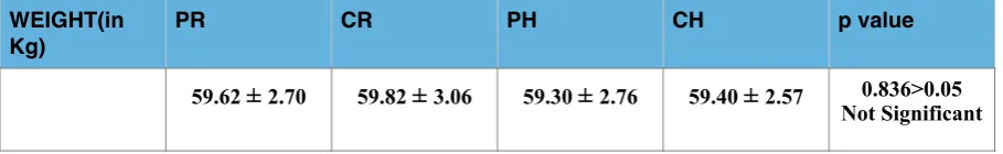
Table 3 : MEAN WEIGHT
The mean weight of the patients in the four groups is shown in the table 3
and graph 3 . The differences among the four groups were found to be
statistically insignificant (p=0.836).
Graph 3 : MEAN WEIGHT
Weight (in Kg)
WEIGHT(in Kg)
PR CR PH CH p value
59.62 ± 2.70 59.82 ± 3.06 59.30 ± 2.76 59.40 ± 2.57 0.836>0.05 Not Significant
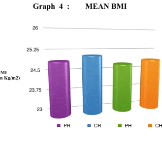
[image:71.595.72.527.188.258.2]BMI
Table 4 : MEAN BMI
Mean BMI among the four groups were compared as shown in
table 4 & graph 4. The differences were statistically insignificant( p = 0.702)
Graph 4 : MEAN BMI
BMI (in Kg/m2)
BMI( in Kg/ m2)
PR CR PH CH p value
24.87 ± 1.17 25.05 ± 1.39 24.72 ± 1.13 24.85 ± 1.21 0.702>0.05 Not Significant.
[image:72.595.113.443.410.697.2]MAXIMAL DERMATOMAL LEVEL OF SENSORY
BLOCK
Table 4 : LEVEL OF SENSORY BLOCK
The maximal level of sensory blockade to pin prick sensation were
compared among the four groups and was found to be in fourth thoracic
dermatome.The results were found to be statistically insignificant, ( p = 0.696 ) .
Level of block PR CR PH CH p value
Thoracic
Dermatome 3.97 ± 0 .42 4.02 ± 0.15 3.95 ± 0.22 3.97± 0.27
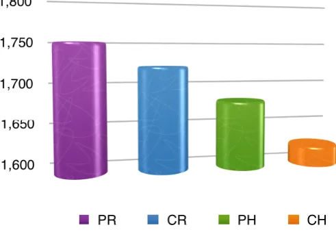
[image:73.595.73.528.213.270.2]TOTAL INTRAVENOUS FLUIDS USED
Table 5 : TOTAL IV FLUIDS USAGE
The total volume of intravenous fluid used were compared among
the four groups. Group PR required more amount of volume - 1748.7500 ±
159.92286 ml compared to Group CH - 1623.9750 ± 118.12784 ml which was
found to be statistically significant, ( p = 0.017) .
Graph 4 :
TOTAL IV FLUIDS USAGE
TOAL IV FLUIDS volume (in ml)
PR CR PH CH p value
1748.7500 ±
159.92286
1721.2500 ±
113.16399
1680.0000 ±
153.50603
1623.9750 ±
118.12784 0.017
HAEMODYNAMIC VARIABLES
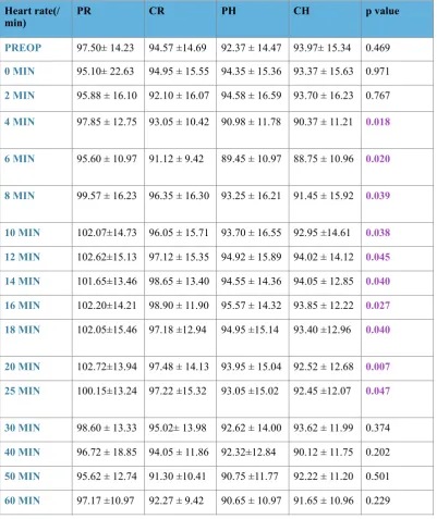
HEART RATE
Table 6 : MEAN HEART RATE
Volume (in ml)
Heart rate(/
min) PR CR PH CH p value
PREOP 97.50± 14.23 94.57 ±14.69 92.37 ± 14.47 93.97± 15.34 0.469
0 MIN 95.10± 22.63 94.95 ± 15.55 94.35 ± 15.36 93.37 ± 15.63 0.971
2 MIN 95.88 ± 16.10 92.10 ± 16.07 94.58 ± 16.59 93.70 ± 16.23 0.767
4 MIN 97.85 ± 12.75 93.05 ± 10.42 90.98 ± 11.78 90.37 ± 11.21 0.018
6 MIN 95.60 ± 10.97 91.12 ± 9.42 89.45 ± 10.97 88.75 ± 10.96 0.020
8 MIN 99.57 ± 16.23 96.35 ± 16.30 93.25 ± 16.21 91.45 ± 15.92 0.039
10 MIN 102.07±14.73 96.05 ± 15.71 93.70 ± 16.55 92.95 ±14.61 0.038
12 MIN 102.62±15.13 97.12 ± 15.35 94.92 ± 15.89 94.02 ± 14.12 0.045
14 MIN 101.65±13.46 98.65 ± 13.40 94.55 ± 14.36 94.05 ± 12.85 0.040
16 MIN 102.20±14.21 98.90 ± 11.90 95.57 ± 14.32 93.85 ± 12.22 0.027
18 MIN 102.05±15.46 97.18 ±12.94 94.95 ±15.14 93.40 ±12.96 0.040
20 MIN 102.72±13.94 97.48 ± 14.13 93.95 ± 15.04 92.52 ± 12.68 0.007
25 MIN 100.15±13.24 97.22 ±15.32 93.05 ±15.02 92.45 ±12.07 0.047
30 MIN 98.60 ± 13.33 95.02± 13.98 92.62 ± 14.00 93.62 ± 11.99 0.374
40 MIN 96.72 ± 18.85 94.05 ± 11.86 92.32±12.84 90.12 ± 11.75 0.202
50 MIN 95.62 ± 12.74 91.30 ±10.41 90.75 ±11.77 92.22 ± 11.20 0.501
[image:75.595.109.511.258.736.2]Graph 5 : MEAN HEART RATE
The mean heart rate of all the four groups were compared at the
preset intervals designed for this study.The differences in the mean heart rate
among the four groups were found to be statistically significant at
4 min (p = 0.018) , 6 min (p = 0.020) , 8 min (p = 0.039) , 10 min (p =0.038 ) ,
12 min (p = 0.045), 14 min (p =0.040 ), 16 min (p = 0.027), 18 min (p =0.040 ),
20 min (p = 0.007) , 25 min (p =0.047 ).
H
e
a
rt
ra
te
/mi
n
85 90 95 100 105 110
time (min)
PREOP 0 MIN 2 MIN 4 MIN 6 MIN 8 MIN 10 MIN 12 MIN 14 MIN 16 MIN 18 MIN 20 MIN 25 MIN 30 MIN 40 MIN 50 MIN 60 MIN
MEAN ARTERIAL PRESSURE
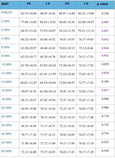
Table 7 : MEAN ARTERIAL PRESSURE
MAP PR CR PH CH p value
PREOP 82.57±10.88 86.85 ±9.26 88.97 ± 6.80 86.22 ± 9.06 0.194
0 MIN 77.90 ± 8.85 84.10 ± 8.82 86.05 ±9.38 82.80±10.87 0.005
2 MIN 64.47±13.26 73.97±10.87 76.32±12.39 79.22 ±11.33 0.047
4 MIN 60.32±10.91 65.60 ±9.52 76.67 ±9.93 76.17 ±9.83 0.042
6 MIN 63.20±10.97 68.40 ±8.43 74.82±10.15 75.12±8.64 0.048
8 MIN 62.52±10.77 65.20 ±9.78 74.87 ±9.31 74.12 ±7.51 0.042
10 MIN 62.70±10.54 67.07±10.24 73.30±10.53 74.32 ±7.92 0.059
12 MIN 59.37±13.32 65.10 ±11.95 73.22±10.85 73.85 ±8.71 0.028
14 MIN 60.62 ±11.07 64.55±10.84 72.85±10.97 72.77 ±7.43 0.186
16 MIN 59.07 ±9.76 63.20±10.14 70.47 ±9.74 72.92 ±7.63 0.037
18 MIN 64.72 ±9.67 67.02 ±9.94 72.57 ±9.10 72.67 ±7.24 0.960
20 MIN 65.95 ±9.00 70.32 ±9.56 72.22 ±8.77 74.05 ±7.92 0.906
25 MIN 68.97 ±9.90 70.15 ±9.69 72.32 ±9.19 73.27 ±7.40 0.710
30 MIN 69.35 ±8.99 71.17 ±8.71 72.12 ±8.42 75.52 ±6.65 0.735
40 MIN 70.77 ±7.36 71.57 ±8.12 74.82 ±8.69 76.07 ±7.94 0.759
50 MIN 71.90 ±6.68 72.72 ±7.09 74.17 ±7.80 76.42 ±7.10 0.525
[image:77.595.134.503.206.714.2]Graph 6 : MEAN ARTERIAL PRESSURE
The mean arterial pressure was compared among the four groups at the
pre-set time intervals for this study. The differences among the four groups were
statistically significant at 0 min ( p =0.005) , 2 min ( p =0.047), 4 min ( p
= 0.042), 6 min ( p =0.048) , 8 min ( p =0.042) , 12 min ( p =0.028 ) & 16 min (
p =0.037).
MA
P
(m
m
o
f
H
g
)
55 65 75 85 95
Time(min)
PREOP 0 MIN 2 MIN 4 MIN 6 MIN 8 MIN 10 MIN 12 MIN 14 MIN 16 MIN 18 MIN 20 MIN 25 MIN 30 MIN 40 MIN 50 MIN 60 MIN
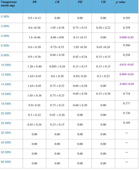
VASOPRESSOR USE
Table 8 : MEAN BOLUS DOSE OF VASOPRESSOR USAGE
Vasopressor
use(in mg) PR CR PH CH p value
0 MIN 0.9 ± 0.15 0.00 0.00 0.00 0.395
2 MIN 0.6 ±0.30 1.05 ± 0.38 0.75 ± 0.33 0.30 ± 0.22 0.359
4 MIN 1.8 ±0.46 0.60 ±.030 0.15 ±0.15 0.00 0.000<0.05
6 MIN 0.6 ± 0.30 0.75± 0.33 1.05 ±0.38 0.45 ±0.26 0.560
8 MIN 0.9 ± 0.36 0.60 ± 0.30 0.45 ± 0.26 0.15 ± 0.15 0.258
10 MIN 1.20 ± 0.40 0.045 ± 0.26 0.15 ± 0.15 0.15 ± 0.15 0.011<0.05
12 MIN 1.65± 0.45 0.6 ± 0.30 0.45± 0.26 0.3 ± 0.22 0.009<0.05
14 MIN 1.65± 0.45 0.75 ± 0.33 0.60 ± 0.30 0.00 .0.002<0.05
16 MIN 1.05 ± 0.38 0.75 ± 0.33 0.60 ± 0.30 0.15 ± 0.30 0.718
18 MIN 0.9± 0.26 0.75 ± 0.33 0.60 ± 0.30 0.00 0.171
20 MIN 0.3 ± 0.22 0.45 ± 0.26 0.00 0.00 0.136
25 MIN 0.45 ± 0.26 0.15 ± 0.15 0.00 0.00 0.105
30 MIN 0.00 0.00 0.00 0.00 —
40 MIN 0.00 0.00 0.00 0.00 —
50 MIN 0.00 0.00 0.00 0.00 —
Graph 7 : MEAN VASOPRESSOR USE
The mean bolus dose usage of the vasopressor ephedrine - 6mg was
compared among the four groups, it was found to be statistically significant at 4
min - (p = 0.000) , 10 min - (p = 0.011) , 12min -(p = 0.009) & at 14 min -(p =
0.002).
Graph 8 : Mean % use of ephedrine
me
a
n
e
p
h
e
d
ri
n
e
u
se
(i
n
mg
)
0.00 0.50 1.00 1.50 2.00
time (in min)
0 MIN 2 MIN 4 MIN 6 MIN 8 MIN 10 MIN 12 MIN 14 MIN 16 MIN 18 MIN 20 MIN 25 MIN 30 MIN 40 MIN 50 MIN 60 MIN
PR CR PH CH
TOTAL VASOPRESSOR USAGE
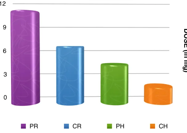
Table 9: TOTAL VASOPRESSOR USAGE
When the total usage of vasopressor was compared among all the
4 groups, group PR had highest mean usage (10.62 ± 7.86 mg) compared to all the other three groups and group CH had lowest mean usage scores ( 1.92 ± 3.12 mg ) and the results were statistically significant, p = 0.000.
Graph 9 : TOTAL VASOPRESSOR USE
Vasopressor
Usage(in mg) PR CR PH CH p value
10.62 ± 7.86 6.6 ± 5.58 4.5 ± 4.44 1.92 ± 3.12 0.000
[image:81.595.144.448.523.738.2]Graph 10 : % TOTAL EPHEDRINE USE
When % total ephedrine use among each group was compared,
greatest usage was found to be in the Group PR (45%) & least in Group
CH(8%) , the differences among the groups were statistically significant with p = 0.000 .
SIDE-EFFECTS
Table 10 : SIDE-EFFECTS
The incidence of side-effects such as nausea, vomiting ,hypotension were
more common among the Group PR & Group CR compared to the Groups PH & CH, with p value of 0.017,0.004 & 0.025 respectively.
SIDE EFFECTS PR CR PH CH
NAUSEA 40% 20% 10% 2.5%
VOMITING 30% 15% 10% 2.5%
HYPOTENSION 77.5% 72.5% 60% 47.5%
RESPIRATORY DEPRESSION
5% 2.5% 2.5% 0
EXCESS BLEED 0 0 0 0
ALLERGIC REACTIONS
0 0 0 0
PRURITUS 0 0 0 0
NEONATAL OUTCOME
Table 11: APGAR SCORE
The APGAR Scores were compared among the 4 groups and the
results were statistically insignificant.
APGAR Score PR CR PH CH p value
DISCUSSION
Spinal anaesthesia is frequently used for caesarean section for its
rapid onset, dense neural blockade and little risk of anaesthetic toxicity and
minimum transfer of