“COMPARATIVE ANALYSIS OF FUNCTIONAL OUTCOME OF
MULTIPLE METATARSAL FRACTURES TREATED WITH PLATE OSTEOSYNTHESIS AND KIRSCHNER WIRE FIXATION”
Dissertation submitted in partial fulfillment of the regulation for the award of M.S. Degree in Orthopaedic Surgery
Branch II
THE TAMILNADU
Dr. M. G. R. MEDICAL UNIVERSITY CHENNAI – 600 032.
APRIL - 2018
CERTIFICATE
This is to certify that the work “COMPARATIVE ANALYSIS OF FUNCTIONAL OUTCOME OF MULTIPLE METATARSAL
FRACTURES TREATED WITH PLATE OSTEOSYNTHESIS AND KIRSCHNER WIRE FIXATION”which is being submitted for M.S.
Orthopaedics, is a bonafide work of Dr.C. CHANDRU, Post Graduate Student at Department of Orthopaedics, Madurai Medical College, Madurai.
The Dean ,
CERTIFICATE
This is to certify that this dissertation titled“COMPARATIVE ANALYSIS OF FUNCTIONAL OUTCOME OF MULTIPLE
METATARSAL FRACTURES TREATED WITH PLATE
OSTEOSYNTHESIS AND KIRSCHNER WIRE
FIXATION”isabonafidework done by
Dr.C. CHANDRU postgraduate student of Madurai Medical College, Govt.
Rajaji Hospital.
Prof.Dr.P.V.PUGALENTHI, M.S Ortho.D.Ortho Professor and Head,
Department of Orthopaedics &Traumatology MaduraiMedicalCollege,
CERTIFICATE
This is to certify that this dissertation “COMPARATIVE ANALYSIS OF FUNCTIONAL OUTCOME OF MULTIPLE METATARSAL FRACTURES TREATED WITH PLATE OSTEOSYNTHESIS AND KIRSCHNER WIRE FIXATION” is the bonafide work done byDr. C. CHANDRU under my direct guidance and supervision in the Department of
Orthopaedic Surgery, Madurai MedicalCollege, Madurai-20.
Prof. Dr. V.R. GANESAN M.S Ortho., D. Ortho Professor and Chief Ortho unit-IV
Department of Orthopaedics &Traumatology MaduraiMedicalCollege,
ACKNOWLEDGEMENT
I am grateful to Prof.Dr.P.V. PUGALENTHI , M.S., Ortho, D.Ortho., Professor and Head, Department of Orthopaedic Surgery and Traumatology,
MaduraiMedicalCollege in guiding me to prepare this dissertation.
I am greatly indebted and thankful to my beloved chief, and my guide
Prof.DR. V.R. GANESAN, M.S.,Ortho, D.Ortho., Ortho-IV unit, Department
of Orthopaedic Surgery and Traumatology, Madurai Medical College for his
invaluable help, encouragement and guidance rendered to me in preparing this
dissertation.
I am most indebted and take immense pleasure in expressing my deep
sense of
gratitudetoProf.Dr.R.ArivasanM.S.Ortho.,Prof.Dr.R.SivakumarM.S.Ortho
.,D.ortho.,Prof.Dr.B.SivakumarM.S.Ortho.,D.Ortho and Prof.Dr.N.ThanappanM.S.Ortho for their easy accessibility and timely suggestion, which enabled me to bring out this dissertation.
I would like to thank PROF.Dr.D.MARUDHUPANDIYANM.S,the Dean, Madurai Medical College and Govt. RajajiHospital, Madurai for
permitting me to carry out this study in this hospital.
I take immense pleasure to thank my co-guide Dr. R. ASHOK
I also take this opportunity to thank
Dr.K.RavichandranM.S.Ortho., Dr.RamanathanM.S.Ortho.,
Dr.M.N.KarthiM.S.Ortho., Dr.K.P.SaravanakumarM.S.Ortho., Dr.J.MaheswaranM.S.Ortho., Dr.T.SaravanaMuthuM.S.Ortho.,
Dr.V.A.PrabhuM.S.Ortho., Dr.R.KarthikRajaM.S.Ortho., Dr.SenthilKumarM.S.Ortho., Dr.S.MadhuM.S.Ortho.,
Dr.GopiManoharDNBortho., DR.Gokulnath M.S Ortho DR.Anbarasan M.S Ortho DR.Karthikeyan M.S Ortho
DR.Singaravelu M.S Ortho
Assistant Professors, Department of Orthopaedics, Madurai Medical College,
for their timely help and guidance given to me during all stages of the study.
DECLARATION
I, Dr.C.CHANDRU, solemnly declare that the dissertation titled“COMPARATIVE ANALYSIS OF FUNCTIONAL OUTCOME OF MULTIPLE METATARSAL FRACTURES TREATED WITH PLATE OSTEOSYNTHESIS AND KIRSCHNER WIRE FIXATION”has been
prepared by me. This is submitted to “The Tamil Nadu Dr.M.G.R.MedicalUniversity, Chennai, in partial fulfilment of the regulations for the award of M S degree branch II Orthopaedics.
.
Place : Dr.C. CHANDRU,
Date : Post Graduate in Orthopaedics,
MaduraiMedicalCollegeHospital,
CONTENTS
PART A
CONTENTS PAGE NO
INTRODUCTION 10
REVIEW OF LITERATURE 15
EPIDEMIOLOGY 17
FUNTIONAL ANATOMY 21
CLINICAL PRESENTATION 31
DIAGNOSTIC PROCEDURES 32
PART –B
CONTENTS PAGE NO
METHODOLOGY 40
CASES 50
STATISTICS 77
OBSERVATIONS AND RESULTS 86
DISCUSSION 86
CONCLUSION 89
ANNEXURES :
a. BIBLIOGRAPHY
b. PATIENT PROFORMA
c. CONSENT FORM
d. MASTER CHART
e. ETHICAL COMMITTEE APPROVAL
INTRODUCTION:
The human foot is a highly developed, biomechanically complex
structure that serves to bear the weight of the body as well as forces many times
the weight of the human body during propulsion.
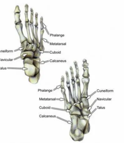
About 26 bones in the human foot provide structural support. They can be
grouped into 3 parts, as follows :
The tarsal bones
The metatarsal bones
The phalanges
Apart from these main bones, the sesamoid bones help to improve function and
are often found as variants of the accessory bones.
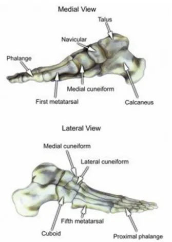
The foot itself can be divided into 3 parts: the hindfoot, the midfoot, and the
forefoot. The hindfoot is composed of 2 of the 7 tarsal bones, the talus, and the
calcaneus; the midfoot contains the rest of the tarsal bones; and the forefoot
The images below depict the bones of the foot. (Fig 1 and fig 2)
The modern understanding of the evolutionary anatomy ofthe human foot
comprises features such as:
(1) a long Achilles tendon to reduce stress and improve energy efficiency;
(2) apassively stabilized longitudinal plantar arch to improve shock absorption
and plantar flexion;
(3) an enlarged calcaneal tuberosity for stress reduction,
(4) a close-packed positioning of the calcaneo-cuboidal joint to improve spring
effectiveness of the plantar arch during running;
(5) the permanent inability to oppose the hallux to increase stability during
plantar flexion;
(6) relatively short phalanges adding to improved lever function.
Based on the functional anatomy of the foot: two segments comprising the
longitudinal arch
1. medial column of the foot (calcaneus, talus, tarsal navicular, cuneiform 1
to 3, and metatarsal 1 to3)
2. lateral column of the foot (calcaneus, cuboid, and metatarsals 4 and 5
Although foot injuries are not life threatening, they have significant impact on
Fractures of the forefoot are common and may result in significant sequelae.
The forefoot as unit provides a broad plantar surface for load sharing.
This platform also is structured to be mobile in the sagittal plane and provides
the forefoot with the ability to alter the position of the individual metatarsal
heads to accommodate uneven ground.
Therefore, injuries to this area can lead to difficulties with ambulation and gait.
Although the forefoot appears to work as a single unit, its parts are distinctly
LITERATURE REVIEW:
In 1995 MK O'Shea et al[1] presented a retrospective study of
fifth metatarsal fractures including Jones fractures, avulsion fractures, spiral
and oblique midshaft fractures, and the author-termed "tulip" fracture
(impaction fracture of the fifth metatarsal head). These fractures were fixed with
the cannulated screw, Kirschner wires, and circlage loop wires combined with
Kirschner wires. A one-way analysis of variance (ANOVA) was performed on
the data to test for any significant difference in the fixation type used and the
overall healing time. The ANOVA was found to be nonsignificant, F(2,10) =
0.379, p < 0.05. Therefore, it can be concluded that all three types of fixation
work equally well.
In 1996 Martin J. O'Malley[2],et al reported Spiral fractures of the distal
shaft of the fifth metatarsal are common injuries and can usually be treated
nonoperatively for these high performance athletes without long-term functional
sequelae.
In 2012 Hyong-Nyun Kim, MD et al[3] reported Closed antegrade
displacedmetatarsal fractures and to allow immediate joint motion and partial
weightbearing in a stiff-soled shoe.
In 2012Daniel Baumfeld et al[4]: described Anterograde percutaneous
treatment of lesser metatarsal fractures. They concluded
thatPercutaneousantegrade surgical treatment is an effective alternative to other
types of treatment for lateral metatarsal fractures, with a lower incidence of
complications.
In 2016 Mahan, Susan T et al[5]study was to review multiple metatarsal
fractures to help refine surgical indications. A total of 98 patients had multiple
metatarsal fractures; displacement greater than 10% shaft width (displaced) was
encountered in 33 (34.0%) patients. Fifteen patients had displacement of more
than 75% shaft width of one metatarsal. Patients older than 14 years of age were
more likely to have surgery for their injury (52.6%) than those younger than 14
years of age (3.7%) (P<0.0001). Younger patients and those with less than 75%
METATARSAL FRACTURES
Definition:
A metatarsal bone fracture is a complete or incomplete break in one of the
five metatarsal bones in each foot. These long thin bones are located
between the toes and the ankle (between the tarsal bones in the hindfoot and
the phalanges in the forefoot).
Epidemiology:
The metatarsals are a common fracture site in the body and account for 35% of
all foot fractures [6].Metatarsal fractures occur most often in patients between 20
and 50 years of age.5 to 6% of fractures treated in primary care are
metatarsal fractures. These are the most common injuries of the foot. They
are about ten times as frequent as Lisfranc-dislocations.They are equally
among men and women .
The distribution of the fractures[6]:
First metatarsal: 5%
Second metatarsal: 12%
Fourth metatarsal: 13%
Fifth metatarsal: 56%
Multiple metatarsal fractures: 15.6%
Metatarsal fractures are common in the pediatric age group, accounting for
close to 60% of all pediatric foot fractures[7]. The most common involved
fracture in childhood is fifth metatarsal [8]followed by the third metatarsal.
The lowest rate is in first metatarsal. Children age below the 5 years are
more likely to have first metatarsal fractures, with a frequency of isolated
first metatarsal fractures of 51%, in contrast to those more than 5 years old,
who are more likely to have fifth metatarsal fractures, depending on the age
group, a frequency as high as 65%[8]. The next most common fracture
finding was a specific combination of second, third and fourth metatarsal
fractures[8][9][10][11]..
Metatarsal fractures may result from direct or indirect violence, and they
display a wide variety of injuries ranging from isolated, simple fractures of
one metatarsal to crush injuries with serial fractures and severe soft tissue
compromise. Direct trauma is common in industrial workers where they
have a heavy object fall on the foot. Indirect trauma happened when the leg
Injury to the metatarsals is common in acute and chronic settings and they
are the most common site for stress fractures in the human
skeleton[12][13][14][15].. Among stress fractures of the metatarsal bones, the
middle and the distal portions of the corpus ossismetatarsalis II or III are
most common. Stress fractures at the base of the first or second metatarsals
are less common[16].. Metatarsal stress fractures are a common in athletes,
particularly in runners, in whom they account for 20% of lower extremity
stress fractures. Increased stresses over the second and third metatarsals
during walking and running, these metatarsals are at greatest risk for stress
fracture.[17][18].
The percentages ofinjuries as follows[6]:
Supination injury: 48%
Fall from height: 26%
Crush injury: 12%
Athletes, individuals who are obese, and with osteoporosis or rheumatoid
It also occurs in sports like jogging, ballet, gymnastics, and high-impact
aerobic activities[18]. Shoe shock attenuation will prevent the metatarsal
stress fractures[19].
It has been shown that the fracture pattern and severity of injury vary
according to age and mechanism of injury[20].This association can further
be correlated with both osseous development and the age-related levels of
activity[10].
The metatarsal can be fractured at 3 locations: on the caput, corpus or on
the basis ossis metatarsalis. Like that we can differentiate multiple different
fractures:
Sub capital fracture.
Fracture of the corpus ossis metatarsalis.
Fracture of the basis ossis metatarsalis.
The arterial arch and the dorsal and plantar metatarsal arteries are more
susceptible to injuries in associated with metatarsal fractures. Compartment
FUNCTIONAL ANATOMY:
First Metatarsal Fractures
Pathoanatomy and Applied Anatomy Relating to
First Metatarsal Fractures
The first metatarsal is stronger than the other 4 metatarsals. It accounts for
1.5% of all metatarsal fractures[21].. Its has large cross-sectional geometric
properties and its role as the preferred ray for loading during walking, running
or turning in a different direction[22].. Its configuration is shorter and wider than
the lesser four metatarsals. The lack of interconnecting ligaments allows
independent motion between the first and second metatarsal.
There are two powerful muscle attachments to its base. The tibialis anterior
inserted on the plantar surface of medial aspect of the first metatarsal base and
the peroneus longus inserted onto the plantar surface lateral base of the first
metatarsal. These muscles exert significant influence on the position of the
head of 1st metatarsal. Averagepeak pressures in metatarsal 1 are the highest or
among the highestlevels of pressure during most activities[23].
First Metatarsal Injury Mechanisms:
First metatarsal fractures can occur from direct or indirect forces. Direct injuries
are common in industrial workers. heavy object falling on the foot. Indirect
First Metatarsal Fracture Imaging:
Three xrayviews are mandatory to judge
shortening,
deviation
angulation,
displacement
intra articular fracture lines and fragments can be seen through computed
tomography. MRI needed for additional information related to soft tissue
injuries.
First Metatarsal Fracture Classification:
The OTA classification:
TYPE A: extra articular simple fractures
TYPE B: partial articular involvement or wedge fracture of shaft
TYPE C: complete articular involvement or comminuted shaft fractures
First Metatarsal Fracture Treatment Options:
Nonoperative Treatment:
No evidence of instability on stress films of the fracture, and no other injuries of
the midfoot and metatarsals and isolated minimally displaced first metatarsal
fractures can be treated non operatively byshort-leg cast with non weight
Operative Treatment of First Metatarsal Fractures.:
Evidence of instability or loss of position of the metatarsal head should be
treated with operative stabilization.
Goal of the treatment:
To restore and maintain the normal position of thatmetatarsal head, the
sesamoids, and metatarsophalangeal joint.
The method of fracture fixation is dependent on fracture configuration.
Percutaneous smooth wires: simple and reducible fractures
Open reduction and internal fixation is needed for: displaced fractures Simple
spiral or oblique fractures: open reduction and lag screw fixation with 2.7mm
screws.
If tarso metatarsal joints are unstable can betreated with contoured one
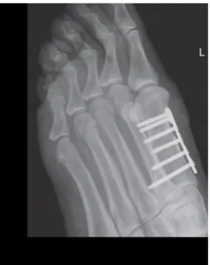
third plate extend between medial cuneiform and distal intact metatarsal shaft.
Plate and screw fixation used for transverse or minimally comminuted fractures
(fig 3). in which inadequate fixation will occur with screws or wires alone.
External fixation can be considered if there is severe midshaft or head
comminution or open injuries(fig 4). These fracture types usually have
Fig 3: : 1stmetatarsal shaft fractures treated with plating
CENTRAL METATARSAL FRACTURES:
Fractures of the central metatarsals account for approximately 10% of all
metatarsal fractures Fractures of the central metatarsals are much more common
than first metatarsal fractures[10].
Central metatarsal fractures can occur from direct or indirectforces. Direct
injuries are common in industrial workers and occur as a result of a heavy
object falling on the foot. Indirect injuries are seen in sport, when the forefoot is
fixed and the leg or foot is twisted Central metatarsal fractures also occur as
stress fractures. Central metatarsals fractures are commonly associated with
injuries to the first ray, and Lisfranc joint injuries.
The 2ndand 3rdmetatarsals are more important because they comprise the
keystone of the foot.The metatarsal bases are of trapezoidal shape. It forms a
“Roman arch” configuration. Base of each central metatarsal having series of
three ligaments (dorsal, central, and plantar), they stabilize and support each
with their neighbour.
MANAGEMENT:
Any fracture displacingmore than 10 degrees of deviation in the sagittal plane
or 3 to4 mm of translation in any plane should be actively corrected. Majority of
isolated central metatarsal fractures can be treated nonoperatively. Isolated
head or neck fractures that deviateeither dorsally or plantarly in the sagittal
the normal alignment. A stable fracture of the base of the third or fourth
metatarsal can be reduced closed without fixation. Hyperextended distal
metatarsal fractures may cause dislocation with the head comes through the
flexorplaten can prevent closed reduction.
OPERATIVE TREATMENT OF CENTRAL METATARSAL
FRACTURES:
Indications :
Unstable base fracture of the second metatarsal requires
Multiple adjacent metatarsal fractures
Severe comminuted fractures
Hyperextended neck fractures
Indication for surgery according to shereff:
Evaluation Parameters
Frontal plane > 3 to 4 mm of deviation
Sagittal plane Angulation > 10 degrees
Metatarsal formula Changes in the metatarsal parabola
Method of fixation based on fracture pattern:
Interfragmentary compression screw : Oblique fracture in diaphyseal region
Intramedullary K-wire fixation : If there is significant soft tissue injury or
open wound, K-wire fixation performed. K-wire fixation is also effective if
there is severe comminution of the shaft fractures . One should be careful not to
shorten the position of the head in relation to its neighbors.
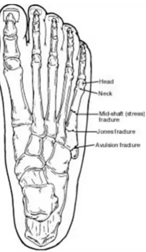
Fifth Metatarsal Fractures
Pathoanatomy and applied anatomy relating to fifth metatarsal fractures:
The base of the fifth metatarsal is a complex anatomic region withthe insertion
of three muscles. The peroneus brevis inserted on the dorsal aspect of the
tubercle of the fifth metatarsaland the peroneus tertius inserted on the dorsal
aspect at theproximalmetaphyseal-diaphysealjunction.Peroneustertius muscle
acts as a balancing force during forefoot dorsiflexioncounteracting the natural
inversion tendency of thetibialis anterior. The peroneus brevis act as more of an
antagonist to posterior tibialis function to maintain the position. The third one is
the abductor digitiquinti. It has a strong attachment of the plantar fascia to the
plantar aspect of the tubercle.
The blood supply to the proximal fifth metatarsal at the
Metaphyseal-diaphyseal junction has been implicated as the leadingcause for
the development of a delayed union or nonunion infractures of the proximal
fifth metatarsal. Fifth metatarsal fracture account for 68% of all metatarsal
Fig 5: Locations of fracture zones for proximal fifth metatarsal fractures
Fifth Metatarsal Fracture Injury Mechanisms:
Majority of injuries are due to twisting of thefoot or a fall from a standing
height.
zone 1 injury usually occurs from an indirect load. Sudden inversion of the
hindfootwithweight placed on the lateral metatarsalproduce tension along the
insertion of the lateral band of the plantar aponeurosis which insertsinto the
proximal base of the fifth metatarsal causing avulsion fracture.
Zone 2 injury is the true Jones fractures caused by adduction of the forefoot will
Zone 3 fracture seen in the proximal fifth metatarsalis now referred to as a
proximal diaphyseal stress fracture.These fractures are rare and seen mainly
inathletes. It mainlyoccurs in the proximal 1.5 cm of the metatarsal shaft.
SPECIFIC CAUSES OF FRACTURES:
Most fractures of the corpus ossismetatarsalis are caused by direct
blows or twisting forces. An abrupt increase in activity or chronic
overload may cause a stress fracture of the metatarsal corpus.
The most common mechanism of injury in fifth metatarsal fractures
involves a fall from standing height or an ankle twist with the forefoot
fixed.
An avulsion fracture of the fifth metatarsal base (‘tennis fracture’) may
occur as a result of inversion injuries to the foot, seen that the base of
the fifth metatarsal is the endpoint of the ‘supination fracture line’.
A tuberosity avulsion fracture usually results from ankle inversion
while the foot is in plantar flexion. The history often suggests a lateral
A diaphysial stress fracture is often due to a chronic overloading,
especially from jumping and pivoting activities in younger athletes.
Fractures from the first through the fourth metatarsals are the kind of
fractures that are less common than other metatarsal fractures. They
warrant special consideration, because they are often associated with
injury to the Lisfranc ligament complex. These crucial ligaments hold
the metatarsal bases rigidly in place, maintaining the arch of the foot
and anchoring the metatarsals to the rest of the body.
Proximal metatarsal fractures are usually caused by crush injuries or
direct blows. They may also result from falling forward over a
plantar-flexed foot. In athletes, the most common mechanism for a Lisfranc
Characteristics/ clinical presentation:
Symptoms and signs are:
Painful and swelling
Palpable crepitus
Axial pressure pain
Patients with metatarsal fractures complain of pain on ambulation and
inability to weight bear. The forefoot is swollen and tender to palpation.
Gross deformities are mainly present with complex injury patterns including
multiple fractures and additional toe dislocations.[23]
shaft fractures: Typically presents with pain, swelling, ecchymosis and
difficulty walking. Initially the pain only occurs with activity. Swelling is
severe if the patient has not elevated the foot. Point tenderness over the
fracture site. Applying an axial load to the head of a metatarsal usually
triggers pain at the injury site. Patients with soft tissue injuries shouldn’t
experience pain with this maneuver.
Tuberosity avulsion fracture, Jones fracture and diaphyseal stress fracture: These fractures cause lateral foot pain and difficulty walking.
Acute fractures also have a sudden onset of swelling and ecchymosis. Stress
with activity. Recognizing the gradual onset of symptoms is key to correctly
diagnosing fifth metatarsal stress fractures.[19]
Stress fractures: Early signs are: pain increased during activity that relieved with resting and pain over a wide area of the foot. Over time the
pain will be present constantly and stronger in one area of the foot. The area
of the foot where the fracture is may be tender when you touched. It might
be swollen as well.
Diagnostic Procedures
A physical examination of the foot with x-rays and bone scans are used to
diagnose the fractures of metatarsal. When the patient having a typical
history and appropriate physical findings, a presumptive clinical diagnosis
can be made. Routine X-rays (anteroposterior, lateral and oblique) are
sufficient to diagnose the fracture. A CT-scan or MRI is used to exclude
other injuries when it isnecessary. When a stress fracture is expected a bone
scan may be helpful.
Acute metatarsal fracture (fracture metatarsal shaft) Radiographic
findings: Fracture position and pattern can be assessed by two views that lie
at a 90° angle to each other. Oblique or modified lateral views are often
the clinical examination and the radiographs should be repeated after one to
two weeks of initial injury.
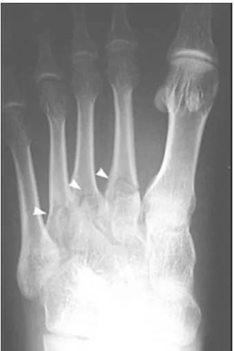
Fractures of the proximal first through fourth metatarsals (figure 6):
Radiographic findings: Proximal fractures are commonly transverse or
oblique and sometimes multiple. In case of Lisfranc ligament injury a
standard radiographic series may be normal in fifty percent of the patients.
In this case weight-bearing anteroposterior and lateral radiographs should
be taken: the anteroposterior view showswidening of the space between the
first and second metatarsal heads (stage II or III) with loss of arch height on
the lateral view in stage III injuries. Radionuclide bone scan: is accurate for
diagnosis in case of stage I injury with clinical suspicion and normal
Figure 6 : Nondisplaced fractures of the proximal portions of
Metatarsal 2 – Metatarsal 4.
Acute fractures of the proximal fifth diaphysis : Using the Ottawa ankle
rule we can exclude a lateral ankle sprain from a tuberosity avulsion
fracture. When point tenderness is present over the fifth metatarsal and the
foot appears to be normal, it could be a sprain.
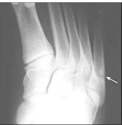
Jones fracture: fig 7: Radiographic findings: Acute fracture of junction
between the proximal diaphysis and the corpus ossis metatarsi quinti. The
fracture line is sharp and extends into the joint between metatarsal 4 and
Fig 7:Jones fracture
Tuberosity (styloid) fracture : (fig 8:)Clinical findings: A radiolucency is
seen perpendicular to the long axis of the 5thMetatarsal. The fracture may be
intraarticularorextraarticular (cuboid-metatarsal articulation) and never
extend into the joint between the fourth or fithmetatarsal (=different from
Jones fractures). It involves the tip of the styloid process at the attachment
of the plantar aponeurosis and peroneus brevis. The peroneus brevis tendon
Outcome measures:
1) Acute metatarsal fracture :
Favourable:
Displacement is often minimal unless more than 1 Metatarsal is
injured, Fractures of a single Metatarsal shaft with lateral or medial
displacement usually heal well without correction.[19]
Unfavourable:
Displacement of more than 3mm or 4mm displacement in a dorsal or
plantar direction or dorsal /plantar angulation exceeds 10 degrees
need reduction. Skin necrosis- due to crush injury- leading to an open
fracture.
2) Fractures of the proximal first through fourth metatarsalsInjury to
the Lisfranc ligament complex can cause chronic disability. [19]
3) Acute fractures of the proximal 5th diaphysis: Jones fractures
With conservative treatment, patients frequently complain about the
lenght of time for nonweight bearing and pain following the first
weeks of rehabilitation as delayed union is surprisingly common.
Complications like nonunion and refracture are reported in literature,
but often conservative treatment is the first option in common
4) Tuberosity (styloid) fracture :
Nondisplaced avulsion fractures usually heal well within 30 to 40
days with symptomatic therapy.
When the fracture is greater than 3 millimeters of displacement or a
step-off of more than one to two millimeters on the articular surface
with the cuboid, surgical treatment is needed. [19]
5)Stress fracture
Stress fractures of the metatarsal shaft usually heal well in case of
discontinuation of the causative activity for 4 to 8 weeks.After 4 to 8
CONSERVATIVE MANAGEMENT:
NON-DISPLACED FRACTURE OF THE CORPUS OSSIS
METATARSI:
- first 24 hours: ice and elevation (higher than the heart). Progressive weight
bearing and treating with soft elastic dressing or a firm supportive shoe
- immobilization in a posterior splint and 3 to 5 days non-weight bearing
DISPLACED METATARSAL FRACTURES:
Displaced more than 3 to 4mm in dorsal or plantar direction:
First 24 hours: ice and elevation (higher than the heart)
Reduction: under local anesthesia, using a regional or hematoma block.
Placing the toes in Chinese finger traps and allowing gravity to accomplish
the reduction. The reduction should be maintained in a molded, bivalve,
below knee cast and postreduction radiographs should be obtained to
confirm proper alignment.
Stress fractures of the metatarsal shaft:
Responds well to cessation of the causative activity (4 to 8 weeks)
Walking causes pain? Crutches and partial-weight bearing
Walking causes severe pain? Non weight-bearing, short leg cast (1 to 3
METATARSAL 5 fracture:
Tuberosity Avulsion:
Symptomatic therapy (3 to 6 weeks)
- soft protective dressing
- short leg cast
Too symptomatic
- a hard-soled shoe or wood-soled postoperative brace or cast
Jones fracture = dancer’s fracture:
- non-weight bearing cast (6 to 8 weeks), crutches are required
METHODOLOGY:
AIM OF THE STUDY:
To compare and evaluate the results of surgical treatment of multiple
metatarsal fractures (> 1 Metatarsal involved) using “Kirschner” wire
fixation and plate osteosynthesis
INCLUSION CRITERIA:
Multiple metatarsal fractures- > 1 Metatarsal involved
Simple fracture
Open fractures grade I
Age More than 16 yrs
EXCLUSION CRITERIA:
Grade II and III open fractures
Single metatarsal
Un co-operative pts(mentally ill)
Associated lower limb long bone fractures
Age less than 16 yrs
MATERIALS AND METHODS
In this study, we have operated on 15 patients after excluding patients according
In this study we selected the patients through randomization by tossing a coin.
After randomization, patients were assigned to the 2 groups – one group
undergoing open reduction and internal fixation with plate osteosynthesis; other
group undergoing Kirschner-wire fixation.
Informed consent was taken from all patients. Surgery was done electively after
assessment under regional anaesthesia. All cases were taken up for surgery
immediately following admission.
Source of Data
Patients with multiple metatarsal fractures simple and compound grade
admitted at Govt. Rajaji hospital in the department of orthopaedics&
traumatology Madurai were taken up for study after obtaining informed
consent. All the patients selected for study were examined according to
protocol, associated injuries were noted and clinical and lab
investigations carried out in order to get fitness for surgery. Consent of
functional out come is achievedClinicalyas well as Radiologically. 15 cases werestudied.
Pre operative preparation: Patients underwent a pre-operative
evaluation including the following parameters : Hb, blood sugar, ECG,
RFT ,x ray chest inorder to get fitness for surgery
FOLLOW UP PERIOD:
AT 3 days
AT 2 weeks
AT 1 ½ month
AT 3 month
AT 6 months
AT 12 months
Kirshnerwire removal at the period of 6 – 10 weeks
IMPLANTS AND INSTRUMENTS:
Anaesthesia:
General anaesthesia or spinal anaesthesia or ankle block
surgical technique:
Antegrade kirshner wire fixation under radiological control.
Open reduction and internal fixation Mini plate system.
surgical approach:
APPROACH- DORSAL APPROACH FOR METATARSALS
The veins are superficial and should be preserved
The approach is in between the long and the short extensor tendons,
staying lateral to the EDL.
Skin incision
The skin incision is made in line with the first ray, starting over the
medial cuneiform and extending to the dorsolateral aspect of the
first proximal phalanx.
Deep dissection
Expose the first metatarsal between the tendons of extensor
hallucislongus and hallucisbrevis
Take care to protect the dorsalispedis artery and the cutaneous
branches of the deep peroneal nerve.
Dorsal approach to 2nd 3rd 4thmetatarsals: Skin incision
Make a longitudinal incision between the second and the third
metatarsal extending it from the metatarso-phalangeal to the
tarsometatarsal joint.
Make a longitudinal incision along the dorsolateral aspect of the 4th
metatarsal, from the head to the tarso-metatarsal joint.
Multiple incisions:
If all lesser metatarsals (2, 3, 4, and 5) are to be approached, we
would advocate three incisions: one between the 2nd and 3rd, the
DORSAL INTERMETATARSAL APPROACH
Deep dissection
Approach goes in-between the long and the short extensor tendon of
the corresponding ray.
Should protect the intermetatarsal nerves and the crossing superficial
veins
For this approach no muscle must be incised. Eventually, the
DEEP DESSECTION
Lateral approach to the 5th metatarsal:
Skin incision:
The incision is made at the junction of the dorsal skin and the plantar
skin
The skin incision starts just proximal to the styloid process of the
base of the fifth metatarsal and proceeds distally, as far as required.
Deep dissection:
Expose of the fascia over the abductor digiti muscle belly, and incise
it longitudinally.
Retract the skin and fascia dorsally, and the muscle belly in a plantar
direction, exposing the underlying fifth metatarsal.
AOFAS MID FOOT SCALE (100 points total)
(AMERICAN ORTHOPAEDIC FOOT AND ANKLE SCORE)
The surveys include a mixture of questions that are both subjective and
objective in nature. The pain category which asks patients a single question
about their level of pain is subjective, while the alignment category (to be
answered by the physician) is objective.
EXCELLENT = 90 -100 POINTS
GOOD = 90-80 POINTS
FAIR = 80- 70 POINTS
PAIN (40 points)
None 40
Mild, occasional 30
Moderate daily 20
Severe almost always present 0
FUNCTIONS: (45 POINTS) Activity limitations , support:
No limitations ,no support 10
No limitations of daily activities, limitations of
recreational activities, no support 7
Limited daily and recreational activities, cane 4 Severe limitations of daily and recreational activities, walker, crutches , wheelchair 0 FOOT WEAR REQUIRMENTS:
Fashionable, conventional shoes, no insert required 5
Comfort footwear , shoe insert 3
Modified shoes or brace 0
MAXIMUM WALKING DISTANCE, BLOCKS
Greater than 6 10
4 – 6 7
1 – 3 4
Less than 1 0
WALKING SURFACES
No difficulty on any surface 10
Some difficulty on uneven terrain, staires, inclines,
ladder 5
Severe difficulty on unev0en terrain, staires,
inclines, ladder 0
GAIT ABNORMALITY
None , slight 10
Obvious 5
Marked 0
ALIGNMENT(15 points)
Good plantigrade foot, mid foot well alighned 15 Fair plantigrade foot. Some degree of midfootmalalignment observed no symptoms 8 Poor plantigrade foot, severe malalignmant,
CASES:
CASE 1:
SERIAL NO: 1
NAME: RAMAKRISHNAN
AGE/SEX: 18/M
DIAGNOSIS: # METATARSAL 2ND 3RDLEFT SIMPLE
Follow up xray:
CASE : 2
SERIAL NO: 2
NAME: RAMESH
AGE/SEX: 43/M
DIAGNOSIS: # METATARSAL 2ND 3RD RT SIMPLE
POST OP:
CASE :3
SERIAL NO: 3
Name : karupaya
Age /sex: 63/m
Diagnosis : # METATARSAL 2nd,3rd lt foot comp.
Preop xray:
Post op:
Postop clinical photo:
CASE:4
SERIAL NO: 5
Name : Eswari
Age/sex: 35/f
Diagnosis: # metatarsal 3rd 4th 5thcomp grade 1 right foot
Post op:
Clinical photo:
CASE: 5
SERIAL NO: 6
NAME: KALIDOSS
AGE/SEX: 23/M
PRE OP:
1 ½ MONTH POST OP:
Follow up xray:
:
Case :6
SERIAL NO: 7
Name : santhana pandy
Age /sex: 18/ m
Diagnosis : # metatarsal 1st2nd3rd left foot simple
Pre op xray:
Post op xray:
Follow up xray:
Case: 7
SERIAL NO: 8
NAME: MUTHURAJA
AGE: 51/M
DIAGNOSIS: # METATARSAL 3RD4TH COMP.
PRE OP: POST OP:
Case: 8
SERIAL NO: 9
Name :Aravindsamy
Age /sex: 23/m
Diagnosis : # metatarsal 2nd3rdsimple
Pre op:
Follow up xray:
CASE : 9
SERIAL NO: 10
NAME : CHOCKALINGAM
AGE/SEX: 40/M
DIAGNOSIS: # METATARSAL 2ND 3RDRT COMP.
PRE OP:
1 ½ MONTH FOLLOW UP:
6 month follow up:
CASE : 10
SERIAL NO: 11
Name : kannan
Age /sex: 32/m
Diagnosis : # metatarsal 1st2nd3rd4thcomp injury right
Pre op xray: post op xray:
Follow up xray:
STATISTICS
I. AGE DISTRIBUTION
Age in years No. of cases Percentage
<25 5 33
26 – 35 3 20
36-60 5 33
>60 2 14
0 1 2 3 4 5 6
<25 26 - 35 36 - 60 > 60
Age in years
II. SEX DISTRIBUTION
Sex No. of cases Percentage
MALE 14 93
FEMALE 1 7
TOTAL 15 100
sex distribution
III. MODE OF INJURY
Mode of injury No. of cases Percentage
RTA 12 80
ACCIDENTAL FALL 3 20
TOTAL 15 100
0 2 4 6 8 10 12 14
RTA ACCIDENTAL FALL
MODE OF INJURY
IV. SIDE OF INJURY
SIDE NO. PERCENTAGE
RIGHT 9 60%
LEFT 6 40%
TOTAL 15 100%
0 2 4 6 8 10
RIGHT LEFT
SIDE OF INJURY
V. METATARSALS INVOLVED
No of metatarsals involved
No. Percentage
2 9 60%
3 3 20%
4 3 20%
0 1 2 3 4 5 6 7 8 9
TWO MT THREE MT FOUR MT
Series 1
VII.PROCEDURE
SURGERY DONE NO.
ORIF WITH PLATE
OSTEOSYNTHESIS
7
KIRSCHNER WIRE FIXATION
8
6.4 6.6 6.8 7 7.2 7.4 7.6 7.8 8 8.2
ORIF K WIRE
PROCEDURE
VI. AOFAS SCORE AND OUTCOME
AOFAS SCORE FOLLOW UP
POOR FAIR GOOD EXCELLENT
1 2 M
3M 6M 1
2 M 3 M 6 M 1 2 M 3 M 6 M 1 2 M
3 M 6 M
ORIF 3 1 1 4 1 0 0 4 2 0 0 4
Kirschner-wire
4 0 0 4 2 0 0 6 2 0 0 6
P VALUE 0.38 (not significant)
AOFAS SCORE AT FINAL FOLLOW UP:
0 1 2 3 4 5 6 7
POOR FAIR GOOD EXCELLENT
VII. AOFAS SCORE AT FINAL FOLLOWUP
PROCEDURE MEAN SD P VALUE
Plate 76.57 3.93 0.38
Kirschner-wire 91.25 3.84
65 70 75 80 85 90 95
ORIF K-WIRE
MEAN
VIII. COMPLICATIONS
INFECTION – 3 TOTAL
PAIN - 0
RESTRICTION OF MOVEMENT – 0
LOSS OF REDUCTION - 0
COMPLICATIONS NO. OF CASES PERCENTAGE
ORIF 2 29%
KIRSCHNER WIRE
FIXATION
1 12%
p value 0.48 (not significant)
0% 5% 10% 15% 20% 25% 30% 35%
ORIF K WIRE
complications
RESULTS
In this study, 7 patients underwent ORIF and 8 underwent K-wire
fixation. The mean AOFAS score at final followupwaslower (76.57)in ORIF
group compared to a higher score of (91.25)in Kwire group. Using unpaired T
test, we tested for the level of significance by calculating p value. However
p-value for the final followup score was 0.38(not significant).
Complications rate was high in ORIF group(29%) compared to the other
group(12%). 2 patients in ORIF group got infected with one patient requiring
implant exit. Only one patient has superficial infection in K-wire group which
healed and had good outcome. Others like loss of reduction, metatarsalgia or
restricted movements were not found in either of the groups. Overall the scoring
was found better in K-wire group in our study.
DISCUSSION
Metatarsal fractures are the most common fractures involving the foot.
Multiple metatarsal fractures most commonly occurred due to high energy
trauma.This study was carried out after obtaining ethical committee clearance.
In this study, we found that most of the fractures were due to RTA and few due
to accidental fall. 80 % of cases occurs due to RTA and 20% of cases occurs
In our study males were most commonly affected. 93% of males were involved
and 7% of females were involved.
In our study particular age groups were not involved, all age groups were
involved. Less than 25 yrs age- 33% cases were involved. 26 to 35 yrs age- 20%
cases were involved. 36 to 60 yrs – 33% cases were involved and more than 60
yrs of age 14% cases were involved.
In our study right side of the foot was involved more, 60% of cases having
fracture in right foot and 40% of cases having fracture in left foot.
In our study 2 metatarsals were fixed with miniplate in single incision. So the
skin stretching was increased while doing surgery..In our study duration of
surgery was prolonged in ORIF with platinggroup compared with kirschner
wire fixation. So this is the reason for getting minimal infection in plating
group.
Kirschner wire was removed after 6 to 10 weeks of surgery. Plate was removed
or planned to be removed after 1 year of surgery.
In our study, we used AOFAS score was used for assessment of the outcome of
surgery. Scoring was done both before and after surgery. After surgery,
assessment done at 1 and half months, 3 months and 6 months as followup to
both groups. Though AOFAS score seemed better with k-wire group at initial
In our study, comparing the two groups using UNPAIRED T-TEST for
testing significance was found to be non-significant p-value=0.38 (>0.05).
Similarly, in study by MK O'Shea et al, a one-way analysis of variance (ANOVA) was performed on the data to test for any significant difference in the
fixation type used and the overall healing time. The ANOVA was found to be
nonsignificant, F(2,10) = 0.379, p >0.05.
Daniel Baumfeld et alconcludedthatPercutaneousantegrade surgical treatment is an effective alternative to other types of treatment for lateral
metatarsal fractures, with a lower incidence of complications. In our study also,
the overall complication rate was found to be lower in K-wire group (12%).
In our study the mean AOFAS score was found better in k-wire group.
Similarly, Hyong-Nyun Kim, MD et al reported Closed antegrade intramedullary pinning was found to be a useful method for treating displaced
metatarsal fractures and to allow immediate joint motion and partial
CONCLUSION
In our study simple and compound grade I multiple metatarsal
fractures treated at emergency within 24 hrs with open reduction and
internal fixation with plate osteosynthesis and kirschner wire stabilization.
Open reduction and internal fixation with plate osteosynthesis done
under tourniquet control. Kirschner wire stabilization done by closed
technique under c arm control.
Both treatment modalities equally good, achieving good fracture
union, decreased incidence of pain and achieve good range of movements
with minimum complication.
Eventhough we had a minimal complications in plating group, we
have achieved good union and excellent functional outcome at end of follow
up.
AOFAS score was used to assess the functional outcome in our study.
At the end of study AOFAS score was equal in both groups. So both
PROFORMA
Name : IP No. :
Age / sex : Occupation :
DOA:
DOS:
DOD:
Diagnosis :
AOFAS SCALE:
Nature of injury: Simple
Comp grade I
Associated injuries :
Postoperative follow up :
N
O NAME A/S Modeof injury Diagnos is Duration of surgery AOFAS score before surgery
Procedure follow up score
in months complications Remarks
1 Ramakri
shnan 18/m RTA #metatars al 2nd3rd simple
90 min 36 ORIF with plate osteosynthe sis
72 86 92 Nil Excellent
2 Ramesh 43/
m Accidental fall #metatars al 2nd3rd simple
95 min 32 ORIF with plate osteosynthe sis
74 82 90 Nil Excellent
3 Karupay
a 65/m RTA #metatars al 2nd3rd comp.
90 min 28 ORIF with plate osteosynthe sis
62 - - Infectio
ns Implantexit done
4 Manikan
dan 30/m RTA #metatars al 2nd3rd comp.
80 min 34 ORIF with plate osteosynthe sis
72 84 92 Nil Excellent
5 Eswari 35/
f RTA #metatars al 3rd 4th 5th comp
100 min 32 ORIF with plate osteosynthe sis
66 76 88 Nil Good
6 Kalidoss 24/
al 2nd3rd
simple osteosynthesis
7 Santhan
apandy 18/m RTA #metatars al 1st2nd 3rd simple
75 min 42 ORIF with plate osteosynthe sis
74 82 92 Nil Excellent
8 Muthura
ja 51/m Accidental fall #metatars al 4th5th simple
40 min 44 K wire
fixation 72 82 94 Nil Excellent
9 Arvinds
amy 23/m RTA #metatars al 2nd3rd comp.
45 min 36 K wire
fixation 64 88 94 Nil Excellent
10 Chockal
ingam 40/m RTA #metatars al 2nd3rd comp.
30 min 38 K wire
fixation 74 84 96 Nil Excellent
11 Kannan 32/
m RTA #metatars al 1st 2nd 3rd 4th simple
50 min 32 K wire
fixation 72 86 92 Nil Excellent
12 Kannan 40/
m RTA #metatars al 2nd3rd 4th5th comp.
50 min 42 K wire
fixation 66 72 88 Nil Good
13 Gopal 25/
m RTA #metatars al 3rd
45 min 36 K wire
4th5th comp. 14 Thattank
aruppan 59/m RTA #metatars al 2nd 3rd4th5th comp.
55 min 42 K wire
fixation 70 82 90 Nil Excellent
15 Minnagi
ri 63/m RTA #metatars al 1st2nd 3rd comp.
45 min 38 K wire
BIBILIOGRAPHY:
1. O'Shea MK, Spak W, Sant'Anna S, Johnson C. Clinical perspective of the
treatment of fifth metatarsal fractures. Journal of the American Podiatric
Medical Association. 1995 Sep;85(9):473-80.
2. O'Malley MJ, Hamilton WG, Munyak J. Fractures of the Distal Shaft of
the Fifth Metatarsal: " Dancer's Fracture". The American journal of sports
medicine. 1996 Mar;24(2):240-3.
3. Kim HN, Park YJ, Kim GL, Park YW. Closed antegrade intramedullary
pinning for reduction and fixation of metatarsal fractures. The Journal of
Foot and Ankle Surgery. 2012 Aug 31;51(4):445-9.
4. Baumfeld D, Macedo BD, Nery C, Esper LE, BaldoFilho MA.
Anterograde percutaneous treatment of lesser metatarsal fractures:
technical description and clinical results. RevistaBrasileira de Ortopedia
(English Edition). 2012 Dec 31;47(6):760-4
5. Mahan ST, Lierhaus AM, Spencer SA, Kasser JR. Treatment dilemma in
multiple metatarsal fractures: when to operate?. Journal of Pediatric
Orthopaedics B. 2016 Jul 1;25(4):354-60.
6. Cakir H, Van Vliet-Koppert ST, Van Lieshout EM, De Vries MR, Van
metatarsalfractures. Archives of orthopaedic and trauma surgery. 2011
Feb 1;131(2):241-5.
7. Rammelt S, Heineck J, Zwipp H. Metatarsal fractures. Injury. 2004 Sep
30;35(2):77-86.
8. Robertson NB, Roocroft JH, Edmonds EW. Childhood metatarsal shaft
fractures: treatment outcomes and relative indications for surgical
intervention. Journal of children's orthopaedics. 2012 May 5;6(2):125-9.
9. Owen RJ, Hickey FG, Finlay DB. A study of metatarsal fractures in
children. Injury. 1995 Oct 1;26(8):537-8.
10.Singer G, Cichocki M, Schalamon J, Eberl R, Höllwarth ME. A study of
metatarsal fractures in children. JBJS. 2008 Apr 1;90(4):772-6.
11.Holubec KD, Karlin JM, Scurran BL. Retrospective study of fifth
metatarsal fractures. Journal of the American Podiatric Medical
Association. 1993 Apr;83(4):215-22.
12.Geyer M, Sander-Beuermann A, Wegner U, Wirth CJ. Stress reactions
and stress fractures in the high performance athlete. Causes, diagnosis
and therapy. Der Unfallchirurg. 1993 Feb;96(2):66-74.
13.Harrington T, Crichton KJ, Anderson IF. Overuse ballet injury of the base
of the second metatarsal: a diagnostic problem. The American journal of
14.O'malley MJ, Hamilton WG, Munyak J, DeFranco MJ. Stress fractures at
the base of the second metatarsal in ballet dancers. Foot & ankle
international. 1996 Feb;17(2):89-94.
15.Torg JS, Balduini FC, Zelko RR, Pavlov HE, Peff TC, Das M. Fractures
of the base of the fifth metatarsal distal to the tuberosity. Classification
and guidelines for non-surgical and surgical management. JBJS. 1984
Feb 1;66(2):209-14.
16.Resnick D. Physical injury: concepts and terminology. Diagnosis of bone
and joint disorders. 1995:2561-692.
17.Anderson RB, Hunt KJ, McCormick JJ. Management of Common
Sports related Injuries About the Foot and Ankle. Journal of the
American Academy of Orthopaedic Surgeons. 2010 Sep 1;18(9):546-56.
18.Wall J, Feller JF. Imaging of stress fractures in runners. Clinics in sports
medicine. 2006 Oct 1;25(4):781-802.
19.Hatch RL, Alsobrook JA, Clugston JR. Diagnosis and management of
metatarsal fractures. Am Fam Physician. 2007 Sep 15;76(6):817-26.
20.Simons SM. Foot injuries in the runner. Textbook of running medicine.
New York: McGraw-Hill. 2001:213-6.
21.Petrisor BA, Ekrol I, Court-Brown C. The epidemiology of metatarsal
22.Griffin NL, Richmond BG. Cross-sectional geometry of the human
forefoot. Bone. 2005 Aug 31;37(2):253-60.
23.Gotha HE, Lareau CR, Fellars TA. Diagnosis and management of lisfranc
injuries and metatarsal fractures. RI Med J. 2013 May 1;96(5):33-6.
24.Theodorou DJ, Theodorou SJ, Kakitsubata Y, Botte MJ, Resnick D.
Fractures of proximal portion of fifth metatarsal bone: anatomic and
imaging evidence of a pathogenesis of avulsion of the plantar aponeurosis
and the short peroneal muscle tendon. Radiology. 2003