White Rose Research Online URL for this paper:
http://eprints.whiterose.ac.uk/138792/
Version: Published Version
Article:
Dix, S.R. orcid.org/0000-0002-6907-1435, Owen, H.J., Sun, R.
orcid.org/0000-0003-3192-7290 et al. (13 more authors) (2018) Structural insights into the
function of type VI secretion system TssA subunits. Nature Communications, 9. 4765.
ISSN 2041-1723
https://doi.org/10.1038/s41467-018-07247-1
[email protected]
https://eprints.whiterose.ac.uk/
Reuse
This article is distributed under the terms of the Creative Commons Attribution (CC BY) licence. This licence
allows you to distribute, remix, tweak, and build upon the work, even commercially, as long as you credit the
authors for the original work. More information and the full terms of the licence here:
https://creativecommons.org/licenses/
Takedown
If you consider content in White Rose Research Online to be in breach of UK law, please notify us by
Structural insights into the function of type VI
secretion system TssA subunits
Samuel R. Dix
1
, Hayley J. Owen
1
, Ruyue Sun
2
, Asma Ahmad
2
, Sravanthi Shastri
2
, Helena L. Spiewak
2,4
,
Daniel J. Mosby
2
, Matthew J. Harris
1,3
, Sarah L. Batters
2
, Thomas A. Brooker
2
, Svetomir B. Tzokov
1
,
Svetlana E. Sedelnikova
1
, Patrick J. Baker
1
, Per A. Bullough
1
, David W. Rice
1
& Mark S. Thomas
2
The type VI secretion system (T6SS) is a multi-protein complex that injects bacterial effector
proteins into target cells. It is composed of a cell membrane complex anchored to a
con-tractile bacteriophage tail-like apparatus consisting of a sharpened tube that is ejected by the
contraction of a sheath against a baseplate. We present structural and biochemical studies on
TssA subunits from two different T6SSs that reveal radically different quaternary structures
in comparison to the dodecameric
E. coli
TssA that arise from differences in their C-terminal
sequences. Despite this, the different TssAs retain equivalent interactions with other
com-ponents of the complex and position their highly conserved N-terminal ImpA_N domain at
the same radius from the centre of the sheath as a result of their distinct domain
archi-tectures, which includes additional spacer domains and highly mobile interdomain linkers.
Together, these variations allow these distinct TssAs to perform a similar function in the
complex.
DOI: 10.1038/s41467-018-07247-1
OPEN
1Department of Molecular Biology and Biotechnology, Krebs Institute, University of Sheffield, Sheffield S10 2TN, UK.2Department of Infection, Immunity and
Cardiovascular Disease, University of Sheffield Medical School, Beech Hill Road, Sheffield S10 2RX, UK.3Present address: Department of Chemistry, King ’s College London, Britannia House, London SE1 1DB, UK.4Present address: Northern Genetics Service, The Newcastle upon Tyne Hospitals NHS Foundation Trust, Institute of Genetic Medicine, International Centre for Life, Newcastle upon Tyne NE1 3BZ, UK. These authors contributed equally: Samuel R. Dix, Hayley J. Owen, Ruyue Sun, Asma Ahmad. Correspondence and requests for materials should be addressed to D.W.R. (email:d.rice@sheffield.ac.uk) or to M.S.T. (email:m.s.thomas@sheffield.ac.uk)
123456789
C
ontractile bacteriophages of the family
Myoviridae
(i.e.
T4), R-type pyocins and the type VI secretion system
(T6SS) of Gram-negative bacteria are evolutionarily
rela-ted nano-scale injection machines that puncture target cell
membranes using a shared contraction mechanism
1–3. These
injection devices are comprised of an inner tube, surrounded by
a contractile sheath, that are both assembled on a platform
known as the baseplate. The inner tube is sharpened with spike
proteins at the baseplate proximal end, which facilitates its
penetration of target cells upon contraction of the sheath against
the baseplate
2–5.
The T6SS secretion machinery is formed from multiple copies
of 12 core subunits (TssA-TssG, TssI-TssM) and a single PAAR
tip protein
6–9and can be subdivided into two main components.
One of these, the membrane complex, consists of 10 subunits
each of TssJ, TssL, and TssM that assemble into a chamber-like
structure with
five-fold symmetry which serves to anchor the
injection machinery at the cell envelope as well as providing an
exit channel for translocated subunits and effectors
10–15. The
other component, the injection machinery, consists of two
sub-complexes. One sub-complex consists of the inner tube, which is
comprised of stacked hexameric rings of TssD (Hcp), capped by
the trimeric hub protein, TssI (VgrG), and sharpened by the
PAAR subunit, surrounded by repeating TssBC heterodimers that
form the contractile sheath
1,3,5,16,17. The latter consists of a
six-start helix that possesses six-fold symmetry, giving a
cogwheel-like appearance when viewed end-on
1,18–21. Both the inner tube
and sheath exhibit the same degree of helical twist thereby
ensuring a six-fold symmetry match along the entire length of the
tube-sheath complex
21. The other sub-complex is the baseplate,
which consists of TssE, TssF, TssG and TssK, and contains a
central channel through which the sharpened inner tube passes
upon contraction of the sheath
3,17,22–24. The sheath is
subse-quently recycled by the AAA
+
ATPase, TssH (ClpV)
1,18,25.
Until recently, relatively little was known about the location
and role of the TssA subunit within the T6SS complex. TssA
subunits are enigmatic as they possess a conserved N-terminal
region of unknown function, previously identified as ImpA_N
(PFAM: PF06812
26), whereas sequences located C-terminal to
this region are highly divergent
6,27,28. Consistent with this,
phy-logenetic analysis has suggested that the TssA family can be
subdivided into three clades (TssA1, TssA2 and TssA3)
28. The
C-terminal regions of TssA1 and TssA2 have been shown to be
required for assembly of these TssA subunits into higher order
oligomers and both subunits are required for T6SS function
27,28.
However, the TssA3 subunit has not been previously investigated.
Recent studies on the TssA2 subunit of enteroaggregative
Escherichia coli
(EAEC), Ec042_4540, have provided structures for
two of its putative three domains (the middle (Nt2) and the
C-terminal domain (CTD)), leaving the structure of the highly
con-served N-terminal domain (Nt1), yet to be determined. These
structural studies showed that the CTD assembles into a
dodeca-mer that resembles a six-pointed star. Further analysis showed that
TssA2 interacts with components of the baseplate, inner tube,
sheath and the T6SS membrane complex
27. This led to the proposal
of a capping model whereby TssA2 initially interacts with the core
TssJLM membrane complex, thereby triggering baseplate
recruit-ment. According to the model, TssA2 subsequently serves to
coordinate the assembly of the inner tube and contractile sheath,
during which it migrates away from the baseplate complex,
remaining in contact with the distal end of the polymerising
tube
27,29. In a separate study, the TssA1 subunit of
Pseudomonas
aeruginosa
, PA0082, was shown to form a ring-like complex of
indeterminate stoichiometry and symmetry, and in contrast to
TssA2, was proposed to serve as the T6SS counterpart of the phage
T4 baseplate protein gp6, in a baseplate-associated model of TssA
function
28. However, in the absence of a high-resolution structure
for TssA1 or TssA3, any relationship between their CTDs and the
Nt2 and CTDs of TssA2 subunits are, as yet, unclear.
In this paper, we present evidence that there are only two main
TssA clades (TssA1 and TssA2) and that each clade can be
sub-divided into two distinct sub-clades, referred to here as A and B.
We provide the
first structure for any TssA1 subunit (TssA1
B) to
show that it is composed of two domains instead of three: the
conserved N-terminal domain (Nt1), containing the ImpA_N
region, that is tethered by a long linker to a distinct CTD that
assembles into a ring containing 32 subunits with 16-fold
sym-metry. Furthermore, we present the
first T6SS subunit interaction
analysis of a TssA1
Bmember. We also provide the structure of an
Nt2-CTD TssA2
Aconstruct, demonstrating that 10 subunits
assemble into a complex with distinct
fi
ve-fold symmetry rather
than the six-fold symmetry seen in EAEC TssA2
B. The structure
further reveals a striking range of conformational mobility of the
TssA2
ANt2 domain with respect to the CTD oligomer. Despite
the differences in symmetry of TssAs belonging to the different
clades, the similar subunit interaction network and disposition of
their conserved domains suggests that they function by a related
mechanism to coordinate inner tube/sheath assembly.
Results
Bioinformatic and proteolytic analysis of TssA family proteins
.
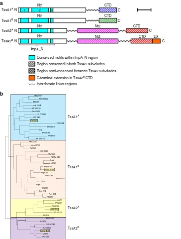
Bioinformatic analysis suggests that all TssA subunits contain an
N-terminal conserved region of ~120 amino acids (ImpA_N)
composed of three shorter motifs of significant sequence
simi-larity (ImpA_N1
–
3) (Fig.
1
a and Supplementary Fig. 1).
How-ever, these proteins possess one of two distinct C-terminal regions
based on amino acid sequence conservation, and are referred to
here as TssA1 and TssA2 (Fig.
1
a and Supplementary Fig. 2). In
members of the TssA1 clade, ImpA_N forms part of a large
N-terminal domain (Nt1) of ~250 amino acids which is connected
by a central region of variable length (40
–
60 amino acids) and
sequence to a conserved C-terminal region of 60
–
75 amino acids
containing a signature EPxxP motif (Supplementary Fig. 2a).
Phylogenetic analysis shows that TssA1 subunits fall into one or
other of two sub-clades, referred to here as A and B (Fig.
1
b). In
TssA1
Amembers, such as
P. aeruginosa
PA0082 (Pa TssA1
A), the
EPxxP motif is EPSxP, whereas in TssA1
Bmembers, such as
B.
cenocepacia
I35_RS01755 (Bc TssA1
B), an alternative EP(H/Q)SP
motif is present (Supplementary Fig. 2a). TssA1
Acorresponds to
TssA1 in the previously proposed nomenclature
28, whereas
TssA1
Bcorresponds to TssA3. In comparison, TssA2 orthologues
are longer (480
–
540 amino acids) and are predicted to contain
three domains: Nt1 (including the ImpA_N region), a middle
domain (Nt2) and CTD, each separated by regions of variable
length and sequence (Fig.
1
a and Supplementary Fig. 1 and 2b).
However, our phylogenetic analysis reveals that TssA2 proteins
are also subdivided into two sub-clades (Fig.
1
b) with members of
sub-clade B, represented by the EAEC TssA2 subunit (EAEC
TssA2
B) having an extension of 20
–
40 amino acids at the
C-terminus compared to sub-clade A members, such as
A.
hydro-phila
AHA1844 (Ah TssA2
A).
To provide support for the bioinformatics, analysis of protein
fragments derived by either limited or adventitious proteolysis
was used to identify putative domain boundaries. Analysis of Bc
His
6.TssA1
Bdegradation products suggested that TssA1
Bpos-sesses a large folded Nt1 domain (~31 kDa) connected to a small
CTD (~8.5 kDa) by an interdomain linker of ~40 amino acids
(Supplementary Fig. 3a, c, and d, Supplementary Table 1).
Products of limited proteolysis carried out on purified Ah His
6.
TssA2
Ashowed that residues in regions corresponding to
sequence variability, were particularly susceptible to protease
cleavage. This is consistent with the predicted three-domain
architecture with approximate boundaries of M1-R178 for Nt1,
D231-L374 for Nt2, and I402-K478 for the CTD (Supplementary
Fig. 3b and e).
Interactions of TssA1
Band its domains with other T6SS
components
. The interactions of a TssA1
Bsubunit with other
components of the T6SS have not been previously investigated. To
address this, a two-hybrid analysis was performed.
Complementa-tion of CyaA funcComplementa-tion was assayed on maltose MacConkey
indi-cator plates and by performing
β
-galactosidase assays, both of
which indicated that TssA1
Bmay interact with TssC, Hcp, TssE,
TssF, the conserved core region of VgrG (VgrG
C), TssK, TssL and
the N-terminal cytoplasmic region of TssM (TssM
N) (Fig.
2
a, b). To
validate these results, co-IP experiments were performed using
FLAG-tagged TssA1
Bin pairwise combinations with other
epitope-tagged subunits. The results supported the in vivo observations.
Thus, epitope-tagged TssC, Hcp, TssF, VgrG
C, TssK, TssL and
SMA2257 N454 00632 BB0799 Avin 26680 BPSS2101 Daci 3848
XOO2886 XCV4202 PA0082
YPO0499 BPSS0515 SciA
BPSS0180 YPO2949 BPSS0110 Pfl01 3401
Avin 50750 PA2360
HCH 04246 VPA1036
PBPRA0665 ImpA Atu4343 BPSL3100
I35 RS01755 RSp0759
Daro 2185 F952 02433 XOO3514
EvpK CV3963 Csal 2257
Psyr 4966 PP3088
Aec29 YPO3602 ECA3433 AHA1844 VCA0119 PP2626
BPSS1493 PA1656
I35 RS17400 Rmet 0634 Ec042 4540
c3399 YPO1483
Conserved motifs within ImpA_N region
Region semi-conserved between TssA2 sub-clades
Interdomain linker regions
Region conserved in both TssA1 sub-clades
a
b
TssA2A TssA1B TssA1A
TssA2B
TssA1A N
CTD
TssA2A N
TssA2B N
Nt1
TssA1B N
C
1 2 3
CTD Nt2
C CTD Nt2
C EX CTD
C
C-terminal extension in TssA2B CTD
Nt1
Nt1
Nt1
ImpA_N
[image:4.595.132.496.49.529.2]TssM
Nwere all co-immunoprecipitated with FLAG.TssA1
B(Fig.
2
c). Demonstration of an interaction between TssA1
Band
TssE required solubilisation of TssE by fusion to the MBP solubility
tag. In summary, the protein-protein interaction analysis suggests
that TssA1
Bmakes contact with components of the baseplate, the
membrane-anchoring complex, the inner tube and its cap, and the
surrounding sheath (Fig.
2
d). Two-hybrid analysis was then
per-formed to investigate the role of the N- and C-terminal regions of
TssA1
Bin interactions with other T6SS subunits. This showed that
TssA1
BCTD interacts with Hcp, TssF and VgrG (and also with
VgrG
C) but interactions with other subunits were not observed for
the Nt1 domain (Supplementary Fig. 6).
TssA1
Bis an essential component of the T6SS
. P.a TssA1
A,
V.
cholerae
TssA2
A(VCA0119) and EAEC TssA2
Bhave been shown
to be essential for T6SS activity
27,28,30. Therefore, the requirement
for TssA1
Bfor a fully functional T6SS was explored by assaying
the ability of a
B. cenocepacia tssA1
Bmutant to secrete Hcp
—
an
indicator of T6SS activity. The results showed that, unlike the
wild-type strain, Hcp was absent from the culture supernatant of
the
tssA1
Bmutant, but it was secreted when
tssA1
Bmutant
bac-teria contained a plasmid expressing
tssA1
B(Fig.
3
), thereby
confirming the requirement for this subunit for a functional
T6SS
31.
Structure determination of the TssA1
BNt1 domain
. SEC and
two-hybrid analysis of the overexpressed Bc TssA1
BNt1 domain
indicated that it is monomeric (Supplementary Fig. 7a, b).
Ana-lysis of the crystal structure of the TssA1
BNt1 domain,
deter-mined to 1.80 Å (Supplementary Table 2), revealed a monomer
composed of 11
α
-helices that are organised into two subdomains,
an N-terminal subdomain 1 (Sd1) connected to a larger
A + + + + + + + + + + + + + + + + + + + + + + + + + + + + + + + + + + + + ++++
d
Hcp TssC TssK VgrG TssB TssL TssM TssJ Cell envelope complex Baseplate complex Sheath PAAR Translocated tube TssA1B TssE TssF TssGc
Tot UB IP Tot UB IP Tot UB IP TssX+ FLAGTssA1B FLAGTssA1B TssX MBP TssCVSVg VSVgHcp MBP-TssE HATssF TssFHA
VSVgVgrGC
VSVgTssK VSVgTssL VSVgTssMN
FLAGTssA1 B
Proteins present in cell lysate
A Z 55 72 26 17 55 43 55 95 72 55 72 95 55 72 72 55 55 72 26 55 43 55 kDa F – – – – + – – – + – – – + – + – – + – + – – – – – – – – – – – – – – – – – – – – – – – – – – – – – – F – + – + + – – + + + – – + + + – – + – + – – + – – – – – – – – – – – – – – – – – – – – – – – – – – – F A1 A1
B C H E F G V J K L MN MC
B C H E F G V J K L MN MC
V
C
F
B C H E F G V J K L MN MC
B C H E F G V J K L MN MC
V
C
A1 A1
a
b
pUT18C-tssXNT18 X C
pKT25-tssX NT25 X C
pUT18-tssX N X T18C
pKNT25-tssX
F
T25 X
N C
pUT18CNT18 C
pKT25 NT25 C
pUT18 N T18C
pKNT25N T25C
F
M
C
G A
G VAV A V
C VCJ A J KAK LAL A
G G
A A V V V
CA VC A J J A K K A L L A A
Z Z M N M N M
N MC
A A M C + + + + + + + + + + + + + + + + + + + + + + + + + + + + + + + + + + + + M C M N M N M N A F 0 1000 2000 3000 β
-galactosidase activity (Miller units)
4000 5000 6000 7000 0 1000 2000 3000 β
-galactosidase activity (Miller units)
4000 5000 6000 7000 TssA1B controls TssA1B + TssB TssA1B + TssG TssA1B + VgrG TssA1B + TssJ TssA1B + TssK TssA1B + TssL TssA1B + TssM TssA1B + TssC TssA1B + Hcp TssA1B + TssE TssA1B + TssF A A B
A B A A
A
A H H A E E A F
A F
A A
F F
Z A A
A A BAB
C C C C
C C H A H
EAE
F A
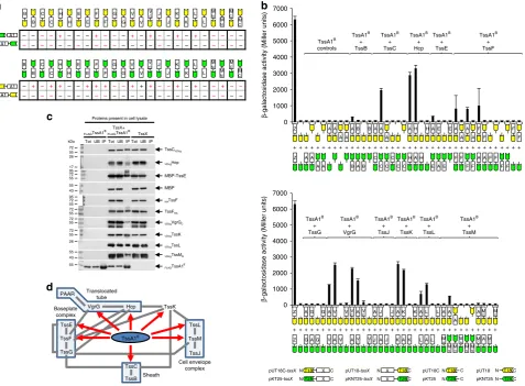
Fig. 2Interaction of TssA1Bwith other T6SS subunits.aTwo-hybrid analysis (maltose phenotypes). Hybrid proteins are represented by a green or yellow coloured motif representing the CyaA fragment (T25 and T18, respectively) linked to a white rectangle labelled according to the fused T6SS subunit, as shown in the key at the bottom of (b). T6SS subunits are indicated by a single letter corresponding to the suffix used in the Tss nomenclature (i.e. A1 corresponds to TssA1B, B corresponds to TssB etc) except for H (Hcp), V (VgrG) and V
C(VgrG core region). MNand MCrepresent the N-terminal
cytoplasmic and C-terminal periplasmic regions of TssM, respectively. The efficiency of complementation of representative combinations (phenotypes shown in red font) were determined byβ-galactosidase assay.bTwo-hybrid analysis (β-galactosidase activity). Data is representative of three independent experiments (n=3) performed in duplicate and values correspond to the mean ± standard deviation. Nomenclature as in (a). Z represents the Zip control. Values are presented in Supplementary Data 1.cCo-immunoprecipitation analysis. FLAG-tagged Bc TssA1Band potential interacting T6SS subunits (TssX) tagged with the VSV-g or HA epitope tags, or with MBP, were co-expressed inE. coli. FLAG.TssA1Bwas immunoprecipitated from cell lysates and recovered prey proteins (IP) were detected with the appropriate epitope antibody by western blotting. MBP was included as a control and was detected with MBP pAb. Proteins present in the cell lysate (Tot) and the unbound material (UB) were also analysed. Blots were also probed with FLAG mAb (an example of such a blot following co-expression of FLAG.TssA1Band VSVg.TssM
Nis shown in the bottom panel). Uncropped images of the blots are shown
[image:5.595.62.540.51.402.2]subdomain 2 (Sd2) (Fig.
4
a, b). Sd1 (M1-P113) is comprised of
five antiparallel
α
-helices (
α
1
–
α
5) and contains the ImpA_N1
(L8-I31) and the ImpA_N2 (D55
–
P113) motifs (Fig.
4
a
–
c).
Sd2 of Bc TssA1
B(L114-Q251) contains six
α
-helices (
α
6
–
α
11),
of which the three longest (
α
6,
α
10 and
α
11) form a
five-helix
bundle with
α
4
–
α
5 of Sd1. Residues from ImpA_N3 (R123
–
R133)
(Fig.
4
a
–
c) form the start of
α
6, a helix which includes a sharp
kink between L124 and G125. The C-terminal helix of Sd2 (
α
11)
terminates at Q251 and is thereafter followed by the connecting
interdomain linker to the CTD. The sequence conservation in the
ImpA_N region across the TssA clades (Supplementary Fig. 1)
implies that all Nt1 domains are closely related and adopt a
similar fold, which includes the entirety of Sd1, and the
N-terminal part of the
first helix of Sd2 of Bc TssA1
B. Mapping of
the 34 conserved residues in the ImpA_N region onto the
structure of Nt1, shows that approximately two-thirds are
involved in packing interactions in the core, with the remainder
located predominantly at one end of the molecule in the loops
between helices
α
1
–
α
2,
α
3
–
α
4, and
α
5
–
α
6 (Fig.
4
a, c).
TssA1
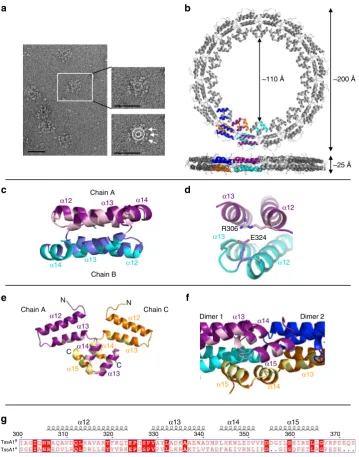
BCTD forms a double-layered ring with D
16symmetry
.
SEC-MALLS analysis of native Bc TssA1
Bindicated an apparent
molecular mass of 1.24 MDa (Supplementary Fig. 7c), suggesting
that the TssA1
Boligomer contains ~30 subunits, and negative
stain EM revealed rosette-like structures in which globular
domains were irregularly distributed around a central ring
(Fig.
5
a). Two-hybrid analysis demonstrated that oligomerisation
of TssA1
Brequired the CTD (Supplementary Fig. 7b), suggesting
that the inner ring is composed of polymerised CTDs. This was
confirmed by negative stain EM of the isolated CTD which was
observed to form rings without peripherally located globular
domains (Supplementary Fig. 7e).
The structure of a Bc TssA1
BCTD domain construct
(I303-S373) was determined to 3.08 Å (Fig.
5
b and Supplementary
Table 3) and the asymmetric unit was observed to contain eight
CTD monomers (chains A
–
H) (Supplementary Fig. 8a
–
c). Each
chain folds into four
α
-helices (
α
12-
α
15) that lie in approximately
the same plane, and form two antiparallel pairs (Fig.
5
e). The
eight monomers in the asymmetric unit further assemble into a
higher order oligomer of 32 subunits, consistent with the
SEC-MALLS analysis of TssA1
Band TssA1
BCTD oligomers
(Supplementary Fig. 7c, d), with D
16symmetry using the
crystallographic two-fold axes to make up a double-layered ring
with 16 subunits in each layer (Fig.
5
b and Supplementary
Figure 8a). In the assembled Bc TssA1
BCTD ring, analysis by
PISA
32revealed an extensive dimer interface (~950 Å
2) between
monomers in the different layers, which principally involves the
interaction of residues located in helices
α
12
–
α
13 with their
two-fold related subunits forming a region of hydrophobic packing
with hydrophilic interactions at the periphery (Fig.
5
c, d). Further
less extensive interactions (~570 Å
2) were identified between
dimers that facilitate formation of the ring, with each pair of
helices
α
14-
α
15 in one dimer, together with a small number of
residues from
α
13, interacting with their equivalents in adjacent
16-fold related dimers on either side (Fig.
5
e, f). This type of
interlocking series of interactions is repeated in a cyclic
arrangement to form a double-layered ring (Supplementary
Fig. 8d). Nested deletion of consecutive helical segments of
TssA1
BCTD revealed that loss of
α
15 resulted in the failure of the
protein to assemble into a high molecular weight ring, and
structure determination of the construct H12
–
H14 (helices
α
12
–
α
14) and of that consisting of
α
12 and
α
13 alone (residues
303
–
347), revealed them both to be dimers (Supplementary
Figure 8e
–
h and Supplementary Tables 4 and 5).
In the crystal structure of the TssA1
BCTD oligomer, the
double ring is ~25 Å thick with a lumenal diameter of ~110 Å and
an outer diameter of ~200 Å (Fig.
5
b), in agreement with the
dimensions of Bc TssA1
Band purified CTD oligomers
deter-mined by negative stain EM analysis (Supplementary Fig. 7e, f).
The C-termini of the TssA1
BCTD monomers are located at the
inner face of the ring, while their N-termini are orientated on the
outside, consistent with EM analysis, which places the Nt1
domain at the periphery of the ring in the intact complex
(Fig.
5
a). The irregular arrangement of the globular domains and
the poor resolution of the region linking them to the inner ring
are consistent with the presence of a
flexible linker connecting the
Nt1 domain to the CTD. Analysis of the CTD sequences of Bc
TssA1
Band Pa TssA1
Aidentified 17 conserved residues, of which
11 are involved in interactions around the subunit interfaces,
including the highly conserved EPxxP motif (Fig.
5
g and
Supplementary Fig. 2a). The Bc TssA1
BCTD structure shows
that this motif defines a loop that connects
α
12 and
α
13, and
includes a salt bridge between E324 from this motif and a
conserved arginine, R306, from a symmetry-related subunit
(Fig.
5
d). The conservation of these residues suggests that the
quaternary structures of TssA1
Aand TssA1
Bare related.
Moreover, although it has been previously proposed that the
C-terminal region of Pa TssA1
Ais structurally similar to the
C-terminal part of the gp6 subunit of the T4 phage baseplate on the
basis of low level sequence similarity
28, the similarity in sequence
between TssA1
Aand TssA1
Band the difference in structure of
the TssA1
BCTD to gp6 indicates that this is not the case.
TssA2
ANt2 is structurally similar to the TssA1
BNt1 domain
.
The structure of an Ah TssA2
ANt2 domain construct
(D231-L374) was solved to 1.76 Å with four subunits (chains A
–
D) in
the asymmetric unit (Supplementary Table 6) arranged as two
independent dimers, consistent with previous SEC analysis
33, that
showed that Ah TssA2
ANt2 exists as dimers in solution. Each
monomer folds into a cluster of seven
α
-helices (
α
1
–
α
7), the
arrangement of which is very similar to that of the EAEC TssA2
BNt2 domain (RMSD
=
2.0 Å) as determined by structural
alignment
27,34. Subsequent comparison using the Dali structural
alignment server
35identified a match between the Sd1 domain of
Bc TssA1
BNt1 and the Nt2 domains of both Ah TssA2
Aand
EAEC TssA2
B(RMSD
=
2.7 and 2.6 Å
,respectively) despite there
being essentially no amino acid sequence similarity between
them. Specifically, the related regions include helices
α
1-
α
5 of Bc
TssA1
BSd1 (i.e. most of the ImpA_N region) and
α
2
–
α
6 of Ah
∆M130 130
17 17 kDa
SN
Hcp WT
VC ptssA VC ptssA tssA::Tp
β-RNAP Hcp
β-RNAP
CA
Fig. 3Effect of TssA1Binactivation on T6SS secretion activity. Cell associated protein (CA) and spent supernatants (SN) from cultures ofB.
cenocepaciaH111 (WT) and the isogenictssA1Bmutant (tssA::Tp), each with
or without pBBR1MCS (VC) or pBBR1MCS-tssA1B(ptssA), were
[image:6.595.79.257.50.154.2]TssA2
ANt2 (Fig.
4
d). This similarity suggests that an ancient
gene duplication event may have played a role in the evolution of
the Nt1 and Nt2 domains of TssA2 subunits.
TssA2
ACTD assembles into an oligomer with D
5
symmetry
.
Two-hybrid and SEC MALLS analysis indicated that Ah TssA2
ACTD self-associates and dictates the overall stoichiometry of the
TssA2
Acomplex (Supplementary Fig. 9a, b, c, d). This is also a
feature of EAEC TssA2
B27. However, unlike EAEC TssA2
B, the
molecular weight estimations of Ah TssA2
A, Ah TssA2
ACTD
and an Ah TssA2
ANt2-CTD fusion construct corresponded to a
subunit stoichiometry of 10 rather than 12. Consistent with this,
negative stain EM showed that Ah TssA2
ACTD and Ah TssA2
ANt2-CTD assembled into complexes that resembled a
five-c
C
N
C
b
a
Sd1
Sd2 N
ImpA_N2
ImpA_N3
ImpA_N1
d
α1
α2
α1
α6
α4 α5
α10
α11
α7
α8
α9
α1
α6
α6
α7 α5
α3 α4
α2
α3
α1
α5
α4
α3
α4 α5
α2 α3
α6
α10
α9
α8
α7
α11
1 10
80
150
220 230 240 250
160 170 180 190 200
Sd1 Sd2 ImpA_N motifs
210
90 100 110 120 130 140
20
ImpA_N1
ImpA_N2 ImpA_N3
30 40 50 60 70
[image:7.595.120.476.50.551.2]pointed star (Fig.
6
a and Supplementary Fig. 9e, f). The structure
of crystals of an Ah His
6.TssA2
ANt2-CTD construct
(R232-K478) was successfully determined to 3.13 Å through molecular
replacement using the coordinates of the Ah TssA2
ANt2 dimer
as a search model (Supplementary Table 7). This identified the
position of
five dimers of the TssA2
ANt2 domain (R232 to L374,
helices
α
1
–
α
7) in the asymmetric unit, which did not appear to be
arranged with any obvious symmetry with respect to each other.
Following refinement, these dimers could be seen to be essentially
identical in structure to that of the dimer formed by the isolated
TssA2
ANt2 domain (RMSD
=
0.4 Å). The remaining C-terminal
residues of each of the 10 subunits (chains A-J) were organised
into a separate domain (the CTD) consisting of
five
α
-helices
(G388-L472,
α
8
–
α
12). The 10 CTDs were, in turn, assembled into
a
flat ring resembling a star with D
5symmetry (Fig.
6
b),
con-sistent with the negative stain EM and the SEC-MALLS analysis.
In the assembled TssA2
ACTD decamer two distinct two-fold
interfaces can be identified (Fig.
6
b). In the
first, a two-fold
related interface (shown by PISA
32to be 530 Å
2) is formed
primarily through interactions between residues from the
TssA1B300 310 320 330 340 350 360 370
TssA1A
~200 Å
~25 Å ~110 Å
Chain A Chain C
N N
C
C
E324 R306
α12
α13 α14
α15
α15 α14
α13
c
d
f
α12 α13α12 α13
α14
α13
α12 α13 α14 α15
α13 α14
α15
α14 α13
α12
α12
α13
α12 α13
α14
Chain A
Chain B
Dimer 1 Dimer 2
g
a
b
e
Fig. 5Structure of the Bc TssA1BCTD ring.aNegative stain EM of TssA1Bparticles. The magnified view indicates the location of the Nt1 domains (arrows) presented around the CTD oligomeric ring (circle). Scale bar corresponds to 50 nm.bBc TssA1BCTD monomers assemble to form a double-layered ring containing 32 subunits exhibiting D16symmetry (PDB: 6HS6 [https://www.ebi.ac.uk/pdbe/entry/pdb/6hs6]). The views shown correspond to a top view
[image:8.595.119.479.48.506.2]extended loop linking
α
11 and
α
12 (Fig.
6
b, c) and the N-terminal
end of
α
10 and their symmetry-related partners. This loop
includes a WEP motif (W461, E462 and P463) that is conserved
across both TssA2 sub-clades, where W461 is partially buried
within a hydrophobic pocket formed by the conserved residues
L456, L458 and L465, and an interaction with F423, a
non-conserved residue, in the two-fold related monomer. The
entrance to this pocket is capped by a salt bridge between E462
of the motif and R420, two residues that are again conserved
(Fig.
6
c and Supplementary Fig. 2b). In the second, more extensive
interface (950 Å
2), the WEP-mediated dimers assemble around
the
five-fold axis through interactions between the surface of
helices
α
9,
α
10 and
α
11 (Fig.
6
b, d) and their two-fold related
symmetry mates to form a mixed interface of hydrophobic/
hydrophilic contacts. The TssA2
ACTD (S381-K478) ring has a
thickness of ~30 Å, an outer diameter of ~110 Å, and a lumenal
h
Chain A
Chain B
a
b
d
c
e
g
f
W461E462 P463
R420
L465 L458
L456 F423
Chain B Chain C
Chain A
Chain B
Chain C ~25 Å
~110 Å
~35°
~30 Å
~110 Å
~110 Å N
C
N
~110 Å 1
2
α9
α8 α9
α8 α12
α11
α10
α8
α8 α13
α14
α12
α11
α10
α8
diameter of 25 Å (Fig.
6
b, g). As in the structure of the Bc TssA1
BCTD ring, the N-terminus of each TssA2
ACTD monomer is
orientated on the outside of the CTD oligomer resulting in the
presentation of the Nt2-CTD interdomain linker at the periphery
of the molecule (Fig.
6
b).
Despite the differences in symmetry between TssA2 CTD
oligomers assembled by members of sub-clades 2
Aand 2
B, the
fold of the Ah TssA2
ACTD monomer is closely related to that of
its counterpart in EAEC TssA2
B(PDB
=
4YO5, RMSD
=
1.6 Å).
Moreover, close inspection shows that the conserved WEP motif
is maintained at the dimer interface in members of both
TssA2 sub-clades, albeit that the two monomers of Ah TssA2
Aare arranged in a slightly different orientation with respect to each
other, corresponding to a rotation of ~35° compared to their
TssA2
Bcounterparts (Fig.
6
e).
The way in which the dimers assemble around their respective
five-fold and six-fold symmetry axes in the Ah and EAEC TssA2
CTDs are strikingly different, in contrast to the similarity seen
around their two-fold axes. In Ah TssA2
A, two monomers,
related by a WEP-mediated two-fold axis, pack adjacent to one
another, parallel to the
five-fold axis, resulting in the formation of
an oligomer which resembles two interpenetrating rings (Fig.
6
g).
The face of Ah TssA2
A, used in subunit assembly around the
five-fold axis (residues from
α
9,
α
10, and
α
11), interacts with a
symmetry related equivalent (Fig.
6
g). In comparison, EAEC
TssA2
BCTD contains a 41 residue C-terminal extension which
folds into two additional helices (
α
13 and
α
14) (Fig.
6
f). These
dominate the interactions around the six-fold axis, packing
against the face of helices
α
9,
α
10 and
α
11 of an adjacent subunit
(Fig.
6
h). Consequently, this causes an incline of the
WEP-mediated dimer by ~50°. Therefore, in EAEC TssA2
B, two
monomers, related by a WEP-mediated two-fold axis, stack on
top of one another, parallel to the six-fold axis, with the dimers
then assembling around the latter to form a double-layered ring
(Fig.
6
h). Interestingly, despite the difference in symmetry we
note that the lumenal diameter and outer diameter of the TssA2
Aand TssA2
BCTD oligomers are similar (Fig.
6
g, h), thereby
placing the Nt2-CTD interdomain linker at the same distance
from the centre of the complex.
A
fl
exible linker in TssA2
ANt2-CTD mediates domain
mobi-lity
. Despite the electron density for the Nt2 and CTD domains
being clear, density was observed for only part of the Nt2-CTD
interdomain linker, and, depending on the chain, between 6 and
13 residues (Q375-A387) could not be identified within this
region. However, restrictions imposed by the maximum length of
the absent linker residues and the relative orientations of the Nt2
and CTD domains allowed the unambiguous assignment of the
Nt2 domains to their corresponding CTD (i.e. in the same
polypeptide chain). This revealed that in the decameric Ah
TssA2
ANt2-CTD complex, the Nt2 domains are not related by
the
five-fold symmetry exhibited by their corresponding CTDs
(Fig.
7
a). Instead, dimers of Nt2 make no contacts with their own
CTDs but rather contact either TssA2
ANt2 or CTD domains of
other decamers in the crystal lattice. Moreover, as a result of the
flexible linker, the Nt2 dimers have quite different orientations
with respect to the
five-fold axis, with angular rotations of up to
~60° about the vertical axis (Fig.
7
b), and ~45° and ~60° about the
horizontal and torsional axes, respectively. Although, the
observed Nt2 domain orientations described above are believed to
be an artefact of crystal packing, the range of motion seen is
believed to reflect the inherent
flexibility between CTD and Nt2
domains. This is consistent with negative stain EM analysis that
revealed a lower calculated correlation coefficient for strict
five-fold symmetry when comparing an Nt2-CTD construct to a CTD
construct (Supplementary Fig. 9e
–
h).
Sequence alignments suggest that the Nt2-CTD interdomain
linker is longer in TssA2
Bsubunits than in TssA2
Asubunits
(Supplementary Fig. 2b). For example, the linker in Ah TssA2
Ais
composed of ~21 residues whereas it contains ~37 residues in
EAEC TssA2
B. The most likely explanation for the presence of a
longer linker in TssA2
Bis to overcome a restricted range of
motion of Nt2 dimers that would be imposed by a shorter linker
due to the possible physical interference between the Nt2 dimer
and the
‘
taller
’
double-layered ring arrangement formed by EAEC
TssA2
BCTD compared to the
‘
squat
’
interpenetrating ring
assembled by Ah TssA2
ACTD (Fig.
6
g, h).
The quite different orientation of the CTD WEP-mediated
dimers in TssA2
Aand TssA2
Boligomers has one further
consequence, which is that the orientation of the interdomain
linker between Nt2 and CTD is necessarily rotated by ~50°
(Fig.
6
g, h). We assume that the
flexibility of the linker allows this
structural difference to be maintained without affecting the
biological role of the protein. The
finding that in the Ah TssA2
ANt2-CTD structure the Nt2 domains consistently occur as dimers
further suggests that the Nt2 domains remain associated as part
of the biological role of TssA, in contrast to the proposal by
Zoued and co-workers
27, who interpreted low resolution negative
stain EM data to suggest that they separate to form monomers
27.
A rigorous test will require a high-resolution EM reconstruction
from TssA particles embedded in vitreous ice, although this could
be challenging given the apparent
flexibility of domain linkers.
Discussion
The structural studies presented here show that TssA1
B, TssA2
Aand TssA2
Bpossess structurally diverse CTDs that lead to distinct
overall architectures for their respective complexes. Nevertheless,
all these subunits contain a conserved N-terminal ImpA_N
region that is peripherally located in the assembled complexes.
The question that arises from this is whether these TssAs play the
same role in the function of the T6SS? The presence of the
ImpA_N region in the Nt1 domain of subunits from all TssA
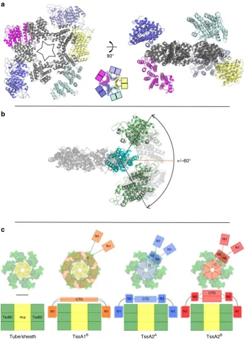
Fig. 6Structure of the Ah TssA2ACTD ring.aNegative stain EM particle averaging of Ah TssA2ACTD indicatingfive-fold symmetry.bStructure of 10 Ah TssA2ACTD monomers assembled to form a decameric oligomer exhibiting D5symmetry (PDB: 6G7Chttps://www.ebi.ac.uk/pdbe/entry/pdb/6g7c).
Hcp TssBC TssBC
+/–60° 90°
a
c
b
Tube/sheath TssA1B TssA2A TssA2B
C N1
N1
C N1
N1
N2 N2
C N2
N2 N1
N1
N1
CTD
N1 N1 N1
N2 CTD N2
CTD
N2 N2
N1 N1
Fig. 7Schematic representation of TssA ring dimensions and domainflexibility.aArrangement of the Ah TssA2ANt2 domains around the CTD oligomer as observed in the asymmetric unit. The Nt2 domains do not follow strictfive-fold symmetry. The decameric CTD ring is shown in dark grey
[image:11.595.119.477.50.545.2]clades strongly suggests that they have a common function that is
of critical importance. This is further supported by the studies
described here which reveal that TssA1
Binteracts with the
baseplate components TssE, TssF and TssK, the inner tube
sub-unit, Hcp, the tip protein, VgrG, the sheath subunit TssC and
cytoplasmic components of the cell envelope-anchoring complex.
This is strikingly similar to the TssA2
Bsubunit that was shown to
interact with the same components except for TssF
27. Indeed the
TssA1
Band TssA2
BCTD oligomers were both shown to interact
with Hcp and VgrG despite their quite different structures. This
would argue for a similarity of function despite the divergent
architecture of the two proteins. Given the sequence similarity
and predicted structural relationship between TssA1
Aand
TssA1
B, this suggests that all three clades of TssA perform related
roles within the T6SS. If the role of these quite different TssA
subunits is similar, then how is this achieved given the structural
variations that are observed?
To date, two distinct models for the role of TssA have been
proposed. In the
first, baseplate-associated model, it was proposed
that TssA is a component of the T6SS baseplate required for
priming the inner tube and sheath for assembly, but not
migrating during polymerisation
28. This model was based partly
on predicted similarities in structure for the CTDs of TssA1
Aand
the phage T4 baseplate protein gp6, to which it was believed that
this TssA subunit was related. The structure we have obtained for
TssA1
B, which is closely related to TssA1
A, reveals that it has a
quite different structure to gp6. This suggests that TssA1
Ais very
unlikely to be a gp6 orthologue. Due to the absence of structural
similarity to baseplate proteins and the conservation of the Nt1
domain among all TssA subunits, it is therefore likely that
TssA1 subunits fulfil a similar role to TssA2 subunits.
In the alternative model proposed for a TssA subunit that
corresponds to TssA2
B, TssA acts as a coordinator of inner tube
and sheath assembly, in which the CTD oligomer of TssA2
Bwould lie close to the centre of the tail assembly, above the
position occupied by Hcp where it would coordinate
poly-merisation of the inner tube and sheath
27,29,36,37. In one version
of this proposal (the cap model) eversion of CTD monomers,
from a closed form of the CTD oligomer to an open form, is
then proposed to permit the passage of Hcp through the
lumen of the open ring. This would allow sequential stacking
of Hcp hexamers, with the N-terminal domains of TssA2
Binteracting with the polymerising sheath. However, as yet, an
open form of TssA2
Bhas not been observed and the proposed
reorientation of monomers in the CTD oligomer remains
speculative.
The studies described here show that the fold of TssA2
Aretains
clear structural similarity to TssA2
Byet generates a decamer
possessing
five-fold symmetry in contrast to the six-fold
sym-metry of the TssA2
Bdodecamer
27. Therefore, while it could be
argued that it might lie in the same position in the complex as
TssA2
B, the difference in symmetry implies there must be
changes to the complementary surfaces of surrounding
TssB/-C/-D subunits to allow pseudo equivalent interactions. This situation
becomes even more extreme given the quite different
organiza-tion of the TssA1
Bring oligomer with its sixteen-fold symmetry.
One possibility is that the structure of TssA1
Bwe have
deter-mined represents an open form of the TssA CTD oligomer,
leaving a closed form of the TssA1
BCTD oligomer, which might
pack on top of a Hcp hexamer, still to be identified. Alternatively,
if some features of the cap model are correct, and TssA1
A/TssA1
Band TssA2
Aalso serve to coordinate tube and sheath assembly,
then, given their quite different symmetries and architectures,
rationalising how they function in an equivalent way to that
proposed for TssA2
Bis problematic. One way for this dilemma to
be resolved is if the different structures of these distinct TssAs
represent different ways in which a common function can be
maintained.
Comparing the TssAs of clades 1 and 2, these structures
indicate that the Nt1 domain is linked either to a large diameter
CTD ring (200 Å) of high symmetry, via a single
flexible segment,
as seen in sub-clade TssA1
B, or to a smaller (100
–
110 Å) diameter
CTD of lower symmetry, through a middle domain (Nt2)
flanked
by two
flexible linkers, as seen in sub-clades 2
Aand 2
B. Despite
these two alternative architectures, the radial position of the Nt1
domain from the centre of the complex is similar in both TssA1
and TssA2 clades (Fig.
7
c). This suggests that a role of the Nt2
domain is to act as a spacer connecting the Nt1 domain of
TssA2
Aand TssA2
Bto their corresponding CTDs.
In light of the above, and given the conservation of the Nt1
domain, we suggest that the common feature shared by all these
TssAs is that the Nt1 domain is positioned around the periphery
of the ring-forming CTD oligomers, furthest away from the
symmetry axis but at an equivalent distance. Given the apparent
symmetry mismatch between the tube/sheath assembly and the
TssA CTD oligomers of some TssA species, it is important to
consider how equivalent interactions between these components
can be maintained. The simplest explanation is to consider a
model whereby the interactions between TssA subunits and the
tube/sheath assembly are solely the property of the conserved Nt1
domains, which have been suggested to participate in assembly of
the latter
27. This would mean that the role of the CTD or
Nt2-CTD oligomers of clades 1 or 2, respectively, are to provide a
platform for the presentation of the Nt1 domains above the
growing tube/sheath complex at a similar distance from the
centre of each TssA complex (Fig.
7
c). In such a model it would
not be necessary for the CTDs to interact directly with the tube/
sheath assembly except perhaps in a transient manner as
indi-cated by interaction studies. This agrees with surface plasmon
resonance data for EAEC TssA2
Bwhere the interaction of the
Nt1-Nt2 region with the TssBC sheath is significantly greater
than that of the corresponding CTD with Hcp
27.
This proposal in which one domain or component of a protein
subunit (CTD/Nt2-CTD) associates into a platform that
facil-itates the role of a second domain (Nt1) in subunit incorporation
by a polymerising protein assembly, is a theme that occurs in the
mechanism of
flagellum assembly by the HAP2 cap
protein
27,29,38,39. An important aspect of the
flagellin-capping
model is the observation of domain mobility, a feature clearly
shown here for both the relative motion of Nt1 with respect to the
CTD in TssA1
Band between the CTD and Nt2 in TssA2
Aand
TssA2
B, and the predicted
flexibility of the linker between Nt2
and Nt1 in TssA2
A/TssA2
B. This would provide the necessary
movement of the Nt1 domains for dynamic interactions with the
TssBC subunits during sheath biogenesis. This might involve the
displacement of Nt1 domains and their linkers, consequently
allowing passage of Hcp, in addition to further TssB/C subunits
to be incorporated into the growing sheath. This model does not
necessarily require eversion of the CTD rings. However, we do
not rule out the possibility that TssA2
A/TssA2
BCTD oligomers
can re-orientate to allow access of Hcp hexamers to the growing
tube. In this regard, we note that the TssA1
BCTD ring oligomer
surface of the TssA ring faces the membrane while the other faces
the cytosol. According to the current model, this asymmetry is
reinforced once the baseplate is recruited to the membrane
complex, whereupon one face of the TssA oligomer is located
adjacent to the cytoplasmic face of the baseplate
27. Here TssA is
ready to coordinate inner tube and sheath assembly once the
first
Hcp ring is recruited to the base of the VgrG trimer located at the
hub of the baseplate
40. Although the precise mode of action of
TssA subunits remains unknown, they are likely to lock the inner
tube/sheath structure in the high energy form prior to
firing
29.
Thus, TssA may possess chaperone-like activity.
A
final question is to consider the mismatch in symmetry
between the assemblies formed by members of different TssA
clades, on the one hand, and other components of the T6SS, on
the other, and how this impacts on the mechanism by which TssA
executes its role. For example, the six-fold symmetry of the
TssA2
Boligomer is consistent with the symmetry of the
tube-sheath structure but not with the
five-fold symmetry proposed for
the TssJLM membrane complex
10,27. In contrast, the symmetry of
the TssA2
Aoligomer matches that of the membrane complex but
not the contractile machinery. In this regard it is interesting to
note that, as seen with the different symmetries of TssA2
A/
TssA2
B, both
five-fold and six-fold symmetrical variants of the
HAP2 capping protein can interact with the common 11-fold
symmetrical
flagellin
filament
41,42suggesting that the apparent
symmetry mismatch between oligomers formed by some TssA
species and the tube/sheath assembly is not necessarily
proble-matic. Based on these considerations we suggest that a
flagellin-like capping model is a plausible mechanism for the role of all
TssA subunits. Ultimately, these proposals need to be confirmed
by the structural analysis of other complexes of secretion systems
from each of the different TssA clades. Once this has been
achieved, the manner in which the difference in structure and
symmetries among TssA subunits resolves to provide equivalent
function will be revealed.
Methods
Bacterial strains and growth conditions. Bacterial strain details are given in Supplementary Table 8.E. colistrains JM83 and DH5αwere used for routine cloning steps.B. cenocepaciastrain H111 andA. hydrophilaATCC 7966 were the source of the TssA1Band TssA2Asubunit genes, respectively. LB was routinely used for growing bacteria except where indicated43. Lennox medium was used with
glucose included at 0.2%44. D-BHI broth was made by dissolving 9.25 g brain-heart
infusion (BBL) in 25 ml water and dialysed (12,000–14,000 MWCO) against 500 ml water overnight45. Antibiotics were used at the following concentrations forE. coli:
ampicillin, 100μg/ml; kanamycin, 50μg/ml; chloramphenicol, 25μg/ml; tri-methoprim, 25μg/ml (selection for trimethoprim resistance was applied in/on iso-sensitest (IST) broth/agar (Oxoid)). Bacto MacConkey agar base used for two-hybrid assays was obtained from Becton, Dickinson and Co.
Plasmids and primers. All oligonucleotides and plasmids used in this study are listed in Supplementary Tables 9,10. Construction of plasmids is also described in Supplementary Table 10. Plasmids were transferred toE. coliby transformation46
and toB. cenocepaciaby conjugation47,48.
Construction of aB. cenocepacia tssA1Bmutant. To construct theB. cenocepacia H111tssA::Tp mutant, a segment of thetssA1BORF was amplified with primers iotAfor3 and iotArev and cloned between theHindIII andBamHI sites of pBBR1MCS (to give pBBR1MCS-‘tssA1B) whereupon it was disrupted by insertion of thedfrB2(TpR) cassette from p34E-Tp (generating pBBR1MCS-‘tssA1B::Tp). ThetssA1B::Tp fragment was then transferred to the suicide vector pSHAFT2 and the resultant plasmid (pSHAFT2-‘tssA1B::Tp) conjugally mobilised into H111 usingE. coliS17–1(λpir). Ex-conjugants were selected on M9-glucose agar con-taining trimethoprim (25μg/ml). Mutants were identified by PCR screening chloramphenicol-sensitive ex-conjugants using primers iotAfor and iotArev2 that anneal to sequences located outside the genomic region contained on the suicide vector. Construction of thetssMdeletion mutant was performed by conjugally mobilising the allelic replacement plasmid, pSNUFF-ΔtssM, containing atssM
deletion allele (codons 506–1216 removed) into strain H111 usingE. coliSM10 (λpir). Ex-conjugants containing the integrated allelic replacement vector were selected on M9-glucose agar containing trimethoprim (25μg/ml) and were then
plated on M9-glucose agar containingp-chlorophenylalanine to identify recom-binants in which a second crossover had resulted in excision of the integrated plasmid49. Mutants were identified by diagnostic PCR using primers tssM-OPfor
and tssM-OPrev2.
Hcp secretion assay. D-BHI broth was inoculated with overnight cultures ofB. cenocepaciastrains to give a starting OD600of 0.03, and grown to an OD600of 1.0 at
37 °C. Cultures were centrifuged to collect the cells and the supernatant wasfi lter-sterilised. To precipitate secreted proteins, sodium deoxycholate was added to the cleared supernatant to afinal concentration of 0.2 mg/ml followed by incubation on ice for 30 min. Trichloroacetic acid was then added to afinal concentration of 10% (v/v), and the suspension was incubated overnight at−20 °C. Precipitated
proteins were recovered by centrifugation for 15 min at 14,000 ×gat 4 °C and washed with ice cold acetone. Pellets were air-dried and resuspended in 0.001 of the original volume of SDS-PAGE sample buffer. Cell-associated proteins were isolated by resuspending the initial bacterial pellet in a volume of SDS-PAGE sample buffer that was 20-fold less than the starting culture volume and transferred to a microcentrifuge tube. All protein samples were boiled for 5 min, centrifuged for 10 min at 14,000 ×gto remove cell debris and then fractionated by electrophoresis in a 15% discontinuous SDS-PA gel followed by electroblotting onto a PVDF membrane (Millipore). Membranes were blocked with 5% (w/v) non-fat skimmed milk in TBS containing Tween 20 (0.05% (v/v)) solution for 1 h. Hcp was detected by probing with a custom polyclonal rat antibody raised to purified His6.Hcp
(1:1000) and goat anti-rat HRP secondary antibody (1:5000). Antibody to the cytoplasmically locatedβsubunit ofE. coliRNA polymerase (1:2000) that cross-reacted to theBurkholderiaspp. subunit was also used as a lysis control, with a rabbit anti-mouse HRP secondary antibody (1:5000). Detection of chemilumi-nescence was performed using an EZ-ECL detection kit and a XRS+imaging system (BioRad). Uncropped blots are shown in Supplementary Fig. 5.
Two-hybrid analysis. The bacterial adenylate cyclase two-hybrid (BACTH) system was employed50.B. cenocepaciaT6SS subunit genes (tssA-tssC,hcp,tssE-tssG,vgrG, tssJ-tssM) or domain coding sequences were fused to the N- and C-terminal coding sequences of theB. pertussisCyaA T25 or T18 fragments present in plasmids pKT25, pKNT25, pUT18 and pUT18C. To avoid membrane or periplasmic tar-geting of CyaA fusion proteins, signal sequences and TMDs were omitted from the TssJ, TssL and TssM hybrid proteins. As a consequence, the cytoplasmic N-terminal region of TssM (TssMN) and the periplasmic C-terminal region (TssMC)
were fused to the CyaA domains separately. Hybrid proteins were generated with full-length VgrG and also with the conserved core region corresponding to the phage T4 gp27 and gp5 proteins (VgrGC). DNA encoding full-lengthA. hydrophila
TssA2Aand the TssA2ANt1 domain were also cloned into all four vectors while DNA encoding the Nt2 and CTDs were cloned into pKT25 and pUT18C only. Further details on plasmid constructions are given in Supplementary Table 10. The
E. coli cya−strain BTH101 was transformed with the two compatible plasmids
expressing fusion proteins and plated on MacConkey indicator agar containing 1% (w/v) maltose, ampicillin (100μg/ml) and kanamycin (50μg/ml). Colony pheno-types were scored after 3 and 5 nights incubation at 30 °C (Mal+(Cya+) colonies
are purple and Mal−(Cya−) colonies are white). The appearance of purple colonies
after 3 days incubation indicated a strong Cya+phenotype. The positive control
combination (Zip control), where each plasmid encoded the leucine zipper segment of the yeast transcriptional activator protein, GCN4, fused to T25 and T1850was
always included as were all negative control combinations (two compatible empty vectors and one empty vector in combination with a plasmid specifying a hybrid protein).
The efficiency of CyaA complementation was quantitated by measurement of theβ-galactosidase activity in broth cultures. Cells were grown in LB broth containing antibiotics and 0.5 mM IPTG at 30 °C with aeration for 14–16 h. The cultures were then chilled on ice before being diluted in LB medium to OD600
0.4–0.6. 50μl of each diluted culture was added to test tubes containing 950μl of Z buffer (0.06 M Na2HPO4, 0.04 M NaH2PO4, 0.01 M KCl, 0.001 M MgSO4and
0.27% (v/v)β-mercaptoethanol), chloroform (30μl) and 0.1% SDS (30μl). The mixtures were vortexed for 10 s and equilibrated at 30 °C for 15 min whereupon 200μl of ONPG (4 mg/ml in Z buffer) was added to initiate the reaction. Reactions were stopped by addition of 500μl 1 M Na2CO3and the absorbance at 420 and 550
nm was measured. Theβ-galactosidase activity (in Miller units) was calculated using the equation 1000 (OD420–1.75 × OD550/txv× OD600) wheretcorresponds
to the duration of the reaction in minutes andvcorresponds to the volume of culture used in the assay43. The background activity measured in the negative
controls (a pair of empty vectors or one empty vector in combination with a plasmid encoding a hybrid protein) was 75–90 Miller units (Mu). Each assay reaction was performed in duplicate on three biological replicates (n=3), and the mean, along with standard deviation, was calculated.