CELL CARCINOMA
A thesis submitted for the degree of
DOCTOR OF PHILOSOPHY
by
FADWA AL-YAMAN
May 1981
Department of Immunology
The John Curtin School of Medical Research Australian National University
The ultrastructural studies of the cultivated tumour cells were done jointly with Professor Bede Morris. Miss W. Trevella assisted with the cannulations of the lymphatic vessels in sheep. The histological sections were prepared by Miss W. Hughes. All the other work reported in this thesis was done by myself.
~
.Al-r
The work described in this thesis was carried out in the Department of Immunology, John Curtin School of Medical Research during the tenure of an Australian National University Ph.D.
Scholarship.
I wish to thank Professor Bede Morris for the opportunity to work in his department and for his help and valuable criticism during the preparation of the manuscript. I also wish to thank Miss Wendy Trevella for her help with the cannulations of the lymphatic vessels in sheep.
Special thanks go to my supervisor Dr. David Willenborg for his help and guidance throughout the performance of the experiments and the preparation of the thesis.·
Finally, I wish to thank Ms. Alice Duncanson and
This thesis reports experiments I have done to investigate some of the immunobiological aspects of a naturally occurring squamous cell carcinoma in sheep. Methods were established for the successful cultivation of a number of pure populations of .tumour cells
in vitro.
The epithelial nature of the establishedcell lines was confirmed by electron microscopic studies . An association was found between the site at which the tumour grew on the host and the rate of success obtained in establishing the tumour in culture. Tumours growing on the muzzle were established in culture most easily followed by those on the ear and the vulva respectively. A similar correlation 'I was observed between the site
of the primary tumour and its capacity to grow in nude mice.
Studies on the tumour cell-lymphocyte interaction indicated that all tumour cell lines that were tested failed to stimulate a proliferative response in allogeneic lymphocytes when mixed
together
in
vitro.
This lack of stimulation was found to be due to an immunosuppressive capacity of the majority of the cell lines tested. This immunosuppressive capacity of the tumours, which was manifestin
vitro~
was not seenin vivo~
sincechallenge of allogeneic normal sheep with tumour cells resulted in significant stimulation of the recipient~ irrrrnune system.
The immune reactivity of tumour-bearing sheep to autochthonous tumour cells was investigated both in the lymph node regional to the tumour and in lymph nodes distant from the tumour site. This was done by cannulating both the afferent and the efferent
remained present, no immune reactivity , as measured by cellular changes in the lymph from the regional nodes was detected whereas a significant response did occur in the lymph from a
distant node when this was challenged directly with tumour cells. The response observed in the distant lymph node was associated with the production of antibodies which bound to the tumour cells but were not cytotoxic for them. Removal of the primary tumour and subsequent sensitization of the host three weeks after the tumour was resected, resulted in the appearance of specific humoral and cellular cytotoxic mechanisms in the efferent lymph
from distant nodes when these were challenged by autochthonous
CHAPTER 1
1.1 1. 2 1.3 1. 4 1.5 1.6
1.7 1.8 1.9 1.10
CHAPTER 2
2.1 2.2
2.3 2. 4 2.5 2.6 2.7 2.8 2.9 2.10
2.11
INTRODUCTION
PATHOLOGY OF SKIN TUMOURS
THE AETIOLOGY OF SKIN CANCER IN MAN OVINE SQUAMOUS CELL CARCINOMA
BOVINE OCULAR SQUAMOUS CELL CARCINOMA MECHANISMS OF CARCINOGENESIS
CLINICAL OBSERVATIONS AND EXPERIMENTAL
EVIDENCE SUGGESTIVE OF A HOST IMMUNE RESPONSE AGAINST TUMOURS
TUMOUR ANTIGENS
THE NATURE OF THE HOST RESPONSE MECHANISMS OF TUMOUR ESCAPE
SCOPE OF THE THESIS
MATERIALS AND METHODS
EXPERIMENTAL ANIMALS AND SURGICAL PHYSIOLOGICAL SOLUTIONS , BUFFERS , AND OTHER SOLUTIONS
TUMOUR CELL LINES
LYMPHOCYTE PREPARATIONS
IRRADIATION OF STIMULATOR CELLS
IN
V
ITRO
ASSAYS FOR CELL-MEDIATED ANTIBODY ASSAYSIN V
IVO
CHALLENGE ELECTRON MICROSCOPYCHAPTER 3
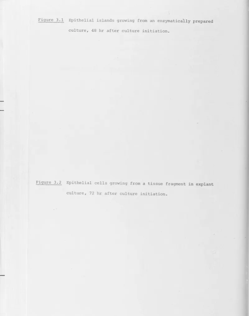
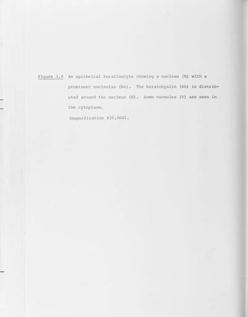
3.1 3.2
3.3 3.4
CHAPTER 4
4. 1 4. 2
4.3 4.4
4.5
CHAPTER 5
5.1 5. 2 5.3
THE ESTABLISHMENT AND CHARACTERIZATION OF THE TUMOUR CELL LINES
IN VITRO
AND IN THE NUDE MOUSEINTRODUCTION
METHODSOF TISSUE CULTURE RESULTS
DISCUSSION
THE
I
N VITR
O STIMULATORY CAPACITY OF TUMOUR
CELL LINESINTRODUCTION
THE ALLOGENEIC TUMOUR CELL-LYMPHOCYTE INTERACTION
DEMONSTRATION OF ALLOANTIGENS ON TUMOUR CELLS SUPPRESSION OF THE MIXED LYMPHOCYTE RESPONSE
(MLR) BY TUMOUR CELLS DISCUSSION
THE
IN VIVO STIMULATORY
CAPACITY OF TUMOUR CELL LINESCHAPTER 6
6.1 6. 2 6. 3
CHAPTER 7
7. 1 7. 2 7.3
CHAPTER 8
REFERENCES
THE
IN
VIVO
REACTIVITY AGAINST AUTOCHTHONOUSTUMOUR
EXPLAPTS
AND CELL LINES
INTRODUCTION RESULTS
DISCUSSION
THE REACTIVITY OF REGIONAL AND DISTANT LYMPH NODES AGAINST AUTOCHTHONOUS TUMOUR CELLS
INTRODUCTION RESULTS
DISCUSSION
GENERAL CONCLUSIONS
FIGURES ARE PLACED AT THE END OF EACH CHAPTER
126 128 142
150 153 158
164
Skin cancer is one of the most common cancers of man. The
incidence of skin cancer in Australia is the highest of any country in the world and represents more than 50% of all the
cancers diagnosed. There is a well established association between the incidence of skin cancer in man, and the degree to which the skin is exposed to sunlight. Skin cancer in sheep is
also common in Australia and this tumour, like the skin tumours
in man, has been attributed to the effect of ultraviolet
irradiation of the sun. Thus skin cancer in sheep provides an excellent model for studying the biology of a cancer which is of
important medical significance(Ladd and Entwistle 1977).
The aim of the work described in this thesis was to investigate
some of the immunobiological aspects of this naturally occurring solid tumour of the skin of sheep. The introduction to the thesis consists of two parts; one part describes the histological
structure of the skin, the alterations in the skin which are a prelude to the development of either basal or squamous cell
carcinomata, and includes a discussion of the natural history
of squamous cell carcinoma in sheep. The other part reviews
the various types of immune reactivities detected in natural and
experimental tumours both
i
n
v
it
r
o
and&
n V&
VO
.
1. 1 PATHOLOGY OF SKIN TUMOURS
The ski n consist s of an outer epidermis and an inner dermis.
The epidermis is divided into four main layers; the basal layer consisting of a single row of columnar epithelial cells (stratum germinativum), a thick prickle cell layer (stratum spinosum), a
keratohyalin (stratum granulosum) and , a surface -layer of dead keratinized cells (stratum corneum) . Cell proliferation in the epidermis occurs mainly in the basal layer. Newly-formed cells leave this layer and move outwards through the more superficial
layers until they reach the surface and are shed. During this process of outward migration, the cells undergo a series of
characteristic morphological changes in which they become
progressively more keratinized. As the cells produce the
insoluble protein keratin, they lose their nuclei and die . Thus, under normal conditions, the production of new cells in the
basal layer is balanced against the death of cells through the
process of keratinization and desquamation. In certain circum-stances, for example .following prolonged exposure of the skin to
UV-light, hyperplasia of the epidermis can occur (Blum l959;
Blum
et al
.
~
1975) . This hyperplastic condition of the skin may, with time1revert to normal or become permanent. Two types of epidermal carcinomata may re~ult from alterations in the balance
between cell proliferation and cell differentiation (keratinization),
and these tumours are classified as basal cell and squamous cell
carcinoma ta.
1
.
1
.
1
Basal Cell Carcinoma
Basal cell carcinoma accounts for over 75% of all skin
cancers (Fitzpatrick and Pathak 1977). These carcinomata arise
from the basal layer of the epidermis and the malignant cells
show little tendency to undergo the usual differentiation into
keratinized squamous cells (Belisario 1959; Klein
et
al.~
1973). Although these tumours very rarely metastasise, they are locallystructures including nerves , bone and brain. This cancer occurs mainly on the head and neck of man, classically distributed around the shade line of the hat. The typical basal cell carcinoma
appears as a non·-inflamed smooth , translucent waxy nodule, which has a variable amount of melanin pigment. Such nodules often ulcerate and a crust forms on their surface . Basal cell
carcinomata may also take other forms, including more subtle
infiltrating lesions that do not produce elevated nodules.
Although there is no readily recognizable premalignant lesion that
precedes a basal cell carcinoma, the cancer is normally seen in people who manifest symptoms of skin damage from sunlight or
X-rays (Klein
et
al
.~
1973).1
.
1
.
2 Squamous
Cell
Carcinoma
Squamous cell carcinoma also arises from the epidermis and
the malignant cells show significant differentiation into squamous
cells. This cancer is characterized histologically by the presence of accumulations of keratinized cells with a characteristically whorled arrangement described as keratin pearls (Klein
et al
.
J
1973; Fitzpatrick and Pathak, 1977) . This pattern is apparent in the early stages of the disease . Squamous cell carcinoma may also grow and infiltrate the deeper layers of the dermis and
subcuticulum as does the basal cell carcinoma or i t may grow to form a raised hyperplastic lesion which may infiltrate the deeper tissue at a later stage . Squamous cell tumours tend to metastasise more readily than basal cell tumours, but the outcome depends on
the size , location and the invasiveness of the tumour.
Iri
commonly arise from a pre-existing actinic or solar keratosis.
These premalignant keratoses are scaly, rough, red plaques that occur in chronically sun-damaged skin. Although very few of these keratoses progress to carcinomata, most squamous-cell carcinomason exposed skin do arise from these keratoses(Klein
e
t al
.
~
1973; Scott and Starf 1977) . The type of squamata which arise from keratotic lesions has a low frequency of metastases(below 2%,Fitzpatrick and Pathak 1977) . Squamous cell carcinomata which arise from mucous membranes , mucocutaneous junctions., burn scars or chronic ulcers have a greater tendency to metastasize
(Klein
et al
.
~
1973). In general however, the malignancy of squamous cell carcinomata is fairly low and metastases occurinfrequently. When the tumours do spread, they do so principally through the lymphatic system although occasionally they do spread by way of the blood. Because of this metastases are seen most frequently in the lymph nodes regional to the tumour but they
occur sometimes in the lungs. Secondary growths are rarely found outside these two sites.
1.2 THE AETIOLOGY OF SKIN CANCER IN MAN
1.
2.
1
Ultraviol
e
t Irradiation
It has been suspected for many years that the ultraviolet component of sunlight has carcinogenic activity (Blum 1948; 1964). Evidence to support this proposition is derived from both
epidemiological and experimental studies. Epidemiological
in outdoor workers is much higher than in people who work indoors
(Upton 1973; Urbach 1975) and that the lesions occur most commonly
on those parts of the body which are most exposed to sunlight.
The areas most commonly affected are the skin of the face,
particularly the ear, lip and nose, the backs of the hands and
the back of the neck (Silverstone and Gordon 1966; Urbach 1975).
The· relationship between the exposure to sunlight and the prevalence
of skin cancer is also highlighted by the high incidence of these
tumours in persons living in the tropics (Upton 1973; Scott and
Starf 1977). The fact that blonde races with little skin
pigmentation are more susceptible to skin cancer than
dark-skinned people also suggests that UV-light is a causative agent
of the disease. The highly susceptible people are those who have
fair complexionsand who sunburn easily, severely and often
(Silverstone
et a
l
.,
1963; Silverstone and Searle 1970).Furthermore, people suffering from xeroderma pigmentosum (XP), an
inherited disease characterized by extreme sensitivity of the skin
to sunlight, show a significantly higher incidence of skin cancer
than the normal population (Robbins
et
al
.,
1974; Burnet 1978; Setlow 1978). Skin cancer has been produced experimentally inlaboratory animals by exposing them to UV-irradiation and this
finding supports the causal r elationship between UV-light and
skin cancer (Blum 1959; Kripke 1974).
1
.
2.
2 Photo en i
li
zat
i
on
Photosensitization is another factor which has been
implicated in the causation of skin cancer (Weisburger 1973).
Cutaneous photosensitivity is a general term used in referring to
manifested by the skin is usually the result of the exposure to
certain chemical ag€nts coupled with sunlight. This can occur if the photosensitizing agents are present either in the interstitial
fluid of the skin or in the epidermal cells themselves. Thus the sensitizing agent can arrive in the skin following absorption into
the blood or following topical application. Photosensitivity reactions are characterized by an abnormal sunburn reaction,
oedema of the skin and an acute eczematous reaction. This may be
accompanied by desquamation and the subsequent hyperpigmentation
of heal d lesions .
Many substances which occur in nature (plants and certain
oils) hav·e a photosensitizing potential. Many other substances which have been prepared synthetically for clinical and commercial
purposes can also cause photosensitivity reactions when the skin i s exposed to sunlight. These include certain chemical agents such as tranquillizers, antibiotics and antimicrobial agents that
are incorporated into soaps and other cosmetic products. These
agents or drugs are generally innocuous to the skin in the absence of exposure to light. However, i f the skin is subjected to a
sufficient concentration of the agent together with the appropriate
wavelength of light, photosensitivity reactions occur and the cells may be damaged or killed.
There are experimental studies as well as clinical
observations which suggest that photosensitization may play a role in carcinoge11esis. Photosensitizing substances reiult in skin damage and damaged skin seems to predispose to skin cancer (Klein
et al
.~
1973; Weisburger 1973). Thus most squamous cell carcinomatanormal (Willis 1948; Belisario 1959; Klein
et al.~
1973).Experimentally, Bungeler (1937) observed an increased incidence of epithelioma in the skin of mice treated with chemical
carcinogens following photosensitization with eosin and
haematoporphyrin. Thus i t seems that contact with photosensitizing substances may provoke lesions of the skin; such lesions may
become cancerous when exposed to other carcinogenic agents. Consequently, photosensitizing substances may play a role in cancer development by acting as co-carcinogens.
1.3 OVINE SQUAMOUS CELL CARCINOMA
1
.
3
.
1
Introduction
Epithelial skin tumours are common neoplasms affecting domestic animals and have been described in a variety of animal species such as cattle (Drabble 1929; Anderson
et al.~
1956;French 1959), dogs (Nielsen and Cole 1960), goats (Ramadan 1975), horses (Feldman 192~) and sheep (Dodd 1923; Davis and Shorten
1952 ; Carter 1958; Lloyd 1961; Vandegraaff 1976; Ladd and
Entwistle 1977). In all these species , as in man, the tumours are common on those parts of the body which lack protective pigmentation and are exposed to the greatest intensity of
sunlight (Dodd 1923; French 1959; Lloyd 1961; Ladd and Entwistle 1977) . ~his epidemiological finding supports the view that solar irradiation is a causative factor in skin cancer in animals as well as in man.
Extensive studies on this tumour were done by Lloyd (1961) and by Ladd and Entwistle (1977) . Lloyd (1961) studied a flock of 7840 Merino sheep in which an increase in the incidence of the tumours was observed following a period of drought conditions. The overall incidence of the tumours in the flock was 1 .75% compared with the usual incidence of approximately 0.2% . Ladd and Entwistle (1977) studied the disease over a period of 4 years in a flock of 8000 Merino sheep maintained at a research station in Queensland.
In the studies of both Lloyd (1961) and Ladd and Entwistle (1977) a number of parameters associated with malignancy were investigated. These are discussed below.
1
.
3
.
2 Age and
Sex
Incidence
of
Ovine Squamous Cell Carcinoma
Both studies showed an increased incidence of tumours in older sheep. The maximum age of sheep in the flock studied by Lloyd (1961) was 6 years . In this age group, the incidence of the tumour was 3.26%. No tumours were observed in sheep between 1-3 years old. The age of the sheep investigated by Ladd and Entwistle extended up to 12 years. In the 12 year old
sheep, the incidence of tumours was as high as 12%. The increased incidence of ovine squamous cell carcinoma in the oldest age group studied was significant and this finding was similar to the increased
incidence of tunours with age in man (Silverstone
et al
.)
1963; Silversto11e and Searle 1970: M6ller and M6ller 1975).Ladd and Entwistle (1977) suggested that ewes were more susceptible to the disease than wethers. However, the unequal distribution of
1.
3.
3 Site
of
Lesions
A total of 69 and 146 tumours were observed by Lloyd (1961) and Ladd and Entwistle (1977) respectively. In both studies i t was found that the most common site for tumours was the ear,
followed by the muzzle, the lower lip, perineal region and finally, the eye . When tumours were present on the ears , they were mainly located on the outer aspect of the auricle and on the distal part (Lloyd, 1961). The sites of the lesions were consistent with the
idea that sunlight is an important factor in the aetiology of the disease , since the outer aspect of the auricle and t he distal part of the ear would be more exposed to sunlight than the inner
aspect of the auricle and the proximal part. Lloyd (1961) has y pointed out that the lower incidence of the tumours on the muzzle
as compared to the ears may be due to the fact that on hot days sheep move around with their heads down, and this reduces the extent to which the skin of the muzzle is exposed to sunlight.
Moreover, the wool on the fronto-parietal region and the wool
covering the cheeks seems to provide protection for the eye from
sunlight and this may explain the low incidence of tumours in that region (Lloyd 1961).
The perineal region of sheep is not usually exposed to much direct sunlight and the incidence of tumours in this region is
low. It is a management practice in some flocks to perform the
Mule 's operation to remove skin wrinkles from each side of the vulva and across the tail so as to produce a woolless perineum which is stretched free of skin folds . This operation is
performed to reduce the incidence of blow-fly strike. The
removal of medial perineal skin folds during the Mulels operation
leads to lateral contraction of the skin and partial opening of
in an increased exposure of this region to sunlight and as a
resul~ up to 3% of 3 year old ewes may have tumours of the vulval
region (Vandegraaff 1976).
1
.
3
.
4 Frequency
of Metastases
Th frequency of metastases of skin tumours in sheep is approximately 11-12% (Lloyd 1961; Ladd and Entwistle 1977). The secondary tumours occur mainly in the regional lymph nodes
particularly in the parotid, retropharyngeal, mandibular and prescapular lymph nodes. A single case was described in which the secondary lesion was found in the lung (Lloyd 1961).
Lloyd (1961) suggested that the actual frequency of
metastases in sheep might be higher than 12% since two factors
would reduce the number of secondary tumours observed, the
early death of the affected animals due to infection or fly strike , and early removal of lesions by the stockowner. These two factors make i t difficult to obtain an accurate figure for
the incidence of secondary tumours in sheep with skin cancer.
1
.
3
.
5 A
e
tiology
There are probably several factors which are responsible for the aetiology of ovine squamous cell carcinoma; however as in man, squamous cell carcinoma in sheep has been mainly attributed
to the effect of solar UV-irradiation (Dodd 1923; Lloyd 1961; Ladd and Entwistle 1977) although other contributing factors
have been implicated.
of the skin by grass seeds was closely associated with the development of skin cancer in sheep. Dodd (1923), examined 47
skin tumours in sheep and observed that such neoplasms were often preceded by a long period of chronic irritation and inflammation. He postulated that chronic irritation and inflammation associated with ear marks may play a part in the development of skin cancer. Similar findings were reported by Ladd and Entwistle (1977) who observed that 39% of the tumours on the ear were associated with
punch holes or ear marks.
The possibility of a co-carcinogenic agent involved in the aetiology of the disease has been suggested by a number of
investigators (Carter 1958; Lloyd 1961; Vandegraaff 1976). The increased incidence of tumours observed by Lloyd (1961) was associated with an outbreak of photosensitization following
ingestion of certain plants growing in the grazing area. Similar observations were reported by Carter (1958) who claimed that
acute photosensitization with dermatitis was common in
flocks with a high incidence of skin cancer. Photosensitization due to topical application of organo-phosphate chemicals for
the treatment of cutaneous myiasis of the perineum has been implicated in the development of skin cancer in that region
(Vandegraaff 1976).
Virological studies carried out on cultivated tumour
material have so far revealed no evidence of viral involvement in the disedse (Ladd and Entwistle 1977).
1.4 BOVINE OCULAR SQUAMOUS CELL CARCINOMA
Bovine ocular squamous cell carcinoma is an example of
UV-irradiation from the sun. This disease has been reported to
occur in a variety of animal species; morbidity is much higher
in cattle than in other species, and the tumour is widely
recognized as a particular disease probl em in certain breeds of
cattle (Russell
et al._,
1956) .The age incidence of this tumour shows a similar trend to
that of skin cancer. in sheep. The disease occurs
infrequently in cattle less than 5 years of age (Anderson
et al._,
1957; Russell
et al
._,
1976); the incidence increases with ageup to a peak at 8 years . The rate of metastasis of this tumour
seems to be higher than that of ovine squamous cell carcinoma
but it, too, spreads primarily by way of the lymphatic system (Russell
e
t al._,
1956).Many factors have been proposed as causes for eye cancer
in cattle, but solar ·radiation has been postulated as being
most important in the causation of the disease. Guilbert
et al.
(1948) have suggested that sunburn of nonpigmented orbital skin
results in the development of ulcers and benign tumours, and
malignant transformation follows with continued irritation.
This is supported by the finding that "cancer eye" is particularly prevalent in areas where solar radiation is high and in cattle
that lack eye pigmentation (Anderson
et
al._,
1956; French 1959;Anderson and Skinner 1961).
In addition to solar radiation , an influence of
hereditary fdctors on the occurrence of ocular squamous carcinoma
has been inferred by many investigators. Thus, although the
disease has been reported to occur in many different breeds of
cattle, it has been frequently stated that the Hereford breed is
Woodward and Knapp 1950).
The possibility of viral involvement in the aetiology of
bovine ocular carcinoma has been argued by some investigators
(Nair and Sastry 1954; Russellet
al
.~
1956). Evidence to supportthis comes from studies by Taylor and Hanks (1969) who isolated
infectious bovine rhinotracheitis virus (IBR) from eye cancer
material.
Other factors have also been suspected to contribute to the
disease. These include irritation caused by sand, insects,
chemicals,· thorny plants and photosensi tization (Russe1J_
et
al.~
1956) .
1.5 MECHANISMS OF CARCINOGENESIS
1
.
5
.
1 Introduction
It is thought that the carcinogenic action of UV-light and
of other physical and chemical agents is due to interactions which
produce damage in the DNA of the target cells. It is well
established that structural defects can be produced in DNA by
UV-irradiation and this causes disruption of the continuity of
the molecule. Other UV damage interferes with replication and
transcription of DNA by causing changes in the hydrogen bonding.
Single-strand breaks and cross-links between the DNA strands are
induced by UV-light, but they occur only at high doses and their
biological significance is questionable (Setlow
et al.)
1968).Modification of the DNA bases which results in the formation of
pyrimidine dimers is the most frequent lesion formed in DNA by
UV-irradiation but again the significance of these alterations in
1
.
5.
2 Mechanisms of DNA Repair
Although a variety of agents can produce lesions in DNA,
living plant and animal cells possess mechanisms which enable DNA damage to be corrected so as to re-establish the integrity of the
molecule . Most of the early work on the mechanisms of DNA repair following UV-induced damage has been done using the bacterium
Eschericia coli
(E
.
coli)
.
In these cells three mechanisms of DNArepair have been identified; ( 1) Photoenzymatic repair, in whi_ch
the damaged part of the molecule can be restored to a functional
state
in situ
.
This is accomplished by an enzymatic mechanism(Sutherland 1975). (2) Excision repair, which is a multi-enzymatic
process in which the damaged part is removed and replaced by the
insertion of the correct sequence of nucleotides necessary to restore the normal function of the original molecule (Cerutti
1974; Regan and Setlow 1974). (3) Post replication repair, in
which the damage may remain in the DNA which is then repaired after
r eplication by a complicated process which involves recombination
of the damaged daughter DNA with paternal strands and
de
novo
DNAsynthesis (Rupp
et al
.~
1971; Lehmann 1974).1
.
5.
3
Faulty DNA R
e
pair and Carcinog
e
n
e
sis
Although UV-induced damage to DNA can be repaired by a variety
of mechanisms , cell mutations caused by UV-light, still occur. One possible explanation is that such mutations originale from errors in thP repair process itself. Recently Witkin (1976), has shown that at least in
E.
coli
~
most of the repair procedures are free of error. Evidence however, has been provided for the existence of an enzyme system which seems to be involved in error-pronesystem is normally suppressed in undamaged
E.
coli
cells. However,when the bacterium is severely damaged by UV-light, the synthesis
of the enzyme system is induced. This system seems to be involved
in error-prone forms of repair which lead to the insertion of
incorrect nucleotides into the damaged DNA strand and subsequently
to mutation. Support for the above view linking error-prone DNA
repair, mutagenesis and carcinogenesis has come from studies by
Latarjet (1977) , who found that the induction of skin cancer in
mice by UV-light is strongly inhibited by caffeine (a specific
inhibitor of error-prone repair process) .
Other evidence for the role of unrepaired DNA damag~ in
mutagenesis and possibly cancer induction is derived from the work
of Cleaver (1968; 1969) on the pathogenesis of xeroderma
pigmentosum. One important clinical finding in patients with
xeroderma pigmentosum is the accelerated appearance of epithelial
tumours of the skin at an early age . Such individuals have
epithelial tumours about 103-104 times more frequently than are
found in normal outdoor workers in tropical areas (Burnet 1978,
Setlow 1978) . Studies on cultured cells from patients with
xeroderma pigrnentosum have enabled these patients to be divided
~nto groups which have different deficiencies in their DNA repair
mechanisms. Cleaver (1968,· 1969) reported that DNA repair in
cultivated skin cells from xeroderma pigrnentosum patients was
defective following exposure to UV-light when compared to normal
cells. This finding was confirmed by other investigators and was
attributed to a defect in the excision-repair process (Setlow
et al
.
J
1969; Lehmannet al
.
J
1975).Xeroderma pigrnentosum patients have also been found who
the cells of these individuals have a slower rate of DNA synthesis compared to cells from normal individuals. In addition, the cells of these patients are defective in post-replication repair
(Lehmann
et
al
.
~
1975, 1977) . It is now thought that such defects in the DNA repair processes in xeroderma pigmentosum patients arepartially responsible for their clinical symptoms and for their high incidence of skin tumours .
The evidence suggests that inJury to DNA is somehow related to carcinogenesis . Epstein
et al
.
(1971) suggested that theproduction of skin cancer by UV-light is initiated by the repair of DNA which while allowing the cell to survive , favours subsequent errors in DNA replication which result in mutagenesis and
subsequently carcinogenesis. Alternatively, i t was proposed that UV-light induced damage leads to neoplasia by promoting
transform-ation by an oncogenic virus (Cleaver and Bootsma 1975).
With regard to the role of photosensitizing chemicals in carcinogenesis, i t is believed that these chemicals result in augmentation of the primary reactions that underlie the sunburn response of the skin. A large amount of radiant energy is absorbed by the skin in the presence of the photosensitizing agents . This
absorbed energy can cause cell damage directly or by creating covalent linkage between the sensitizing molecule and the
pyrimidines in the cellular DNA. In addition, the photosensitizing molecules can transfer the absorbed energy and oronote the
increasing the susceptibility of the affected areas to the harmful effects of UV-light.
1.6 CLINICAL OBSERVATIONS AND EXPERIMENTAL EVIDENCE SUGGESTIVE OF A HOST IMMUNE RESPONSE AGAINST TUMOURS
One of the functions of the immune system is to recognize and respond to foreign materials introduced into the body and to develop resistance against that foreign material. In the last forty years , evidence has been accumulating suggesting that the immune system is involved in controlling tumour growth. Support
for this view is based on both clinical observations and experimental evidence (Baldwin and Price 1976b). The clinical observations
include the following: (1) The demonstration that many tumours are infiltrated by large numbers of inflammatory cells. The presence of cellular infiltrates within the tumour is sometimes associated with a better prognosis (Black
et
al
.
J
1971; Underwood 1974;Dipaola
et al
.
J
1977). ( 2 ) The increased reactivity of the regional lymph node in some tumour systems, which again is often associated with a better prognosis (Black and Speer 1958; Tsakraklideset al.J
1973, 1974; Berlinger
et
al
.
J
1976). ( 3) Spontaneous remissionsare sometimes seen in patients with melanoma, neuroblastoma and r enal cell carcinoma which are suggestive of an involvement of immune mechanisms (Sumner ·and Foraker 1960; Smith and Stehlin 1965; Everson and Cole 1966). (4) There is a well documented
increase in the incidence of certain types of tumours in people with impaired immune reactivity, eg. in people with
and Starzl 1972; Penn 1975), and in aged people (Cairns 1975a, 1975b; Teasdale
et al
.~
1979).Experimental evidence supporting the view that immune
mechanisms are involved in controlling tumour growth~ was first offered by Gross (1943) , when he described the existence of tumour specific antigens. The demonstration of such antigens was crucial to sustain the concept of immune reactivity to tumours.
1.7 TUMOUR ANTIGENS
The existence of specific antigens on tumour cells induced by chemical , physical and viral agents has been established by the
use of several different methods. The term tumour antigen has been used to describe two different classes of molecules ·
(Alexander 1976a); those which are present on the tumour cells and can elicit an immunological reaction in the host, and those which are associated with the tumour and can be found on normal cells, and arc only immunogenic in species other than the host. Based on their immunogenicity the antigens can define two distinct types of tumours; potentially immunogenic and weakly immunogenic tumours.
1
.
7
.
1 P
o
t
en
ti
all
y Immuno
gen
i
c
Tum
o
urs
A striking characteristic of the TSTA of chemically
induced tumours of various histologic types , is the existence of
individually distinct TSTA. Thus immunization with other
tumours induced by the same chemical is usually ineffective in
offering protection against subsequent challenge with the first
tumour (Old
et al
.
~
1962; Klein 1966; Baldwin 1973) . In contrastto the diversity of TSTA expressed on chemically induced tumours,
tumour cells induced by DNA and RNA oncogenic viruses possess
antigens that cross- react with other tumours induced by the same
virus . This group specific cross-reactivity extends across the
species and the histologic type of neoplasms and strongly suggests
viral induction or coding of TSTA (Habel 1961, 1962). Most of
these neoplasms do not contain the replicating virus particles,
and once the tumour is established , i t does not depend upon virus
proliferation for its continued growth.
It has been proposed that unique TSTA of chemically induced
tumours are anomalously expressed H-2 or minor histocompatibility
antigens. This was based on the finding that mice immunized with
allbgeneic tissue are sometimes protected against chemically induced
syngeneic tumours (Invernizzi and Parmiani 1975). Anomalous
reactions of H-2 antisera with chemically induced tumours have been
also observed (Garrido
e
t al.~
1976) . Finally, i t has been reportedthat unique TSTA of rat
hepatomata resemble the major histocompatibility
c
o
mplex (MHC)antigens in molecular weight and amino acid composition
(Bowen and Baldwin 1975).
1
.
7.
2 W
eakly Immunogen
i
c Tumo
urs
This category includes tumours with 2 classes of tumour
expressed only by foetal tissue, and differentiation antigens which
are normally expressed by certain differentiated adult cells.
Oncofoetal antigens can be found on tumour cell membranes or they
may represent tumour products secreted into the serum of
tumour-bearing animals . Oncofoetal antigens are expressed by a wide
range of tumour cells including those induced by chemicals and
viruses (Baldwin
et al.
~
1974; Evans 1976) . The expression of afoetal product by tumours was first demonstrated by Abelev
et al
.
(1963) , who showed that chemically induced rat
h
e
pat
o
mata synthesize
a-foetoprotein which is present in the serum of new-born mice but
not of normal adults. Alpha-foetoproteins were subsequently
identi·fied in the serum of patients with
he
p
at
o
ma
t
a (Abelev 1971)
and in those with germ cell tumours such as ovarian and testicular
teratocarcinomas (Smith and O'Neill 1971; Abelev 1974).
Alpha-foetoproteins are poorly irnrnunogenic and only a low degree of
protection has been obtained by immunization with foetal tissues
(Baldwin
et
al.~
1974).Another foetal product which was found to be associated with
malignancy is termed carcinoembryonic antigen (CEA).
Carcino-embryonic antigen was first described by Gold and Freedman
(1965a, 1965b) as a substance that was present in adenocarcinomata
of the human digestive tract and in the digestive organs of foetuses
up to 6 months old, but not in normal adult tissues. These
investigators suggested that these antigens represent cellular
constituents which are r epressed during the course of differentiation
of the normal digestive system epithelium and reappear in the
corresponding malignant cell by a process of dedifferentiation.
It was originally thought that both CEA and a-foetoprotein
respectively. However, i t was subsequently found that elevated
levels of these tumour-associated products can be detected in other
malignancies as well as in non-malignant diseases (Abelev 1974;
Terry
et
al
.,
1974). Accordingly, i t is unlikely that the presenceof these tumour products will be of great value in cancer diagnosis.
However, monitoring the levels of a-foetoprotein and CEA in patients
with hepatoma and colon carcinoma may be of clinical value since any
variation in their concentrations is likely to reflect changes in the
behaviour of the tumour .
Another group of tumour-associated antigens include~ those
antigens expressed only by certain normal differentiated adult
cells , and tumour cells. The best example is the thymus leukaemia
(TL) antigen of mice . TL antigen is structurally and functionally
homologous to the H-2 antigens (Anundi
et
al.,
1975). In addition,tumours of lymphoid origin can express anomalously certain other
normal cell-surface antigens. Some tumours were found to express
antigens characteristic of both Band T cells, such as surface
immunoglobulin and Thy-1 antigen (Schwartz
et al.
,
1977).1.8 THE NATURE OF THE HOST RESPONSE
The discovery of the existence of tumour specific antigens
led to a search for the effector mechanisms involved in the immune
response to tumours. A wide variety of
in
vitro
assays weredeveloped to detect both humoral and cellular immune responsiveness
directed against tumours. These will be briefly discussed here
together with their relevance to the bioloqy of the tumour.
1
.
8
.
1 Humoral
Respon es
It has been generally held that anti-tumour antibodies play
little or no role in controlling neoplastic growth. This
proposition has been based on findings from both experimental and
human tumour systems . Attempts to transfer passive protection
against tumours with immune anti-tumour sera have been unsuccessful
(M~ller 1964; Baldwin and Barker ·1967). Cytotoxic anti-tumour
antibodies have been detected in some human tumours at an early
stage of tumour growth and have been eluted from tumour cells of
various histologic types
in vivo
(Irieet
al
.
,
1974; Gupta andMorton 1975). These antibodies apparently have no protective
effect for the host and consequently their role in controlling
neoplastic growth has been discounted.
There are studies, however, that have indicated that
antibodies may have a role in controlling tumour growth. In a
murine l eukaemia system, Old and Boyse (1964) found that the
injection of immune sera inhibited tumour growth. Immune sera
were also found to be effective in the treatment of tumours
induced by Moloney virus (Fefer 1969; 1970). Moreover, in studies
with induced primary sarcomata in rats, Alexander and co-workers
(1970) found that the removal of antibodies from animals carrying
subcutaneous implants increased the formation of metastases , while
the passive transfer of these antibodies provided good protection
against an intravenous injection of the tumour cells.
In some human tumour systems, a positive correlation between
the presence of anti-tumour antibodies and the clinical course
of the disease has been well documented. Malignant melanoma has
attracted considerable interest because of reported instances of
1965) which suggests that immunological control may be involved.
Studies with malignant melanoma have shown a clear correlation
between the appearance of antibodies and the clinical course of
the disease. Lewis
et al.
(
1969) -found that anti-melanomaantibodies were only detected in patients in which the disease
was restricted and these antibodies disappeared when the disease
became disseminated. One interpretation of this finding is that
antibodies may be responsible fGr controlling the potential of
tumours to metastasize. Once metastasis occurs, antibodies may
become absorbed by circulating antigens and consequently are no
longer detectable.
Recent studies have indicated that anti-tumour antibodies
may be involved in protection against cancer, through an
antibody-dependent cellular cytotoxic mechanism (ADCC) . This is a
cytotoxic reaction mediated by non-immune e·ffector cells in the
presence of specific antibody (MacLennan 1972 ; Perlmann
et al.,
1972) . Some aspects of this reaction have been characterized.
The relevant antibodies were found to be of the IgG class,
although IgM has also been shown to mediate ADCC in some situations
(Lamon
et
al.,
1975). The exact nature of the cell involved isless clearly defined. It has been established, however, that the
effector cell is not a T-lymphocyte (van Boxel
et al.
,
1972) andthat i t carries the Fe receptor for the immunoglobulin IgG
(van Boxel
et
al.,
1973). The cytotoxic reaction is not complementdependent (van Boxel
et a
l
.
.
,
1974) and does not involve phagocytosis(Greenberg
et
a
l.)
1973b). Cells from a variety of sources havebeen shown to be capable of mediating ADCC. These include adherent
cells (Dennert and Lennox, 1973), and null cells (Greenberg
et al.,
Indirect evidence which is accumulating both from
experimental animal models and clinical studies of human cancer
suggests that ADCC may be an important defence mechanism
&n V&VO
against cancer. The Moloney sarcoma virus system (MSV) has been
studied most extensively
in
vivo
with regard to ADCC. MSV inducestumours which frequently undergo regression (Fefer
et al.)
1967).Although immune T-cells are generally believed to be responsible
for mediating the destruction of MSV-induced tumours , cells which
are not T-cells have also been shown to be able to inhibit tumour
growth
in vitro
,
especially when obtained from animals in whichthe tumour has regressed (Lamon
et
al
.
)
1973, 1974) . Moreover,i t has been reported that sera from animals which have previously
carried a tumour are capable of transferring immunity to MSV induced tumours. In general , sera collected from mice in which
tumours have regressed, were most effective (Pearson
et al.)
1973) .
It was subsequently shown that sera, collected from mice
in which tumours had regressed, were able to mediate ADCC
&n
vitro)
whereas sera taken from mice in which tumours wereprogressing had no detectable ADCC activity (Harada
et
al.)
1975). This correlation between the amount of tumour present and thelevel of ADCC has been demonstrated in other virus-induced
tumours. Prather and Lausch (1976) found that with tumours caused
by PARA-adenovirus in hamsters, ADCC activity was detected in the
first week after tumour isografting but was no longer detectable
at three weeks . Following surgical removal of the tumour, ADCC levels increased to a maximum at two weeks and persisted at a
significant level for a month. Similar findings were obtained in
was closely associated with regression of the tumour (de Landazuri
et
al
.
.,
1974).Due to the extreme sensitivity of the ADCC assay in detecting low levels of antibodies (Moller and Svehag 1972; Kiessling and Klein 1973) , i t has been used extensively in human tumour systems. Data obtained so far have indicated that ADCC may be of prognostic value. Pearson
et
al.
(1978) and Chanet
al.
(1979) found acorrelation between ·ADCC titres to Epstein-Barr'virus (EBV) membrane antigens and the rate of survival in patients with' nasopharyngeal carcinoma . Patients with high ADCC titres at diagnosis survived significantly longer following therapy than those with low ADCC titres. Similar findings were obtained in patients with Burkitt' s lymphoma (Granlund
et al
. .,
1979; Pearsonet
al
.
.,
1979). Patients with high ADCC titres showed partial orcomplete regression of lymphoma when treated by chemotherapy, while the lymphoma grew progressively in those with low titres. Other investigators have demonstrated a correlation between ADCC titres and the stage of the disease. Faldt and Ankerst (1979) in their studies of patients with acute myelogenous leukaemia detected low leve ls of ADCC in all patients tested. They found that ADCC
levels increased after therapy and were highest during clinical
r emission.
Although ADCC has been used extensively in detecting tumour antigens in a variety of experimental and human tumour systems ,
the validity of this assay in detecting tumour-specific antigens is questionable. Studies by different groups of investigators have shown that most of the cytotoxicity detected when lymphoid
similar type of cytotoxicity was obtained when lymphocytes from healthy donors were cultivated with these tumour cell lines
(Takasugi
e
t al
.~
1973; Papee
t al
.~
1977b). More studies by the same research group provided evidence for the existence ofantibodies capable of mediating ADCC in the serum of both cancer patients (disease related antibodies) and healthy controls
(natural antibodies) (Troye
et
al
.
J
1980a, 1980b). Mccredie and MacDonald (1980) found that ADCC activity was the same in healthy individuals as i t was in those with benign diseases. It was decreased in patients with advanced cancer but also in patientswith septicaemia
1
.
8.
2 C
e
llular R
e
s
p
onses
There is a substantial body of evidence which shows that lymphoid cells from turnour-bea_ring animals or from cancer patients are toxic for tumour cells
i
n vi
t
ro
.
Furthermore, adoptivetransfer experiments in which various cell populations have been shown to offer protection against subsequent challenge with
specific tumours suggest that the cellular response to a tumour is important in controlling its growth. Techniques which identify cells according to their surface markers and functional properties have implicated a variety of cells of lymphoreticular origin in
tumour destruction.
(
a
) Th
ymu
-
de
r
ived Lymp
h
ocytes
(
T
-
::el
ls)
The role of T-cells in controlling tumour growth has been determined mainly from studies with virally and chemically induced tumours. In tumours induced by Moloney sarcoma virus (MSV) ,
infection with the virus, when the tumour was developed and again
at the time i t was regressing (Lamon
et
al
.
J
1973). Similarly,Leclerc
et
al
.
(1973) and Plata and Levy (1974) demonstratedT-cell-mediated cytotoxicity against MSV induced tumours.
Cytotoxicity could be detected early after virus infection;
cytotoxic activity reached a peak around tbe time when the tumour had
begun to regress . In contrast, cytotoxic activity disappeared
within a few days in younger mice which were unable to reject
their tumours. The killing of tumour cells by T-lymphocytes has
also been implicated in plasmacytoma and in EL- 4 leukaemia in the
mouse (Rouse
et al
.
J
1972, Wagner and Rollinghoff 1973). Rouseet al
.
(1972) have demonstratedin vivo
that cells capable oftransferring immunity to syngeneic murine plasma cell tumours
were sensitive to treatment by anti-8 serum and complement.
In man the role of T-cells in controlling neoplastic growth
is conjectural. Only a few reports have indicated that the
cytotoxicity detected against tumour cells
in vitro
was mediatedby T-cells. Such reports include observations on melanoma
(Wybran
et
al
.
J
1974), lung cancer (Voseet al.J
1978) andbladder carcinoma (Troye
et al.J
1980a). However, in some of these studies , there was only suggestive evidence that thereaction is mediated by T-cells and consequently tumour cell
death being mediated by natural killer cells (NK) remains a
possibility.
Another assay which has been used extensively in studies
of cellular responses to antigens on transformed human cells is the blastogenesis assay. The responsiveness of lymphocytes to
tumour antigens detected using this assay, has been shown to be
responses were obtained with lymphocytes exposed to Burkitt's lymphoma (Stjernsward
et
al
.~
1970) human leukaemia (Guttermanet al
.~
1972) and other lymphoblastoid cell lines (Svedmyret al.~
1974) . In solid human tumours the significance of theblasto-genesis assay has been more difficult to assess. Low positive responses were obtained with melanoma cells (Nagel
et
al.~
1971), sarcoma cells (Vankyet
al
.~
1971) , cells from breast tumours(Stjernsward
et al.
~
1973) and from other cancers (Vankyet al
.~
1975). The difficulty in demonstrating blastogenic responses with solid tumour cells may be a function of the stage of the disease at which the test is done. It has been shown by Lamon andco-workers (1973: 1974), that different cell populations are involved in the host 's response to tumour cells during different stages of the disease. Alternatively, the· difficulty in demonstrating
blastogenesis may be related to the nature of the antigen on the tumour cells.
(b)
Macrophage
Responses
Evidence from experimental tumour models has identified
macrophages as important anti-tumour effector cells . Macrophages have been shown to inhibit the growth of tumour cells and also to kill th
min vitro
by a non-phagocytic mechanism that requirescontact between the macrophages and the tumour cells. Two types of effector macrophages have been described, the non-specifically
"activated" macrophages and the specifically "armed" macrophages. The "activated" macrophages were described by Hibbs
et
al.
(1972)as a non-specific cytotoxic cell from animals infected with
parasitic organisms. A number of microbes and microbial products have been shown to activate macrophages and although the
immunological sense, i t is nevertheless selective. Thus
cytotoxicity is most readily detected against transformed cells,
but not against normal cel~s (Hibbs 1973; Kaplan
et
al.,
1974;Meltzer
e
t al
.
,
1975; Mantovaniet al
.~
1979). The armedmacrophages are cells that can participate in immunologically
specific reactions by acting in co-operation with immune
lymphocytes (Evans and Alexander 1970). Macrophages can be armed
by contact with either immune lymphoid cells or with cell-free
supernatant from cultures of immune T-cells incubated with antigen
(Evans and Alexander 1972a and b). Macrophages so armed acquire
the ability to kill the specific target. The difference between
"activated" and "armed" macrophages lies in the method by which
they are generated and the specificity of the cytotoxic reaction
(Krahenbuhl and Remington 1974). The two terms are not mutually
exclusive, however, since "armed" macrophages, following specific
interaction with antigen become activated and kill in both a
specific as well as non-specific way.
Based on results derived from experimental tumour models,
i t has been suggested that macrophages may play a role not only
in the regulation of tumour growth but also in controlling its
metastatic spread (Evans 1972; Zarling and Teveth-ia 1973). A
clear correlation has been demonstrated between the content of
macrophages in the primary tumour, the immunogenicity of the tumour
and its metastatic potential (Eccles and Alexander 1974). In
these studies with a chemically-induced fibrosarcoma in rats,
Eccles and Alexander (1974) found that tumours with a high content
of macrophages were highly immunogenic ·and showed little tendency
to metastasize. Similarly Wood and Gillespie (1975) found that
in an increase in the metastatic potential of these tumours and
the recipient animals had lower rates of survival. In addition
the administration of activated macrophages has been reported to
prolong the survival of mice inoculated with tumour (van Loveren
and den Otter 1974).
The mechanism(s) by which "armed" or "activated" macrophages
inhibit tumour growth and kill tumour _cells is not clear.
Although Granger and Weiser (1966) demonstrated large amounts
of irnrnunoglobulins on the surface of cytotoxic macrophages
this finding provides no conclusive evidence for the involvement
of cytophilic antibodies in the cytotoxic process since inhibition
of tumour growth was found to be unaffected by anti-Ig antiserum
(Evans and Alexander 1972c) . Recently, Currie (1978) reported
that activated macrophages exert their effect on tumour growth
through the release of arginase which cleaves the essential
amino acid arginine.
Macrophages appear therefore to play an important part in
the body 's defence against neoplastic growth. The fact that
"activated" macrophages have selective toxicity against transformed
cells
in
v
itro
suggests that they may be important in providingan early surveillance mechanism which pr ecedes or parallels immune
r ecognition (Hibbs 1974; Alexander 1976b) .
( c
)
Nalu1
1al
Kille
r
Cell Responoes ( NK
)
Natural killer cel ls are lymphoid cells which are
spontaneously cytotoxic
in vitro
to a wide variety of cultivatedtumour cel l lines . As they occur naturally in animals which have
not been pr eviously sensitized and because they have the ability
these cells act &n V&VO as a mechanism of surveillance against
potentially neoplastic cells (Haller
et
aZ
.J
1977; Kiessling andWigzell 1979). Evidence to support this proposition comes mainly
from observations in mice. A positive correlation has been
observed between NK cell activity
in
vitro
and tumour resistancein vivo
(Zarlinget
aZ
.
J
1975; Minatoet aZ
.J
1979); eg.mousestrains with a high incidence of leukaemia have been found to
express little or no NK activity. Also, mice genetically
deficient in NK activity
t
Beige
mice-), have poor resistance to thegrowth of small doses of a variant of B16 melanoma, and large
numbers of metastases form following the inoculation of tumour
cells (Talmadge
et
aZ
.J
1980). Furthermore , mice which have lowlevels of NK cells can be made resistant to tumours by the transfer
of bone-marrow cells from strains that have high levels of NK
cells (Haller
et aZ
.
J
1977) .Several extraneous agents including bacterial adjuvants
(Wolfe
et
aZ
.
J
1976) and viruses (Welsh and Zinkernagel 1977)produce increased NK activity in V&VO and at the same time this
increases resistance to the growth of transplantable tumours.
Since several of these agents are also known to induce the
production of Interferon , the possibility has been raised that
Interferon may be the mediator or regulator by which NK a~tivity
is expressed and this leads, in turn, to an increased resistance
to tumours. Support for this idea rests on results of experiments
in mice which showed that, after the injection of substances
which induce the production of Interferon, similar kinetics were
demonstrated for the appearance of both NK cells and Interferon.
Also the injection of mice with anti-Interferon globulin inhibited