nutrient stressed endodontic multispecies biofilm
‐
.
White Rose Research Online URL for this paper:
http://eprints.whiterose.ac.uk/80825/
Version: Accepted Version
Article:
Niazi, SA, Clark, D, Do, T orcid.org/0000-0002-5668-2181 et al. (4 more authors) (2014)
The effectiveness of enzymic irrigation in removing a nutrient stressed endodontic
‐
multispecies biofilm. International Endodontic Journal, 47 (8). pp. 756-768. ISSN
1365-2591
https://doi.org/10.1111/iej.12214
[email protected]
Reuse
See Attached
Takedown
If you consider content in White Rose Research Online to be in breach of UK law, please notify us by
The effectiveness of enzymic irrigation in
removing a nutrient-stressed endodontic
multispecies biofilm
S. A. Niazi1,2, D. Clark2, T. Do2, S. C. Gilbert2, F. Foschi1, F. Mannocci1 & D. Beighton2
1
1Department of Restorative Dentistry, King’s College London, London, UK; and2Department of Microbiology, Dental Institute, King’s College London, London, UK
2
Abstract
Niazi SA, Clark D, Do T, Gilbert SC, Foschi F,
Mannocci F, Beighton D.The effectiveness of enzymic
irrigation in removing a nutrient-stressed endodontic multispecies biofilm.International Endodontic Journal. Aim To establish a nutrient-stressed multispecies model biofilm and investigate the dynamics of biofilm killing and disruption by 1% trypsin and 1% protein-ase K with or without ultrasonic activation.
Methodology Nutrient-stressed biofilms ( Propioni-bacterium acnes,Staphylococcus epidermidis, Actinomyces radicidentis, Streptococcus mitisandEnterococcus faecalis
OMGS 3202) were grown on hydroxyapatite discs and in prepared root canals of single-rooted teeth in modified fluid universal medium. The treatment groups included trypsin, proteinase K, 0.2% chlorhex-idine gluconate and 1% sodium hypochlorite (NaOCl) (with and without ultrasonics). NaOCl and chlorhexi-dine were the positive controls and untreated group, and sterile saline was the negative control. The 3
biofilms were investigated using confocal laser scan-ning microscopy (CLSM) with live/dead staiscan-ning and quantitative microbial culture.
Results Nutrient stress in the multispecies biofilm was apparent as the medium pH became alkaline, glucose was absent, and serum proteins were degraded in the supernatant. The CLSM showed the percentage reduction in viable bacteria at the biofilm surface level due to nutrient starvation. On the disc model, trypsin and proteinase K were effective in killing bacteria; their aerobic viable counts were sig-nificantly lower (P<0.01) than the negative control and chlorhexidine. NaOCl was the most effective agent (P<0.001). In the tooth model, when compared to saline, trypsin with ultrasonics caused significant killing both aerobically and anaerobically (P<0.05). Chlorhexidine (1.460.42), trypsin (3.56 1.18) and proteinase K (4.21.01) with ultrasonics were significantly effective (P<0.05) in reducing the substratum coverage as compared to saline with ultrasonics (12%4.9).
Conclusion Trypsin with ultrasonic activation has a biofilm killing and disrupting potential.
Keywords: stressed biofilm model.
Received 24 February 2012; accepted 13 November 2013
Introduction
The main objective of root canal treatment is the elimination of bacteria from the root canal system by chemomechanical means (Bystr€om & Sundqvist 1981, Dalton et al. 1998). Root canal irrigants having tissue lytic and microbicidal activity facilitate the removal of unwanted organic and inorganic material (Bystr€om & Sundqvist 1983, Sj€ogren et al.
1997, Shuping et al. 2000, Siqueira et al. 2002). 4 Correspondence: David Beighton, Department of
Microbiol-ogy, The Henry Wellcome Laboratories for Microbiology and Salivary Research, KCL Dental Institute, Floor 17, Guys Tower, London Bridge SE1 9RT, UK (Tel.: 44 0 2071887465; Fax: 44 0 2071887466; e-mail: david. [email protected]).
1
2 3
4
5
6
7
8
9
10
11
12
13
14
15
16
17 18
19
20
21
22
23
24
25
26
27
28
29
30
31 32
33
34
35
36
37
38
39
40
41
42
43
44
45
46 47
48
49
50
51
doi:10.1111/iej.12214
Journal Code Manuscript No. No. of pages: 13 PE: Sujitha
-Irrigants act by flushing loose debris and lubricating the dentinal walls (Siqueira et al. 2002). Sodium hypochlorite (1–5.25%) and chlorhexidine (0.2–2%) solutions are the most commonly used irrigants (Zehnder 2006, Giardino et al. 2007, Siqueira et al.
2007) and their effects can be enhanced by their use in combination with ultrasonic cleaning (Weller
et al. 1980, Lee et al. 2004, van der Sluis et al.
2006). To exert their antimicrobial effect, irrigants should be able to disrupt the biofilm matrix, because the complex anatomy of the root canal system with lateral canals, isthmuses and apical deltas provides the bacterial biofilm with niches to escape these antimicrobial irrigants.
Proteinase K and trypsin are proteolytic enzymes, which have been used in studies to degrade the extracellular matrix leading to dissolution of staphy-lococcal and Gardnerella vaginalis biofilms (Chaignon
et al. 2007, Patterson et al. 2007). Given their bio-film disruption potential, these proteases may have a possible role as advanced endodontic irrigants either as an alternative or as an adjunct to NaOCl and chlorhexidine. However, these agents need to be assessed initially in relevant biofilm models that simulate the in vivo environment as closely as possi-ble. A variety of biofilm models have been used to test numerous endodontic irrigants (Hubble et al.
2003, Gulabivala et al. 2004, Hems et al. 2005, Kishen et al. 2006, Kowalski et al. 2006, Sena et al.
2006, Shen et al. 2009, 2010, Pappen et al. 2010). However, all models are at best approximations, and these models may not be the most appropriate as biofilms within infected root canals are multispecies and are subject to nutritional stress due to a limita-tion in the nutrient supply (Siqueira 2002). Under nutritional stress, organisms alter their cell morphol-ogy and cell surface, with enhanced adherence (Bowden & Li 1997).
In this study, the development of a nutrient-stressed multispecies biofilm model is reported, using bacteria isolated from refractory endodontic infections (Niazi
et al. 2010). The model was used to investigate the killing and disruption of biofilm by trypsin and proteinase K with or without ultrasonic irrigation.
Materials and methods
Development of a multispecies biofilm
Propionibacterium acnes, Staphylococcus epidermidis,
Actinomyces radicidentis and Streptococcus mitis
recovered in a previous study as predominant taxa (Niazi et al. 2010) from the root canals of teeth with refractory endodontic infections were selected. In addition, Enterococcus faecalis strain OMGS 3202 reported to be present in refractory lesions (Dahlen
et al.2000) was also included.
To establish the biofilms, the strains were cultured anaerobically at 37 °C on Fastidious Anaerobe Agar (FAA, Lab M, Lancashire, UK) supplemented with 5% defibrinated horse blood. Individual starter cultures of each species were established in filter-sterilized modi-fied fluid universal medium (mFUM) (Gmur & Gug-genheim 1983) and incubated anaerobically at 37°C for 3 h. The absorbance was adjusted with fresh mFUM to 0.5 at 540 nm to obtain 107 cells mL 1. (Labsystems iEMS Reader MF, Basingstoke, UK). Hydroxyapatite discs (9 mm diameter, Clarkson Chro-matography Products Inc., South Williamsport, PA, USA) were autoclaved at 121 °C for 15 min, placed in 1 mL of mFUM and pre-reduced in an anaerobic atmosphere (80% nitrogen, 10% hydrogen and 10% carbon dioxide). The hydroxyapatite discs were seeded with 400lL (49106cells) of each of the five starter
cultures. The discs were divided into three groups (n=5 discs group 1) and named as T0, T1 and T2. In the T0 group, the biofilms were grown anaerobi-cally with regular medium change after every 24 h for the first 7 days. T1 and T2 biofilms were further grown in the same medium without changing the medium for an additional 7 and 14 days, respectively. The medium pH (Corning pH meter 240, Sigma-Aldrich, Dorset, UK) and glucose concentration (Glucose HK kit, Sigma-Aldrich, Dorset, UK) were determined, and protein profiles were examined using sodium dodecyl sulphate polyacrylamide gel electro-phoresis (SDS-PAGE) using the Laemmli method (Laemmli 1970).
To enumerate the numbers of bacteria in the bio-films, half of the biofilm on each disc was removed using a sterile scalpel blade (Swann-Morton, Sheffield, UK) under magnification and dispersed into 1 mL of BHI (Brain-Heart infusion Broth, Lab M, Lancashire, UK), by vortexing. After serial dilution in BHI, aliqu-ots (100 lL) were plated onto duplicate FAA plates
and incubated anaerobically for 7 days, and the colo-nies were counted. The remaining half of the biofilm was stained with live/dead Baclight bacterial viability kit (Invitrogen, Paisley, UK) and visualized under a Leica SP2 confocal laser scanning microscope. The biofilm structure was analysed using bioImage_L (Chavez de Paz 2009).
1
2 3
4
5
6
7
8
9
10
11
12
13
14
15
16 17
18
19
20
21
22
23
24
25
26
27
28
29
30
31 32
33
34
35
36
37
38
39
40
41
42
43
44
45
46 47
48
49
50
The effect of irrigants on the model biofilm grown on hydroxyapatite discs
The effects of four irrigants, 1% NaOCl, 0.2% w/v chlorhexidine gluconate, 100lg mL 1 trypsin
(Sigma) in mFUM and 100lg mL 1 proteinase K in
mFUM, and an mFUM only control, were determined on the stressed biofilm (n=5 biofilms group 1). The concentration of trypsin and proteinase K of 100lg
mL 1was based on the concentration of these
prote-ases used in previous studies (Moscoso et al. 2006, Patterson et al. 2007). The irrigation was performed using a 27-gauge side-vented irrigating needle and 3-mL syringe (Monoject, Tyco Healthcare, Gosport, UK). The biofilm disc was placed in 9-cm Petri dishes (SLS, UK), and then, 6 mL of the test solution was gently poured on top of the disc, so that it flowed off and away from the biofilm at a constant rate over 2 min. To determine the effects of the irrigants, half of the biofilm was removed and processed as described above. They were plated onto duplicate FAA plates and incubated aerobically for 2 days and anaerobi-cally for 7 days along with duplicate MacConkey agar plates incubated aerobically for 2 days. The relative proportions of each species were determined by identi-fying their individual colony morphology on selective medium (FAA aerobically, anaerobically and MacCon-key) after respective treatments. The proportions of the untreated control group represent the relative pro-portions of each species before the treatment. The remaining portion of the biofilms were analysed by CLSM as described above.
Development of the stressed biofilm in single-rooted teeth
Fifty-five freshly extracted single-rooted teeth were obtained, after ethical approval was granted from the United Kingdom National Health Service (Research Ethics Committee Reference Number 10/H0804/056) and autoclaved individually at 121°C for 15 min. To prepare the root canal, the crown of the tooth was removed, and the root length standardized to 15 mm. The canal was prepared as described previously to produce hemi-sectioned teeth, which could be reas-sembled for endodontic instrumentation (Bhuva et al.
2010). The modification to this model was the crea-tion of 2 lateral canals; one in the apical 1/3 and another in the middle 1/3 of the root laterally on the side of the main canal of the chosen root half using an F1 ProTaper rotary file (Dentsply Maillefer). The
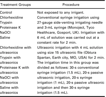
stressed biofilms were developed on the hemi-sec-tioned teeth using the above-menhemi-sec-tioned protocol. After that, the root halves were reapproximated, the teeth were randomly allocated to eleven treatment groups, and their respective irrigation procedures were carried out (Table 1). For conventional syringe irrigation, a rubber stop was placed at a length of 14 mm from the tip of the irrigating needle. The nee-dle was moved back and forth in the canal gently, ensuring that it did not bind in the canal. In case of ultrasonic irrigation, the power setting on the ultrasonic unit (Piezon Master 400; Electro Medical Systems, Nyon, Switzerland) was kept at one quarter of maximum for all of the roots. The size 15 ultra-sonic file (Endosonore File, Dentsply Maillefer) was inserted into the canal as far as possible without obvious constraint of file oscillation.
[image:4.595.281.493.483.676.2]After irrigation, the tooth was taken out of the silicon index, and the chosen root half with the two lateral canals was place in the 9 cm Petri dish (SLS, Nottingham, UK). For microbial analysis, the samples were taken from the upper and lower half of the root canal, and the medial (close to the main root canal) and distal (close to the outer surface of the tooth) portion of middle 1/3 and the apical 1/3 of the artifi-cially created lateral canal using sterile paper points (Protaperâ Universal F3 Paper point Dentsply, Maille-fer). Viable counts of all species were determined as described above, and the biofilm was also visualized using CLSM and analysed usingbioImage_L.
Table 1 Treatments and procedures applied to the stressed biofilms developed in single-rooted teeth
Treatment Groups Procedure Control Not exposed to any irrigant. Chlorhexidine
Trypsin Proteinase K NaOCl Saline
Conventional syringe irrigation using 27-gauge side-venting irrigating needle and 3-mL syringe (Monoject, Tyco Healthcare, Gosport, UK). Irrigation with 6 mL of solution was carried out at a constant rate for 2 min.
Chlorhexidine with ultrasonics Trypsin with ultrasonics Proteinase K with ultrasonics NaOCl with ultrasonics Saline with ultrasonics
Ultrasonic irrigation with 4 mL solution using size 15 ultrasonic file (Obtura Spartan, Earth city, MO, USA) for 2 min. The irrigation time in this group was divided as follows: 30-s conventional syringe irrigation (1.5 mL), 20-s passive ultrasonic irrigation, 20-s syringe irrigation (1 mL), 20-s passive ultrasonic irrigation and then 30-s syringe irrigation (1.5 mL).
Statistical analysis
The statistical analysis was performed in SPSSPC (Ver 16.0)
5 using a general linear model for multivariate analysis of variance single factor: ANOVA was used to
compare the quantitative viable counts between the treatment groups. bioImage_L (Chavez de Paz 2009) was used to compare the biovolume and biofilm substratum coverage in each treatment group by performing two-way analysis of variance (ANOVA).
Results
The characterization of the stressedin vitro multispecies biofilm
The mean number of bacteria (SD) as log10 (CFU
per sample) in the biofilm at T0 was 6.89 (0.47) and 6.7 (0.31) at T1 and was reduced significantly (P<0.05) to 5.89 (0.5) at T2. The pH value of the uninoculated mFUM was 7.19. At T0, it was as low as 5.58 (0.1), which significantly increased to 5.94 (0.13) at T1 (P<0.05) and further increased to 6.13 (0.1) at T2 (P=0.05); indicating extensive proteolysis. The glucose concentration of the uninocu-lated mFUM was 1.05 mg mL 1 but was reduced to
0.22 mg mL 1 at T0 and was <0.01 mg mL 1 at both T1 and T2, respectively. The SDS-PAGE of
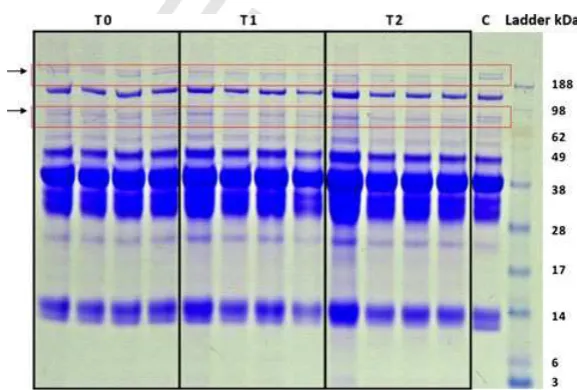
culture supernatants revealed loss of bands and the change in their mobility indicating degradation and loss of serum proteins at T1 and T2 (Fig. 1).
The CLSM analysis indicated that at T0, 99% of the biofilm bacteria at all levels throughout the biofilm were viable. At T1, the proportion of viable cells was significantly reduced, but at T2, the proportion of viable cells was>90% at all levels (Fig. 2). The distri-bution at T1 and T2 was different to that observed at T0. It was decided to use cells at T1 for all the other parts of the study.
The effectiveness of irrigants on biofilms on hydroxyapatite discs
The mean number of bacteria (SE) grown from the control discs, as log10 (CFU per sample), was 8.07
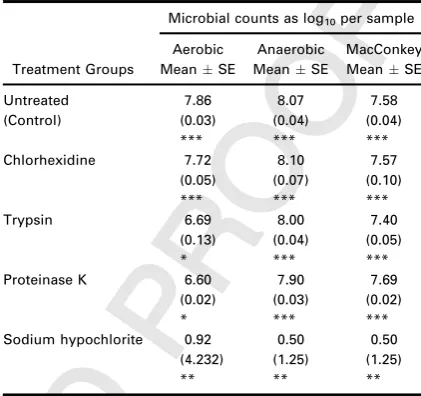
[image:5.595.178.467.421.616.2](0.04) anaerobically, 7.86 (0.03) aerobically and 7.58 (0.04) on the MacConkey agar (Table 2). Chlorhexidine had no significant effect on the recov-ery of bacteria. Although trypsin and proteinase K also did not show any significant effect on the recov-ery of bacteria anaerobically or on MacConkey agar, they both significantly (P<0.05) reduced the recov-ery aerobically. NaOCl was the most effective, signifi-cantly (P<0.001) reducing the recovery on all media with mean values of <10 CFU per sample (Table 2).
Figure 1 Comparison of total protein in the broth supernatant of three groups of biofilms T0, T1 and T2 separated by sodium dodecyl sulphate polyacrylamide gel electrophoresis. Each group had four biofilms. T0 shows the protein separation of the broth supernatant for the biofilms grown for 7 days with regular medium change, whereas T1 and T2 represent the protein separation of broths of the biofilms left in the medium for another 7 and 14 days, respectively. Control (C) is the modified fluid universal medium.
COLOR
1
2 3
4
5
6
7
8
9
10
11
12
13
14
15
16
17 18
19
20
21
22
23
24
25
26
27
28
29
30
31 32
33
34
35
36
37
38
39
40
41
42
43
44
45
46 47
48
49
50
According to the CLSM results, trypsin was most effective in bacterial killing amongst all four treat-ment groups as the mean per cent biovolumes for red (dead) populations were highest (55%) (Fig. 3a). The chlorhexidine group mean per cent biovolume of red (dead) populations was significantly less (21%) than that of both trypsin (55%) and proteinase K (37%) (P<0.05). Compared with the control group, chlorh-exidine exhibited no significant effect in the mean per-centage biovolumes for green (75%) and red (21%) populations (P>0.05). The mean per cent substra-tum coverage of the proteinase K group (74.1%) was significantly higher than the control group (54.3%) (P<0.05), but there was no significant difference between the test groups (Fig. 3b).
Effects of irrigants on microbial killing on single-rooted teeth
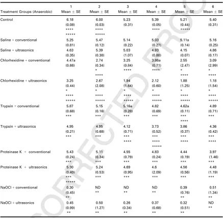
The quantitative viable counts showed that the pas-sive ultrasonic irrigation was significantly more effec-tive in killing the biofilm bacteria as compared to the conventional syringe irrigation using the same irri-gant (P<0.05) (Table 3). NaOCl used with either conventional or ultrasonic irrigation proved to be the best irrigant with the viable counts significantly less than those of control, chlorhexidine, trypsin and pro-teinase K in all six sample sites (P<0.001).
The viable counts for saline used with conventional or ultrasonics were significantly reduced (P<0.05) as compared to the untreated control group. When
compared to saline, proteinase K with conventional or ultrasonic irrigation and trypsin with conventional syringe irrigation caused no significant reduction in the viable counts (P>0.05); however, trypsin with ultrasonics caused significant killing (P<0.05) (Table 3).
In the case of chlorhexidine, the viable count values for chlorhexidine used with conventional or ultrasonic irrigation were significantly less than those of saline (P<0.05). Moreover, chlorhexidine with ultrasonic irrigation was significantly more effective than both trypsin and proteinase K with conventional or ultra-sonic irrigation (P<0.05). Chlorhexidine with ultra-sonic activation was more effective than conventional syringe irrigation in the medial portion of apical 1/3 of the artificially created lateral canal (Table 3).
Effects of irrigants on biofilm disruption on single-rooted teeth
[image:6.595.50.263.69.187.2]The effects of the irrigants with or without ultrasonics on the tooth model are shown in Fig. 4(a,b). The sub-stratum coverage of all the treatment groups was sig-nificantly less than the untreated control (P<0.001) (Fig. 4b), and the majority of bacteria remaining after treatments were viable (Fig. 4a). This was in contrast to the effects of the irrigants on the substratum Figure 2 The distribution of the viable bacteria in all three
[image:6.595.279.490.93.291.2]groups of biofilm (group T0 is the biofilms grown for 7 days with regular medium change, whereas T1 and T2 represent the biofilms left in the medium for another 7 and 14 days, respectively).The x-axis shows the 9 levels of the z stack (level 0 is the deepest level of biofilm and level 9 is the most superficial level of the biofilm), and they-axis gives the mean of the percentages of the living bacteria in all biofilms in each group.
Table 2 Quantitative viable counts of the five disc model biofilm experimental groups
Treatment Groups
Microbial counts as log10per sample
Aerobic Anaerobic MacConkey MeanSE MeanSE MeanSE Untreated (Control) 7.86 (0.03) *** 8.07 (0.04) *** 7.58 (0.04) *** Chlorhexidine 7.72 (0.05) *** 8.10 (0.07) *** 7.57 (0.10) *** Trypsin 6.69 (0.13) * 8.00 (0.04) *** 7.40 (0.05) *** Proteinase K 6.60
(0.02) * 7.90 (0.03) *** 7.69 (0.02) *** Sodium hypochlorite 0.92
(4.232) ** 0.50 (1.25) ** 0.50 (1.25) ** *Values significantly less than control and chlorhexidine groups but greater than sodium hypochlorite (P<0.001). **Values significantly less than all other groups (P<0.001). ***No significant difference in the values between the groups (P>0.05).
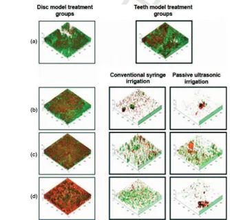
coverage on discs (Figs 5 and 6), where the substra-tum coverage was more than that of control after irri-gant treatment. Chlorhexidine (1.46 0.42), trypsin (3.56 1.18) and proteinase K (4.21.01) with ultrasonics were significantly effective (P<0.05) in reducing the substratum coverage as compared to sal-ine with ultrasonics (12% 4.9) (Fig. 5b). The bio-film disrupting efficacy of the irrigants using tooth model biofilms is more apparent than the disc model in the representative images of the treated root canals (Fig. 6).
Discussion
The success of root canal treatment depends largely on chemomechanical procedures facilitating
debride-ment of the root canal system (Bystrom & Sundqvist€ 1983, Sj€ogren et al. 1997, Siqueiraet al. 2002). The major cause of refractory endodontic infections is the persistence of bacteria in the root canal space (Moorer & Wesselink 1982). The biofilm communities of refractory endodontic infections are surviving under nutritionally stressful environmental conditions that possibly render them resistant to the chemomechani-cal disinfecting procedures (Siqueira 2002). A nutri-ent-stressed in vitro biofilm model was successfully established. As fermentation of glucose results in the production of acid, a low pH of broth supernatant was seen after an initial 7 days of regular medium replenishment. Leaving the biofilms for 7 days in the same medium led to the complete consumption of glu-cose from broth supernatant; thus, leaving the biofilm (a)
[image:7.595.177.470.65.423.2](b)
Figure 3 The effect of irrigant treatment on biofilms grown on hydroxyapatite discs (a) the effect of selected irrigant treatments 10 on the mean percentages of dead (red), live (green) and unknown (blue) biovolumes, of the biofilms in their respective treat-ment groups and (b) the effect of irrigant treattreat-ment on the mean percentage of substratum coverage of the biofilms.
LOW
RESOLUTI
ON
COLOR
FIG
1
2 3
4
5
6
7
8
9
10
11
12
13
14
15
16
17 18
19
20
21
22
23
24
25
26
27
28
29
30
31 32
33
34
35
36
37
38
39
40
41
42
43
44
45
46 47
48
49
50
bacteria starved of the depleted fermentable carbohy-drates. After elimination of glucose from the broth, to obtain energy, the biofilm bacteria started degrading
[image:8.595.53.491.133.560.2]the proteins as was seen by SDS-PAGE results. The degradation of proteins results in the release of ammonia, which led to the rise in the pH of the broth Table 3Anaerobic quantitative viable counts of all six sites taken from five biofilms each, in the nine experimental groups after their respective irrigant treatment. (1) upper half of root canal, (2) lower half of root canal, (3) medial portion of middle 1/3 of the artificially created lateral canal close to the main root canal, (4) distal portion of middle 1/3 of the artificially cre-ated lateral canal close to the outer surface of the tooth, (5) medial portion of apical 1/3 of the artificially crecre-ated lateral canal close to the main root canal and (6) distal portion of apical 1/3 of the artificially created lateral canal close to the outer surface of tooth. The values are given as the mean number of bacteria (standard error) as log10(CFU per sample)
Treatment Groups (Anaerobic)
1 2 3 4 5 6
MeanSE MeanSE MeanSE MeanSE MeanSE MeanSE
Control 6.18 (0.08) **** ***** 6.00 (0.03) **** ***** 5.23 (0.31) 5.39 (0.05) **** 5.21 (0.44) ***** 5.40 (0.31)
Saline+conventional 5.25 (0.81) 5.47 (0.12) 5.14 (0.22) 5.03 (0.27) 5.11a (0.14) 5.16 (0.25) Saline+ultrasonics 4.63
(0.35) 5.39 (0.38) 5.03 (1.07) 4.93 (0.80) 4.15 (0.07) 4.86 (0.17) Chlorhexidine+conventional 4.47a
(0.66) 2.74 (0.34) * **** 3.25 (0.84) **** 3.86a (0.71) **** 2.55 (2.47) * **** 3.09 (2.99) * **** Chlorhexidine+ultrasonics 3.25
(0.44) * **** ***** 2.67 (2.08) * **** ***** 1.94 (1.64) * **** ***** 2.12 (0.60) * **** ***** 1.88 (1.25) * **** ***** 1.18 (1.54) * **** ***** Trypsin+conventional 5.87
(0.68) *** 5.15 (0.16) *** 5.16a (0.28) *** 4.82 (0.69) *** 4.62a (0.11) *** **** 4.89 (0.71) ***
Trypsin+ultrasonics 4.95 (0.21) *** 4.95 (0.68) *** 4.12 (0.71) *** **** 3.73 (0.52) *** **** ***** 3.86 (0.37) *** **** 4.36 (0.42) *** ****
Proteinase K + conventional 5.43 (0.24) *** 5.11 (0.34) *** 4.55 (0.79) *** 4.83 (0.24) *** 4.44 (0.19) *** 3.97 (1.46) *** Proteinase K + ultrasonics 5.30
(0.40) *** ***** 5.24 (0.53) *** 4.69 (0.95) *** 4.6 (2.09) *** 4.56 (0.56) *** 4.48 (1.19) ***
NaOCl+conventional 0.30 (0.45) ** ND ** ND ** ND ** 0.39 (0.76) ** 0.51 (1.34) ** NaOCl+ultrasonics 0.45
(0.99) ** 0.50 (1.27) ** 0.26 (0.34) ** 0.37 (0.68) ** 0.32 (0.51) ** ND **
aUltrasonic irrigation better than conventional syringe irrigation using same irrigant (P<0.05). *Values significantly (P<0.05) less than all other groups, except NaOCl.
**Values significantly less than all other groups (P<0.001).
***No significant difference in the values between the groups (P>0.05).
****Values significantly (P<0.05) less than saline+conventional at the same sampling site. *****Values significantly (P<0.05) less than saline+ultrasonics at the same sampling site. ND (not detected)=0.00.
supernatant during the starvation period. The viable counts of the biofilm also decreased during the period of nutritional stress.
To test the biofilm killing capacity of selected chem-ical solutions and enzymic preparations, a study was carried out using the stressed in vitro biofilm model grown on hydroxyapatite discs. The results of the quantitative viable counts clearly showed that NaOCl was the most effective chemical in killing biofilm bac-teria. This finding is consistent with previous studies where NaOCl was proven to be the most efficient irrigant (Bystr€om & Sundqvist 1983, Siqueira et al.
1997). Trypsin and proteinase K were associated with greater bacterial killing than chlorhexidine and the
untreated control when the biofilm was cultured aero-bically on FAA plates after treatment; however, when cultured aerobically on MacConkey agar and anaero-bically on FAA, there was no significant difference between the killing efficacy of chlorhexidine, trypsin, proteinase K and untreated controls. It is probable that trypsin and proteinase K had an antibacterial effect on the aerobically growing organisms of the multispecies model biofilm (i.e. E. faecalis, S. mitis,
S. epidermidis or A. radicidentis). It is less likely that
E. faecaliswould have been affected by these enzymes, because MacConkey agar culturing did not yield any significant viability difference between the four irri-gants. Therefore, these enzymes were more effective (a)
[image:9.595.179.469.68.449.2](b)
Figure 4 The effect of irrigant treatment on biofilms grown on teeth model (a) the effect of selected irrigant treatments on the 11 mean percentages of dead (red), live (green) and unknown (blue) biovolumes, of the biofilms in their respective treatment groups and (b) the effect of irrigant treatment on the mean percentage of substratum coverage of the biofilms.
LOW
RESOLUTI
ON
COLOR
FIG
1
2 3
4
5
6
7
8
9
10
11
12
13
14
15
16
17 18
19
20
21
22
23
24
25
26
27
28
29
30
31 32
33
34
35
36
37
38
39
40
41
42
43
44
45
46 47
48
49
50
in killing S. mitis, S. epidermidis and A. radicidentis
whereasE. faecalisandP. acneswere more resistant. Interestingly, in the hydroxyapatite disc model, chlorhexidine appeared to be inefficient in eradicating
the biofilm. This is in contrast to the existing litera-ture (Evanovet al.2004, Senaet al.2006, Shenet al.
2009). This may be due to the resistance of the biofilm model developed during the nutritional stress phase.
The CLSM results of bacterial killing efficacy on disc model biofilm revealed that both trypsin and protein-ase K had an effect, whereas there was no difference in chlorhexidine from the control group. The CLSM results seemed to be inconsistent with the case of anaerobic and MacConkey quantitative viable counts, where there was no significant difference between chlorhexidine, trypsin, proteinase K and the untreated control group. Previous studies using live–dead stain have revealed that staining with SYTO 9 and propidi-um iodide (PI) does not always produce distinct ‘live’ and ‘dead’ populations, whereas an intermediate cellular state is also observed (Christiansen et al.
[image:10.595.47.266.123.273.2]2003, Hoefelet al. 2003, Berneyet al. 2007). This is referred to in the manufacturer’s manual as ‘unknown’ (http://probes.invitrogen.com/media/pis/ Figure 5 Comparison of the biofilm disrupting efficacy
between disc model biofilm and tooth model biofilm using chlorhexidine, trypsin and proteinase K with conventional syringe irrigation.
(a)
(b)
(c)
(d)
Figure 6 3-D view of the disc and teeth model biofilms. (a) control; (b) chlorhexidine; (c) proteinase K; and (d) trypsin. 12
LOW
RESOLUTIO
N
COLOR
FIG
COLOR
1
2 3
4
5
6
7
8
9
10
11
12
13
14
15
16
17 18
19
20
21
22
23
24
25
26
27
28
29
30
31 32
33
34
35
36
37
38
39
40
41
42
43
44
45
46 47
48
49
50
[image:10.595.86.421.340.659.2]mp34856.pdf) in which the partially damaged bacte-rial membranes can allow the penetration of red fluorescing PI dye even when the cells are viable. It is possible that these cells are capable of revival. The discrepancy between the cultural and CLSM results can be attributed to these intermediate cellular state bacteria. These were detected dead by CLSM results, but survived once plated and grown onto the rich FAA and MacConkey medium under appropriate growth conditions.
The bleaching effect of 1% NaOCl was confirmed on the SYTO 9 and PI dyes of live/dead stain using a planktonic E. faecalissuspension andE. faecalis single-species biofilm. Based on these findings, it was not possible to analyse these biofilms using CLSM imag-ing. Sodium thiosulphate has been used to neutralize NaOCl when assessing its effect on biofilms using confocal microscopy (Bryceet al.2009)
6 ; however,
fur-ther studies are needed to evaluate the effect of such compounds on bacterial biofilms.
Based on the findings of this initial study, a closed apex tooth model was used to determine the effects of these irrigants using conventional syringe irrigation and passive ultrasonic irrigation in the root canal space in the hope of making the findings more rele-vant. Every effort was made to make the tooth model as realistic as possible, although the flow of the irri-gant within the root canal space remains different from that of a real root canal due to the inevitably imperfect seal of the two reapproximated root halves.
Bhuva et al. (2010), using a similar experimental model with a single-species biofilm, showed that ster-ile saline used as a control group was less effective as compared to NaOCl. The quantitative viable count results of the present study using a tooth model showed that NaOCl had the best biofilm killing effi-cacy with no significant difference when used with either ultrasonic irrigation or conventional syringe irrigation. Chlorhexidine with either conventional syringe or ultrasonic irrigation had significantly higher killing than saline. Moreover, chlorhexidine when used with ultrasonics achieved a biofilm killing significantly higher than those of both trypsin and proteinase K. The killing potential of proteinase K with or without ultrasonics and trypsin with conven-tional syringe irrigation was no different than those of saline. However, the results of both quantitative viable counts and CLSM showed that trypsin when used with ultrasonic activation had better killing as compared to saline with conventional or ultrasonic irrigation.
The CLSM results suggest that in the tooth model biofilm, even inactive saline was significantly effective in disrupting the biofilm and reducing the substratum coverage as compared to the untreated control group. This could be attributed to the mechanical flushing out of the biofilm from the root canal. However, the biofilm disruption of chlorhexidine, trypsin and pro-teinase k used with ultrasonics was significantly more than that of saline with ultrasonics.
Confocal laser scanning microscopy results revealed that chlorhexidine with ultrasonics proved to be the most effective approach for disrupting the biofilm. However, in contrast to their quantitative viable count results, the biofilm killing efficacy of this group was found to be lower than even untreated control, proteinase K and trypsin with conventional or ultra-sonic irrigation. Another observation in this group was the high percentage of the unknown biovolume that was not differentiated by thebioImage_L software as either red or green during analysis. This suggests the possibility that the portion of this unknown biovo-lume may be the dead population left unrecognized during analyses resulting in inconsistent results of the quantitative viable counts and the CLSM.
In this study was tested the efficacy of two enzymes (proteinase K and trypsin) in disrupting and killing biofilm bacteria. Recent studies have shown that both these proteases were efficient in removing the biofilm
of S. aureus 383 (Chaignon et al. 2007). Moscoso
et al. (2006), using 100lg mL 1 of both these 7
enzymes for 1 h, successfully demonstrated their inhibitory effects on biofilm development of Streptococ-cus pneumoniae (Moscoso et al. 2006). Patterson et al.
(2007)used 100lg mL 1 of proteinase K to deter- 8 mine the effect on biofilm viability. Moreover, it was also reported that a concentration as low as 32 lg
mL 1 and 6.4lg mL 1 of proteinase K and trypsin,
respectively, effectively disrupted the mature biofilm of three different strains of Gardnerella vaginalis (Patter-sonet al.2007). The enzymes have different substrate specificities: proteinase K endolytically cleaves the peptide bonds of aliphatic, aromatic or hydrophobic amino acids, whereas trypsin is specific for the peptide bonds of lysine and arginine (Chaignon et al. 2007). In the present study, trypsin as well as proteinase K with ultrasonic activation was efficient in disrupting the biofilm although less than chlorhexidine. This may be attributed to the combined mechanical effect of ultrasonics complemented with the hydrolytic activity of these proteases by acting on the protein fraction of the biofilm. Although there is little evi-1
2 3
4
5
6
7
8
9
10
11
12
13
14
15
16
17 18
19
20
21
22
23
24
25
26
27
28
29
30
31 32
33
34
35
36
37
38
39
40
41
42
43
44
45
46 47
48
49
50
dence on the exact antibacterial effects of these enzymes, in this study, trypsin especially when used with ultrasonics caused bacterial killing when com-pared to saline. It is possible that the protease acts by degrading the protein components of the bacterial cell walls and membranes, causing cellular damage and ultimately cell lysis and death. Furthermore, the pro-teolytic enzymes might disrupt the extracellular matrix, which is also made of proteins secreted by the bacteria, thus reducing the cohesion of the biofilm. Recent studies suggest that cell surface proteins might act as co-aggregation factors during the biofilm formation as these surface structures were found to be protease sensitive (Furukawa et al. 2011). None-theless, the killing efficacy of the two proteases was lower than that of NaOCl.
For all irrigants except NaOCl, the results of quanti-tative viable counts as well as CLSM analysis showed that passive ultrasonic was more effective than con-ventional syringe irrigation. Previous studies sug-gested that the heat generation by ultrasonics (Zeltner et al. 2009) may also have an enhancing effect on the antibacterial properties of chlorhexidine (Evanov et al. 2004) and NaOCl (Cunningham & Balekjian 1980, Abou-Rass & Oglesby 1981, Cunn-inghamet al. 1982). The heat generated by ultrason-ics may also have enhanced the antibacterial properties of trypsin.
Greater biofilm disruption by flushing out and shear stress is achieved in the tooth model as compared to the disc model biofilm because the mechanical effects of irrigation protocol are better simulated in a con-fined root canal space. Moreover, this tooth model offers the potential advantage to allow the investiga-tion of the efficacy of different endodontic instrumen-tation techniques.
Conclusion
This study suggests that trypsin used as an irrigant with ultrasonic activation has biofilm killing and disrupting potential. Even though the antibacterial properties were not as good as NaOCl, it certainly has a biofilm disrupting ability. Further studies are neces-sary to assess its compatibility with other irrigants and to determine its potential in a disinfection regi-men, where enzymes can help disrupt the biofilm and render single bacterial cells more susceptible to killing by other stronger antimicrobials. Investigations are also required to determine its impact and toxicity on dentine and periapical tissues.
Acknowledgments
We acknowledge support from the Higher Education Commission of Pakistan; from the Dental Institute, King’s College London. We thank Professor Gunnar Dahlen (Department of Oral Microbiology, Faculty of Odontology, University of G€oteborg, Sweden) for pro-viding the E. faecalis strain and Dr. Luis Chevaz de Paz (Department of Oral Biology, Malm€o University, Sweden) for providingBioImage_L software.
References
Abou-Rass M, Oglesby SW (1981) The effects of tempera-ture, concentration, and tissue type on the solvent abil-ity of sodium hypochlorite. Journal of Endodontics 7, 376–7.
Berney M, Hammes F, Bosshard F, Weilenmann HU, Egli T
et al. (2007) Assessment and interpretation of bacterial viability by using the LIVE/DEAD BacLight Kit in combina-tion with flow cytometry.Applied Environmental
Microbiol-ogy73, 3283–90. 9
Bhuva B, Patel S, Wilson R, Niazi S, Beighton D, Mannocci F (2010) The effectiveness of passive ultrasonic irrigation on intraradicular Enterococcus faecalis biofilms in extracted single-rooted human teeth.International Endodontic Journal
43, 241–50.
Bowden GH, Li YH (1997) Nutritional influences on biofilm development.Advances in Dental Research11, 81–99.
Bryce G, O’Donnell D, Ready D, Ng YL, Pratten J, Gulabivala K (2009) Contemporary root canal irrigants are able to disrupt and eradicate single- and dual-species biofilms.
Journal of Endodontics35, 1243–8.
Bystr€om A, Sundqvist G (1981) Bacteriologic evaluation of the efficacy of mechanical root canal instrumentation in endodontic therapy.Scandinavian Journal of Dental Research
89, 321–8.
Bystr€om A, Sundqvist G (1983) Bacteriologic evaluation of the effect of 0.5 percent sodium hypochlorite in endodon-tic therapy.Oral Surgery, Oral Medicine, and Oral Pathology
55, 307–12.
Chaignon P, Sadovskaya I, Ragunah C, Ramasubbu N, Kap-lan JB, Jabbouri S (2007) Susceptibility of staphylococcal biofilms to enzymatic treatments depends on their chemi-cal composition. Applied Microbiology Biotechnology 75, 125–32.
Chavez de Paz LE (2009) Image analysis software based on color segmentation for characterization of viability and physiological activity of biofilms. Applied Environmental Microbiology75, 1734–9.
Christiansen T, Michaelsen S, Wumpelmann M, Nielsen J (2003) Production of savinase and population viability of
Bacillus clausii during high-cell-density fed-batch cultiva-tions.Biotechnology Bioengineering83, 344–52.
1
2 3
4
5
6
7
8
9
10
11
12
13
14
15
16
17 18
19
20
21
22
23
24
25
26
27
28
29
30
31 32
33
34
35
36
37
38
39
40
41
42
43
44
45
46 47
48
49
50
Cunningham WT, Balekjian AY (1980) Effect of temperature on collagen-dissolving ability of sodium hypochlorite end-odontic irrigant. Oral Surgery, Oral Medicine, and Oral Pathology49, 175–7.
Cunningham WT, Martin H, Pelleu GB Jr, Stoops DE (1982) A comparison of antimicrobial effectiveness of endosonic and hand root canal therapy.Oral Surgery, Oral Medicine, and Oral Pathology54, 238–41.
Dahlen G, Samuelsson W, Molander A, Reit C (2000) Identi-fication and antimicrobial susceptibility of enterococci isolated from the root canal. Oral Microbiology and Immu-nology15, 309–12.
Dalton BC, Orstavik D, Phillips C et al. (1998) Bacterial reduction with nickel-titanium rotary instrumentation.
Journal of Endodontics24, 763–7.
Evanov C, Liewehr F, Buxton TB, Joyce AP (2004) Antibac-terial efficacy of calcium hydroxide and chlorhexidine glu-conate irrigants at 37°C and 46°C.Journal of Endodontics
30, 653–7.
Furukawa S, Nojima N, Yoshida K, Hirayama S, Ogihara H, Morinaga Y (2011) The Importance of inter-species cell-cell Co-Aggregation betweenLactobacillus plantarum ML11-11 andSaccharomyces cerevisiae BY4741in mixed-species biofilm formation. Bioscience, Biotechnology, and Biochemis-try75, 1430–4.
Giardino L, Ambu E, Savoldi E, Rimondini R, Cassanelli C, Debbia EA (2007) Comparative evaluation of antimicrobial efficacy of sodium hypochlorite, MTAD, and Tetraclean against Enterococcus faecalisbiofilm.Journal of Endodontics
33, 852–5.
Gmur R, Guggenheim B (1983) Antigenic heterogeneity of Bacteroides intermedius as recognized by monoclonal anti-bodies.Infection and Immunity42, 459–70.
Gulabivala K, Stock CJ, Lewsey JD, Ghori S, Ng YL, Spratt DA (2004) Effectiveness of electrochemically activated water as an irrigant in an infected tooth model. Interna-tional Endodontic Journal37, 624–31.
Hems RS, Gulabivala K, Ng YL, Ready D, Spratt DA (2005) Anin vi-troevaluation of the ability of ozone to kill a strain ofEnterococcus faecalis.International Endodontic Journal38, 22–9.
Hoefel D, Grooby WL, Monis PT, Andrews S, Saint CP (2003) Enumeration of water-borne bacteria using viability assays and flow cytometry: a comparison to culture-based techniques.Journal of Microbiological Methods
55, 585–97.
Hubble TS, Hatton JF, Nallapareddy SR, Murray BE, Gillespie MJ (2003) Influence of Enterococcus faecalis proteases and the collagen-binding protein, Ace, on adhesion to dentin.
Oral Microbiology and Immunology18, 121–6.
Kishen A, George S, Kumar R (2006) Enterococcus faecalis -mediated biomineralized biofilm formation on root canal dentine in vitro. Journal of Biomedical Materials Research Part A77, 406–15.
Kowalski WJ, Kasper EL, Hatton JF, Murray BE, Nallapared-dy SR, Gillespie MJ (2006) Enterococcus faecalis adhesin,
Ace, mediates attachment to particulate dentin.Journal of Endodontics32, 634–7.
Laemmli UK (1970) Cleavage of structural proteins during the assembly of the head of bacteriophage T4. Nature
227, 680–5.
Lee SJ, Wu MK, Wesselink PR (2004) The effectiveness of syringe irrigation and ultrasonics to remove debris from simulated irregularities within prepared root canal walls.
International Endodontic Journal37, 672–8.
Moorer WR, Wesselink PR (1982) Factors promoting the tissue dissolving capability of sodium hypochlorite. Interna-tional Endodontic Journal15, 187–96.
Moscoso M, Garcia E, Lopez R (2006) Biofilm formation by
Streptococcus pneumoniae: role of choline, extracellular DNA, and capsular polysaccharide in microbial accretion.
Journal of Bacteriology188, 7785–95.
Niazi SA, Clarke D, Do T, Gilbert SC, Mannocci F, Beighton D (2010) Propionibacterium acnesandStaphylococcus epide-rmidis isolated from refractory endodontic lesions are opportunistic pathogens. Journal of Clinical Microbiology
48, 3859–69.
Pappen FG, Shen Y, Qian W, Leonardo MR, Giardino L, Haapasalo M (2010)In vitroantibacterial action of Tetra-clean, MTAD and five experimental irrigation solutions.
International Endodontic Journal43, 528–35.
Patterson JL, Girerd PH, Karjane NW, Jefferson KK (2007) Effect of biofilm phenotype on resistance of Gardnerella vaginalis to hydrogen peroxide and lactic acid. American Journal of Obstetrics and Gynecology197(170), e171–7.
Sena NT, Gomes BP, Vianna MEet al.(2006)In vitro antimi-crobial activity of sodium hypochlorite and chlorhexidine against selected single-species biofilms. International End-odontic Journal39, 878–85.
Shen Y, Qian W, Chung C, Olsen I, Haapasalo M (2009) Evaluation of the effect of two chlorhexidine preparations on biofilm bacteria in vitro: a three-dimensional quantita-tive analysis.Journal of Endodontics35, 981–5.
Shen Y, Stojicic S, Qian W, Olsen I, Haapasalo M (2010) The synergistic antimicrobial effect by mechanical agita-tion and two chlorhexidine preparaagita-tions on biofilm bacte-ria.Journal of Endodontics36, 100–4.
Siqueira JF Jr (2002) Endodontic infections: concepts, para-digms, and perspectives. Oral Surgery, Oral Medicine, Oral Pathology, Oral Radiology, and Endodontics94, 281–93.
Siqueira JF Jr, Machado AG, Silveira RM, Lopes HP, de Uzeda M (1997) Evaluation of the effectiveness of sodium hypo-chlorite used with three irrigation methods in the elimina-tion of Enterococcus faecalis from the root canal, in vitro.
International Endodontic Journal30, 279–82.
Siqueira JF Jr, Rocas IN, Santos SR, Lima KC, Magalh~aes FA, de Uzeda M (2002) Efficacy of instrumentation techniques and irrigation regimens in reducing the bacterial popula-tion within root canals.Journal of Endodontics28, 181–4.
as an irrigant and Calcium Hydroxide/Camphorated Para-monochlorophenol paste as an intracanal dressing.Journal of Endodontics33, 667–72.
Sj€ogren U, Figdor D, Persson S, Sundqvist G (1997) Influ-ence of infection at the time of root filling on the outcome of endodontic treatment of teeth with apical periodontitis.
International Endodontic Journal30, 297–306.
van der Sluis LW, Gambarini G, Wu MK, Wesselink PR (2006) The influence of volume, type of irrigant and flush-ing method on removflush-ing artificially placed dentine debris
from the apical root canal during passive ultrasonic irriga-tion.International Endodontic Journal39, 472–6.
Weller RN, Brady JM, Bernier WE (1980) Efficacy of ultra-sonic cleaning.Journal of Endodontics6, 740–3.
Zehnder M (2006) Root canal irrigants.Journal of Endodontics
32, 389–98.
Zeltner M, Peters OA, Paque F (2009) Temperature changes during ultrasonic irrigation with different inserts and modes of activation.Journal of Endodontics35, 573–
Journal:
IEJ
Article:
12214
Dear Author,
During the copy-editing of your paper, the following queries arose. Please respond to these by mark-ing up your proofs with the necessary changes/additions. Please write your answers on the query sheet if there is insufficient space on the page proofs. Please write clearly and follow the conventions shown on the attached corrections sheet. If returning the proof by fax do not write too close to the paper’s edge. Please remember that illegible mark-ups may delay publication.
Many thanks for your assistance.
Query reference Query Remarks
1 AUTHOR: Please identify and encircle the fore-name and surfore-name of all authors.
2 AUTHOR: Please confirm that all affiliations are correct.
3 AUTHOR: The term “LIVE/DEAD” has been changed to “live/dead” throughout the article. Please check whether the edit is correct.
4 AUTHOR: Shuping et al. 2000 has not been included in the Reference List, please supply full publication details.
5 AUTHOR: Please give manufacturer informa-tion for ‘SPSSPC’: company name, town, state (if USA), and country.
6 AUTHOR: Bryce 2009 has been changed to Bryce et al.2009 so that this citation matches the Reference List. Please confirm that this is correct.
7 AUTHOR: Moscoso (2006) has been changed to Moscoso et al. (2006) so that this citation matches the Reference List. Please confirm that this is correct.
Reference Berney et al. (2007), please supply all of their names. If there are 7 or more authors, please supply the first 3 author names then et al. Please check and update all such references found in the list.
10 AUTHOR: Figure 3 has been saved at a low resolution of 202 dpi. Please resupply at 600 dpi. Check required artwork specifications at http://authorservices.wiley.com/bauthor/illus-tration.asp
11 AUTHOR: Figure 4 has been saved at a low resolution of 171 dpi. Please resupply at 600 dpi. Check required artwork specifications at http://authorservices.wiley.com/bauthor/illus-tration.asp
Please correct and return this set
Instruction to printer
Leave unchanged
under matter to remain
through single character, rule or underline
New matter followed by
or
or
or
or
or
or
or
or
or
and/or
and/or
e.g.
e.g.
under character
over character
new character
new characters
through all characters to be deleted
through letter or
through characters
under matter to be changed
under matter to be changed
under matter to be changed
under matter to be changed
under matter to be changed
Encircle matter to be changed
(As above)
(As above)
(As above)
(As above)
(As above)
(As above)
(As above)
(As above)
linking
characters
through character or
where required
through character or
where required
or
indicated in the margin
Delete
Substitute character or
substitute part of one or
more word(s)
Change to italics
Change to capitals
Change to small capitals
Change to bold type
Change to bold italic
Change to lower case
Change italic to upright type
Change bold to non-bold type
Insert ‘superior’ character
Insert ‘inferior’ character
Insert full stop
Insert comma
Insert single quotation marks
Insert double quotation marks
Insert hyphen
Start new paragraph
No new paragraph
Transpose
Close up
Insert or substitute space
between characters or words
Insert in text the matter
Textual mark
Marginal mark
Please use the proof correction marks shown below for all alterations and corrections. If you
in dark ink and are made well within the page margins.
USING e-ANNOTATION TOOLS FOR ELECTRONIC PROOF CORRECTION
Required software to e-Annotate PDFs: Adobe Acrobat Professional or Adobe Reader (version 8.0 or
above). (Note that this document uses screenshots from Adobe Reader X)
The latest version of Acrobat Reader can be downloaded for free at:
http://get.adobe.com/reader/
Once you have Acrobat Reader open on your computer, click on the Comment tab at the right of the toolbar:
1.
Replace (Ins)
Tool
Î
for replacing text.
Strikes a line through text and opens up a text box where replacement text can be entered.
How to use it
‚ Highlight a word or sentence.
‚ Click on the Replace (Ins) icon in the Annotations section.
‚ Type the replacement text into the blue box that appears.
This will open up a panel down the right side of the document. The majority of tools you will use for annotating your proof will be in the Annotations section, rkevwtgf"qrrqukvg0"YgÓxg"rkemgf"qwv"uqog"qh"vjgug"vqqnu"dgnqy<
2.
Strikethrough (Del)
Tool
Î
for deleting text.
Strikes a red line through text that is to be deleted.
How to use it
‚ Highlight a word or sentence.
‚ Click on the Strikethrough (Del) icon in the Annotations section.
3.
Add note to text
Tool
Î
for highlighting a section
to be changed to bold or italic.
Highlights text in yellow and opens up a text box where comments can be entered.
How to use it
‚ Highlight the relevant section of text. ‚ Click on the Add note to text icon in the
Annotations section.
‚ Type instruction on what should be changed regarding the text into the yellow box that appears.
4.
Add sticky note
Tool
Î
for making notes at
specific points in the text.
Marks a point in the proof where a comment needs to be highlighted.
How to use it
‚ Click on the Add sticky note icon in the Annotations section.
‚ Click at the point in the proof where the comment should be inserted.
USING e-ANNOTATION TOOLS FOR ELECTRONIC PROOF CORRECTION
For further information on how to annotate proofs, click on the
Help
menu to reveal a list of further options:
5.
Attach File
Tool
Î
for inserting large amounts of
text or replacement figures.
Inserts an icon linking to the attached file in the appropriate pace in the text.
How to use it
‚ Click on the Attach File icon in the Annotations section.
‚ Enkem"qp"vjg"rtqqh"vq"yjgtg"{qwÓf"nkmg"vjg"cvvcejgf" file to be linked.
‚ Select the file to be attached from your computer or network.
‚ Select the colour and type of icon that will appear in the proof. Click OK.
6.
Add stamp
Tool
Î
for approving a proof if no
corrections are required.
Inserts a selected stamp onto an appropriate place in the proof.
How to use it
‚ Click on the Add stamp icon in the Annotations section.
‚ Select the stamp you want to use. (The Approved
stamp is usually available directly in the menu that appears).
‚ Enkem"qp"vjg"rtqqh"yjgtg"{qwÓf"nkmg"vjg"uvcor"vq" appear. (Where a proof is to be approved as it is, this would normally be on the first page).
7.
Drawing Markups
Tools
Î
for drawing shapes, lines and freeform
annotations on proofs and commenting on these marks.
Allows shapes, lines and freeform annotations to be drawn on proofs and for comment to be made on these marks..
How to use it
‚ Click on one of the shapes in the Drawing Markups section.
‚ Click on the proof at the relevant point and draw the selected shape with the cursor. ‚ To add a comment to the drawn shape,
move the cursor over the shape until an arrowhead appears.