www.advhealthmat.de
The Biomedical Use of Silk: Past, Present, Future
Chris Holland,* Keiji Numata,* Jelena Rnjak-Kovacina,* and F. Philipp Seib*
DOI: 10.1002/adhm.201800465
1. Prologue
Before we begin to define the current state of the art surrounding the field of silk-based biomaterials for (bio)medical use and look toward their future, we feel it is important to spend some time considering the motivation of the research and the his-tory of the material that has led us to today. The issue of motivation for studies in the field of silk research is generally divided into bottom-up, curiosity-driven fundamental research and top-down, chal-lenge-based activities.
Fundamental silk research hinges on the question, “what can we learn from nature?” This is clearly a wider topic than silk itself, but the overall approach helps frame scholarly activities in the area. We certainly have more to learn beyond under-standing the silk fiber itself, and dozens of cross-disciplinary researchers worldwide are using both simula-tion and experimentasimula-tion[1] to make concerted efforts to
under-stand the evolution,[2] processing,[3] and performance of silk,[4]
from the molecule[5] to the material.[6] However, as we broaden
our interpretive horizons, we must remember that silks are bio-logical materials, and thus are defined by their biology, before we attempt to transfer this knowledge to biomaterials, which are defined through their application.
2. Introduction
For the purpose of this progress review, we use the term silk to refer to protein-based fiber-forming materials spun by living organisms. We also include in our terminology silk-inspired proteins produced by recombinant approaches.
When studying silks, one must always appreciate that the results derived from testing any naturally obtained bio logical material are a product of both nature (its evolution) and nurture (its environment), with the latter typically constraining the property space of the former (although exceptions exist[4a,7]).
The biological definition of a silk is a structural protein that is spun into a fiber for use outside the body.[4a,8] In the wild,
silks have undergone over 400 million years of “research and development” via natural selection, and after solutions to bio-logical challenges that range from predation (spider webs) to housing (honey bees and wasps) and protection (silkworm cocoons).[4a,8] The ubiquity and widespread use of silk is a clear
testament to its success, especially as it has arisen numerous times in independent convergent evolutionary events.[2c] Hence,
Humans have long appreciated silk for its lustrous appeal and remarkable physical properties, yet as the mysteries of silk are unraveled, it becomes clear that this outstanding biopolymer is more than a high-tech fiber. This progress report provides a critical but detailed insight into the biomedical use of silk. This journey begins with a historical perspective of silk and its uses, including the long-standing desire to reverse engineer silk. Selected silk structure–function relationships are then examined to appreciate past and current silk challenges. From this, biocompatibility and biodegradation are reviewed with a specific focus of silk performance in humans. The current clinical uses of silk (e.g., sutures, surgical meshes, and fabrics) are dis-cussed, as well as clinical trials (e.g., wound healing, tissue engineering) and emerging biomedical applications of silk across selected formats, such as silk solution, films, scaffolds, electrospun materials, hydrogels, and particles. The journey finishes with a look at the roadmap of next-generation recombinant silks, especially the development pipeline of this new industry for clinical use.
Dr. C. Holland
Department of Materials Science and Engineering The University of Sheffield
Sir Robert Hadfield Building, Mappin Street, Sheffield, South Yorkshire S1 3JD, UK
E-mail: [email protected] Dr. K. Numata
Biomacromolecules Research Team
RIKEN Center for Sustainable Resource Science 2-1 Hirosawa, Wako, Saitama 351-0198, Japan E-mail: [email protected]
Dr. J. Rnjak-Kovacina
Graduate School of Biomedical Engineering The University of New South Wales Sydney, NSW 2052, Australia E-mail: [email protected] Dr. F. P. Seib
Leibniz Institute of Polymer Research Dresden Max Bergmann Center of Biomaterials Dresden Dresden 01069, Germany
E-mail: [email protected] Dr. F. P. Seib
Strathclyde Institute of Pharmacy and Biomedical Sciences University of Strathclyde
Glasgow G4 0RE, UK
The ORCID identification number(s) for the author(s) of this article can be found under https://doi.org/10.1002/adhm.201800465.
Silk
looking at how silk materials have evolved can not only deter-mine their performance in the present, but can also reveal common design criteria and molecular “blueprints” for high performance biological materials.[4a,9]
In unraveling the properties of silk, we have also begun to address common misconceptions regarding biological materials and their potential for industrial application. These are often tarred with a brush of sample variability, suggesting that they are unsuitable for engineering or medical applications where consistency is key. However, recent studies now show that the variation previously observed is typically a manifestation of a silk’s exquisite responsiveness to its surroundings (making silks incredibly “smart” materials).[4d,7a,10] Yet for uninitiated
researchers, this can sometimes become unwanted variation if the they fail to ensure consistent sample preparation or testing environments. Hence, biological diversity and plasticity offer several important lessons for those wishing to make the best use of silk for their own applications.
At the other end of the spectrum is the widely held belief that biological materials automatically qualify as “biocompat-ible” materials. While these materials, including many silks, are often biocompatible, simply labeling silk as “biocompatible” without context specific biocompatibility testing and critical assessment of the available evidence is not in the best interest of the field or, ultimately, patients. This mindset also perme-ates into the assumption that all natural materials are “green” which without appropriate and carefully considered environ-mental analysis, the use of the phrase ultimately detracts from any potential impact of these materials.
Once past our prejudices, at the interface of fundamental and challenge-based activities sits biomimetics. This specifi-cally looks to nature to reveal concepts, processes, and systems that can be applied to solve human challenges.[11] While the
term “biomimetics” was only coined by American biophysicist Otto Schmitt in the latter half of the 20th century,[11] humans
have been looking to translate silk’s natural utility for their own use for millennia.[12] The simplest, most primitive forms
of mimicry are examples of imitation of the spider’s use of silk to catch prey, as seen in the Australian Aborigines’ use of spider silk as fishing lines and New Guinean natives’ develop-ment of fishing nets and bags.[13] However, the biological
diver-sity of silk soon inspired humans to adapt silk for their own needs (e.g., ref. [14]), extending the silk phenotype beyond its
natural remit. Some of the first examples were the use of silks medicinally by ancient Greeks and Romans, who bundled up spider silk to treat wounds (Figure 1).[13] This was even noted
by Shakespeare’s character, Nick Bottom, in A Midsummer
Night’s Dream, who says, “I shall desire you of more
acquaint-ance, good Master Cobweb. If I cut my finger, I shall make bold of you.”[15]
However, the above examples describe the use of silk in its unprocessed, natural state. A step forward in the utilitarian evolution of silk came about with the realization that silk could be readily reprocessed into different forms. This was first performed at the macroscale by unwinding fibers from the nonwoven composite cocoons of the silkworm Bombyx
mori to create textiles. This skill originated in China, and direct
archeological evidence confirms human interactions with silk-worm silk originating from the Neolithic period of the 4th mil-lennium BC, with the discovery of examples of cut cocoons and rudimentary looms at numerous archeological sites.[16] Further
archeological evidence suggests that the Indus Valley civiliza-tion (in what is now Northern Pakistan) was also developing silk materials based on Antheraea silk. Therefore, sericulture—the act of rearing silkworms specifically for their silk—can be esti-mated to have spread across South Asia from 5000–2000 BC.[17]
Textiles produced from silk were truly a disruptive product, as they required both a unique material and highly sophisti-cated processing (programmable looms for weaving that were, in essence, the progenitor of modern computing).[18] As such, silk
textiles were sufficiently valuable to become a formal currency for Chinese soldiers at the edges of the empire and were used to barter with the locals for goods.[16] Nevertheless, silk production
remained a closely guarded secret within the Chinese empire for several thousand years, and when asked, traders would say it was “derived from the wool of sheep sprinkled with water and exposed to sunshine.” However, this product monopoly could not go unchallenged for long, and the establishment of trade routes (the “silk roads”), and the apocryphal industrial espionage that ensued, made silk technology available throughout the world. As a result, Bombyx mori silk has developed hand in hand with humans, through domestication and artificial selection of the moths for over 4000 years.[16] This extensive history is a testament
[image:2.595.58.539.69.223.2]to the success and suitability of this animal for large-scale indus-trial agricultural development, as ≈980 billion animals are raised each year to produce ≈400 megatonnes of commercial silk.[19]
Across millennia, silkworm has been a luxury item for the elite. However, Claudius Galenus of Pergamon (c. 131 to c. 211 AD) was the first to document a potential medical application of the silk thread. Galenus gained a reputation for treating gladiators whose tendons were severed in hand-to-hand combat and noted in his book De Methodo Medendi (150 AD) the use of several materials as sutures, including linen. He writes, “in many places under Roman rule you can obtain silk, especially in large cities where there are many wealthy women. If there is no such oppor-tunity, choose from the material where you were living the least putrescible such as thin catgut.” Galenus’s teaching persisted for centuries after his death but was eventually lost.[20] The war
surgeon Ambroise Paré (1510–1590) avoided cauterizing open wounds with boiling oil and reverted to using vascular ligatures made of silk or fine linen strips. However, only in 1869 did Joseph Lister introduce the first sterile silk suture into clinical practice.[20]
Throughout history, several alternative sources for textile silk beyond the domesticated silkworm have been sought, from the wild silkworms of India and Africa[21] to the more esoteric
source represented by spider silk. The quest to commercialize spider silk, due to its favorable mechanical properties, seem-ingly began with the inventions of Abbé Ramon de Termeyer in the 18th century for his reeling device.[22] Over the years, these
inventions were followed by others, such as those of the civil war surgeon Burt G. Wilder.[23] The most successful attempts
are probably those made by the Madagascan spider silk industry, which has produced, to date, only a handful of items destined for the elite.[24] Yet, while producing arguably mechanically
superior materials compared to those made of silkworm silk, none of these endeavors were ultimately found to be scalable.
Hence, given the coveted nature of silk, the fact that indus-trialists wished to replicate it may come as no surprise. In fact, nearly every single industrial fiber produced in the latter half of the 19th and throughout the 20th century, from rayon to nylon to Kevlar, has been developed in the hope that it would provide a suitable alternative to silk.[25] Nevertheless, even after
150 years of concerted research and development, and although replication of the properties and performance of silkworm silk is now possible, similar success with spider silk, and specifi-cally dragline silk, remains elusive. A complete overview of the history, progress, and trends in artificial silk spinning from a fiber performance perspective is available in a recent review that comprehensively covers this topic.[25]
Fortunately, the attempts to replicate various silks resulted in several distinctly important innovations that led the bioma-terials field to consider silk as more than just a fiber.[12] Akin
to the ancient Chinese realizing that a silkworm cocoon can be unspun, early attempts at creating artificial silk led to the conclusion that the silk fiber itself could be “unspun” back into a processable protein feedstock, which could then be solidified into a variety of forms. According to the original patents, this finding was largely motivated by a need to utilize the waste streams from the industry,[26] as the last tens to a hundred
meters of silk from a cocoon could not be unraveled. (Today, this would be labelled an exercise in sustainability.)
To the best of our knowledge, the first attempt to create an artificial silk feedstock appeared at the turn of the 20th century, 110 years ago. It began with the work of Baumann and Diesser, who proposed the dissolution of whole silk glands in formic
acid.[27] In subsequent years, a notable race began between
Japanese and German researchers in the 1920s, with patents granted in 1924–1927[26a,28] and 1928,[29] respectively, for the
successful dissolution (and respinning) of artificial fibers using ZnCl2, Mg(NO3)2, and orthophosphoric acid as the main
chao-tropic agents. However, not until the 1930s did today’s familiar degumming using Na2CO3[30] and dissolution in LiBr appear.[26b]
The latter report clearly noted the potential of silk regeneration/ reconstitution: “These solutions containing, if at all, only a small amount of salt, may be used in the known manner to produce artificial articles, such as fibers, films, or plastic masses.”
Beyond the replication of silk fibers for textile use, these feed-stocks were originally intended for reprocessing into solid form to harness silk’s excellent insulating properties[26b] and enable
the casting of films (to make fabrics water and air imperme-able[31]). This was mainly because naturally derived materials
were still superior in many aspects when compared to those arising from the burgeoning field of industrial polymers.[32]
Interestingly, nearly three decades passed before the first bio-medical use for a regenerated silk was reported in the patent lit-erature. In the 1960s, Bloch and Messores, of Ethicon Inc. (NJ, USA), were the first to propose the use of a LiSCN/LiBr recon-stituted silk as a replacement for the standard wax coating used on silk sutures to reduce their limpness, fraying, and unwanted capillary action.[33] In the following years, while developments
continued in the suture field, another two decades passed before the first examples of nonfibrous silk-based biomate-rial patents were reported. In 1986, a silk fibroin:fibrinogen glue, based on the “standard” LiBr reconstitution approach, was developed by a Japanese firm.[34] This was followed by the
first patent for a silk based porous scaffold in 1987, again from Japan, produced from a freeze-dried native silk solution (i.e., silk extracted directly from the silk gland).[35] The 1990s saw
more patent applications from Japan, including powdered silk for wound dressings,[36] reconstituted silk films and molded
gels for skin, blood vessel, and corneal coatings,[37] and colloidal
silk for consumption in medicine.[38] However, in the 2000s,
an explosion occurred in the USA in research and commer-cialization activity around this area with the emergence of large patent families (>100) focused on the future medical exploita-tion of these materials.[39]
In summary, looking back, the ability to unspin silk, and thereby reconstitute it, has been a monumentally disruptive development in the field. It represents a platform technology for the development of biomimetic structures that are built with silk but are not built to replicate silk. While gaps still undoubtedly exist in our knowledge surrounding the process of reconstitution and how this affects the integrity and application of the silk proteins undergoing it,[40] the unspinning process
has been widely adopted throughout the biomaterials field. This is perhaps best evidenced by the impact of the landmark review of Altman et al.[41] 15 years ago and the more recent protocol of
Rockwood et al.,[42] which leads us in the present day.
3. Silk: Hierarchical and Crystal Structures
subsequent secondary, tertiary, and quaternary structures, govern the protein’s overall function. In nature, silk and silk-like proteins are made by several organisms such as spiders, silkworms, scorpions, mussels, bees, and ants. However, the silk fibroins and silk-like proteins of each organism exhibit dif-ferent physical and biological characters due to their difdif-ferent amino acid sequences, spinning conditions, and hierarchical structures.[7b,43] The hierarchical structures of silk proteins vary
among silk types.[43a,44] Silk proteins produced by spiders and
insects are referred to as silk spidroin and silk fibroin, respec-tively. The term “silk fibroin” is commonly used to differentiate “virgin” silk (silk filament still encased by sericin) or silk cocoons (i.e., the sericin-coated silk thread arranged into a cocoon) from purified silk (i.e., degummed; see Section 6). For the purpose of this progress review, we will use the term silk fibroin to refer to degummed (Bombyx mori) silk unless otherwise stated.
As discussed in the previous section, the silk fibroin of the domesticated silkworm (Bombyx mori) is the most well studied silk for biomedical applications due to its established supply chain, abundance, and clinical track record. The Bombyx mori
protein fiber is a composite material comprising a semi-crystal-line silk core (i.e., silk fibroin), which is mainly responsible for the load-bearing capacity, and an outer layer of sericin, which functions as a gumming agent.[45] However, emerging evidence
suggests that sericin also inhibits the premature conversion of soluble silk (silk I) into β-sheet-rich silk.[46]
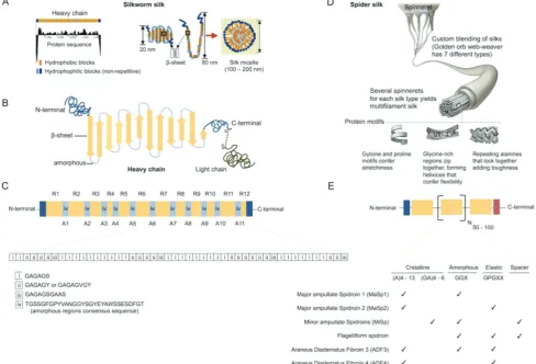
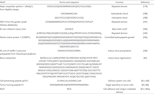
The Bombyx mori silk protein (i.e., silk fibroin) is very large and can be subdivided into light (≈26 kDa) and heavy (≈391 kDa) chains that are linked by a single disulfide bond at the C-ter-minus[47] (Figure 2). The C-terminal and N-terminal capping
sequences are completely nonrepeating amino acid residues. The mechanical properties of silk fibroin arise due to the block copolymer-like arrangement of the silk heavy chain, which con-tains 11 short hydrophilic regions typically 31 amino acid long and 12 hydrophobic blocks that account for 94% of the silk heavy chain. These hydrophobic blocks contain predominately glycine-X (GX) repeats, where X is alanine (A) (65%), serine (S) (23%), or tyrosine (Y) (9%).[47a] These GX blocks can be broadly
[image:4.595.51.540.308.640.2]classified into three groups: i) a highly repetitive GAGAGS sequence that contributes to the bulk of the crystalline regions
Figure 2. Silk structure. A) Solution conformation of Bomyx mori silk. Hydrophobicity pattern of the heavy chain with possible chain folding and micelle assembly of silk fibroin in water. Adapted with permission.[50] Copyright 2003, Macmillan Publishers. B) 2D silk fibroin schematic. Adapted with
permis-sion.[51] Copyright 2018, American Chemical Society. C) Primary structure of the Bombyx mori silk heavy chain. R01 to R12 and A01 to A11 represent
the arrangement of 12 repetitive and 11 amorphous regions, respectively. The approximate amino acid sequence of the R10 is shown by combination of sequences of i, ii, and iii. Adapted with permission.[52] Copyright 2005, American Chemical Society. D) Hierarchal structure of spider silk. Adapted
with permission.[53] Copyright 2011, Elsevier. E) Primary structure of spider silks. Adapted with permission.[54] Copyright 2017, American Association
and is typically found at the beginning of each motive, ii) a relative less repetitive sequence containing hydrophobic and/ or aromatic residues, namely, GAGAGY, GAGAGV, and GAGA-GVGY, which form semi-crystalline regions, and iii) motifs similar to i) except for the presence of an AAS motif, which typically exists at the C-terminus of each motif and may play a role for sheet-breaking.[48] Bombyx mori silk fibroin lacks the
tripeptide sequence arginine, glycine, and glutamic acid (RGD) that is typically exploited by cells to mediated cell–substrate attachment via integrin engagement; however, the N terminal of the silk heavy chain contains a fibroblast growth-promoting peptide.[49] Nevertheless, a sequence specificity exists between
different silkworm silks; for example, the Indian non-mulberry tasar silkworm (Antheraea mylitta) contains RGD sequences that are absent in Bombyx mori silk.
Spider dragline silk, one of the toughest materials known to humankind, is composed of a skin layer and a bundle of micro-fibrils (Figure 1).[5b,55] The microfibrils are composed of aligned
granules, and their silk molecules form an amorphous phase and β-sheet-rich crystalline regions.[5b] In both spider and
silk-worm silk fibers, the aligned β-sheet structure provides cross-links between the β-sheet domains embedded in an amorphous matrix that consists of less orderly structures in the form of random coils, helices, and β-turns.[56] These β-sheet crystals are
critical structures in the hierarchical structures of silk fibers, because they play an essential role as cross-linking points and realize the stiffness, strength, toughness, and characteristic deformation behaviors.[56a,b,57]
The amino acid sequences that form the β-sheet are 7–9mer alanine sequences for Nephila clavipes dragline silk and GAGAGS for Bombyx mori,[58] whereas other silkworm silk
spe-cies use polyalanine sequences to form the β-sheet structure.[6a]
The influence of the number of alanine residues on the sec-ondary structure and assembly behaviors of silk molecules has been studied using wide angle X-ray crystallography as well as solid-state nuclear magnetic resonance (NMR) spectroscopy. Those data from X-ray and NMR analyses reveal that short poly(alanine) sequences, such as 6mers or shorter, form a packed rectangular arrangement, while poly(alanine) sequences longer than 7mers pack in a staggered arrangement.[59]
The β-sheet is the most fundamental secondary structure in silk-based (bio)materials. The predominant β-sheet structure plays a key role in stabilizing silk materials via physical cross-links, as the β-sheet behaves as a cross-linking point. Crystal structures of silk β-sheets have been characterized using wide-angle X-ray analysis. The crystal structure of Bombyx mori silk fiber has a unit cell with the space group P21-C22.[60] The crystal
lattice of the Bombyx mori silk fiber reported by Marsh et al. had unit cell dimensions of a = 9.40 Å, b = 9.20 Å, and c (fiber axis) = 6.97 Å, while Takahashi et al. reported cell dimensions of
a = 9.38 Å, b = 9.49 Å, and c (fiber axis) = 6.98 Å.[60] The lattice of
other silks, such as Antheraea yamamai (Japanese silk moth), has been characterized and reported by many groups.[57b] The unit
cells contain four molecular chains, a pair of which symmetrically forms a β-sheet structure via hydrogen bonds. The up-molecular and down-molecular chains also alternate with each other in an antiparallel manner. Each silk has a different crystal lattice, which can be attributed to differences of the silk amino acid sequences. However, the relationship between the crystal lattices of different
silks and the subsequent characteristics of silk fibroin as a biomaterial remains largely unexplored, despite the fact that crys-tallinity (i.e., the amount of crystalline region) affects the physical and biological properties of silk-based biomaterials.
4. Hydration State
Silk and regenerated silk fibroin materials are expected to exhibit high toughness and ductility because of the excel-lent mechanical characters of spider (dragline) silks found in nature.[9,61] However, in addition to sequence specificity, the
hydration state of silk is critical for its performance.[62] For
example, most native spider silks show significant fiber con-traction when transitioned from a dry state to a high humidity environment. Exposure to humidity facilitates the rearrange-ment of the noncrystalline GPGXX sequence of orb web silks and the glycine-glycine-X 310 helices in nonorbicularian species
(which lack the GPGXX sequence). This occurs due to disrup-tion of hydrogen bonding by these sequences, which facilitates the transition from a parallel arrangement for the fiber axis to a lower energetic configuration that is accompanied by fiber shrinkage and thickening.[63] Thus, water is a key component
that enables spiders to tailor the properties of their silks during spinning and for in situ web tightening (a phenomenon also known as “supercontraction”).[63]
In nature, silkworm cocoons and spider webs/draglines are tough structural materials that perform their function; for example, to capture prey in the spider’s web or to protect the devel-oping moth from predators and infection.[57b] The mechanical
robustness of the native silk fiber has been exploited by humans for biomedical applications both in preclinical (e.g., ref. [64]) and clinical trials (detailed below). For example, silk fibroin scaf-folds proposed for bone repair have shown a high compressive strength of ≈13 MPa when reinforced with Bombyx mori silk fibers.[64] A similar approach has been taken to enhance the
mechanical properties of Bombyx mori silk hydrogels for car-tilage tissue engineering.[65] Recently, a high relative humidity
of >97% was found to cause a dramatic increase in the tough-ness and crystallinity of silk films.[6a] This finding exemplifies
how an appropriate hydration of silk molecules and materials can achieve crystallization and plasticization simultaneously, resulting in a high-strength and tough silk material.
5.1. Silk Biocompatibility
The exact set of biocompatibility requirements is application specific, although many preclinical studies simply cite that silk can meet all the necessary requirements, or they make refer-ence to silk as a “clinically approved” biomaterial for use in humans. However, this ignores our appreciation that a uni-versal biocompatibility does not exist: a material needs to be fit for its intended use[66] (as documented by dedicated
bio-compatibility studies); thus, its performance is context specific. The clinical approval of silk typically refers to its load bearing applications; degummed Bombyx mori silk fibers processed into a knitted surgical mesh (SERI Surgical Scaffold manufactured by Sofregen Inc., Medford, MA, USA), silk sutures (coated with waxes, Ethicon Inc. and several other manufacturers), and silk garments to treat dermatological conditions are in wide use today in the clinical setting. Therefore, their performance in humans is becoming better documented in the literature[41,67]
and is accompanied by a cadre of clinicians with experience working with these silk materials.
Dedicated biocompatibility assessment is critical when gen-erating novel silk formats (e.g., (nano)particles, hydrogels, scaf-folds, films, coatings, etc.) to address areas of unmet clinical need or when applying existing silk technologies to new indica-tions. Any nonautologous material will elicit an initial foreign body response that reflects the first steps of tissue repair.[68]
Therefore, ensuring that the foreign body response is transient rather than chronic is a prerequisite to ensure that clinical end-points can be met. Overall, biomaterial performance depends on the implantation site, size, geometry, surface topography, and physical characteristics.[68] A systematic literature review[69]
exam-ining the performance of silk constructs (e.g., vascular grafts, ligaments, and wound dressings for skin grafting) in small and large animal studies overwhelming showed that a variety of dif-ferent Bombyx mori silk constructs performed well across the broad spectrum of indications and animal models.[69] Direct
in vivo comparison of silk with commonly used natural (e.g., collagen) and synthetic (e.g., polycaprolactone, polylactic acid, poly[lactide-co-glycolic acid]) biomaterials indicates that Bombyx
mori silk fibroin is typically at least as good as these synthetic
materials and often superior than other natural biopolymers.[69]
As new applications for silk emerge, appropriate biocompati-bility studies must be performed to support these developments. For example, silk nanoparticles for anticancer drug delivery are typically designed for intravenous administration[70] and thus
require hemocompatibility assessments because biological performance cannot be deduced by extrapolating results from macroscopic films[71] to nanoscale particles.[72] An initial proof of
biocompatibility is a first step to translate silk technologies from the bench to the clinical setting. For example, regulatory frame-works imposed by the Pharmaceuticals and Medical Devices Agency Japan, the Food and Drug Administration (FDA, USA), the Medicines and Healthcare Products Regulatory Agency (UK), and the European Medicine Regulatory Agency (EU) for medical devices (e.g., Regulation (EU) 2017/745 to obtain CE marking analogous to the Class III Premarket Approval/510(k) in the USA, and the Australian Register of Therapeutic Goods certificate of inclusion) stipulate that a biological safety assess-ment needs to be conducted first (by an ISO certified laboratory,
in conjunction with a notified body) before progressing the device to first-in-man clinical assessment. Materials of animal or allogeneic origin need to fulfill additional safety require-ments (e.g., absence of infectious agents such as retroviruses, etc.) before use in humans. However, from a regulatory per-spective, Bombyx mori silk is regarded as a non-animal product (EU Council Directive 93/42/EEC, rule 17).
Reports on the biocompatibility of silk in humans come pri-marily from silk sutures (reviewed in ref. [41]) that have been in use for several centuries[20] and from SERI Surgical
Scaf-fold that obtained 510(k) clearance by the FDA in 2008 and underwent a market launch in 2013. Histological evidence of 69 breast tissue samples (by 60 patients) taken at stage 2 in patients undergoing two-stage breast reconstruction with SERI Surgical Scaffold showed a mild inflammatory response in 59 patients, as confirmed by histology. This consisted of an infil-tration of mostly macrophages and occasional multinucleated giant cells that phagocytosed the silk fibers, as well as occa-sional lymphocytes and, rarely, neutrophils or polymorphonu-clear cells.[73] Ordered collagen deposition was observed, with
minimal or no encapsulation of the silk surgical mesh. These clinical trial data[73] were similar to observations made in a
sheep study.[74] However, one patient had a postoperative
hema-toma that led to mesh removal.[73]
Synthetics are now the most widely used suture material, but silk sutures are still in demand for specialized applications where exquisite handling is of paramount importance (e.g., eye surgery). Silk sutures are strong, are easy to handle, lie flat on the tissue surface, and allow for secure knots. Adverse reactions to silk sutures are typically reported for virgin silk, where the silk filaments are still coated with sericin (and often with addi-tional waxes or silicones).[41] There is an ongoing debate about
the potential role of sericin in these adverse reactions. However, emerging evidence suggests that sericin on its own shows a low allergenic and immunogenic profile in mice; in fact, this pro-file is similar to that seen for silk fibroin or alginate.[75] These
observations are supported by in vitro data with macrophages: extracted sericin from Bombyx mori silk cocoons showed no significant release of the inflammatory marker TNF-α; sim-ilar observations were made with silk fibroin.[76] However,
extracted sericin in combination with bacterial lipopolysaccha-ride induced TNF-α release (but not for the silk fibroin group). Furthermore, recoating of silk fibroin with sericin showed no macrophage response, while virgin silk induced a high level of TNF-α release.[76] These data suggest that other leachable
compound(s), or these compounds combined with sericin, may be responsible for the adverse clinical reactions reported for silk.[69,76] For example, patients subjected to bilateral
cata-ract surgery showed no suture reaction on the first eye but a severe reaction on the second eye when it was treated six to three months later. This suggested that these patients had undergone a sensitization toward virgin silk. Prompt removal of the offending silk suture resulted in significant clinical improvement.[77] Examining the clinical literature regarding
silk sutures and identifying the exact cause of the adverse reac-tion are challenging because often little (or no) informareac-tion is provided about the exact nature of the silk suture (e.g., virgin silk, type of coatings, etc.). Nonetheless, an allergic response to
domestic setting (reviewed in ref. [78]). For example, exposure to virgin silk fibers and repurposed silk waste (e.g., silk floss incor-porated into rugs and bedding) has been linked to the develop-ment of asthma in silk weavers[79] and children,[80] mounted by
an IgG and IgE immune response.[81] Textile workers have an
increased risk of developing chronic obstructive pulmonary dis-ease, and this risk is highest in silk workers.[82]
Complete and reproducible sericin removal from Bombyx
mori silk (a.k.a. degumming) is an essential step in silk
utiliza-tion. Clinically acceptable limits for residual sericin levels for marketed silk products have not been released into the public domain (note that SERI Surgical Scaffold is described by the manufacturer as highly purified silk with ≥95% purity). Cur-rent evidence from both preclinical in vivo studies and clinical experience in humans across a range of applications indicates that Bombyx mori silk fibroin is biocompatible, provided that all other contaminants are successfully removed.
Sericin has traditionally been linked to the adverse effects reported for virgin silk (reviewed in ref. [78]). However, over the past decade, sericin has emerged as an interesting biopolymer (reviewed in ref. [83]), and dedicated biocompatibility studies are now showing encouraging results in relation to the allergenic and immunogenic profile of sericin (e.g., ref. [75]). An increasing number of studies report the biomedical use of the biopolymer sericin. For example, the development of composite sericin/ silicone nerve guides[84] or sericin/polyacrylamide hydrogels
pro-posed for dermal repair.[85] Preliminary Phase I clinical trials using
sericin composite wound dressings for split-thickness skin grafting are on going and the results are eagerly awaited (NCT01539980 and NCT02643680 reported at www.ClinicalTrials.gov).
5.2. Silk Biodegradation
Silk sutures are classified by regulatory agencies as non-biode-gradable because regulatory guidelines expect a loss of most tensile strength within 60 days postimplantation. Over this time scale, silk sutures do not lose their mechanical performance, as they require longer time frames to degrade in humans.[41]
In patients undergoing two-stage breast reconstruction, histo-logical evidence of breast tissue samples taken from 60 patients at stage 2 (median 152 days after initial scaffold implantation, range 74 to 357 days) showed consistent SERI Surgical Scaffold degradation (although this was not quantified). The one excep-tion was a patient that had a postoperative hematoma, which was accompanied by an apparent lack of silk degradation.[73]
The silk protein is known to degrade in vitro and in vivo in response to proteolytic enzymes,[69] as exemplified by studies
with silk films (e.g., ref. [86]) and porous silk scaffolds (e.g., ref. [87]). Experience with SERI Surgical Scaffold in a sheep model of two-stage breast reconstruction showed progressive degradation and vascularization of the silk mesh: at 1 month postimplantation, tissue ingrowth and marked vascularization were evident; at 4 months, the mesh was no longer felt through the skin; and at 12 months, the mesh degradation and vascu-larization were scored as mild but with substantial silk loss that precluded mechanical testing of the remaining SERI Surgical Scaffold.[74] At 12 months, the SERI Surgical Scaffold had
stim-ulated extensive type I collagen deposition and the resulting
tissue was mechanically strong.[74] Clinical hernia repair in a
horse showed incomplete SERI Surgical Scaffold degradation at 2 years postimplantation, but no hernia relapse.[88]
The time scale for silk degradation depends on a number of factors, including, but not limited to: i) the amount of material, ii) gross morphology, iii) silk secondary structure, iv) silk treatment history, v) mechanical environment, and vi) implantation site (or final destination). The implantation site directly impacts the type of proteolytic enzyme encountered by the silk, because these enzymes vary between tissues, cells, and subcellular location.
Silk fibroin sequence alignment indicates a susceptibility to a number of proteases (e.g., protease XIV, α-chymotrypsin, proteinase K, papain, matrix metalloproteinases, collagenase, etc.)[51,89]). Nonetheless, predicting silk fibroin degradation simply
based on the primary sequence is unreliable; for example, chymo-trypsin has 434 cleavage sites in the silk heavy chain and 81 in the light chain, while protease XIV has 348 in the heavy chain and 41 in the light chain. Despite numerous cleavage sites, chymotrypsin treatment for 20 days had no quantifiable effect on silk fibroin, while protease XIV significantly degraded silk fibroin in vitro.[51]
Papain, a cysteine protease enzyme that mimics the activity of lysosomal enzymes, has 26 cleavage sites in the silk heavy chain (albeit exclusively in the amorphous regions) and 15 in the light chain, and it caused significant silk fibroin degradation over 20 days but at a slower rate than protease XIV. Similar observations were made with isolated lysosomal enzyme preparations.[51]
Overall, these studies exemplify that the structure beyond the primary sequence is of critical importance for silk degra-dation. The current working model supports the notion that, for Bombyx mori silk, degradation begins with the 11 hydro-philic amorphous segments in the silk heavy chain, as well as the C-terminal and N-terminal and the silk light chain, which consist of completely nonrepeating amino acid sequences; this is then followed by degradation of the more crystalline sequences.[51,61c,89] The tightly packed crystalline domains are
degraded last.[90] Furthermore, the silk format is a critical factor
in determining degradation rates, as in vivo studies in rodent models indicated faster degradation for open silk structures than for tightly packed monolithic silk fibroin films (rank order: hydrogel > silk scaffold > monolithic film).[69]
Protease XIV is a useful model enzyme for studying silk degra-dation and for comparing with earlier studies. However, protease XIV is a nonmammalian enzyme cocktail, so it must not be used to deduce or predict biocompatibility performance. Silk, as a pro-tein-based biopolymer, is commonly considered to yield harm-less biodegradation products; however, a more critical inspec-tion of silk and its degradainspec-tion products is timely. For example, silk fibrils have molecular-level similarity to amyloid fibrils,[91]
and they were also reported to enhance amyloidosis of amyloid protein through a mechanism based on cross-seeding effects.[92]
Formation of β-amyloid structures is a concern because amy-loid beta fibrils are a hallmark of Alzheimer’s disease.[61c,93]
Preliminary studies in mice injected with self-assembling silk fibroin hydrogels into the caudate putamen (striatum) showed no decline in cognitive function or animal behavior over the 6 week study period.[94]
6. Processing of Silk Cocoons—Generating
Silk for Biomedical Use
Unspinning the Bombyx mori silk cocoon and degumming to remove sericin are two crucial steps that yield silk suitable for biomedical use. Sericin can be removed by enzymatic methods (i.e., digesting sericin but not silk) or chemical processing (e.g., alkaline treatment). The latter approach is widely used and typi-cally involves boiling silk in sodium bicarbonate for 20–60 min.[42]
Degumming times as short as 5 min are also sufficient to remove sericin while minimizing silk damage, which usually occurs due to cleavage of the disulfide bond between the silk heavy and lights chain and fragmentation of the amorphous silk sequences in the silk heavy chain, which results in polydispersed silk.[95]
The degummed silk fibers can be fully reverse engineered by dissolving them in a high concentration chaotropic agent (for example, 9.3 m lithium bromide) at 60 °C over several hours
to dissemble the higher order silk structure. The resulting silk fibroin solution is then dialyzed extensively against water to yield an aqueous silk solution that is stable at room tem-perature for weeks and at 4 °C for several months.[42] When
compared to native silk feedstock, this reverse engineered silk fibroin solution has a reduced solution conformation[96] and
changed rheological properties.[40d]
The reverse engineered aqueous silk fibroin solution is com-monly used to generate novel silk formats; for example films, fibers, scaffolds, and (self-assembling) silk hydrogels, as well as (nano)particles and (nano)coatings, and these formats are often achieved using an all aqueous processing under ambient condi-tions. These mild processing conditions are ideal for preserving the activity of biologics.
7. Present Routine Clinical Use of Silk
The silk surgical mesh SERI Surgical Scaffold, silk sutures, and silk clothing to treat dermatological conditions are the only available products in routine clinical use today. All these prod-ucts are manufactured by unwinding Bombyx mori silk cocoons and working with the silk thread. The clinical performance of silk sutures, their adverse effects, and the developments and potential solutions to improve suture performance have been reviewed previously.[41]
The SERI Surgical Scaffold technology is based on work con-ducted by David Kaplan and co-workers at Tufts University, Med-ford, MA, USA.[67a,97] The resulting patent portfolio and
proprie-tary silk processing technologies formed the basis of the spin-out company, Serica Technologies Inc. (Medford, MA, USA). Serica Technologies Inc. was able to prove to the FDA that SERI Sur-gical Scaffold was “substantially equivalent” to existing surSur-gical meshes and thus received 510(k) clearance to market the device.
Serica Technologies Inc. was subsequently acquired by Allergan Inc., and the SERI Surgical Scaffold became commercially avail-able for soft tissue support and repair in 2013 and was since then acquired by Sofregen Medical Inc. (Medford, MA, USA).
The current SERI Surgical Scaffold indications are for abdominal wall reconstruction[98] and investigational plastic
sur-gery applications, including total body contouring, brachioplasty, abdominoplasty, mastopexy, and breast reconstruction (Table 1).
The clinical performance of SERI Surgical Scaffold has been reported in the literature, which includes open label clinical trials and case reports (Table 1). Many of these encouraging clinical studies have been sponsored by Allergan Inc. A few independent retrospective clinical reports of small patient cohorts are reporting side effects (e.g., poor scaffold integration, see Table 1, Figure 3), often requiring surgical removal of the mesh.[67b,105,106] Therefore, some clinicians are abandoning the
use of SERI Surgical Scaffold in their clinical practices,[105] and
caution has been raised by others.[108] In 2013, Allergan Inc.
vol-untary withdraw several SERI Surgical Scaffold batches due to concerns about product sterility. How this might have affected the reported adverse events is not known.
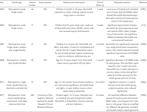
We are familiar with silk for the textile industry, although silk garments are also used clinically to treat dermatological conditions, especially atopic dermatitis[109] and acne vulgaris[110]
(Table 2). Mechanical skin irritation by harsh, rough (e.g., wool), and short (e.g., cotton) textile fibers is thought to contribute to atopic dermatitis. Furthermore, the skin of atopic dermatitis patients is often colonized with Staphylococcus aureus and the extent of colonization correlates with the severity of the disease. Silk fibers are very long (up to 1500 m) and smooth, so they minimize mechanical irritation when knitted into clothing. This silk clothing has been chemically modified to achieve antibacterial properties with the aim of reducing Staphylococcus
aureus colonization of the skin. Sericin-free silk has also been
covalently functionalized with 3-trimethylsilylpropyl-dimeth-yloctadecyl ammonium chloride (AEM 5700/5772; AEGIS), resulting in commercial products (e.g., DermaSilk) for the treatment of atopic dermatitis. These silk garments use highly purified silk to minimize the risk of contact dermatitis.[111]
The silk garments are also knitted in a specific fashion to improve transpiration of sweat through the fabric (unlike eve-ryday silk, which can worsen atopic dermatitis by trapping moisture). A randomized double-blind study in 30 patients with atopic dermatitis on both arms received an AEM 5700/5772 functionalized silk sleeve and a silk-only sleeve. Patients treated with the silk sleeve showed a rapid improvement within 2 weeks but remained similar until the end of the study. The contralat-eral arm treated with the AEM 5700/5772 functionalized silk showed similar results at 2 weeks but reached a greater level of improvement over 4 weeks.[109d] Other clinical trials using AEM
5700/5772 functionalized silk garments in small patient cohorts reported substantial improvements in skin conditions (Table 2). By contrast, a randomized, controlled, observer-blinded clinical trial in 300 children showed only a 3% reduction in skin infec-tion compared to control and was therefore not regarded as providing a significant clinical benefit.[109e] Overall, these
Ta
bl
e 1
.
Published data reporting the clinical use of SERI Surgical Scaffold in humans.
Year
Article type
Patient number
Study sponsor
Intervention
Clinical follow up
(months)
Reported outcome
Reference
2013
Retrospective case report
1
Allergan
Abdominoplasty and use of scaffold to provide soft-tissue support to the abdominal fascia in
patient with massive weight loss
24
C
ontour and flatness of the anterior abdominal wall was maintained
[99] 2014 Retrospective study Multi center 141 Allergan
Revision of breast augmentation (
n
=
40); revision of
breast reconstruction (
n
=
24); mastopexy augmen
-tation (
n
=
20); mastopexy augmentation-revision
(n
=
16); hernia repair (
n
=
11); other (
n
=
30)
0 to 12
Adverse side effect reporting voluntary
Surgeons rated the ease of use a mean of 2.86 (scale 0–3) Surgeons rated their satisfaction a mean of 9.31 (scale 0–10)
[100]
2014
Prospective study Multi center
139
Allergan
2-stage implant-based breast reconstruction
6
75 subjects undergone stage 2, subject satisfaction score 4.3
±
0.91
(5 best). Investigator satisfaction score was 9.4
±
0.84 (10 best). Adverse
effects in 214 breasts: tissue necrosis (6.1%), seroma (6.1%), hematoma (2.8%), breast infection (1.9%), cellulitis (1.9%), implant loss (1.9%),
capsular contracture (0%)
[101]
2014
Retrospective case report
1
Allergan
Abdominoplasty and lower body lift of in patient with massive weight loss. Scaffold implantation on
left lower body only
7
No complications reported, improve patient satisfaction
[102]
2014
Retrospective case report
1
Allergan
Brachioplasty
6
No complications reported, perceived faster maturation process and a
better-quality scar [103] 2014 Retrospective study Multi center 172 Not disclosed
77 patients (71 women, 6 men) underwent
abdominal wall fascial repair or reinforcement. The
remaining 95 patients not reported on
18.4
±
7.5
The overall complication rate (N77) was 6.5%, consisting of 2 wound
dehiscences, 1 with device exposure, 1 seroma, 1 infection with explanta
-tion, and a perioperative bulge requiring reoperation
[98] 2014 Retrospective study Single center 15 No
Direct-to-implant after skin-sparing mastectomy
6 to 13
C
apsular contraction (35%); loss of scaffold due to necrosis (
n = 1); seroma ( n =
1); hematoma self-limiting (
n
=
1); patient satisfaction
(5.77 out of 10)
[104] 2015 C orrespondence Retrospective study Single center 5 No
Unilateral skin-sparing mastectomy and imme
-diate reconstruction
Not reported
Late infection (6 weeks and 3.5 months postsurgery) in 2 breasts leading to scaffold and implant removal. In 2 patients successful completion
with tissue integration and vascularization
[105]
2015
Prospective study Multi center
139
Allergan
71 patients undergoing 2-stage breast
reconstruction
12
Investigator satisfaction score was 9.4
±
0.91 (10 best) and patient
scores was 4.5
±
0.82 (5 best). C
omplication rates in 105 breasts were
tissue necrosis (6.7%), seroma (5.7%), hematoma (4.8 percent), implant
loss (3.8%), capsular contracture (1.9%), breast infection (1.0%)
[73] 2015 C orrespondence Retrospective study Single center 4 No Breast reconstruction 12
Late infection with
Ps. aeruginosa
in 2 patients at 5 months resulting in
implant replacement. Lack of mesh integration (or degradation) in all 4
patients
[106]
2017
Prospective study Multi center
103
Allergan
2-stage implant-based breast reconstruction
24
Investigator satisfaction high
[107]
2018
Prospective study Single center
16 No Direct-to-implant reconstruction with surgical scaf
-fold after skin-sparing mastectomy
24 to 37
No intraoperative complications. Adverse effects in 22 breasts: Postopera
-tive bleeding, that required reoperation occurred in 5% breast, postopera
-tive seroma in 45% and surgical site infection in 9%. Scaffold-related
complications occurred in 14% breasts, lack of scaffold integration in all, resulting in skin ulceration in 2 and the scaffold lying free in the breast
pocket surrounded with seroma in one
of patients with only moderate eczema (typically not treated with these types of garments) and the often limited wearing of the garments are not in line with the clinical recommendations. Therefore, these difficulties might result in underestimation of treatment effects. Overall, improving these silk garments to maximize their clinical performance warrants more research.
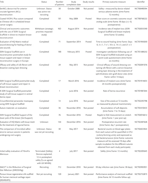
8. Clinical Trials Using Silk
The renewed interest in silk for biomedical use over the past 20 years has resulted in a number of clinical trials; however, historically, these data sets have been difficult to source. Since 2007, the FDA has mandated that drug and device manufac-turers register clinical trials (www.ClinicTrials.gov) (Table 3).
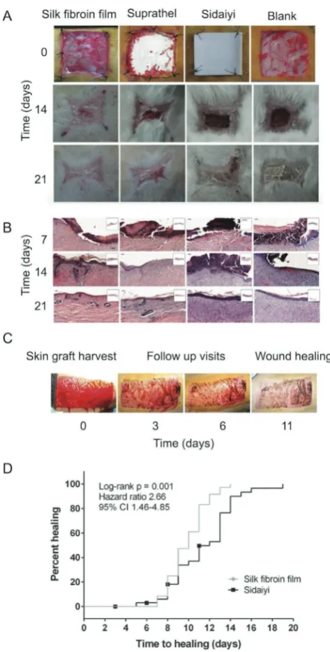
Silk-based biomaterials show particular promise for skin wound healing due to their hemostatic properties, low inflam-matory potential, and permeability to oxygen and water, as well as their ability to function as a barrier to bacterial coloni-zation (Table 3). Sidaiyi, a silk fibroin sponge attached to a sili-cone membrane, is a first generation wound dressing currently approved by the China Food and Drug Administration for clinical use in that country.[113] The Sidaiyi platform was first compared
to silk fibroin films for wound healing applications in preclinical animal models, followed by a randomized, single blinded Phase I clinical trial. Silk films were made by casting an aqueous Bombyx
mori silk fibroin solution in a mold and treating it at 65 °C and 90% relative humidity for 100 min. The resulting 64.9 µm films were water-resistant (albeit their ability to bind a small amount
of water that acts as a plasticizer) and formed an effective barrier against bacterial infection in vitro. The films were found to be biocom-patible for their intended use according to ISO 10 993 tests for the biological evaluation of medical devices. In a rabbit full-thickness wound healing model, healing was three days faster in wounds treated with silk films than in wounds treated with Suprathel, a poly-urethane-based synthetic wound dressing, and seven days faster than in wounds treated with the Sidaiyi wound dressing or phos-phate buffered saline (PBS) treated controls (Figure 4). Silk-film-treated wounds showed the development of an organized epidermis by day 14 post-treatment and showed a mature and organized collagen matrix, hair follicles, and blood vessels histologically by day 21.[113]
These results were further verified in a por-cine full-thickness wound healing model prior to initiation of a Phase I clinical trial that ran from August 2013 to September 2014. This clinical trial enrolled 71 patients (36 randomly assigned to a silk film group and 35 to a Sid-aiyi group). The silk-based wound dressings were used to cover donor sites following sur-gical harvesting of split-thickness skin grafts; healing was significantly faster in the silk film group than in the Sidaiyi wound dressing group and 100% of the wounds were healed by day 14 postinjury in the silk film group. One case of inflamma-tory reaction to the silk film was noted but the exact etiology was not determined. No cases of wound exudation were observed, indicating that the silk films maintained a clean wound environ-ment with suitable moisture levels. The films showed good adhe-sion to the wound surface and no changes of the wound dressing were required. As the wound healed, the silk films spontaneously detached from the regenerated skin areas. The exact mechanism underlying the improved clinical performance observed for the silk film group over the Sidaiyi group is currently unknown.
However, this study demonstrated the ability to manufacture silk films under Good Manufacturing Practice requirements and their successful use as wound dressings for skin repair and regeneration.[113] The potential for relatively easy modification
of silk films for additional functionality, such as the incorpo-ration of pores or the introduction of bioactive molecules,[114]
makes silk films particularly attractive as wound dressings. Thin silk films have also been used in prospective human clinical trials to repair acute and chornic tympanic membrane perforations.[115] These silk patches (Tympasil, Daewoong-Bio,
Seoul, South Korea) were generated using reverse engineered
Bombyx mori silk fibroin. The process leading to stabilization
of these silk patches has not been established, but physical cross-linking is most likely because the brittle patches were first wetted in PBS to plasticize them to facilitate their trimming to the required size and surgical placement.
[image:10.595.51.362.70.309.2]The first clinical trial involved 52 patients with acute trau-matic tympanic membrane perforation who were treated with either a silk film or a paper-based membrane.[115b] A number of
Figure 3. Examples of SERI Surgical Scaffold implant loss in humans. A) Silk fibroin surgical mesh prior to implantation. B) Intraoperative view showing a free lying scaffold in the breast pocket. C) Retrieved scaffold surrounded with seroma. D) Interaoperative view of surgically removed scaffold with interpenetrated granulation tissue/scar plate (at > 5 months), and E) histology of retrieved sample showing granulation tissue with neutrophiles and giant cells at the material (1) interface (dotted line). (B,C) Reproduced with permission.[67b] Copyright
conservative treatment modalities had been explored to support the (often spontaneous) healing of an acutely perforated tym-panic membrane, including the placement of a “patch” on top of the damaged tympanic membrane. In this trial, the silk or paper patches were surgically placed and removed 7 days later, when the tympanic membrane appeared fully regenerated. The closure rate was similar for both the silk film and paper mem-brane (92.3 and 84.6%, respectively), but the silk patch signifi-cantly shortened the healing time from 16.7 to 13.7 days.
A similar improved healing with a silk patch was also pre-viously reported in animal studies.[116] A follow-up study of 40
patients with chronic traumatic tympanic membrane perfora-tion showed that patients treated with a silk patch (Tympasil) had lower otorrhea, minor complication rates, and high patient satisfaction when compared with conventional perichon-drium myringoplasty.[115a] The silk and autologous patches
were removed one week after placement, and the postoperative hearing outcomes were not significantly different between the
two treatment groups. However, the surgical time for the silk patch was very short (13.7 vs 29.5 min) and no sourcing of con-nective tissue was required for the graft.[115a]
9. Preclinical Use of Silk—The Future
As we move from the routine clinical use of silk fibers to human clinical trials, the line between a silk thread-based medical device and other forms of silk starts to blur. A very wide spectrum of silk materials and formats is now emerging in preclinical studies.[167]
We will first review silk solutions derived from reverse engineered
Bombyx mori silk, followed by more complex formulations.
9.1. Silk Solution
Bombyx mori silk in its solubilized aqueous form has been
[image:11.595.52.544.89.462.2]investigated for a range of therapeutic applications, including Table 2. Published data reporting the clinical use of silk garments for the treatment of atopic dermatitis in humans.
Year Article type Patient number
Study sponsor Patients and intervention Clinical follow up
Reported outcome Reference
2004 Retrospective study Single center
46 ND Children 4 months to 10 years. DermaSilk treatment or cotton clothing, topical
moistur-izing cream or emulsion
1 week Local score of treated and untreated area of same child (SCORAD index) Overall DermaSilk decreased atopic dermatitis severity and local
improve-ment of silk treated areas
[109c]
2006 Retrospective study Single center
15 ND Children 0.6–9.2 years, body suits made out of DermaSilk and cotton (50:50). Cotton side
also received topical corticosteroid
3 weeks Significant improvement of atopic dermatitis by assessing eczema area
and severity (EASI index) irrespec-tive of intervention. No significant difference between DermaSilk and
corticosteroid treatment
[112]
2007 Retrospective study Single center,
random-ized, single-blinded
22 ND Children 5 to 12 years old. DermaSilk, silk fabric, and cotton. Control on contralateral arm
(silk for first for 2 weeks followed by cotton for rest of study period), topical moisturizing
cream or emulsion, antihistamines prn
3 months DermaSilk arm significantly improved over study period when compared to cotton. Silk control treatment showed significant improvement of SCORAD
index
[109b]
2008 Retrospective, random-ized, double-blinded
30 ND Age 3 to 31 years (mean 14.2). DermaSilk sleeve versus equivalent silk only fabric
1 month Significant decrease in SCORAD index for both groups. Silk only fabric rapid reduction over 2 weeks only while DermaSilk over 4 weeks; decrease in pruritus values similar during first 2 weeks but further decrease for
Der-maSilk group until end of study
[109d]
2013 Retrospective, single center, double blind ran-domized controlled trial
22 ND Age 4 to 18 months. Acute phases treated as per international guidelines. DermaSilk body and tights, or wear clothes in pure cotton;
expect May to September
24 months Significant reduction in topical steroid use for DermaSilk group;
subjective pruritus also reduced significantly
[109a]
2017 Retrospective, multi-center parallel-group, randomised, controlled,
observer-blind trial
300 University of Not-tingham, National Institute for Health Research Clinical Research Network
Children aged 1 to 15 years. Standard care or standard care plus silk garment (either DermaSilk or DreamSkin). Eczema outcome
and skin infections
24 weeks No statistical difference between groups for eczema area and severity (EASI index). Less frequent skin infec-tions in silk group. Data not stratified for different silk garments. Included cost-benefit analysis rejects garments
[109e]
Table 3. Reported human clinical trials using silk.
Title Status Number
of patients
Study completion date
Study results Primary outcome measure Identifier
SeriACL device trial for anterior cruciate ligament (ACL) reconstruction
Unknown. Status was active not
recruiting
30 October 2008 Not posted Safety—measured by device related serious adverse events (time frame:
12 months)
NCT00490594
Coated VICRYL Plus suture compared to Chinese silk in scheduled breast cancer surgery
Completed 101 May 2009 Posted Mean score on cosmetic outcome visual analog scale (time frame: 30 days (+/−5)
postoperative)
NCT00768222
Clinical and economic outcomes with the use of SERI Surgical Scaffold in direct-to-implant breast reconstruction
Withdrawn (strategic priorities impacted
study)
ND August 2014 Not posted Incidence rate of implant loss (SERI Surgical Scaffold and breast implant)
(time frame: 52 weeks)
NCT02033590
Evaluation of HQ Matrix medical wound dressing for healing of donor site wounds
Completed 71 September 2014 Posted Time to wound healing (time frame: Days 0, 3 ± 1, 7 ± 1, 10 ± 2, 14 ± 3, and 21 ± 3
postoperation)
NCT01993030
SERI Surgical Scaffold use in reconstruction postmarket study for tissue support and repair in breast reconstruction surgery in Europe
Completed 100 February 2015 Posted Investigator satisfaction following use of SERI Surgical Scaffold evaluated using an 11-point scale questionnaire (time frame:
six months)
NCT01389232
Efficacy and safety of silk fibroin with bioactive coating layer dressing
Completed 29 May 2015 Not posted Clinical efficacy of wound dressing con-taining silk fibroin with a sericin bioactive coating layer dressing in the treatment of split-thickness skin graft donor sites (time
frame: within 14 days)
NCT02091076
SERI Surgical Scaffold postmarket study of soft tissue support and repair in breast reconstruction
Completed 17 March 2016 Not posted Incidence of Implant Loss (time frame: 24 months postoperatively)
NCT01914653
A SERI Surgical Scaffold postmarket study of soft tissue support in ventral hernia repair
Completed 1 June 2016 Not posted Rate of hernia recurrence NCT01981044
Circumferential periareolar mastopexy using SERI Surgical Scaffold
Completed 14 June 2016 Not posted Size of the areola at 12 months as measured by physical mammometry
NCT02293798
Suture materials: an evaluation Completed 36 November 2016 Not posted Accumulation of soft deposits (time frame: 7 to 14 days)
NCT03410433
SERI Surgical Scaffold Support of the lower pole of the breast (SeriSupport)
Completed 76 November 2016 Posted Nipple to fold measurement on stretch (time frame: 1 year post op)
NCT02016612
Evaluation of HQ Matrix soft tissue mesh for the treatment of inguinal hernia
Unknown 144 December 2016 Not posted Postoperative recurrent rate (time frame: day 1 postoperation)
NCT02487628
The comparison of microbial adher-ence to various sutures in patients undergoing oral surgery
Unknown. Status was not yet recruiting
30 May 2017 Not posted Bacterial counts on blood agar plates from each suture will be quantified in CFU
(colony-forming units) and expressed as total bacteria/suture (time frame: outcome
measure will be assessed 10 days after sample incubation for the different sutures
obtained from each study participate)
NCT02653924
Initial safety evaluation of FibroFix Meniscus
Terminated (Safety). Devices explanted.
12 m postexplant safety f/u as agreed
with UK MHRA)
4 July 2017 Not posted Safety (time frame: 12 months) NCT02205645
DACCa) in the REduction of Surgical
Site INfection (DRESSINg)
Recruiting 712 December 2018 Not posted 30 day infection rate (time frame: 30 days) NCT02992951
Porous tissue regenerative silk scaffold for human meniscal cartilage repair (REKREATE)
Not yet recruiting 120 January 2021 Not posted Performance analysis of meniscal scaffold (time frame: At 12 months follow up)
NCT02732873
treatment of diabetes,[117] chronic wounds,[118] and
inflamma-tion.[119] Recent studies have investigated the utility of
regen-erated silk fibroin solution in preclinical animal models for
the treatment of ocular conditions, including dry eye[120] and
corneal injuries.[5b] Blindness from corneal disease affects over
50 million people worldwide, while another 337 million people suffer from dry eye disease, representing a significant health-care burden.[5b,121]
For example, silk fibroin treatment resulted in increased tear production and reduced the corneal irregularities observed in the absence of treatment in a mouse dry eye model (consisting of NOD.B10.H2b mice exposed to desiccation stress and
scopol-amine hydrobromide treatment for 10 days). Silk fibroin treat-ment inhibited detachtreat-ment of corneal epithelial cells, increased the number of conjunctival goblet cells, and inhibited the secre-tion of inflammatory factors in the lacrimal gland of the eye, resulting in recovery of the tear film and mucus layer of the eye, improved corneal health, and reduced dry eye symptoms. Other anti-inflammatory agents, such as cyclosporine and corticoster-oids, are available on the market for the treatment of dry eye, but silk has demonstrated a potential multitarget therapeutic effect that lacks the common side effects, such as pain and irritation, and other complications associated with long-term corticosteroid use.[44] Silk fibroin was demonstrated to stabilize
the tear film through anti-inflammatory effects in the lacrimal gland and increased number of conjunctival goblet cells, but the mechanisms underpinning these observations are unknown.
Clinical approaches to treat corneal injuries are relatively limited and predominantly involve topical application of anti-inflammatory or antimicrobial agents that do not promote tissue regeneration. A rabbit corneal injury model, which involved removal of a 7 mm diameter section of the central cor-neal epithelium, was used to study the effects of an aqueous
Bombyx mori silk fibroin solution (deemed “silk-derived
pro-tein” due to an additional autoclaving step during solubilization in lithium bromide that results in a heterogeneous population of low molecular weight silk fragments) on corneal epithe-lial healing.[5b] All treatments showed corneal wound closure
by 48 h postinjury, as indicated by fluorescein staining; how-ever, treatment with silk accelerated the rate of wound healing threefold in the first 6 h postinjury. Relative to a PBS-treated control, silk treatment resulted in a significant increase in the numbers of proliferating Ki-67 positive epithelial cells, a dose dependent increase in epithelial cell attachment to the under-lying basement membrane (as indicated by focal adhesion kinase staining), and a dose-dependent reduction in MMP-9, a metalloprotease involved in matrix remodeling and corneal repair. Finally, compared to the PBS-treated control, the silk-treated group showed the formation of epithelial layers with tight junctions (ZO-1 staining) that more closely resembled those of healthy corneas.[5b] The potential of the silk solution
in aiding wound healing is clearly demonstrated, but the exact mechanism of action is yet to be determined.
9.2. Silk Films
Silk films are among the most extensively explored biomaterials due to the ease of their fabrication and characterization and their versatility. Silk films have been explored for their potential in drug delivery.[61b] wound healing,[113,114,122] corneal
[image:13.595.51.288.66.533.2]replace-ment, and flexible electrode[123] applications, among others.